P40
Does Patient Age Impact Survival after Radical Cystectomy? Polat Turker1, Peter J. Bostrom1, Marcelo L. Wroclawski1, Tuomas Mirtti2,
Martti Nurmi3, Neil E. Fleshner1, Antonio Finelli1, Michael A. Jewett1,
Alexandre R. Zlotta4.
1University Health Network, Princess Margaret Hospital, Toronto, ON,
Canada, 2Helsinki University Hospital, Helsinki, Finland, 3University of
Turku, Turku, Finland, 4Mount Sinai Hospital, University Health Network,
Toronto, ON, Canada.
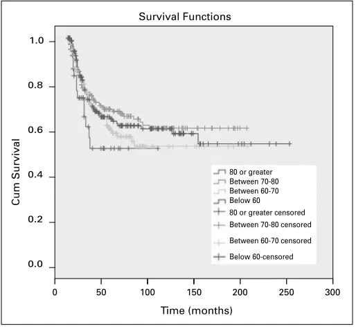
Background: With increased longevity in the population, urologists treat and will treat more and more often senior patients with bladder cancer (BC). The aim of the current study was to analyze the impact of patient’s age on survival after radical cystectomy (RC).
Methods: Two ethics approved RC databases including consecutive mus-cle invasive BC patients undergoing radical cystectomy at UHN, Toronto, Canada (1992-2008) and University of Turku, Turku, Finland (1986-2005) were included. Six hundred thirty five patients who underwent RC were analyzed. Patients were separated according to their age into four groups and compared for their characteristics, recurrence-free survival (RFS), dis-ease-specific survival (DSS) and overall survival (OAS).
Results: Patients age and gender distribution is presented in Table 1. An ileal conduit was the preferred diversion type for all patients >80 years . Although elderly patients had worse ASA scores (p<0.0001), most of the patients undergoing RC were in a good ASA group. No statistical difference between age groups was observed in terms of RFS and DSS (p=0.59 and 0.23) (Fig. 1), although OAS was statistically different (p<0.0001).
Conclusions: Although RC is an operation with significant morbidity, it is a viable treatment strategy for patients with a good health status irrespective
of their age. Senior patients (>80 years) should not be denied RC if they are deemed fit solely based on age criteria.
P41
Multicenter Phase III REDECT Trial with 124I-Girentuximab Positron Emission Tomography/Computed Tomography for the Presurgical Detection of Clear Cell Renal Cell Carcinoma
Robert G. Uzzo1, Paul N. Russo2, David Chen1, Steven Larson2, Robert R.
Bahnson3, John A. Libertino4, Paul Bevan5, Norman Neville5, Silke Treutner5,
Roman Bartz5, Norman LaFrance6, Chaitanya Divgi7.
1Fox Chase Cancer Center, Philadelphia, PA, USA, 2Memorial
Sloan-Kettering Cancer Center, New York, NY, USA, 3Ohio State University,
Columbus, OH, USA, 4Lahey Clinic, Burlington, MA, USA, 5Wilex AG,
Munich, Germany, 6IBA Molecular Tracers, Dulles, VA, USA, 7Kreitchman
PET Center, Columbia University Medical Center, New York, NY, USA.
Background: Clear cell renal cell carcinoma (ccRCC) represents 60%-70% of all renal neoplasms, displays a more aggressive phenotype, and is associated with a worse prognosis vs other RCC subtypes. The ability to distinguish preoperatively between ccRCC and other renal masses may help patients avoid surgical overtreatment. Currently, presurgical tumor type identification via biopsy is underutilized and associated with pathologic limitations (ie, undergrading) and risks. The antibody girentuximab (cG250) binds to carbonic anhydrase-IX (CAIX), which is expressed in >95% of ccRCC. A pilot study demonstrated that 124I-girentuximab-positron emission
tomography/computed tomography (PET/CT) accurately determined ccRCC with a sensitivity of 15/16 (94%; 95% CI, 70%-100%) and a specificity of 9/9 (100%; 95% CI, 66%-100%) (Divgi et al. Lancet Oncol. 2007;8:304-310). Results are presented from a recently completed US multicenter phase III study to determine the specificity and sensitivity of presurgical PET/CT using 124I-girentuximab for detecting ccRCC vs multiphasic diagnostic CT.
Methods: 14 study sites includedpatients with a renal mass and no metas-tases other than from primary tumor. Patients received thyroid blockade for 2 weeks, beginning 24 hours before infusion. Fifteen-minute infusions with 5 mCi/mL 124I-girentuximab were given and PET/CT occurred 2-6 days after
infusion (before surgery and within 21 days of imaging). State-of-the-art multiphasic diagnostic CT of the abdomen was performed immediately after PET/CT. Tumor specimens were evaluated by a central pathologist who was blinded to the results of local pathology and imaging studies. PET/CT and diagnostic CT scans were assessed by 3 blinded central evaluators for each imaging modality.
Results: 226 patients were enrolled in the study. 63% were men, mean age was 56 years (range, 24-87 y), and mean body mass index was 29.8 kg/m2 (range, 18-65 kg/m2). Surgery was conducted in 202 patients; 196
had evaluable PET/CT and diagnostic CT imaging and accessible renal
Moderated Poster Session III: Oncology 2
Friday, October 28, 2011, 10:15 am – 12:00 pm
Table 1. P40. Patients age and gender distribution
Age
< 60 60-70 70-80 >80
Patient
number 169 198 218 50
Gender (%) Female Male
37 (22) 132 (78)
42 (21) 156 (79)
44 (20) 174 (80)
15 (30) 35 (70)
Fig. 1. P40. Survival Functions
Cum Survival
0 50 100 150 200 250 300
0.0 0.2 0.4 0.6 0.8 1.0
Time (months)
80 or greater Between 70-80 Between 60-70 Below 60 80 or greater censored Between 70-80 censored
Between 60-70 censored
Below 60-censored
tumor tissue for histopathologic diagnosis. In total, 146 patients had ccRCC (72.3%), of which 143 had evaluable images; 56 did not have ccRCC (27.7%), of which 53 had evaluable images. Diagnostic efficacy data show specificity of PET/CT at ~87% vs ~47% for CT (P<0.001) and sensitivity of ~86% vs 76%, respectively. 124I-girentuximab was well
tolerated; most serious adverse events (SAEs) were due to postsurgical complications (30/46 SAEs).
Conclusions: PET/CT with 124I-girentuximab reliably distinguished ccRCC
from less-aggressive subtypes/benign lesions and improved presurgical his-tologic diagnosis vs CT. Detection of ccRCC using 124I-girentuximab-PET/
CT might reduce unnecessary surgeries and guide patient management.
P42
Oncologic Results of Percutaneous Renal Cryoablation at a Mean Follow-up of 22 Months
Anees Fazili1, Sriram Venigalla1, Louis Eichel2.
1University of Rochester, Rochester, NY, USA, 2Rochester General
Hospital, Rochester, NY, USA.
Background: Percutaneous renal cryoablation is becoming increasingly offered for patients with suspicious renal lesions who are poor surgi-cal candidates, yet there are few large studies that have examined its long-term efficacy. The purpose of the present study was to examine the oncologic outcomes of percutaneous renal cryoablations over a 5 year period at our institution.
Methods: We retrospectively reviewed the charts of all patients who underwent a percutaneous renal cryoablation at our institution from 2005-2010. This included 90 patients being treated by 7 urologists & 3 interventional radiologists. Post-ablation follow-up imaging was reviewed for areas of contrast enhancement concerning for residual or recurrent dis-ease. Residual disease was defined as enhancement on follow-up imaging within the initial 3 months following treatment. Recurrence was defined as new enhancement found on imaging after this 3 month period. Two patients were ultimately excluded from our final analysis due to insuf-ficient follow-up imaging.
Results: Mean and median follow-up were 22 and 18.5 months, respec-tively (range 3- 88, SD 15.8). Mean tumor size was 2.60 cm (range 1-4.5, SD 0.92). Only 1/90 patients had a major complication from the procedure. 9/88 patients (10.2%) had residual disease, and another 4/88
patients (4.5%) ultimately had recurrence of disease. Among these 13 patients, 9 underwent repeat percutaneous cryoablation, 3 underwent partial or radical nephrectomy, and 3 were ultimately referred to medical oncology for metastatic disease. Of the 9 patients that underwent repeat cryoablation, 6 were subsequently disease free at a mean follow-up of 22.5 months. Using the Student’s t-test, there was no statistically sig-nificant difference in mean tumor size between patients who remained disease free and those patients who had residual or recurrent disease (2.56cm vs. 2.88cm, respectively, p = 0.25). Endophytic tumors, how-ever, were associated with failure of local control as 4/7 patients with such lesions experienced residual or recurrent disease, compared to 9/81 patients with exophytic lesions (p=0.046).
Conclusions: Percutaneous renal cryoablation remains a safe and effective method of local tumor control in properly selected patient populations. Endophytic tumors were associated with an increased risk of residual or recurrent disease in our study, although larger trials are still needed to verify these results.
P43
The Novel Opioid Receptor Antagonist Alvimopan Minifies Post-operative Ileus following Radical Cystectomy
Joel D. Bigley, Jeffrey J. Tomaszewski, M.D., Jeffrey R. Gingrich, Benjamin
J. Davies, Ronald L. Hrebinko.
University of Pittsburgh School of Medicine, Pittsburgh, PA, USA.
Background: Post-operative ileus contributes significantly to the mor-bidity associated with radical cystectomy and urinary diversion (RCx). Alvimopan (Entereg) is a novel peripherally acting µ-opioid receptor antagonist approved by the FDA to accelerate upper and lower gastro-intestinal recovery in the management of postoperative ileus following bowel resection. Our objective was to evaluate the effect of alvimopan on time to return of bowel function and hospital length of stay in a retrospec-tive matched-cohort analysis of patients undergoing radical cystectomy.
Methods: 24 consecutive patients who had undergone RCx and received perioperative Alvimopan were matched chronologically for surgeon and type of urinary diversion (1:2) to patients not receiving alvimopan (n=48). Patients in the treatment group received Alvimopan 12mg twice daily until hospital discharge. Patients were excluded if they had undergone radical cystectomy as part of a larger surgical oncology operation or did
Time (Days
)
Discharge Order
*
*
0 2 4 6 8 10
Actual Discharge
T
ime (Hour
s)
GI-2
* indicates p<0.05 Alvimopan Control
*
*
0 50 100 150 200
1b 1a
GI-3
not survive to hospital discharge. Primary outcomes of interest included time to GI-2 (toleration of solid food and first bowel movement) and GI-3 (toleration of solid food and first flatus or bowel movement) recovery, hos-pital discharge order written, actual hoshos-pital discharge, opioid consump-tion, and overall postoperative ileus-related morbidity. Categorical and linear variables were compared using Fisher’s exact and Mann Whitney U tests, respectively.
Results: There was no significant difference in mean patient age, gender distribution, type of urinary diversion, or PCA usage between groups. There were no significant side effects reported in patients receiving alvim-opan. Alvimopan significantly decreased time to return of bowel function (GI-2: 104±33hr vs. 142 ± 53hr, p<0.01; GI-3: 102±34hr vs. 140±53hr, p<0.01) (Fig. 1, part a). Hospital length of stay was significantly shorter in patients treated with alvimopan (5.8±1.3d vs. 7.5±2.9d, p<0.05) (Fig. 1, part b). The 30-day readmission rate for vomiting, adynamic ileus, or partial small bowel obstruction in patients receiving alvimopan was 0% compared to 10.4% in the control group.
Conclusions: Consistent with clinical trial data in general surgery series, alvimopan use following radical cystectomy shortened hospital length of stay and decreased time to return of bowel function by approximately 1.5 days. These results support the clinical benefit of alvimopan in patients undergoing radical cystectomy.
P44
FGFR3 and Beta HCG in the Progression of TCC Zurab Davili, Jay Reeder, Alfredo Valente, Imad Nsouli.
SUNY Upstate Medical University, Syracuse, NY, USA.
Introduction: Bladder cancer is the 4th and 9th most common malignancy
in men and women, respectively. There is a small population of patients who progress from superficial disease to muscle invasive disease. Our aim in this study was perform a molecular analysis of two markers that play a role in bladder cancer stage progression, FGFR3 and β-HCG. Our hypothesis is that certain patients who progress to muscle invasive disease do so because they have polyclonal tumors, while others experience stage progression of the original tumor.
Methods: After approval by the Institutional Review Board, a list of pathol-ogy reports from the years 2003-2005 was generated using transitional cell carcinoma as the diagnosis. A retrospective single center chart review was performed on 100 patients diagnosed with transitional cell carci-noma. Survival methods were used to determine a recurrence rate. Of the 100 patients, we then identified a sub-set of patients who progressed from superficial disease to muscle invasive disease. After obtaining sections of tumor blocks, DNA amplification/sequencing and immunohistochemistry were performed to look for mutations in the FGFR3 gene and positive staining of β-HCG in the pairs of superficial and invasive tumors of those who have progressed.
Results: Out of the 100 patients with adequate follow up diagnosed with transitional cell carcinoma, 67 developed a recurrence. Median time to recurrence was approximately 1.5 years. Of the 67 patients who developed a recurrence, 11 had progressed from superficial disease to muscle invasive disease. Two of these 11 patients were found to have mutations of the FGFR3 gene in both the superficial and muscle invasive component. Two additional patients had an FGFR3 mutation only in the muscle invasive component. Only 1 patient was positive for β-HCG in both components of their tumor.
Conclusion: Based on our molecular analysis of the patients who pro-gressed in stage, we believe that certain patients exhibit polyclonal characteristics of TCC, while others do not. These findings could have potential benefits in the future of molecular targeted therapies as well as molecular markers in TCC.
P45
Upstaging of Urothelial Cancer at the Time of Radical Cystectomy-Associated Factors and Effect on Outcome
Polat Turker1, Peter J. Bostrom1, Marcelo L. Wroclawski1, Bas W. G.
van Rhijn1, Hannes Kortekangas2, Cynthia Kuk1, Tuomas Mirtti3, Neil E.
Fleshner1, Michael A. Jewett1, Antonio Finelli1, Theodorus H. van der
Kwast4, Andrew Evans4, Joan Sweet4, Matti Laato2, Alexandre R. Zlotta5. 1University Health Network, Princess Margaret Hospital, Toronto, ON,
Canada, 2University of Turku, Turku, Finland, 3Helsinki University
Hospital, Helsinki, Finland, 4University Health Network, Toronto General
Hospital, Toronto, ON, Canada, 5Mount Sinai Hospital, University Health
Network, Toronto, ON, Canada.
Background: Staging is a crucial step when evaluating treatment strategies for bladder cancer (BC). Limited data is available on staging errors and especially on clinical factors associated with staging errors after radical cystectomy (RC).
Methods: Patients undergoing RC at University Health Network, Toronto, Canada (1992-2010) and University of Turku, Turku, Finland (1986-2005) were analysed. In addition to rate and trends to upstaging, clinicopatho-logical features associated with upstaging were evaluated.
Results: Among 602 RC patients, 306 (51%) had a discordance in clinical and pathological stages. Upstaging occurred in 240 (40%) patients and 192 (32%) patients were upstaged from organ confined (OC) to non-organ confined (n-OC) disease. During the study period upstaging became more common in both study centers. In multivariate analyses, T2 disease at initial presentation (p=0.001, HR=2.62, 95%CI: 1.44-4.77), high grade disease (p=0.01, HR=2.85, 95%CI: 1.21-6.7), lymphovascular inva-sion (p<0.0001, HR=5.17, 95%CI: 3.48-7.68), female gender (p=0.038, HR=0.6, 95%CI=0.38-0.97, and histological variants (p<0.0001, HR2.77, 95%CI 1.6-4.8) were associated with a risk of upstaging from OC to n-OC disease. Upstaged patients had worse survival when compared to patients with correct staging. This was especially significant among patients with muscle-invasive disease prior to RC (16% vs 46% 10 year disease specific mortality, p<0.0001).
Conclusions: Upstaging is common for patients undergoing RC and unfortunately no improvements have been observed during the last two decades. Lymphovascular invasion and the presence of histological vari-ants are strong predictors of upstaging at the time of RC.Pathologists should be encouraged to report LVI and any histological variant at the time of TURBT.
P46
Outcomes and Tumor Growth Rate of Renal Masses are Dependent on Size at Presentation in a Watchful Waiting Cohort Naji J. Touma, Patrick McGarry, Jun Kawakami, Richard Sowery, Michael
Leveridge, Robert Siemens.
Queen’s University, Kingston, ON, Canada.
Introduction: Conservative management of small renal masses (SRM) is an emerging concept and has been advocated as an alternative form of management to extirpative and ablative techniques. The natural history of SRM and the role of watchful waiting is currently an active area of research with an important focus on indicators of progression. Herein we report our long-term results of a prospectively followed cohort of patients managed on a watchful waiting protocol.
Methods: A previously reported watchful waiting protocol at our cen-tre was established for patients with any renal mass <7 cm. Patients enrolled were highly selected: either too infirm to undergo surgery or declined any intervention/clinical trial involvement. The program was established in November 1997 and 53 patients have enrolled up to May 2009. Parameters reported here include tumour growth rate, metastases-free and overall survival dichotomized by mass size at presentation.
in the> 4cm and < 4cm cohort respectively (OR: 9.1, p=0.11). Overall survival in patients with masses > 4cm was 54.5% versus 83.3% in those with SRM (< 4cm) (OR: 4.2, p= 0.06) underscoring the selectivity of this cohort, especially those with larger masses.
Conclusions: This longitudinal cohort study demonstrates tumour growth rates consistent with other reports on SRM (< 4cm); however, there is significant differences in growth rate depending on size at presentation, particularly if greater than 4 cm. Ongoing prospective investigations to validate predictive tools for progression are critical. Conservative man-agement of renal masses should be reserved for highly selected patients with limited life expectancy, particularly those masses larger than 4cm.
P47
Impact of Folate Intake on Prostate Cancer Recurrence Following Definitive Therapy: Data from CaPSURE
Jeffrey J. Tomaszewski,1, Erin L. Richman2, Natalia Sadetsky2, Denise S.
O’Keefe1, June M. Chan2, Peter R. Carroll2, Benjamin J. Davies1. 1University of Pittsburgh School of Medicine, Pittsburgh, PA, USA, 2University of California San Francisco, San Francisco, CA, USA.
Background: Secondary data analysis of a randomized placebo-controlled clinical trial of folic acid supplementation for the chemoprevention of colorectal adenomas revealed an increased incidence of prostate cancer in the treatment group. In vivo, high levels of dietary folate intake are associated with more aggressive prostate cancer, while observational epidemiologic studies of folate/folic acid and prostate cancer incidence have been mixed, with most studies reporting null or inverse associations. Limited data exists on post-diagnostic folate/folic acid intake and risk of prostate cancer progression. We prospectively examined the association between post-diagnostic consumption of folate and the risk of prostate cancer recurrence following radical prostatectomy (RP), external beam radiation therapy (EBRT), and brachytherapy (BT).
Methods:This study was conducted among 1153 men treated with RP, EBRT, and BT with clinical stage T1-T3a prostate adenocarcinoma who participated in the Diet and Lifestyle sub-study of CaPSURE (Cancer of the Prostate Strategic Urologic Research Endeavor) by completing a semi-quantitative food frequency questionnaire (FFQ) in 2004-2005. Recurrence was defined as two consecutive prostate-specific antigen (PSA) values of 0.2 ng/ml or greater or any second treatment. We utilized Cox proportional hazard regression to analyze the association between folate intake (total folate, dietary folate, and folate from supplements and fortified foods) and prostate cancer progression.
Results: In the overall cohort, prostate cancer progression occurred in 101 (8.76%) men over a mean follow-up time of 34.33 (20.7) months. 69 of them (68.3%) were treated with RP, 12 (5.74%) with EBRT and 20 (19.23%) with BT. Mean follow up time from date of FFQ to progression was 33.7 months for RP, 38.1 months for EBRT, and 31.2 months for BT. After adjusting for age at diagnosis, Gleason score, caloric intake, and time from diagnosis to the date of FFQ we observed no evidence of an association between intake of total folate, dietary folate, or folate from supplements and fortified foods with prostate cancer recurrence. In a sub-analysis by treatment, RP patients in the lowest decile of dietary folate intake had a 2.7-fold increase risk of recurrence (HR: 2.63; 95% CI: 1.2, 5.35; p=0.01) compared to the rest of the men treated via RP. In patients treated with EBRT and BT, we observed no evidence of an association between prostate cancer progression and increased intake of folate.
Conclusions: Our results suggest that the consumption of folate containing foods and multivitamins is not associated with prostate cancer progression following definitive treatment.
P48
Critical Analysis of Active Surveillance Inclusion Criteria Vitor da Silva, Luke Lavallee, Steve Ducette, Chris Morash, Rodney Breau, Ilias Cagiannos.
The Ottawa Hospital, University of Ottawa, Ottawa, ON, Canada.
Background: Many men diagnosed with prostate cancer are considered at low risk of progression based on current prognostic indicators. An increasing proportion of men are opting for active surveillance, however active surveillance criteria require further validation. This study compares
clinical and pathological outcomes of post-prostatectomy patients who would have qualified for active surveillance to other low-risk patients that did not fully meet these criteria.
Methods: From a database of 9915 post-prostatectomy patients, 2617 low risk prostate cancer patients were divided into two groups. Patients who met the criteria for active surveillance (AS candidate group) (clinical stage T1/T2, Gleason <= 6, PSA < 10, <= 2 positive biopsy cores, PSA Density (PSAD) < 0.2) were compared to other low risk patients (clinical stage T1/T2, Gleason <= 6, PSA < 10) (LR group). Univariate and multivariate analysis was used to evaluate which pre-operative factors were predictive of specific post-operative pathological and clinical outcomes.
Results: Greater than 2 positive biopsy cores was predictive for extra-prostatic extension (EPE) (OR 1.62, p=0.008), and >3 positive biopsy cores was predictive for positive surgical margins (SM) (OR 1.84, p=0.006). PSAD >0.2 was found to be predictive of EPE (OR 0.43, p=0.05), positive SM (OR 0.33, p=0.007) and time to PSA >0.2 (HR 6.85, p=0.001). When the AS candidate group was compared to the LR group, a significant difference was found in EPE (OR 1.97, p=0.01) and positive SM (OR 1.79, p=0.03), though no difference was found between the groups with respects to time to PSA >0.2, or time to death.
Conclusions: This study confirms the role of PSA density and number of positive biopsy cores as prognostic factors in low-risk prostate cancer thus validating their use in selecting men suitable for active surveillance. In a radical prostatectomy cohort, patients who meet the full criteria for active surveillance have better pathological outcomes though similar clinical outcomes to other low risk patients. Given their worse pathological out-comes, it is possible that low risk patients who did not meet the criteria for AS may have had worse clinical outcomes than candidates meeting strict AS criteria had they not received definitive treatment. Finally, our study demonstrates that a large proportion of patients who may benefit from an initial active surveillance strategy continue to be treated with radical prostatectomy.
P49
Ghrelin Receptor as a Novel Imaging Target for Prostatic Neoplasms
Chen Lu, Mark McFarland, Andrew Williams, Susanne Chan, Ana Maria
Autran, Ali Al Zahrani, Jose Gomez-Lemus, Joseph Chin, Jonathan Izawa, Leonard Luyt, John Lewis.
University of Western Ontario, London, ON, Canada.
Background: Ghrelin is a natural growth hormone secretagogue. The co-expression of ghrelin and its receptor GHS-R, a G protein-coupled receptor, has been demonstrated in human prostate cancer cell lines. We wished to investigate the ghrelin receptor as a target for molecular imag-ing strategies in human prostate cancer. A fluorescent ghrelin analogue was synthesized and its specificity was assessed in the human prostate cancer PC-3 cell line and human prostate tissue ex vivo.
Methods: A ghrelin analogue was synthesized using the APEX 396 pep-tide synthesizer and labeled with fluorescein. Purity was determined by reverse phase HPLC. Ghrelin probes were incubated with PC-3 cells and, to assess specificity, a blocking study was performed in the presence of 10 times concentration of unlabeled ghrelin analogue. The detection of ghrelin probe binding was amplified with an anti-fluorescein antibody, and relative intensities were quantified using image analysis. To assess the selectivity of the ghrelin analogue for prostate cancer over prostate intraepithelial neoplasia(PIN) or benign tissue, an ex vivo binding study was performed. Prostate core biopsy samples were collected from fresh surgery specimens of 20 patients undergoing radical prostatectomy, and serial frozen sections were made for both HE staining and ghrelin probe hybridization. Prostate tissue was categorized into prostate cancer (PCa), prostate intraepithelial neoplasia(PIN) or benign tissue based on the pathological changes in HE staining. Ghrelin staining was captured by fluorescence confocal microscopy and quantified by image analysis.
p<0.01; PIN vs. Benign, p=0.017; PCa vs. PIN, p=0.001). In addition, the hybridization of ghrelin probe to prostate tissue ex vivo was blocked in the presence of excess unlabeled probe.
Conclusions: We present here a novel ghrelin probe that, in our ex vivo analysis, was able to distinguish benign tissue from PIN and PIN from prostate cancer in patient tissues. This reveals ghrelin receptor targeting as a potentially useful strategy for molecular imaging of prostatic neoplasms in both localized and metastatic disease.
P50
The Relationship of Penile Bulb Radiation Dosing and Erectile Dysfunction in the Treatment of Prostate Cancer with Rapid Arc Radiation Therapy
Tony Leung, MD, Brian D. Rambarran, K Kent Chevli, Michael Duff. SUNY Buffalo, Buffalo, NY, USA.
Background: There has been concern that radiation therapy may lead to degradation of erectile function. Different series have linked this to the dose delivered to the penile bulb. We present our retrospective experi-ence on post-treatment sexual function following Rapid Arc volumetric radiation therapy.
Methods: The study cohort comprised 250 patients with prostate cancer who underwent volumetric radiation therapy from 2008 to 2009 using Rapid Arc (median dose 77.4 Gy, to the planning target volume). 3 gold fiducial markers were placed transrectally. Imaging consisted of a planning magnetic resonance imaging (MRI) and CT to better define the prostatic apex. During the treatment process daily cone beam CT scanning was uti-lized. Initial target volume was defined as the prostate plus a 7mm margin in all directions except the posterior margin where a 4mm margin was applied. We collected data on patients’ prostatic volume, penile bulb volume, and radiation dose to the penile bulb. The penile bulb was determined volu-metrically, and dose-volume histograms (DVHs) were calculated. Erectile function was determined by the International Index of Erectile Function (IIEF) questionnaire prior to treatment and one year post-treatment.
Results: Mean radiation doses to D10, D25, D50, D70, D75, D90, D95 (doses to
percent volume of penile bulb) were 44.3 Gy, 35.0 Gy, 22.9 Gy, 15.9 Gy, 14.6 Gy, 11.3 Gy, and 10.2 Gy, respectively. Patients were stratified in low (<22.8 Gy) and high (>22.8 Gy) dose groups using D50. A 2 sample T- test was applied and no statistical significance was noted between the groups (15.2 vs. 12.7 and 15.1 vs. 11.9 respectively, p= 0.38). Patients were further stratified into low (<22.8 Gy), medium (22.8 Gy-52.5 Gy), and high dose (>52.5 Gy) groups based on D50, and using a one way ANOVA, no statistical difference was noted between pre-treatment and post-treatment IIEF scores (15.2 vs. 12.7, 14.9 vs. 11.9, and 15.7 vs. 11.9 respectively, p= 0.61).
Conclusions: Past protocols have utilized 52.5 Gy as the mean dose to the penile bulb where significant degradation of erectile function may occur. In our modern series using Rapid Arc volumetric radiation therapy, the mean dose to the penile bulb is well below 52.5 Gy. Our series showed no significant differences based on doses to the penile bulb.
P51
Histopathological Changes and Treatment Failure Following Salvage High Intensity Focused Ultrasound (HIFU) for Prostate Cancer
Linda Lee, Anamaria Autran, Susanne Chan, Jose Gomez Lemus, Joseph
Chin.
The University of Western Ontario, London, ON, Canada.
Background: Recurrence after radiation therapy for localized prostate can-cer is a challenging problem and occurs in 30-40% of patients. Treatment options for salvage therapy include radical prostatectomy, cryotherapy, high-intensity focused ultrasound (HIFU), androgen deprivation therapy and observation. Salvage HIFU is the subject of ongoing investigation. In our paper, we will discuss histological and biochemical failure after salvage HIFU. We present the first study describing the histopathologi-cal changes and histologihistopathologi-cal failure in patients undergoing salvage HIFU following radiation therapy. In addition, the application of biochemical failure using ASTRO, Phoenix and Stuttgart criteria will be discussed in this context.
Methods: From April 2006 to September 2009, 49 patients with biopsy-proven recurrent localized prostate adenocarcinoma following primary external beam radiation or brachytherapy were treated with salvage HIFU using Sonablate® 500 by a single surgeon. Follow-up biopsies were per-formed at 180 days post-HIFU. All biopsies were reviewed by a single pathologist.
Results: Histopathological features following salvage HIFU including benign gland, stromal, vascular, inflammatory necrotic changes are examined. At 180 days post-HIFU, 14 (28%) of patients were found to prostate adenocarcinoma on biopsy. With regards to biochemical failure, ASTRO criteria had the highest sensitivity and Stuttgart definition had the highest specificity.
Conclusions: HIFU may be offered as salvage treatment for local recur-rence following external beam radiotherapy or brachytherapy. The combi-nation of radiation and HIFU causes many unique histopathologic changes to the prostate tissue. Our data shows that 72% of patients biopsied had no evidence of disease on biopsy at 180 days post-HIFU. Biochemical failure has also yet to be standardized in this context. Diagnosis of treat-ment failure following salvage HIFU presents an enormous challenge to pathologists and urologists. Ongoing work in this area is important in order to identify patients who fail salvage therapy.
P52
Predicting Urinary Morbidity after Prostate Brachytherapy Benjamin T. Ristau, Sushil Beriwal, Ryan P. Smith, Ronald M. Benoit.
UPMC, Pittsburgh, PA, USA.
Background: Prostate brachytherapy (PB) has been accepted as an onco-logically effective treatment for clinically localized prostate cancer. A sig-nificant downside associated with PB is the bothersome urinary symptoms that occur after the procedure. The present study attempts to identify men more likely to suffer a greater degree of postoperative urinary morbidity.
Methods: In the first part of the study, the percent change between pre-operative and 2-week postpre-operative Expanded Prostate Cancer Index Composite (EPIC) urinary summary was calculated. Patients with the highest and lowest quartile changes were compared. Preoperative and perioperative factors were compared between groups to determine their relationship to the change in EPIC urinary summary. In the second part, patients were stratified into those who had returned to their preoperative urinary baseline by 3 months after the procedure and those who had not. The same set of preoperative and perioperative factors was compared between groups.
Results: In the first portion of the study, 285 men were included. The mean percent decrease in EPIC urinary summary scores for the upper and lower quartiles was 6.9% and 52.8%, respectively. Men with the greatest change had larger ultrasound prostate volumes (p=0.001), more total seeds implanted (p<0.001), higher total radioactivity (p<0.001), and greater number of needles used (p<0.001). Prostate height (p=0.03) and width (p=0.01) were related to greater change in EPIC urinary summary, however, the relationship to prostate length did not reach statistical signifi-cance (p=0.08). There was a trend for men with worse preoperative EPIC urinary summary scores to report a greater change in urinary symptoms (p<0.1). In the second part, 106 men were included. Thirty-two patients (27.4%) reported they had returned to their baseline urinary pattern by 3 months after the procedure. Men who had returned to clinical baseline voiding patterns scored higher (indicating better urinary function) preop-eratively on the subset of EPIC that measures urinary function (p<0.05). No other factor reached statistical significance.
P53
Accuracy of the Charlson Comorbidity Index (CCI) in Predicting In-hospital Mortality (IHM) in Patients Treated with Radical Cystectomy (RC)
Daniel Liberman1, Orchidee Djahangirian1, Claudio Jeldres1, Maxine Sun1,
Firas Abdollah2, Jan Schmitges3, Kevin C. Zorn1, Hugues Widmer1, Daniel
Pharand1, Shahrokh F. Shariat4, Paul Perrotte1, Pierre I. Karakiewicz1. 1University of Montreal Health Center, Montreal, QC, Canada, 2Department of Urology, Vita-Salute San Raffaele University, Milan,
Italy, 3Martini-clinic, University Hospital Hamburg-Eppendorf, Hamburg,
Germany, 4Department of Urology, Weill Cornell Medical Center, New
York, NY, USA.
Introduction: CCI was developed to predict one-year mortality in medical patients. Currently, its use is widespread in surgical patients, including those treated with RC for urothelial carcinoma of the bladder. However, its accuracy in these patients was never tested. To address this limitation we tested the accuracy of CCI in predicting IHM after RC.
Methods: We relied on 12274 RC records for patients treated between 1998 and 2007, within the Nationwide Inpatient Sample. IHM rates were stratified according to CCI. Univariable and multivariable logistic regres-sion analysis tested the independent predictor status of CCI as a determi-nant of IHM. Predictive accuracy estimates were quantified with the area under the curve (AUC), after 200 bootstrap resamples.
Results: Average patient age was 68.5 years (median 70, range 40-95). CCI was 0 vs. 1 vs. 2 vs. 3 vs. ≥4 in respectively 68.1 vs. 22.9 vs. 4.9 vs. 3.0 vs. 1.1% of patients. The overall IHM rate was 2.4%, and it was 1.8 vs. 3.1 vs. 4.0 vs. 3.8 vs. 12.7% for patients with CCI of respectively 0 vs. 1 vs. 2 vs. 3 vs. ≥4 (p<0.001). The accuracy of CCI to predict IHM was 59.6% vs. 64.9% for age vs. 49.9% for gender vs. 48.3% for race vs. 52.8% for year of surgery vs. 56.6% for annual hospital volume vs. 54.3% for hospital teaching status. In multivariable analyses, CCI achieved independent predictor status of IHM. The accuracy of the final model was 68.1%.
Conclusions: Although CCI is an independent predictor of IHM after RC, its ability to identify individual IHM event is relatively poor, as it misclas-sifies virtually 40% of IHM events. The consideration of other variables resulted in an important improvement in the ability to predict IHM. In consequence, a RC-specific comorbidity index may be warranted.
P54
Smaller Tumor Size is a Risk Factor for Positive Margins in Partial Nephrectomy
Michael Feuerstein, Stephen Hasak, Ronald Kaufman. Albany Medical College, Albany, NY, USA.
Background: With the widespread use of cross-sectional imaging, there has been a well-recognized rise in the incidence and treatment of small renal masses. During this period, there has been a dramatic increase in the indications for nephron-sparing surgery and the use of minimally-invasive surgery. In some malignancies, such as prostate cancer, tumor character-istics such as grade and volume can be risk factors for positive surgical margins (PSM). We aimed to determine which tumor characteristics and surgical factors were risk factors for PSM in our partial nephrectomy series.
Methods: We examined 79 consecutive partial nephrectomies per-formed by a single surgeon at our institution. Patient demographics and pre-operative evaluation including imaging and biopsy results were recorded. Surgery type (open, hand-assisted laparoscopic, or robotic), intra-operative ultrasound, frozen section findings, and final pathology were examined. Chi-sqaure and student T-test were used for statistical analyses when appropriate.
Results: Sixty-seven patients (85%) were diagnosed with renal cell carci-noma, including 27 open, 16 hand-assisted laparoscopic and 24 robotic cases. Seventeen patients (25%) had a PSM. Type of surgery approached statistical significance in favor of open or hand-assisted laparoscopic surgery (p=0.69). Frozen section and intra-operative ultrasound did not decrease PSM rate. Pathology and grade were not significant factors. Interestingly, patients with a PSM had a smaller tumor size on pre-oper-ative imaging (2.2cm versus 2.9cm, p<0.05). This was true for patients who had open and robotic surgery (p<0.05 for both). No patients have
had a recurrence with mean followup of 16 months.
Conclusions: The significance of a PSM in renal cell carcinoma is con-troversial. However, a PSM is an undesirable oncological outcome and may reflect a tumor’s inherit aggressiveness. Based on our high PSM rate, particularly in our early robotic series, we advise larger margins of resection when possible, particularly for smaller renal masses which may be underestimated on pre-operative imaging.
P55
Androgen Deprivation Therapy and Depression: Interim Results of a Prospective Cohort Study
Kirk Roth, Laura Katz, Jessica Ginting, Dean Tripp, D. Robert Siemens. Kingston General Hospital, Kingston, ON, Canada.
Background: The presence, timing and magnitude of effect of androgen deprivation therapy (ADT) on anxiety and major depressive illness are con-troversial. This is an interim analysis of a 2-year prospective cohort study investigating the influence of ADT on depression and mental quality of life.
Methods: Three cohorts of men were enrolled in this prospective, longi-tudinal study: those initiating ADT for advanced prostate cancer, those undergoing adjuvant ADT in a combined-modality curative protocol for higher risk cancer, and a control group consisting of patients on a watch-ful waiting approach. Patients are assessed every 3 months from baseline (i.e., before the onset of ADT) for
years. Questionnaire data was collected to assess depression (CESD) and mental and physical Quality of Life (QoL; SF-36) at every time point.
Results: The mean age of patients was 71.53 ± 7.55 years in this an interim analysis of short-term (3 month) ADT. There were no significant group differences at baseline in terms of age (F=1.68, p=.20), marital status (F=1.12, p=.34), depression (F=1.42, p=.25), mental QoL (F=.59, p=.56) or physical QoL (F=3.51, p=.04) indicating time comparisons across groups was appropriate. A 2x3 (Time x Group) Repeated Measures ANOVA was employed to determine if there were group differences in depression, mental QoL and physical QoL from baseline. Results show no significant main effects for time (F=2.34, p=.14), group (F=9.78, p=.39) or the interaction (F=.34, p=.71) on depression. Similarly, no significant main effects were found for time (F<.00, p=.95), group (F=1.3, p=.29), or the interaction (F=1.80, p=.12) on mental QoL. Lastly, no significant main effects were found for time (F=.32, p=.58), group (F=1.10, p=.35) or the interaction (F=.87, p=.43) on physical QoL.
Conclusions: In this prospective, detailed analysis there was no demon-strable effect of ADT on developing depression in the short-term. Although these results may be subject to change overtime, they provide novel infor-mation in regard to the onset of depression in patients following initial stages of ADT. Future reports will monitor and report on longitudinal changes across patient groups.
P56
Small Renal Mass Needle Biopsy: Outcomes of Non-Diagnostic Percutaneous Biopsy and Rate of Repeat Biopsy
Michael Leveridge, John R. Kachura, Andrew J. Evans, Michael AS Jewett,
Antonio Finelli.
Princess Margaret Hospital, University of Toronto, Toronto, ON, Canada.
Introduction: Percutaneous needle core biopsy is becoming established in the management of small renal masses s(SRMs) that were <4cm in diameter, mainly solid and enhancing. Numerous series have reported success rates of ≥80% with excellent accuracy. Indeterminate or non-diagnostic results continue to be a problem. We present our results of a large biopsy series with outcomes of these non-diagnostic biopsies
Methods: A prospective database of patients undergoing SRM biopsy was analyzed. Pathology reports of “indeterminate” included insufficient material, normal kidney or non renal tissue. We have recently recom-mended repeat biopsy in these cases. The success rate and pathological findings were analyzed.
of cases respectively. Seventeen of the non-diagnostic (25.3%) were taken at the time of radiofrequency ablation. Repeat biopsy was performed in 12 of the 67 non-diagnostic cases and a diagnosis was possible in 10 (83.3%). Eight lesions wee malignant and 2 were oncocytic neoplasms. Larger tumor size and a more solid nature were found to predict for suc-cess of repeat biopsy. Minor complications were experienced in 10.4% of cases but no major bleeding or tumor seeding was experienced.
Conclusions: Percutaneous biopsy can be performed safely and accu-rately for the investigation of SRMs. In those cases in which the biopsy is indeterminate, repeat biopsy can be performed with a similar success rate as for initial biopsy which provides a diagnosis in the majority of these patients.
P57
Is Post-chemotherapy Retroperitoneal Lymphadenectomy Necessary after Complete Response?
Michael AS Jewett, Jeremy FG Sturgeon, Lynn Anson-Cartwright, Padraig R. Warde, Mary Gospodarowicz, Malcolm J. Moore.
Princess Margaret Hospital, University of Toronto, Toronto, ON, Canada.
Background: Post chemotherapy retroperitoneal lymph node dissection (pcRPLND) for residual retroperitoneal disease is the standard of care for non-seminomatous germ cell tumours. The management of men whose retroperitoneal disease completely responds(CR) by imaging to chemotherapy is more controversial. Some centres recommend RPLND to remove microscopic disease that may progress to late relapse. We have retrospectively evaluated our experience with the management of patients who presented with retroperitoneal(RP) metastases and who underwent initial chemotherapy.
Methods: The charts of the 299 consecutive patients from the Princess Margaret Hospital(PMH) Testis Tumor Clinic who presented to the clinic with RP adenopathy and received initial chemotherapy at PMH were reviewed.
Results: Of these 299 men, 126(42%) achieved a CR in the RP as assessed by imaging(defined as residual adenopathy <1 cm in maxi-mal axial dimension) and were observed. Ten(8%) later relapsed and all were salvaged(6 RPLND only, 3 salvage chemotherapy+RPLND, 1 RPLND+chemotherapy). Full bilateral pcRPLND with postganglionic sympathetic nerve preservation where possible was done in 143 men with residual RP disease. Thirty(30) did not achieve an initial response of whom 50% died of disease.
Conclusions: Our experience in unique in that we report the outcomes of all men who present with RP adenopathy managed by initial che-motherapy and not just those who either undergo RPLND or are man-aged expectantly. In our total experience, 42% achieved a CR in the RP. Without further therapy (pcRPLND), 8% of these patients relapsed and all were salvaged. Our experience strongly supports continuing surveil-lance as opposed to surgery in this population of CR after chemotherapy.
P58
Active Surveillance of Small Renal Masses: Progression Patterns of Early Stage Kidney Cancer
Michael AS Jewett, Kamal Mattar, Joan Basiuk, Christopher G. Morash, Stephen E. Pautler, D. Robert Siemens, Simon Tanguay, Ricardo A. Rendon, Martin E. Gleave, Darrel E. Drachenberg, Raymond Chow, Hannah Chung, Joseph L. Chin, Neil E. Fleshner, Andrew J. Evans, Brenda L. Gallie, Masoom A. Haider, John R. Kachura, Ghada Kurban, Kimberly Fernandes, Antonio Finell.
Princess Margaret Hospital, University of Toronto, Toronto, ON, Canada.
Introduction: Most early stage kidney cancers are renal cell carcinoma (RCC) and most are diagnosed incidentally by imaging as small renal masses (SRMs) (<4 cm in diameter). Indirect evidence suggests that most small RCCs grow slowly and rarely metastasize. To determine the growth and progression rates for newly diagnosed SRMs stratified by needle core biopsy pathology.
Methods: A multi-center prospective phase II clinical trial of active sur-veillance of 209 SRMs in 178 elderly and/or infirm patients was
con-ducted from August 2004 until December 2009 with treatment delayed until progression.
Intervention: Patients underwent serial imaging and needle core biopsies Measurements: Tumor progression, time to progression, biopsy histology and rate of tumor growth
Results: 127 subjects with 151 SRMs with >12 months follow-up and
≥2 images, were followed for a mean of 28 months and their tumor(s) diameters increased by an average of .13 cm/year. The presence of biopsy proven RCC did not significantly change the rate of tumor growth. Local progression occurred in 25 patients (12%) and 2 patients dev
Conclusions: SRMs appear to grow very slowly, even if biopsy proven to be RCC. Many patients with SRMs can therefore be initially man-aged conservatively with serial imaging avoiding the morbidity of surgi-cal or ablative treatment. Until better prognostic factors for progression are defined, the lack of curative and salvage therapy limits this clinical approach to patients who do not have a long life expectancy. Further research into the events of RCC initiation and progression is warranted to better define patients who require early treatment.
P59
Adjuvant Chemotherapy for Upper Tract Urothelial Carcinoma Treated with Nephroureterectomy: Assessment of Adequate Renal Function and Impact on Outcome
Faysal A. Yafi1, Simon Tanguay2, Ricardo Rendon3, Niels Jacobsen4, Adrian
Fairey4, Jonathan Izawa5, Anil Kapoor6, Peter Blac7, Louis Lacombe8, Joe
Chin5, Alan So7, Jean-Baptiste Lattouf9, David Bell3, Yves Fradet8, Fred
Saad9, Ed Matsumoto6, Darrel Drachenberg10, Ilias Cagiannos10, Wassim
Kassouf2.
1suite 1101, McGill University, Montreal, QC, Canada, 2McGill University,
Montreal, QC, Canada, 3Dalhousie University, Halifax, NS, Canada, 4University of Alberta, Edmonton, AB, Canada, 5University of Western
Ontario, London, ON, Canada, 6McMaster University, Hamilton, ON,
Canada, 7University of British Columbia, Vancouver, BC, Canada, 8Laval
University, Quebec, QC, Canada, 9University of Montreal, Montreal, QC,
Canada, 10University of Ottawa, Ottawa, ON, Canada.
Introduction and Objective: Upper tract urothelial carcinoma (UTUC) is a rare and aggressive disease associated with poor outcomes following nephroureterectomy. We sought to evaluate the role of adjuvant chemo-therapy (AC) in a multi-institutional, contemporary Canadian series of patients with invasive UTUC in a universal health care system.
Materials and Methods: We collected and pooled a database of 1,029 patients treated with nephroureterectomy in 10 Canadian academic cen-ters. Collected variables included various clinico-pathological param-eters, the use of perioperative chemotherapy and pre- and three months post-operative creatinine values as well as the pre- and post-operative estimated glomerular filtration rates (eGFR). Outcomes were then stratified according to the use or non-use of adjuvant chemotherapy.
Results: Median age of the population was 70 years with a median follow-up of patients alive of 26 months. The median pre- and post-operative eGFR rates were 59 mL/min/1.73m2 and 47mL/min/1.73m2, respectively. Using a cutoff eGFR of 60, 49% of all patients and 48% of patients with >pT2 and/or pTxN+ disease would have been eligible for cisplatin-based chemotherapy pre-operatively and only 18% and 21% of patients, respectively remained eligible post-operatively. Finally, of patients who received adjuvant chemotherapy, 75% had an eGFR<60. Of the 312 patients with >pT2 and/or pTxN+ disease 19% received adjuvant chemotherapy. On multivariate analysis, adjuvant chemotherapy was not an independent prognostic factor for improved overall, disease-specific or recurrence-free survival.