Clinical Ophthalmology
Dove
press
O R I G I N A L R E S E A R C H
open access to scientific and medical research
Open Access Full Text Article
Femtosecond lasers for laser in situ keratomileusis:
a systematic review and meta-analysis
Anne Huhtala1
Juhani Pietilä1,2
Petri Mäkinen1,2
Hannu Uusitalo1–3
1Silmäasema Eye Hospital, 2SILK,
Department of Ophthalmology, School of Medicine, University of
Tampere, 3TAUH Eye Center, Tampere
University Hospital, Tampere, Finland
Purpose: The aim of this study was to review and meta-analyze whether there are differences
between reported femtosecond (FS) lasers for laser-assisted in situ keratomileusis (LASIK) in terms of efficacy, predictability, and safety as primary outcomes and corneal flap thickness measurements and pre- and postoperative complications as secondary outcomes.
Methods: A comprehensive literature search of PubMed, Science Direct, Scopus, and Cochrane
CENTRAL Trials Library databases was conducted to identify the relevant prospective random-ized controlled trials of FS lasers for LASIK. Thirty-one articles describing a total of 5,404 eyes were included.
Results: Based on efficacy, IntraLase FS 10 and 30 kHz gave the best results. Based on
pre-dictability and safety, there were no differences between various FS lasers. FEMTO LDV and IntraLase FS 60 kHz produced the most accurate flap thicknesses. IntraLase and Wavelight SF200 had the fewest intraoperative complications. IntraLase, Visumax, and Wavelight FS200 had the most seldom postoperative complications.
Conclusion: There were dissimilarities between different FS lasers based on efficacy and intraoperative and postoperative complications. All FS lasers were predictable and safe for making corneal flaps in LASIK.
Keywords: femtosecond laser, laser in situ keratomileusis, LASIK, meta-analysis
Introduction
Laser-assisted in situ keratomileusis (LASIK) is the most commonly used refractive surgery technique for the correction of myopia, hyperopia, and astigmatism.1,2 The
first phase of LASIK, the creation of a corneal flap, is the most critical step of LASIK, and it affects the visual outcome of the whole procedure. The flap creation is followed by excimer laser ablation of the exposed stroma after which the flap is repositioned. The technological evolution of flap creation has emerged from mechanical manually guided microkeratomes to automated microkeratomes and single-use microkeratomes, and most recently to femtosecond (FS) laser technology.3,4 In the FS laser technology,
FS laser photodisrupts tissue at a preset depth and produces microcavitation bubbles consisting of water and carbon dioxide.5 The expansion of these bubbles separates the
corneal lamellae and forms a resection plane.5
There are several FS lasers on the market. IntraLase, now produced by Abbott Medical Optics Inc. (Santa Ana, CA, USA), was the first FS laser keratome introduced in the USA in 2001.6 Technolas Femtosecond Workstation, formerly known as Femtec, by Technolas
Perfect Vision (Munich, Germany), was introduced immediately after the market launch of IntraLase.7 FEMTO LDV by Ziemer Ophthalmic Systems (Port, Switzerland) was
introduced in the late 2005, and Visumax by Carl Zeiss Meditec AG (Jena, Germany) in the fall 2006.7 Wavelight FS200 by Alcon Laboratories, Inc. (Fort Worth, TX, USA)
Correspondence: Anne Huhtala Silmäasema Eye Hospital Tampere, Hämeenkatu 6, FIN-33100 Tampere, Finland
Tel +358 50 304 0458
Email annemhuhtala@gmail.com
Journal name: Clinical Ophthalmology Article Designation: ORIGINAL RESEARCH Year: 2016
Volume: 10
Running head verso: Huhtala et al
Running head recto: Femtosecond lasers for LASIK DOI: http://dx.doi.org/10.2147/OPTH.S99394
Clinical Ophthalmology downloaded from https://www.dovepress.com/ by 118.70.13.36 on 21-Aug-2020
For personal use only.
Number of times this article has been viewed
This article was published in the following Dove Press journal: Clinical Ophthalmology
Dovepress Huhtala et al
received the US Food and Drug Administration clearance for marketing in the USA in the late 2010.8
FS laser technology has been increasingly used in LASIK. According to a poll conducted in 2006, .30% of the LASIK flaps were created by FS laser,9 while according to a
MarketScope’s second-quarter survey for the year 2010, ∼70% of the LASIK flaps were created using an FS laser.10 As the
published meta-analysis studies for FS lasers for LASIK have been concentrated only on IntraLase,10,11 the current study
was undertaken to review and meta-analyze whether there are differences between reported FS lasers for LASIK in terms of efficacy, predictability, and safety as primary outcomes and corneal flap thickness measurements (difference from the target flap thickness and combined standard deviation [SD]) and complications as secondary outcomes.
Materials and methods
Literature search strategy
Comprehensive literature searches of PubMed and Science Direct databases were first conducted on December 2, 2013. The literature search of Scopus database was conducted on March 10, 2014, and another literature search of the Cochrane CENTRAL Trials database on October 4, 2014. After the review process, all the databases were rechecked on December 28, 2015, for newer publications. The litera-ture searches were conducted by using the following terms: “femtosecond laser lasik clinical controlled randomized” and “femtosecond laser lasik flap thickness controlled ran-domized” without date limitations. Language was restricted to English. Citations initially selected were first retrieved as titles by one reviewer (AH). After the initial screening, potentially relevant articles were retrieved as abstracts and screened by all authors. After this step, relevant articles were retrieved as complete papers and assessed for compliance.
Quality scoring
The quality of each study was assessed using the Jadad et al12 score with a scale of 0–5. Each study was assessed by
three main aspects of study design: randomizing, masking, and participant withdrawals/dropouts. One point was given for the presence of randomizing, masking, and participant withdrawals/dropouts. If randomizing and blinding were appropriate, one additional point was added to each. Studies scoring ,3 points were considered to be of low quality.
Outcome measures
The primary outcome measures were efficacy, predictability, and safety. The efficacy measure was the proportion of eyes
achieving an uncorrected distance visual acuity (UDVA), an UDVA of 20/20 or better. The predictability measure was refraction within ±0.5 diopters (D) of mean target spherical equivalent refraction. The safety measure was a loss of $2 Snellen lines of CDVA, a corrected distance visual acuity. Secondary outcome measures were flap thickness measure-ments (mean flap thickness and flap predictability as SD), and intraoperative and postoperative complication rates. For primary outcome measures, hyperopic eyes were excluded from the analysis.
Statistical methods
The data were pooled together for different FS lasers. Percent-age values were calculated back to basic count values in order to make nonparametric analysis possible. Statistical significance for efficacy, predictability, safety, and intra- and postoperative complications was evaluated with the chi-square test. Corneal flap thickness measurements were also pooled together and analyzed with the one-way analysis of variance (ANOVA) (SigmaPlot; Systat Software Inc., San Jose, CA, USA). In this meta-analysis, which included several previously published studies, P-value ,0.001 was considered significant.
Results
Results of the literature search
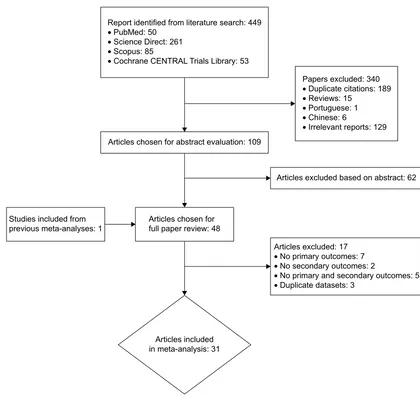
Figure 1 shows the flowchart of the studies from the initial literature search to the final inclusion. Based on the full paper review, 21 controlled randomized trials and ten prospective or retrospective nonrandomized studies were included in the meta-analysis.
Primary outcome measures
Table 1 shows the main preoperative characteristics of the 31 studies describing a total of 5,404 eyes included in the meta-analysis. Studies written in bold (nine studies) have been included in the previously published meta-analysis studies10,11 concerning FS laser for LASIK. Postoperative
characteristics for primary and secondary outcomes are presented in Table 2.
Efficacy
Among the different IntraLase types, the efficacy ranged from 85.1% (IntraLase FS 60 kHz, seven studies included13–19) to 100% (IntraLase FS 1020 and 30 kHz,21
Figure 2). For the most commonly reported IntraLase FS 60 kHz, the average efficacy was 85.1%. In one study, IntraLase FS 60 or 150 kHz22 was used and its efficacy
was 98.3%. For IntraLase 150 kHz,19,23–25 the average
Clinical Ophthalmology downloaded from https://www.dovepress.com/ by 118.70.13.36 on 21-Aug-2020
Dovepress Femtosecond lasers for LASIK
5HSRUWLGHQWLILHGIURPOLWHUDWXUHVHDUFK •3XE0HG
•6FLHQFH'LUHFW •6FRSXV
•&RFKUDQH&(175$/7ULDOV/LEUDU\
$UWLFOHVH[FOXGHG •1RSULPDU\RXWFRPHV •1RVHFRQGDU\RXWFRPHV
•1RSULPDU\DQGVHFRQGDU\RXWFRPHV •'XSOLFDWHGDWDVHWV
3DSHUVH[FOXGHG •'XSOLFDWHFLWDWLRQV •5HYLHZV
•3RUWXJXHVH •&KLQHVH
•,UUHOHYDQWUHSRUWV
$UWLFOHVH[FOXGHGEDVHGRQDEVWUDFW $UWLFOHVFKRVHQIRUDEVWUDFWHYDOXDWLRQ
$UWLFOHVFKRVHQIRU IXOOSDSHUUHYLHZ
$UWLFOHVLQFOXGHG LQPHWDDQDO\VLV 6WXGLHVLQFOXGHGIURP
SUHYLRXVPHWDDQDO\VHV
Figure 1 Flowchart of study selection.
efficacy was 93.7%. For FEMTO LDV,26,27 the average
efficacy was 91.3%. For Visumax18,24,28 and Wavelight
FS200,29,30 the average efficacy rates were 79.1% and
83.6%, respectively.
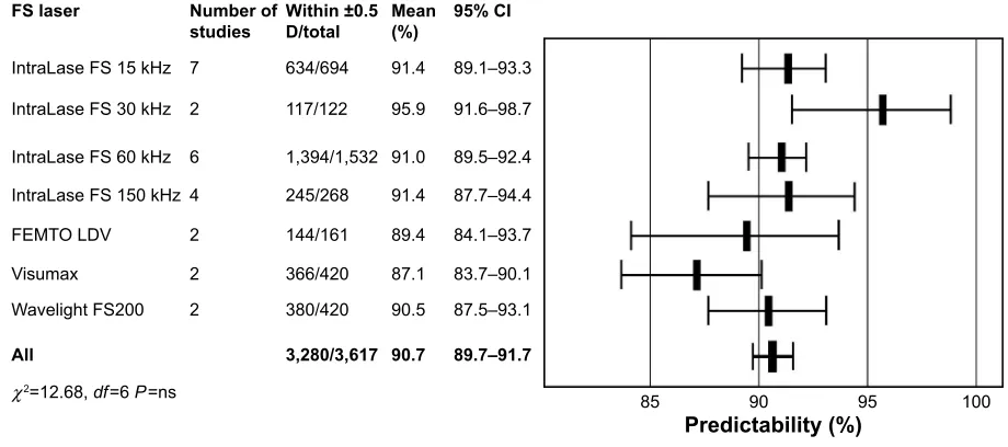
Predictability
The average predictability for the different IntraLase types ranged from 91.0% (IntraLase FS 60 kHz14–19) to 95.9%
(IntraLase FS 30 kHz;21,31 Figure 3). FEMTO LDV26,27 had
the average predictability of 89.4%. For Visumax18,24 and
Wavelight FS200,29,30 the average predictabilities were 87.1%
and 90.5%, respectively.
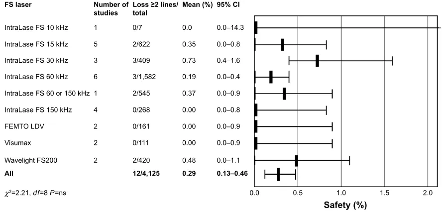
Safety
The average safety percentage was zero or very close to zero for all the studied FS laser types (Figure 4). For IntraLase FS 1532–36 and 30 kHz,21,31,37 the average safety was
0.4% and 0.7%, respectively. For IntraLase FS 60 kHz,13–18 it
was 0.2%. For Wavelight FS 200,29,30 it was 0.5%.
Secondary outcome measures
Flap thickness measurements
The average difference from the target flap thickness for IntraLase FS 3031,37 and 60 kHz13,14,16,38–40 was +0.8
and +0.6 µm, respectively (Figure 5). For IntraLase FS 15 kHz,33,34,41 the average difference from the target flap
thickness was much bigger, +6.7 µm. For FEMTO LDV,26,40,42
the average difference from the target flap thickness was 5.3 µm less than intended. For Visumax,28,40 the difference
from the target was 10.6 µm more than intended. Wavelight FS20029,30 produced corneal flaps that were close to the
intended value (difference from the target flap thickness was +0.03 µm). The average SD for IntraLase FS 15 and 30 kHz was 15.5 and 13.8 µm, respectively. For the most
Clinical Ophthalmology downloaded from https://www.dovepress.com/ by 118.70.13.36 on 21-Aug-2020
Dovepress Huhtala et al
Table 1
Characteristics of the studies included in the meta-analysis of femtosecond lasers for LASIK
References (author, year)
Surgical procedure
Country
Eyes
(n)
Preop mean SE (D)
±
SD
Follow-up (mo)
Design
Jadad et al
12
quality score
Tran et al, 2005
20
IntraLase FS 10
kHz USA 9 − 2.58 3 Randomized 2
Durrie and Stahl, 2004
32
IntraLase FS 15
kHz USA 30 − 4.66 ±
1.73 (Custom Cornea)
1 Randomized 3 30 − 4.83 ± 1.71 (Zyoptix)
Javaloy et al, 2007
33
IntraLase FS 15
kHz Spain 100 − 3.98 ± 1.89 3 Randomized 4
Chan et al, 2008
34
IntraLase FS 15
kHz USA 51 − 3.76 ± 1.41 12 Randomized 3
Durrie and Kezirian, 2005
50
IntraLase FS 15
kHz USA 51 − 3.59 3 Randomized 3
Montes-Mico et al, 2007
21
IntraLase FS 30
kHz Spain 100 − 2.85 ± 1.79 6 Randomized 4
Patel et al, 2007
41
IntraLase FS 15
kHz USA 21 − 4.02 ± 1.61 6 Randomized 3
Munoz et al, 2010
35
IntraLase FS 15
kHz Spain 48 3.98 ± 2.35 (defocus) a 48 Randomized 4
Stonecipher and Kezirian, 2008
36
IntraLase FS 15
kHz
USA
188
– (WF guided)
3
Randomized
1
186
– (WF optimized)
Alio and Pinero, 2008
31
IntraLase FS 30
kHz Spain 22 − 4.11 ± 1.10 3 Randomized 5
Pfaeffl et al, 2008
37
IntraLase FS 30
kHz Germany 287 − 4.1 ± 3.6 Intraop
Prospective interventional case study
1
Rosa et al, 2009
38
IntraLase FS 60
kHz Portugal 20 − 4.48 ± 2.55 3 Prospective cohort 1 20 − 5.60 ± 1.05
Slade et al, 2009
13
IntraLase FS 60
kHz USA 50 − 3.96 6 Randomized 3
Prakash et al, 2010
14
IntraLase FS 60
kHz India 60 − 5.70 1 Randomized 5 60 − 6.09 60 − 5.98 60 − 5.13
Hatch et al, 2011
15
IntraLase FS 60
kHz USA 26 − 4.34 6 Randomized 5
Prakash et al, 2011
16
IntraLase FS 60
kHz India 385 − 6.08 ± 2.7 12 Randomized 5 385 − 5.99 ± 2.8
He et al, 2014
17
IntraLase FS 60
kHz USA 55 − 4.75 ±
2.22 (WF guided)
12 Randomized 3 55 − 4.81 ±
1.95 (WF optimized)
Zhai et al, 2013
39
IntraLase FS 60
kHz
People’s Republic of China
59 − 7.15 ± 2.87 1 Randomized 4
Tanzer et al, 2013
22
IntraLase FS 60 or 150
kHz USA 544 − 2.56 3 Prospective noncomparative two-site study 1 57 − 0.34 30 + 1.86
Yu and Manche, 2015
19
IntraLase FS 60
kHz USA 61 − 4.66 ± 2.30 12 Randomized 3
IntraLase FS 150
kHz 61 − 4.62 ± 2.32
Sales and Manche, 2013
23
IntraLase FS 150
kHz USA 36 − 4.04 ±
1.67 (WF guided)
12 Randomized 3 36 − 3.99 ±
1.71 (WF optimized)
Yu and Manche, 2014
25
IntraLase FS 150
kHz USA 50 − 3.89 ±
1.67 (Allegretto Wave Eye-Q)
12
Prospective comparative case series
3
50
−
4.18
±
1.73 (Visx Star Customvue S4 IR)
Vryghem et al, 2010
26 FEMTO LDV Belgium 111 − 4.91 ± 2.45 6 Prospective, consecutive 1
Zhou et al, 2012
42
FEMTO LDV
People’s Republic of China
360 − 6.58 ± 2.86 1 wk Randomized 2
Rosman et al, 2013
24 Visumax 500 kHz Singapore 45 − 4.94 ± 2.08 3 Randomized 5
IntraLase FS 150
kHz 45 − 4.91 ± 2.09
Ang et al, 2013
18 Visumax 500 kHz Singapore 381 − 5.41 ± 2.22 3
Retrospective case review
1
IntraLase FS 60
kHz 362 − 5.34 ± 2.28
Clinical Ophthalmology downloaded from https://www.dovepress.com/ by 118.70.13.36 on 21-Aug-2020
Dovepress Femtosecond lasers for LASIK
Lim et al, 2013
28 Visumax 200 kHz Korea 36 − 5.25 ± 1.53 6 Randomized 5 36 − 4.89 ± 1.40
Kymionis et al, 2013
29
Wavelight AG/Alcon FS200
Greece 50 − 4.15 ± 1.69 6
Prospective nonrandomized interventional case series
3
Cummings et al, 2013
30 Wavelight FS200 Ireland 378 − 4.03 ± 2.29 3
Retrospective consecutive case series
1 53 + 2.20 ± 1.23
Ahn et al, 2011
40
IntraLase FS 60
kHz Korea 50 K = 42.37 ± 2.0 2
Comparative case series
3 Visumax 40 K = 43.38 ± 1.77 FEMTO LDV 64 K = 43.31 ± 1.48
Arbelaez et al, 2009
27
FEMTO LDV
Sultanate of Oman
50 − 3.08 ± 2.32 (defocus) a 6 Retrospective 1 Notes:
aMean manifest defocus refraction as reported by the authors.
Studies written in bold have been included in the previously published meta-analyses.
10,11
Abbreviations:
D, diopters; LASIK, laser-assisted in situ keratomileusis; SD, standard deviation; SE, refractive spherical equivalent; mo, months; WF, wave front; wk, weeks; preop, preoperative; intraop, intraoperative.
Table 2
Postoperative outcome measures in the studies included in the meta-analysis of femtosecond lasers for LASIK
References (author, year)
Surgical procedure
Primary outcomes
Secondary outcomes
Eyes (n)
Efficacy (%)
Predictability (%)
Safet
y (%)
Eyes (n)
Target
flap
thickness (µm)
Measured flap thickness (
µ m) ± SD (range) Method
Intraop complication (%)
Postop complication (%)
Tran et
al, 2005
20
IntraLase FS 10
kHz 7 100 – 0 8 120 – – 0 37. 5
Durrie and Stahl, 2004
32
IntraLase FS 15
kHz 30 93 83 0 30 110 – – – – 30 90 93 0 30 110 Javaloy et al, 2007 33
IntraLase FS 15
kHz 100 80 a 83 1 20 120 130. 14 ± 1.70 CM 0 17 Chan et al, 2008 34
IntraLase FS 15
kHz 40 98 93 3 51 120 118. 7 ± 12. 2 SUP 0 28 ( , 1 mo)
Durrie and Kezirian, 2005
50
IntraLase FS 15
kHz 51 98 90 – 51 118 – – – – Montes -Mico et al, 2007 21
IntraLase FS 30
kHz 100 100 98 0 100 120 – – – – Pa tel et al, 2007 41
IntraLase FS 15
kHz 21 0.01 ± 0.11 LogMar (20/ 20) 90 0.06 ± 0.09 LogMar (20/15) 21 120 143 ± 16 (110 – 172) CM 0 0
Munoz et al, 2010
35
IntraLase FS 15
kHz 48 85. 4 97. 9 0 48 120 – – 0 0
Stonecipher and Kezirian, 2008
36
IntraLase FS 15
kHz
188
93 (WF guided)
93 0 374 100–130 – – – – 186
93 (WF optimized)
94
0
Alio and Piner
o,
2008
31
IntraLase FS 30
kHz 22 – 86. 4 a 13. 6 a 22 110 115. 95 ± 6.22 VHF 0 0
Pfaeffl et al, 2008
37
IntraLase FS 30
kHz 287 – – 0 287 100 100.4 ± 13.6 (57–138) OCP 0 2.8
Rosa et al, 2009
38
IntraLase FS 60
kHz 20 0.02 ± 0.04 LogMar – – 20 120 143.1 ± 18.4 (107.2 – 172.5) SUP – – 20 0.04 ± 0.03 LogMar 20 120 115. 5 ± 12. 5 (91. 6 – 147. 8) 20 minutes ( Continued )
Clinical Ophthalmology downloaded from https://www.dovepress.com/ by 118.70.13.36 on 21-Aug-2020
Dovepress Huhtala et al
Table 2 ( Continued ) References (author, year) Surgical procedure Primary outcomes Secondary outcomes
Eyes (n)
Efficacy (%)
Predictability (%)
Safety (%)
Eyes (n)
Target flap thickness (µm)
Measured flap thickness (
µ m) ± SD (range) Method
Intraop complication (%)
Postop complication (%)
Slade et al, 2009
13
IntraLase FS 60
kHz 50 92 – 0 25 100 112 ± 5 (87–118) OCT 0 0
Prakash et al, 2010
14
IntraLase FS 60
kHz 60 97 91.6 0 60 90 89.97 ± 4.12 OCT 3.3 1.7 60 80 90 0 60 100 99.96 ± 5.30 5 3.3 60 82 91.6 0 60 110 110.01 ± 4.50 6.7 1.7 60 87 93.3 0 60 120 119.26 ± 4.94 3.3 1.7
Hatch et al, 2011
15
IntraLase FS 60
kHz 25 92 88 0 26 90 – – 11.5 23.1
Prakash et al, 2011
16
IntraLase FS 60
kHz 368 82 95.1 0 368 90 90.1 ± 6.7 (76–102) OCT 0 0 368 81 94.02 0 368 100 100.6 ± 6.9 (85–114) 0 0
He et al, 2014
17
IntraLase FS 60
kHz 54 87 80 0 54 105
– (WF guided)
SUP 0 0 54 78 72 1.9 54 105
– (WF optimized)
Zhai et al, 2013
39
IntraLase FS 60
kHz 59 – – – 59 110 111 ± 3 (104–120) OCT – –
Tanzer et al, 2013
22
IntraLase FS 60
kHz or 150 kHz 497 98.2 – 0.4 544 – – – 0.2 0.2 48 100 – 0 57 – – – 23 b 95.7 b – 0 b 30 – – –
Yu and Manche, 2015
19
IntraLase FS 60
kHz 61 93 95 2 61 105 – – 0 0
IntraLase FS 150
kHz 61 95 90 0 61 105 – – 0 0
Sales and Manche, 2013
23
IntraLase FS 150
kHz 34 97 94 0 34 105
– (WF guided)
SUP – – 34 97 85 0 34 105
– (WF optimized)
Yu and Manche, 2014
25
IntraLase FS 150
kHz 50 97.9 100 0 50 105
– (Allegretto Wave Eye-Q)
SUP – – 50 91.5 92 0 50 105
– (Visx Star Customvue S4 IR)
Vryghem et al, 2010 26 FEMTO LDV 111 94.6 95.5 0 111 110 106.68 ± 12.68 (84–149) SUP 17.1 14.4
Zhou et al, 2012
42 FEMTO LDV 360 – – – 360 110 103.95 ± 6.11 OCT – – Ros
man et al,
2013
24
Visumax 500
kHz
IntraLase FS 150
kHz 39 79.5 89.7 0 45 115 – SUP 13.3 0 39 82.1 84.6 0 45 110 – 2.2 0
Ang et al, 2013
18 Visumax 500 kHz 381 75.5 86.9 – 381 110–120 – – 2.1 0.3
IntraLase FS 60
kHz 362 75.0 87.3 – 362 100–110 – 0.3 0.6
Lim et al, 2013
28 Visumax 200 kHz 36 95.8 100 c 0 36 80 83.46 ± 3.50 (73–95) OCT Only OBL 36 100 97.1 0 36 120 122.93 ± 3.55 (107–131) Only OBL
Kymionis et al, 2013
29
Wavelight AG/Alcon FS200
42 86 86 0 42 105 102.98 ± 6.33 (91–114) OCT 4.8 0
Cummings et al, 2013
30 Wavelight FS200 378 83.4 91.0 0.3 431 120 120.23 ± 13.94 (73–176) SOP 0 0 53 b 46.4 b 71.0 b 7.5 b
Ahn et al, 2011
40
IntraLase FS 60
kHz 50 – – – 50 110 103.3 ± 13.2 OCT – – Visumax 40 – – – 40 110 133.9 ± 13.9 FEMTO LDV 64 – – – 64 110 105.8 ± 8.2 Arbelaez et al, 2009 27 FEMTO LDV 50 84 76 d 0 50 110 – SOP 0 0 Notes: –, not reported; aestimated from a figure; bhyperopic eyes were excluded; crefraction within the ± 0.25 D of target refraction; donly astigmatism correction. Studies written in bold have been included in the previously published meta-analyses. 10,11 Abbreviations: CM, confocal microscopy; LASIK, laser-assisted in situ keratomileusis; OBL, opaque bubble layer; OCP, optical coherence pachymetry; OCT, optical coherence tomography; SD, standard deviation; SOP, subtraction online pachymetry, difference between preoperative corneal thickness and the thickness of corneal bed after flap lifting; SUP, subtraction ultrasound pachymetry; VHF, very-high-frequency digital ultrasound; WF, wave
front; postop, postoperative; intraop, intraoperative; mo, month.
Clinical Ophthalmology downloaded from https://www.dovepress.com/ by 118.70.13.36 on 21-Aug-2020
Dovepress Femtosecond lasers for LASIK
3UHGLFWDELOLW\ )6ODVHU
,QWUD/DVH)6N+]
,QWUD/DVH)6N+]
,QWUD/DVH)6N+] ,QWUD/DVH)6N+]
)(072/'9
9LVXPD[ :DYHOLJKW)6
$OO
χ GI 3 QV
1XPEHURI VWXGLHV
:LWKLQ 'WRWDO
0HDQ
±
±
±
± ±
±
± ±
&,
Figure 3 Predictability, the proportion of eyes within ±0.5 D of target refraction after FS laser for LASIK.
Abbreviations:χ2, chi-square statistic; CI, confidence interval; D, diopters; df, degrees of freedom; FS, femtosecond; LASIK, laser-assisted in situ keratomileusis;
ns, nonsignificant.
(IILFDF\
)6ODVHU
,QWUD/DVH)6N+] ,QWUD/DVH)6N+] ,QWUD/DVH)6N+] ,QWUD/DVH)6N+] ,QWUD/DVH)6RUN+] ,QWUD/DVH)6N+] )(072/'9 9LVXPD[ :DYHOLJKW)6
$OO
χ GI 3
1XPEHURI VWXGLHV
8'9$ WRWDO
0HDQ
± ± ± ± ± ± ± ± ±
± &,
Figure 2 Efficacy, the proportion of eyes within UDVA $20/20 after FS laser for LASIK.
Abbreviations:χ2, chi-square statistic; CI, confidence interval; df, degrees of freedom; FS, femtosecond; LASIK, laser-assisted in situ keratomileusis; UDVA, uncorrected
distance visual acuity.
studied IntraLase FS 60 kHz, the average SD was 12.4 µm. For FEMTO LDV, the reproducibility, the average SD was 8.2 µm. For Visumax, the average SD was 23.3 µm and for Wavelight FS200 14.3 µm.
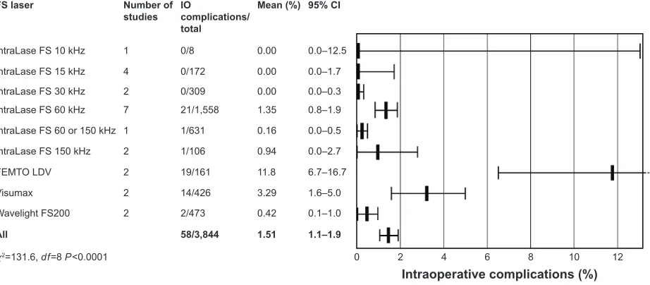
Complication rates
Among the different IntraLase types, IntraLase FS 10 kHz20 had
no intraoperative complications, but postoperative complica-tion rate was 37.5% (one study was included; Figures 6 and 7). IntraLase FS 15 kHz33–35,41 had no intraoperative complications,
but the average postoperative complication rate was 18.0%.
IntraLase FS 30 kHz31,37 had no intraoperative complications,
while the percentage for postoperative complication was 2.6%. IntraLase FS 60 kHz13–19 had the average intraoperative and
postoperative complication rates of 1.4% and 3.2%, respec-tively. IntraLase 150 kHz19,24 had the average intraoperative
complication rate of 0.9% and postoperative complication rate of 0%. FEMTO LDV26,27 had the average intraoperative
complication rate of 11.8%. The postoperative complication rate for FEMTO LDV averaged 9.9%. For Visumax,24,28
intra-operative and postintra-operative complication rates averaged 3.3% and 0.2%, respectively. Wavelight FS20029,30 had the lowest
Clinical Ophthalmology downloaded from https://www.dovepress.com/ by 118.70.13.36 on 21-Aug-2020
Dovepress Huhtala et al
±
)6ODVHU
,QWUD/DVH)6N+]
,QWUD/DVH)6N+]
,QWUD/DVH)6N+]
)(072/'9 9LVXPD[
:DYHOLJKW)6
$OO
) 3 QV
1XPEHU RIVWXGLHV
1XPEHU RIH\HV
±
±
'LIIHUHQFHIURP WDUJHWIODS WKLFNQHVVP
'LIIHUHQFHIURPWDUJHWIODSWKLFNQHVVP
6' P
Figure 5 Corneal flap measurements after FS laser for LASIK.
Abbreviations:F, F factor; FS, femtosecond; LASIK, laser-assisted in situ keratomileusis; ns, nonsignificant; SD, standard deviation.
6DIHW\ )6ODVHU
,QWUD/DVH)6N+]
,QWUD/DVH)6N+]
,QWUD/DVH)6N+]
,QWUD/DVH)6N+]
,QWUD/DVH)6RUN+]
,QWUD/DVH)6N+]
)(072/'9
9LVXPD[
:DYHOLJKW)6 $OO
χ GI 3 QV
1XPEHURI VWXGLHV
/RVVOLQHV WRWDO
0HDQ
±
±
±
±
±
±
±
±
± ± &,
Figure 4 Safety, the proportion of eyes losing two or more Snellen lines of CDVA after FS laser for LASIK.
Abbreviations: CDVA, corrected distance visual acuity; χ2, chi-square statistic; CI, confidence interval; df, degrees of freedom; FS, femtosecond; LASIK, laser-assisted in situ
keratomileusis; ns, nonsignificant.
intraoperative and postoperative complication rates of 0.4% and 0%, respectively.
Discussion
This meta-analysis compared different types of FS lasers for LASIK. In the literature search, we found 109 potentially relevant abstracts for review, but only 47 were suitable for a full paper review. Additionally, we included one study33
from the previously published meta-analyses.10,11 From
these 48 articles, we excluded 17 papers based on lacking or duplicate data, and the final analysis included 31 articles. For primary outcome measures, hyperopic eyes were excluded. No studies were excluded from the analysis due to the low-quality scoring by Jadad et al system.12
The different FS laser systems can be classified into two groups: one group is characterized with high pulse energy–low pulse frequency (such as IntraLase and Femtec) and the other with low pulse energy–high pulse frequency (such as FEMTO LDV and Wavelight FS200).7
In the FS laser technology in the high pulse energy–low pulse frequency group, pulse energies are in the range of 1 µJ and repetition rates on the order of kilohertz.7 The
low pulse energy–high pulse frequency system delivers only pulse energy on the order of nano-joule and uses megahertz repetition rates.7 From all FS lasers on the
market, IntraLase was the first introduced and different IntraLase FS types (10, 15, 30, 60, and 150 kHz) are the most commonly reported.
Clinical Ophthalmology downloaded from https://www.dovepress.com/ by 118.70.13.36 on 21-Aug-2020
Dovepress Femtosecond lasers for LASIK
,QWUDRSHUDWLYHFRPSOLFDWLRQV )6ODVHU
,QWUD/DVH)6N+] ,QWUD/DVH)6N+] ,QWUD/DVH)6N+] ,QWUD/DVH)6N+] ,QWUD/DVH)6RUN+] ,QWUD/DVH)6N+] )(072/'9 9LVXPD[ :DYHOLJKW)6 $OO
χ GI 3
1XPEHURI VWXGLHV
,2
FRPSOLFDWLRQV WRWDO
0HDQ
± ± ± ± ± ± ± ± ± ± &,
Figure 6 Intraoperative complications after FS laser for LASIK.
Abbreviations:χ2, chi-square statistic; CI, confidence interval; df, degrees of freedom; FS, femtosecond; IO, intraoperative; LASIK, laser-assisted in situ keratomileusis.
The pooled primary outcome results showed that there was a statistically significant difference in the proportion of eyes within a UDVA of 20/20 or better (efficacy). Based on efficacy, IntraLase and FEMTO LDV gave the best results. There were also statistically significant differences in the mean spherical equivalent refraction within ±0.5 D of target refraction (predictability). Based on predictability, IntraLase types FS 15, FS 30, and FS 60 kHz were the best. There was no statistically significant difference in the loss of $2 Snellen lines of CDVA (safety) between different FS lasers.
It was difficult to combine the results of randomized con-trolled trials because of different follow-up times. In the 31
studies chosen for this meta-analysis, the follow-up time was #1 month in five studies.14,32,37,39,42 The most commonly reported
follow-up times were 3 months (ten studies) 18,20,22,24,30,31,33,36,38,50
and 6 months (eight studies).13,15,21,26–29,41 The follow-up time
was $1 year only in seven cases.16,17,19,23,25,34,35 Long follow-up
times should be recommended for reporting refractive results, especially in controlled randomized studies. In the meta-analysis, different excimer laser choices used in LASIK made it also more difficult to compare refractive results. Another drawback of this meta-analysis is that due to limited reporting, the results were pooled together from standard, wavefront-guided, and wavefront-optimized treatments, and there was no compensation for this.
3RVWRSHUDWLYHFRPSOLFDWLRQV
)6ODVHU
,QWUD/DVH)6N+] ,QWUD/DVH)6N+] ,QWUD/DVH)6N+] ,QWUD/DVH)6N+] ,QWUD/DVH)6RUN+] ,QWUD/DVH)6N+
)(072/'9 9LVXPD[ :DYHOLJKW)6
$OO
χ GI 3
1XPEHURI VWXGLHV
32
FRPSOLFDWLRQV WRWDO
0HDQ
± ± ± ± ± ±
± ± ±
± &,
Figure 7 Postoperative complications after FS laser for LASIK.
Abbreviations:χ2, chi-square statistic; CI, confidence interval; df, degrees of freedom; FS, femtosecond; LASIK, laser-assisted in situ keratomileusis; PO, postoperative.
Clinical Ophthalmology downloaded from https://www.dovepress.com/ by 118.70.13.36 on 21-Aug-2020
Dovepress Huhtala et al
Mechanical microkeratomes typically create meniscus-shaped flaps that are thinner in the center and thicker in the periphery, whereas FS laser flaps have been found to be typically more uniformly planar.43–47 Preliminary studies
with the IntraLase FS laser have demonstrated that free flaps, irregular flaps, microperforations, decentered flaps, epithelial defects, and abrasions were significantly reduced or eliminated.48–50 The SD of achieved flap thickness with
FS lasers has also been found to be narrower than with mechanical systems.4,46
Although in this meta-analysis flap thickness measure-ments and complication rates were classified as secondary outcomes from the surgeons’ point of view, they are in fact the areas of major concern in LASIK. In the meta-analysis, the pooled secondary outcome results showed some varia-tions between different FS lasers. Based on the SD of the measured flap thicknesses, FEMTO LDV reproduced the most accurate flap thicknesses.
Certain complications have been shown to be unique to the FS laser, such as transient opaque bubble layer (OBL),51–55
especially with the IntraLase, transient light sensitivity syndrome,56,57 increased corneal backscatter,41 and rainbow
glare.58–60 The incidence of transient OBL, transient light
sen-sitivity syndrome, and rainbow glare has reduced with lower energies used. In this meta-analysis, IntraLase and Wavelight SF200 had the fewest intraoperative complications. IntraLase FS 60 kHz, Visumax, and Wavelight FS200 had the most seldom postoperative complications. In the meta-analysis, it was difficult to compare the complications based on the percentage of complications reported in the studies. In general, there were very few intraoperative complications reported. The most common intraoperative complications were a loss of suction, OBL, and adhesions. The most frequently reported postoperative complications were diffuse lamellar keratitis and microstriae. No ectasia was found in these studies. How-ever, there seemed to be big differences between authors in reporting complications in LASIK. For instance, in the first FEMTO LDV study that reported the results of a preproduc-tion FS laser system, its intraoperative complicapreproduc-tion rate was 17.1% and postoperative complication rate was 14.4%.26
Furthermore, there was a mild epithelial sloughing in 11.8% of the eyes. Yet, in another FEMTO LDV study, there were no complications.27 Therefore, we suggest a standardized system
for reporting complications in refractive surgery. Intraopera-tive side effects that do not have any effects on the refracIntraopera-tive outcome should also be reported, such as OBL, decentered, incomplete or free flaps, and flap adhesion. Bleeding from the limbal vessels should also be reported.
Conclusion
In conclusion, there were dissimilarities between different FS lasers based on efficacy and intraoperative and postoperative complications. All FS lasers were predictable and safe for making corneal flaps in LASIK.
Acknowledgment
Dr Matti Kataja is acknowledged for his statistical and graphical expertise.
Disclosure
Dr Pietilä has financial interest in the Ziemer Ophthalmic Systems. The other authors report no conflicts of interest in this work.
References
1. Sugar A, Rapuano CJ, Culbertson WW, et al. Laser in situ keratomileu-sis for myopia and astigmatism: safety and efficacy – a report by the American academy of ophthalmology. Ophthalmology. 2002;109(1): 175–187.
2. Varley GA, Huang D, Rapuano CJ, Schallhorn S, Wachler BSB, Sugar A. LASIK for hyperopia, hyperopic astigmatism, and mixed astigmatism – a report by the American Academy of Ophthalmology.
Ophthalmology. 2004;111(8):1604–1617.
3. Stonecipher K, Ignacio TS, Stonecipher M. Advances in refractive surgery: microkeratome and femtosecond laser flap creation in relation to safety, efficacy, predictability, and biomechanical stability. Curr Opin Ophthalmol. 2006;17(4):368–372.
4. Pietilä J. Flap Creation in Laser-Assisted in Situ Keratomileusis: From Microkeratome to Femtosecond Laser. Tampere: Tampere University Press; 2012.
5. Kurtz RM, Liu X, Elner VM, Squier JA, Du D, Mourou GA. Photodis-ruption in the human cornea as a function of laser pulse width. J Refract Surg. 1997;13(7):653–658.
6. Sugar A. Ultrafast (femtosecond) laser refractive surgery. Curr Opin Ophthalmol. 2002;13(4):246–249.
7. Lubatschowski H. Overview of commercially available femtosecond lasers in refractive surgery. J Refract Surg. 2008;24(1):S102–S107. 8. Farjo AA, Sugar A, Schallhorn SC, et al. Femtosecond lasers for LASIK
flap creation: a report by the American Academy of Ophthalmology.
Ophthalmology. 2013;120(3):e5–e20.
9. Slade SG. The use of the femtosecond laser in the customization of corneal flaps in laser in situ keratomileusis. Curr Opin Ophthalmol. 2007;18(4):314–317.
10. Zhang ZH, Jin HY, Suo Y, et al. Femtosecond laser versus mechanical microkeratome laser in situ keratomileusis for myopia: metaanaly-sis of randomized controlled trials. J Cataract Refract Surg. 2011; 37(12):2151–2159.
11. Chen S, Feng Y, Stojanovic A, Jankov MR, Wang Q. IntraLase femto-second laser vs mechanical microkeratomes in LASIK for myopia: a sys-tematic review and meta-analysis. J Refract Surg. 2012;28(1):15–24. 12. Jadad AR, Moore RA, Carroll D, et al. Assessing the quality of reports
of randomized clinical trials: is blinding necessary? Control Clin Trials. 1996;17(1):1–12.
13. Slade SG, Durrie DS, Binder PS. A prospective, contralateral eye study comparing thin-flap LASIK (sub-Bowman keratomileusis) with photo-refractive keratectomy. Ophthalmology. 2009;116(6):1075–1082. 14. Prakash G, Agarwal A, Yadav A, et al. A prospective randomized
comparison of four femtosecond LASIK flap thicknesses. J Refract Surg. 2010;26(6):392–402.
Clinical Ophthalmology downloaded from https://www.dovepress.com/ by 118.70.13.36 on 21-Aug-2020
Dovepress Femtosecond lasers for LASIK
15. Hatch BB, Moshirfar M, Ollerton AJ, Sikder S, Mifflin MD. A prospec-tive, contralateral comparison of photorefractive keratectomy (PRK) versus thin-flap LASIK: assessment of visual function. Clin Ophthalmol. 2011;5:451–457.
16. Prakash G, Agarwal A, Kumar DA, et al. Femtosecond sub-bowman keratomileusis: a prospective, long-term, intereye comparison of safety and outcomes of 90- versus 100-µm flaps. Am J Ophthalmol. 2011; 154(4):582–590.
17. He L, Liu A, Manche EE. Wavefront-guided versus wavefront-optimized laser in situ keratomileusis for patients with myopia: a prospective Randomized Contralateral Eye Study. Am J Ophthalmol. 2014;157(6):1170–1178.
18. Ang M, Mehta JS, Rosman M, et al. Visual outcomes comparison of 2 femtosecond laser platforms for laser in situ keratomileusis. J Cataract Refract Surg. 2013;39(11):1647–1652.
19. Yu CQ, Manche EE. Comparison of 2 femtosecond lasers for flap cre-ation in myopic laser in situ keratomileusis: one-year results. J Cataract Refract Surg. 2015;41(4):740–748.
20. Tran DB, Sarayba MA, Bor Z, et al. Randomized prospective clinical study comparing induced aberrations with IntraLase and Hansatome flap creation in fellow eyes: potential impact on wavefront-guided laser in situ keratomileusis. J Cataract Refract Surg. 2005;31(1):97–105. 21. Montes-Mico R, Rodriguez-Galietero A, Alio JL. Femtosecond laser
versus mechanical keratome LASIK for myopia. Ophthalmology. 2007;114(1):62–68.
22. Tanzer DJ, Brunstetter T, Zeber R, et al. Laser in situ keratomileusis in United States Naval aviators. J Cataract Refract Surg. 2013;39(7): 1047–1058.
23. Sales CS, Manche EE. One-year outcomes from a prospective, ran-domized, eye-to-eye comparison of wavefront-guided and wavefront- optimized LASIK in myopes. Ophthalmology. 2013;120(12): 2396–2402.
24. Rosman M, Hall RC, Chan C, et al. Comparison of efficacy and safety of laser in situ keratomileusis using 2 femtosecond laser plat-forms in contralateral eyes. J Cataract Refract Surg. 2013;39(7): 1066–1073.
25. Yu CQ, Manche EE. Comparison of 2 wavefront-guided excimer lasers for myopic laser in situ keratomileusis: one-year results. J Cataract Refract Surg. 2014;40(3):412–422.
26. Vryghem JC, Devogelaere T, Stodulka P. Efficacy, safety, and flap dimensions of a new femtosecond laser for laser in situ keratomileusis.
J Cataract Refract Surg. 2010;36(3):442–448.
27. Arbelaez MC, Vidal C, Arba-Mosquera S. Excimer laser correc-tion of moderate to high astigmatism with a non-wavefront-guided aberration-free ablation profile: six-month results. J Cataract Refract Surg. 2009;35(10):1789–1798.
28. Lim DH, Keum JE, Ju WK, Lee JH, Chung TY, Chung ES. Pro-spective contralateral eye study to compare 80- and 120-mum flap LASIK using the VisuMax femtosecond laser. J Refract Surg. 2013; 29(7):462–468.
29. Kymionis GD, Kontadakis GA, Grentzelos MA, et al. Thin-flap laser in situ keratomileusis with femtosecond-laser technology. J Cataract Refract Surg. 2013;39(9):1366–1371.
30. Cummings AB, Cummings BK, Kelly GE. Predictability of corneal flap thickness in laser in situ keratomileusis using a 200 kHz femtosecond laser. J Cataract Refract Surg. 2013;39(3):378–385.
31. Alio JL, Pinero DP. Very high-frequency digital ultrasound measure-ment of the LASIK flap thickness profile using the IntraLase femto-second laser and M2 and Carriazo-Pendular microkeratomes. J Refract Surg. 2008;24(1):12–23.
32. Durrie DS, Stahl J. Randomized comparison of custom laser in situ keratomileusis with the Alcon Custom Cornea and the Bausch & Lomb Zyoptix systems: one-month results. J Refract Surg. 2004;20(5): S614–S618.
33. Javaloy J, Vidal MT, Abdelrahman AM, Artola A, Alio JL. Confocal microscopy comparison of intralase femtosecond laser and Moria M2 microkeratome in LASIK. J Refract Surg. 2007;23(2):178–187.
34. Chan A, Ou J, Manche EE. Comparison of the femtosecond laser and mechanical keratome for laser in situ keratomileusis. Arch Ophthalmol. 2008;126(11):1484–1490.
35. Munoz G, Albarran-Diego C, Ferrer-Blasco T, Garcia-Lazaro S, Cervino- Exposito A. Long-term comparison of corneal aberration changes after laser in situ keratomileusis: mechanical microkeratome versus femtosecond laser flap creation. J Cataract Refract Surg. 2010;36(11): 1934–1944.
36. Stonecipher KG, Kezirian GM. Wavefront-optimized versus wavefront-guided LASIK for myopic astigmatism with the ALLEGRETTO WAVE: three-month results of a prospective FDA trial. J Refract Surg. 2008;24(4):S424–S430.
37. Pfaeffl WA, Kunze M, Zenk U, Pfaeffl MB, Schuster T, Lohmann C. Predictive factors of femtosecond laser flap thickness measured by online optical coherence pachymetry subtraction in sub-Bowman keratomileusis. J Cataract Refract Surg. 2008;34(11):1872–1880. 38. Rosa AM, Neto MJ, Quadrado MJ, et al. Femtosecond laser
versus mechanical microkeratomes for flap creation in laser in situ keratomileusis and effect of postoperative measurement interval on estimated femtosecond flap thickness. J Cataract Refract Surg. 2009; 35(5):833–838.
39. Zhai CB, Tian L, Zhou YH, Zhang QW, Zhang J. Comparison of the flaps made by femtosecond laser and automated keratomes for sub-bowman keratomileusis. Chin Med J (Engl). 2013;126(13):2440–2444. 40. Ahn H, Kim JK, Kim CK, et al. Comparison of laser in situ
ker-atomileusis flaps created by 3 femtosecond lasers and a microkeratome.
J Cataract Refract Surg. 2011;37(2):349–357.
41. Patel SV, Maguire LJ, McLaren JW, Hodge DO, Bourne WM. Femto-second laser versus mechanical microkeratome for LASIK: a random-ized controlled study. Ophthalmology. 2007;114(8):1482–1490. 42. Zhou Y, Zhang J, Tian L, Zhai C. Comparison of the Ziemer FEMTO
LDV femtosecond laser and Moria M2 mechanical microkeratome.
J Refract Surg. 2012;28(3):189–194.
43. Holzer MP, Rabsilber TM, Auffarth GU. Femtosecond laser-assisted corneal flap cuts: morphology, accuracy, and histopathology. Invest Ophthalmol Vis Sci. 2006;47(7):2828–2831.
44. Krueger RR, Dupps WJ Jr. Biomechanical effects of femtosecond and microkeratome-based flap creation: prospective contralateral examina-tion of two patients. J Refract Surg. 2007;23(8):800–807.
45. von Jagow JB, Kohnen T. Corneal architecture of femtosecond laser and microkeratome flaps imaged by anterior segment optical coherence tomography. J Cataract Refract Surg. 2009;35(1):35–41.
46. Vaddavalli PK, Yoo SH. Femtosecond laser in-situ keratomileusis flap configurations. Curr Opin Ophthalmol. 2011;22(4):245–250. 47. Srivannaboon S, Sunlakaviset P, Kosrirukvongs P, et al. Refractive
outcomes of femtosecond LASIK for myopic correction at Siriraj Hospital, Thailand. J Med Assoc Thai. 2012;95(suppl 4):S18–S23. 48. Kezirian GM, Stonecipher KG. Comparison of the IntraLase
femto-second laser and mechanical keratomes for laser in situ keratomileusis.
J Cataract Refract Surg. 2004;30(4):804–811.
49. Binder PS. Flap dimensions created with the IntraLase FS laser.
J Cataract Refract Surg. 2004;30(1):26–32.
50. Durrie DS, Kezirian GM. Femtosecond laser versus mechanical keratome flaps in wavefront-guided laser in situ keratomileusis: prospective con-tralateral eye study. J Cataract Refract Surg. 2005;31(1):120–126. 51. Lifshitz T, Levy J, Klemperer I, Levinger S. Anterior chamber gas
bubbles after corneal flap creation with a femtosecond laser. J Cataract Refract Surg. 2005;31(11):2227–2229.
52. Srinivasan S, Rootman DS. Anterior chamber gas bubble formation during femtosecond laser flap creation for LASIK. J Refract Surg. 2007;23(8):828–830.
53. Srinivasan S, Herzig S. Sub-epithelial gas breakthrough during femtosecond laser flap creation for LASIK. Br J Ophthalmol. 2007; 91(10):1373.
54. Kaiserman I, Maresky HS, Bahar I, Rootman DS. Incidence, possible risk factors, and potential effects of an opaque bubble layer created by a femtosecond laser. J Cataract Refract Surg. 2008;34(3):417–423.
Clinical Ophthalmology downloaded from https://www.dovepress.com/ by 118.70.13.36 on 21-Aug-2020
Clinical Ophthalmology
Publish your work in this journal
Submit your manuscript here: http://www.dovepress.com/clinical-ophthalmology-journal
Clinical Ophthalmology is an international, peer-reviewed journal covering all subspecialties within ophthalmology. Key topics include: Optometry; Visual science; Pharmacology and drug therapy in eye diseases; Basic Sciences; Primary and Secondary eye care; Patient Safety and Quality of Care Improvements. This journal is indexed on
PubMed Central and CAS, and is the official journal of The Society of Clinical Ophthalmology (SCO). The manuscript management system is completely online and includes a very quick and fair peer-review system, which is all easy to use. Visit http://www.dovepress.com/ testimonials.php to read real quotes from published authors.
Dovepress
Dove
press
Huhtala et al
55. Kanellopoulos AJ, Asimellis G. Digital analysis of flap parameter accuracy and objective assessment of opaque bubble layer in femtosec-ond laser-assisted LASIK: a novel technique. Clin Ophthalmol. 2013; 7:343–351.
56. Stonecipher KG, Dishler JG, Ignacio TS, Binder PS. Transient light sensitivity after femtosecond laser flap creation: clinical findings and management. J Cataract Refract Surg. 2006;32(1):91–94.
57. Munoz G, Albarran-Diego C, Sakla HF, Javaloy J, Alio JL. Transient light-sensitivity syndrome after laser in situ keratomileusis with the femtosecond laser incidence and prevention. J Cataract Refract Surg. 2006;32(12):2075–2079.
58. Krueger RR, Thornton IL, Xu M, Bor Z, van den Berg TJ. Rainbow glare as an optical side effect of IntraLASIK. Ophthalmology. 2008;115(7): 1187–1195.
59. Bamba S, Rocha KM, Ramos-Esteban JC, Krueger RR. Incidence of rainbow glare after laser in situ keratomileusis flap creation with a 60 kHz femtosecond laser. J Cataract Refract Surg. 2009;35(6):1082–1086. 60. Gatinel D, Saad A, Guilbert E, Rouger H. Unilateral rainbow glare
after uncomplicated femto-LASIK using the FS-200 femtosecond laser.
J Refract Surg. 2013;29(7):498–501.
Clinical Ophthalmology downloaded from https://www.dovepress.com/ by 118.70.13.36 on 21-Aug-2020