| INVESTIGATION
Analysis of Repeat Induced Point (RIP) Mutations in
Leptosphaeria maculans
Indicates Variability in the RIP
Process Between Fungal Species
Angela P. Van de Wouw,1Candace E. Elliott, Kerryn M. Popa, and Alexander Idnurm
School of BioSciences, The University of Melbourne, Parkville, Victoria 3010, Australia ORCID IDs: 0000-0001-5147-0393 (A.P.V.d-W.); 0000-0002-9174-0244 (C.E.E.); 0000-0002-7156-3201 (K.M.P.); 0000-0001-5995-7040 (A.I.)
ABSTRACT Gene duplication contributes to evolutionary potential, yet many duplications in a genome arise from the activity of “selfish”genetic elements such as transposable elements. Fungi have a number of mechanisms by which they limit the expansion of transposons, including Repeat Induced Point mutation (RIP). RIP has been best characterized in the SordariomyceteNeurospora crassa, wherein duplicated DNA regions are recognized after cell fusion, but before nuclear fusion during the sexual cycle, and then mutated. While“signatures”of RIP appear in the genome sequences of many fungi, the species most distant fromN. crassain which the process has been experimentally demonstrated to occur is the DothideomyceteLeptosphaeria maculans. In the current study, we show that similar toN. crassa, nonlinked duplications can trigger RIP; however, the frequency of the generated RIP mutations is extremely low in L maculans (,0.1%) and requires a large duplication to initiate RIP, and that multiple premeiotic mitoses are involved in the RIP process. However, a single sexual cycle leads to the generation of progeny with unique haplotypes, despite progeny pairs being generated from mitosis. We hypothesize that these different haplotypes may be the result of the deamination process occurring post karyogamy, leading to unique mutations within each of the progeny pairs. Thesefindings indicate that the RIP process, while common to many fungi, differs between fungi and that this impacts on the fate of duplicated DNA.
KEYWORDSascomycete; genome defense; RIP timing; repetitive DNA
O
NE of the major mechanisms for gene and genome evolution involves DNA duplication, which allows the divergence in sequences between a pair of duplicated genes, potentially giving rise to genes with new functions (Ohno 1970; Zhang 2003). However, duplications within the ge-nome can arise from the activity of mobile elements, such as transposable elements, with deleterious effects. Some fungi have evolved a genome defense mechanism, Repeat Induced Point mutation (RIP), which is predicted to protect against multicopy DNA elements such as transposons through the generation of mutations leading to gene inacti-vation (Kinseyet al.1994). This process wasfirst identified and is best characterized in the model filamentous fungus,Neurospora crassa (Selker et al. 1987; Selker and Garrett
1988; Selker 1990). RIP generates G to A and C to T transi-tions, which often lead to the introduction of stop codons within genes. It also leads to changes in methylation patterns of the DNA in N. crassa, effectively further inhibiting the expansion of multicopy genes by preventing the expression of any remnant coding regions (Singeret al.1995b).
RIP occurs during crossing, and affects DNA that can in-clude genes or noncoding regions that are duplicated in the genome. In N. crassa, the duplicated DNA is mutated only during the sexual cycle, and analysis of octads,i.e., a set of eight ascospores derived from a meiosis and one round of mitosis enclosed in an ascus, indicates that progeny emerge carrying no more than two different haplotypes of mutations (Watterset al.1999). The observation of two rather than four progeny types indicates that the mutation event inN. crassa
occurs after cell fusion but before nuclear fusion (Selkeret al.
1987; Watterset al.1999), hence the original name of“ re-arrangement induced premeiotically”for RIP. In N. crassa, RIP acts on duplications that are .400 bp in length when
Copyright © 2019 by the Genetics Society of America doi:https://doi.org/10.1534/genetics.118.301712
Manuscript received July 2, 2018; accepted for publication October 24, 2018; published Early Online November 2, 2018.
Supplemental material available at Figshare: https://doi.org/10.25386/genetics. 7221974.
in tandem or.1 kb when duplications are unlinked (Selker and Garrett 1988; Watterset al.1999). When the repeats are unlinked, a proportion of the sexual spores still show alter-ations in the unlinked duplicalter-ations, although the frequency of RIP in the unlinked duplications can vary considerably (Selker 2002). Since RIP inactivates both copies of the dupli-cated genes, although at varying frequencies, some re-searchers used RIP as a strategy for mutating genes prior to the development of highly efficient methods for gene disrup-tion (Selker 2002; Ninomiyaet al.2004).
Although inN. crassaRIP acts against the deleterious ef-fects of multicopy DNA such as transposable elements (Kinseyet al.1994), it appears also to have minimized the evolution of gene families, as genes with.80% identity un-dergo RIP (Galagan and Selker 2004). Therefore, RIP has been proposed to also impede genome evolution because it restricts the potential for new gene evolution from duplica-tions (Galagan and Selker 2004).
The RIP process has been experimentally shown to occur in only a few fungal species. These include ascomycetes
Fusarium graminearum, Nectria hematococca, Podospora
anserina,Magnaporthe oryzae, and Leptosphaeria maculans
(Graïa et al.2001; Ikeda et al.2002; Idnurm and Howlett 2003; Cuomo et al.2007; Colemanet al.2009; Pomraning
et al. 2013). Although not shown experimentally, the
hallmarks of RIP detected as RIP signatures have been re-ported in numerous fungi based on analyses of transpos-able elements or whole genomes (Clutterbuck 2011). Fungi with these signatures include other ascomycetes such as
Aspergillusspecies,F. oxysporum, andCochliobolus
heterostro-phus, and basidiomycete species likeRhizoctonia solaniand members of the Pucciniomycotina subphylum (Clutterbuck 2011; Horns et al. 2012; Hane et al. 2014; Santana et al.
2014). However, although RIP signatures have been detected in fungi this does not automatically imply that it occurs,e.g.,
inFusarium species, RIP signatures have been detected
al-though these species are generally considered to be predom-inatly asexual in reproduction (Waalwijket al.2017).
As previously mentioned, RIP has been experimentally demonstrated to occur inL. maculans, where tandem inser-tions of plasmid DNA were mutated during crossing (Idnurm and Howlett 2003). L. maculans is a plant pathogen that infects oilseed Brassicas worldwide (Fittet al.2006). In some countries, such as Australia, the life cycle includes a sapro-phytic stage of growth on the residual stubble fromBrassica
napuscrops, during which the two mating types of this
het-erothallic fungus initiate the sexual cycle (Van De Wouw
et al.2016). In Australia, the start of the autumn rain triggers
the release of ascospores that infect the newly planted crops. The sexual cycle produces ascospores within asci, that in laboratory crosses can be analyzed as octads; a set of eight progeny arising from meiosis and one round of mitosis, resulting in four sets of identical pairs (Gall et al. 1994).
L. maculans, although less experimentally tractable than
N. crassa, is a useful fungus in which to investigate RIP as it
can be crossed in vitro, can be transformed, and has
genome-sequencing resources available. Furthermore, RIP is highly relevant to the evolution of pathogenicity of isolates within field populations ofL. maculans. Analysis of the ge-nome of this fungus revealed that many genes encoding known or putative avirulence and effector proteins, involved in disease, are embedded within or near highly repetitive DNA elements that appear to have been subject to RIP (Rouxelet al.2011). In addition to RIP acting on multicopy regions inL. maculans, RIP can“leak”into nearby single-copy sequences, thereby mutating these avirulence genes (Fudal
et al. 2009; Van de Wouw et al. 2010) and subsequently
conferring on these isolates the ability to cause disease on formerly resistant cultivars of canola.
In this paper, we report that the RIP process inL. maculans
has a number of differences to those characterized in
N. crassa and other Sordariomycete species. This provides
new insights into how RIP may not be as restrictive a force in fungal evolution as previously thought.
Materials and Methods
Isolates
L. maculansisolates are listed in Table 1. Wild-type isolates
IBCN18, Lm691, D3, and D9 were transformed with con-structs generated to analyze RIP patterns in progeny of crosses. Details of the constructs and fungal transformations are described in the following sections. The resulting trans-formants were used as parents in crosses (Table 2). All iso-lates were cultured on 10% Campbell’s V8 juice agar.
In vitro crossing of L. maculans
The mating-type genotype of each isolate was determined by PCR as described in Cozijnsen and Howlett (2003) (primers listed in Supplemental Material, Table S1). Isolates of oppo-site mating type were set up for crossing as previously de-scribed (Cozijnsen et al. 2000). Briefly, agar plugs of each parent were placed 5-mm apart on mating media (20% V8 juice, 2% agar, and 0.2% CaCO3) and allowed to grow under 12-hr dark/light cycles for 7 days. After 7 days, plates were overlayed with 1% Difco (Detroit, MI) water agar. Plates were then placed at 14°, with 12-hr dark/blue–black UV light cycles for 4–6 weeks. After this time, plates were examined under a dissecting microscope for pseudothecia (sexual fruit-ing bodies). Usfruit-ing a scalpel blade, pseudothecia were re-moved from the plate and placed in a drop of sterile water to allow asci to be ejected. Octads were then dissected by placing an individual ascus on 2% water agar in a droplet of sterile water. A glass coverslip was placed over the ascus and then, while visualizing under the dissecting microscope, pressure was gently applied to the coverslip to release the eight individual ascospores from the ascus. Each individual ascospore was then placed on a separate agar plate and allowed to germinate.
maintained. Hence, each of the resulting progeny from the octad was analyzed with molecular markers or by Southern blot analysis (see details below) to identify the progeny pairs from within a single octad. Specifically, primers were used to amplify between four and six independently segregating genes in the progeny whereby the parental isolates have different alleles. Pairs of progeny with the same genotype across each of the genes were determined as progeny pairs. The genes amplified were; the MAT locus, which has two
alternative forms, MAT1-1 or MAT1-2, which are detected by a size polymorphism (Cozijnsen and Howlett 2003);
AvrLm1, which is a presence/absence polymorphism
de-tected by PCR (Goutet al.2006);AvrLm4, which is a single-base pair change detected by PCR and then digest with restriction enzyme HaeIII (Van de Wouw and Howlett 2012);AvrLm6, which is a presence/absence polymorphism detected by PCR (Fudalet al.2007);AvrLm5, which is a single-base pair change detectable by PCR and then digest with
Table 1 L. maculansisolates used in this study
Isolate Mating type Purpose Relevant genotype information Reference
IBCN18 MAT1-2 Transformation ContainsAvrLm1andAvrLm4 Marcroftet al.(2012)
Lm691 MAT1-1 Transformation and crosses ContainsAvrLm1andAvrLm4 Haydenet al.(2007) D9 MAT1-1 Transformation and crosses Marcroftet al.(2012) D3 MAT1-1 Transformation and crosses Marcroftet al.(2012) D13 MAT1-2 Crosses ContainsAvrLm4 Marcroftet al.(2012) IBCN18+AvrLm1 MAT1-2 Crosses Isolate IBCN18 transformed withAvrLm1
resulting in nontandem duplication of AvrLm1
This study
IBCN18+AvrLm4#8 MAT1-2 Crosses Isolate IBCN18 transformed withAvrLm4 resulting in tandem duplication of AvrLm4
This study
IBCN18+AvrLm4#9 MAT1-1 Crosses Isolate Lm691 transformed withAvrLm4 resulting in nontandem duplication of AvrLm4
This study
IBCN18+Lema006030 MAT1-2 Crosses Isolate IBCN18 with an additional copy of Lema006030fused to a marker confer-ring nourseothricin resistance, resulting in a nontandem duplication of the Lema006030gene.
This study
D3-IpR+hos1 MAT1-1 Crosses Isolate was isolated as resistant to ipro-dione afterin vitroselection and then transformed with wild-typehos1and a G418 selectable marker
Idnurmet al.(2017)
D9+double-hph#4 MAT1-1 Crosses Isolate D9 transformed with a double-hph construct resulting in a nontandem in-sertion.
This study
D9+double-hph#7 MAT1-1 Crosses Isolate D9 transformed with a double-hph construct resulting in a nontandem in-sertion.
This study
IBCN18+double-hph#8 MAT1-2 Crosses Isolate IBCN18 transformed with a dou-ble-hphconstruct resulting in a nontan-dem insertion.
This study
IBCN18+double-hph#9 MAT1-2 Crosses Isolate IBCN18 transformed with a dou-ble-hphconstruct resulting in a nontan-dem insertion.
This study
D3+double-hph#2 MAT1-1 Crosses Isolate D3 transformed with a double-hph construct resulting in a nontandem in-sertion.
This study
D3+double-hph#5 MAT1-1 Crosses Isolate D3 transformed with a double-hph construct resulting in a nontandem in-sertion.
This study
691+NPS10 MAT1-1 Crosses Isolate 691 transformed with NPS10
hair-pin construct.
This study
sirP MAT1-2 Crosses sirPgene-disruption strain Gardineret al.(2004) LopP MAT1-2 Crosses Loss-of-pathogenicity mutant with tandem
insertion of plasmid pUCATPH (hph gene for hygromycin resistance)
Idnurm and Howlett (2003)
LopC MAT1-2 in vitropassaging Loss-of-pathogenicity mutant with tandem insertion of plasmid pUCATPH (hph gene for hygromycin resistance)
restriction enzymeAvaII (Van de Wouwet al.2014b, 2018); and primers specific for the selectable marker of the con-struct,i.e., the hygromycin B phosphotransferase (hph) gene (selectable marker). All primers and markers were previously published, and are listed in Table S1.
Details of all crosses are in Table 2. The nomenclature used for the progeny is a cross number, followed by a letter to indicate an octad, followed by a number 1–8 for each of the members of the octad. For example, 22A1 refers to progeny one from octad A of cross number 22, while 22B1 refers to progeny one from a different octad, but still from cross num-ber 22.
Construct development and fungal transformations
To test whether RIP could occur in nontandem duplicated sequences, two constructs that had previously been developed were used to transform wild-type isolates (Van de Wouwet al.
2014a,c). These constructs, pPZPHyg_AvrLm1 containing
AvrLm1 and pPZPHyg_AvrLm4 containing AvrLm4, were
transformed into isolate IBCN18 or Lm691, which already contains wild-type copies of these genes. Agrobacterium -mediated transformation and selection with hygromycin was carried out as previously described (Gardiner and Howlett 2004). The resulting transformants, IBCN18+AvrLm1, IBCN18+AvrLm4, and 691+AvrLm4isolates, contained du-plicate copies of theAvrLm1orAvrLm4genes (Figure S1A). The IBCN18+AvrLm1 and IBCN18+AvrLm4 isolates were crossed to Lm691, while 691+AvrLm4was crossed to isolate D13. Octad progeny, defined as the individual progeny col-lected from a single ascus, were colcol-lected and screened for the presence of RIP (further details below).
To trigger RIP by using a large fragment of DNA, two different constructs were generated, and used for transfor-mation and crossing. For the first, a plasmid containing a 4596-bp fragment of theLema006030gene encoding a puta-tive transcription factor was generated. The gene was am-plified with primers KCP014 and KCP015, and cloned
downstream of theL. maculansactin promoter in the pPZPNat vector. The resultant plasmid was transformed into isolate IBCN18 to create transformant IBCN18+Lema006030 (Table 2). This transformant was then crossed with isolate D9, and progeny were collected and analyzed.
A second construct was generated previously, and harbors the wild-type copy of the hos1 gene (encodes a histidine kinase) and the selectable marker for G418 resistance (Idnurm et al.2017). This construct was transformed into isolate D3-IpR, which contains a spontaneous mutation in thehos1gene that confers resistance to the fungicide ipro-dione (Idnurmet al.2017). As a consequence of this trans-formation, the resulting D3-IpR + hos1 isolate contains a repeat region of 5736 bp (two copies of thehos1gene and surrounds). The construct was sequenced within isolate D3-IpR + hos1 and shown to contain no truncations of thehos1gene from the transfer DNA (T-DNA) transforma-tion (data not shown). This transformant was crossed to D13, and the resulting 19 progeny were screened for both G418 resistance and resistance to iprodione. For a progeny to be resistant to both these chemicals, the progeny must harbor the complementation construct but have undergone RIP to inactivate the construct copy of the hos1 gene. A single progeny, 63R15, was detected with this phenotype, and the hos1gene was amplified and sequenced to deter-mine whether mutations were present. Although the inser-tion site was not specifically determined for this construct, because all phenotypic categories were detected in the progeny of the cross, it shows that the construct was inde-pendently segregating and cannot be genetically linked, and therefore not in tandem, with the endogenous copy of thehos1gene.
A construct harboring two copies of the hygromycin (hph) resistance gene in tandem was generated and used in crosses as a means of reliably triggering RIP. The hph coding se-quence was amplified from theAvrLm1construct mentioned above using the hph-CloningF and hph-CloningR primers,
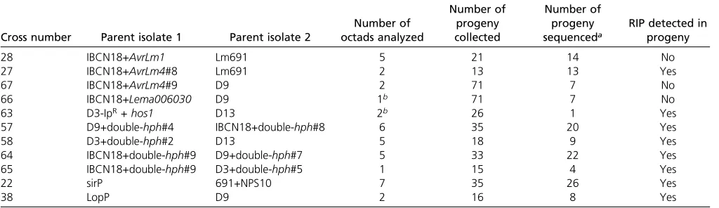
Table 2 Crosses carried out to analyze RIP mutations inL. maculans
Cross number Parent isolate 1 Parent isolate 2
Number of octads analyzed
Number of progeny collected
Number of progeny
sequenceda RIP detected inprogeny
28 IBCN18+AvrLm1 Lm691 5 21 14 No
27 IBCN18+AvrLm4#8 Lm691 2 13 13 Yes
67 IBCN18+AvrLm4#9 D9 2 71 7 No
66 IBCN18+Lema006030 D9 1b 71 7 No
63 D3-IpR+hos1 D13 2b 26 1 Yes
57 D9+double-hph#4 IBCN18+double-hph#8 6 35 20 Yes
58 D3+double-hph#2 D13 5 18 9 Yes
64 IBCN18+double-hph#9 D9+double-hph#7 5 33 22 Yes
65 IBCN18+double-hph#9 D3+double-hph#5 1 15 4 Yes
22 sirP 691+NPS10 7 35 26 Yes
38 LopP D9 2 16 8 Yes
RIP, Repeat Induced Point.
aAll progeny were genotyped for the presence of the relevant construct (as indicated by the amplification of thehphgene). Only progeny harboring the relevant constructs were sequenced and analyzed for the presence of RIP mutations.
which contained restriction sites on the ends. The 1026-bp fragment was cloned into plasmid pPZP-Hyg (Elliott and Howlett 2006), which contained restriction sites forAcc65I and EcoRV at the ends. The resultant plasmid, pPZPHygx2 (3816 bp, Figure S2A), was transformed into isolates D9, IBCN18, and D3 (Table 1). The resulting transformed isolates (Table 1) were crossed with isolates of opposite mating type, and progeny collected and analyzed (Table 2). To sequence across the entire construct and guarantee that the correct copy of the repeat was being amplified and sequenced, over-lapping PCR products were amplified and sequenced. Primer pairs for each PCR were selected so that at least one primer bound to single-copy regions within the construct, for exam-ple the right border sequence, thetrpCpromoter, or thetrpC
terminator (Table S1). Alternatively, primer pairs with spe-cific orientations were selected so that only the correct re-gions could be amplified, e.g., the 59hph primer (CE245) used as a reverse primer with the 39trpCT primer used as a forward primer. The resulting PCR products were then sub-jected to sequencing using multiple primers (not necessarily from the single-copy regions) so that the full length of the construct could be determined, while resolving each re-peated DNA region.
A hairpin construct had previously been designed to silence a nonribosomal peptide synthetase (NPS10) gene ofL.
mac-ulansusing the pHYGGS vector, and was used to investigate
the impact of this gene on virulence (Fox et al. 2008). A 620-bp region of the NPS10 gene (GenBank accession CCT61194.1) was amplified from genomic DNA ofL. macu-lans isolate IBCN18, using attB1- and attB2- tailed primers NPS10RNAiF and NPS10RNAiR, and cloned using Gateway recombination into pDONR207. The fragment was then moved from pDONRnps10 into the gene-silencing vector pHYGGS in two opposing orientations using LR Clonase (Invitrogen, Carlsbad, CA) to create the final NPS10 gene silencing vector, pNPS10RNAi (Figure S1A). The vector was transformed intoAgrobacterium tumefaciensstrain LBA4404 (Invitrogen).L. maculansisolate 691 was transformed using
the Agrobacterium strain containing pNPS10RNAi as
de-scribed in Gardiner and Howlett (2004). Isolates were assessed for silencing of theNPS10gene using quantitative reverse transcriptase PCR to compare the expression of
NPS10 relative to actin in wild-type and silenced isolates.
The transformant with the lowest expression of NPS10, named 691+NPS10, was selected and crossed to a sirodes-min biosynthesis gene-disruption mutant, referred to as sirP since thesirPgene had been disrupted (Gardiner and Howl-ett 2004). This experiment was initially designed for the lab-oratory direction of creating double mutants affected in the synthesis of secondary metabolites, but both the construct used and parents of the cross served as a useful combination to detect RIP. The progeny of this cross were sequenced to detect RIP. As mentioned above, to sequence across the entire construct, a series of overlapping PCR fragments were gen-erated using primer combinations designed to amplify spe-cific fragments of the construct. The resulting PCR products
were then sequenced using primers with unique binding sites so that the entire length of the construct could be sequenced without confusing the identities of the different repeat regions.
Determining the copy number of T-DNA insertions
Southern blot analysis was carried out on the transgenic parents of crosses 27, 28, 57, 58, 64, 65, and 67 to determine the copy number of the plasmid within each parent isolate (Figures S1 and S2). Genomic DNA was digested with re-striction enzymes as indicated in thefigures and DNA frag-ments resolved on a 0.7% agarose Tris-acetate-EDTA gel. The gels were stained with ethidium bromide to visualize digested DNA before transfer. DNA was transferred to a nylon mem-brane using downward alkaline capillary transfer, as de-scribed in Sambrook and Russell (2001). Blots were hybridized with a digoxigenin-11-dUTP-labeled PCR frag-ment corresponding to 579 bp of thehphgene amplified with primers CE249 and CE250 (Table S1). The probe was gener-ated using a PCR DIG Probe synthesis kit from Roche; blots were hybridized using conventional buffer [63 SSC, 0.1% SDS, and 13 blocking buffer (Roche kit)], 53 Denhardt’s solution, and 40mg salmon sperm DNA, and processed using DIG wash and a blocking buffer set followed by DIG lumines-cence detection. Signal detection was carried out using a Bio-Rad (Hercules, CA) Chemidoc imaging system equipped with Image Lab software using high-sensitivity detection with sig-nal accumulation mode.
A single hybridizing band in lanes whereHindIII was used to digest the DNA, and two hybridizing bands in lanes wherePstI was used, indicate that the T-DNA had inserted in single copy (Figure S1, IBCN18+AvrLm1 and IBCN18+AvrLm4#9). Two or more bands in theHindIII lanes, and three or more bands in thePstI lanes, indicated multiple or tandem insertion of the T-DNA (IBCN18+AvrLm4#8 and 691+NPS10). For parents containing pPZPHygx2, one copy of the T-DNA was confirmed by digestion with two independent enzymes that showed two hybridizing bands corresponding to each of the hygromycin resistance genes (Figure S2).
Identification of T-DNA or plasmid insertion sites in the L. maculans genome
Two methods were used to identify where T-DNA or plasmids had inserted in the genome. For the T-DNA insertions, inverse PCR was used. Genomic DNA (2mg) was digested withTaqaI restriction enzyme, the fragments circularized with T4 DNA ligase, and the ligation mix used for PCR with primers M13F-ai076. Amplicons were purified from agarose gels and used as a template for a nested PCR with primers M13F-MAI0324. Amplicons were sequenced and the sequences compared to theL. maculansgenome sequence. The other side of the insertion was identified by amplification with an isolate-specific primer.
undergo RIP during mating (Idnurm and Howlett 2003), were sequenced using next-generation sequencing (100-bp paired end reads on an Illumina HiSeq 2500 instrument) at the Australian Genome Research Facility (AGRF) in Mel-bourne. The sequences (3.65 GB for LopC and 3.58 GB for LopP) were mapped with reiterations to the pUCATPH plas-mid sequence using Geneious version 8.1.7, to provide cov-erage of the plasmid insertion andflanking regions.
Detection and analysis of RIP signatures
Genomic DNA of parental isolates and progeny was prepared as described previously (Gardineret al.2004). In progeny of the crosses, the genes and regions of interest were amplified using primers in Table S1, and then sequenced at the AGRF. Sequence chromatograms were visualized using Geneious version 9.1 (Kearse et al. 2012) or Sequencher version 5.4.1 (Gene Codes Corporation, Ann Arbor, MI) and G/A and C/T transitions identified. RIPCAL was used to de-termine CpA4TpA dominance scores (Hane and Oliver 2008). For all analyses, the DNA sequence obtained from the parental isolate was used as the reference (non-RIP) sequence.
Culturing in the absence of hygromycin selection to trigger RIP
Two isolates, LopP and LopC, were subcultured into 10 start-ing cultures without hygromycin selection. Each week for 10 weeks, mycelial fragments were passaged on media with-out hygromycin. After the 10 weeks, spores from each culture were plated at low density to yield colonies. Of these, 20 were selected and grown on V8 media with or without hygromycin to detect whether RIP could be triggered during mitotic replication to inactivate thehphgene.
All strains and plasmids are available upon request. All primer information can be found in Table S1.
Data availability
The authors state that all data necessary for confirming the conclusions presented in the manuscript are represented fully within the manuscript. All strains and plasmids are available on request. Supplemental material available at Figshare:
https://doi.org/10.25386/genetics.7221974.
Results
The frequency of RIP in L. maculans when duplications are unlinked is extremely low and requires large repeated regions
InN. crassa, duplication of DNA can result in RIP mutations
being generated in both copies of the DNA repeat, even if in unlinked parts of the genome. This has been used inN. crassa
as a tool for mutating genes (Selker and Garrett 1988), as well as in other species such as Trichoderma reesei, where copies of genes in two different parts of the genome are both triggered for mutation during mating (Liet al.2017). To test whether RIP mutations can be triggered and used as a way
of mutating genes in L. maculans, two constructs were designed, one harboring theAvrLm1 gene (1805 bp) and the other theAvrLm4gene (1645 bp). An isolate ofL.
mac-ulansthat already harbored theAvrLm1andAvrLm4genes
(IBCN18) was individually transformed with these con-structs to create isolates, with either two copies of
AvrLm1 or two copies of AvrLm4. Transformants with a
random insertion of the construct resulted in unlinked du-plications of the avirulence genes (IBCN18+AvrLm1 or IBCN18+AvrLm4) and were crossed to an isolate of oppo-site mating type, Lm691, also harboring these avirulence genes. Octad progeny were collected from the resulting crosses. The endogenous copy of the avirulence genes was sequenced from all progeny, while the copy of the avirulence gene within the construct and the hph select-able marker were sequenced in the progeny of the crosses that harbored the construct.
For the cross with isolate IBCN18+AvrLm1(cross 28), no mutations were detected in any progeny in the endogenous or construct copy of theAvrLm1gene, nor thehphgene of the construct (Table S2). Southern analysis and inverse PCR showed that there was a single insertion of the construct within the parental isolate (IBCN18+AvrLm1) and that this insertion site, located on supercontig 8, was unlinked to the endogenousAvrLm1gene located on supercontig 6 (Figures S1 and S3).
Testing the potential of RIP using a second gene, duplicated in isolate IBCN18+AvrLm4#8 (cross 27), RIP mutations were detected throughout thehphgene and a few RIP mu-tations within the construct copy of theAvrLm4gene, but not within the endogenous copy in any of the progeny (Ta-ble S2). Southern blot and PCR analysis showed a tandem insertion of theAvrLm4construct, which was probably trig-gering RIP in this situation (Figure S1). Therefore, a second isolate, IBCN18+AvrLm4#9, was generated, confirmed to have a single insertion using Southern analysis and inverse PCR, and crossed to isolate D9, also harboring AvrLm4
(cross 67) (Figure S1). No mutations were detected in any progeny collected from this cross in either the endogenous or construct copy of the AvrLm4 gene, nor the hph gene (Table S2).
Transforming a second copy of eitherAvrLm4orAvrLm1
intoL. maculans and crossing those isolates did not trigger
RIP in endogenous genes. These genes and the amount of homologous DNA that is duplicated are small (repeat re-gion of,2 kb), which may not be big enough to trigger RIP
inL. maculans. Therefore, additional crosses were assessed
whereby larger constructs [transcription factorLema006030= 4569 bp (cross 66) and fungicide resistance gene hos1
observed in this region of the gene, suggesting that RIP had not occurred.
The second cross involved one parent harboring a sponta-neous mutation within the hos1gene, which confers resis-tance to the fungicide iprodione when mutated, plus a complementation construct containing the wild-type hos1
gene and the G418 antibiotic resistance marker. This isolate was crossed with a wild-type; 19 progeny were isolated, and screened for resistance to both G418 and iprodione, which is impossible unless the hos1complementation construct was mutated. A single progeny, 63R15, was identified with this phenotype, and the construct copy of thehos1gene was am-plified and sequenced. The construct containedfive RIP mu-tations across the entire 5736 bp region, suggesting that RIP can be triggered using nontandem repeats but at a frequency of , 0.1% (Figure S4). Furthermore, only one of the five substitutions is likely responsible for the iprodione resistance phenotype (of a conserved methionine to isoleucine at amino acid position 538). Taken together, these data suggest that targeted mutations of an endogenous gene using RIP may be difficult since the frequency of RIP is extremely low in non-tandem repeats, which contrasts to other fungal species. Therefore, we were interested in determining what similari-ties and differences exist for RIP mechanisms inL. maculans
compared to other species.
RIP mutations leak from repeat regions into single-copy sequences
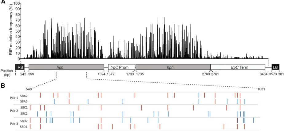
To investigate the mechanisms leading to RIP, a construct har-boring two copies of thehphgene (referred to as double-hph)
was generated, transformed into isolates, and then the result-ing transgenic isolates used for crossresult-ing. Southern analysis showed that single copies of the construct had inserted into each isolate (Figure S2). Four different crosses (crosses 57, 58, 64, and 65; Table 2), using a combination of seven differ-ent pardiffer-ental isolates, were established. A total of 55 progeny, representing 17 different octads, were analyzed for the pres-ence of RIP mutations. A 3342-bp region, spanning almost the entire construct, was sequenced from all progeny harboring the double-hphconstruct (Figure 1). The frequency of nucle-otides that had undergone RIP mutations for the individual progeny ranged from 0.1–6.0%, with the GC content decreas-ing by up to 4.8% (Table S3). All sequences were also sub-jected to RIPCAL analysis, a software tool used to compare alignments and calculate RIP indexes, to determine which dinucleotide transitions were dominant. The CpA/TpA tran-sitions were the most dominant RIP mutations, with an aver-age RIP dominance score of 1.28, while CpG/TpG were the second most dominant with an average score of 0.81 (Figure S5 and Table S4).
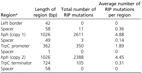
The frequency of RIP mutations at each nucleotide position across the construct was determined in the 55 progeny (Figure 1A). The frequency of RIP was highest within the hph re-peats; however, RIP was also detected within the single-copy regions of thetrpCpromoter, spacer regions, andtrpC termi-nator (Table 3). Although RIP mutations were detected within these single-copy regions, the average number of RIP mutations for these regions was much lower (between 0 and 0.36%) than the repeated hph regions (4.45 and 4.88%) (Table 3).
RIP mutations differ in octad pairs in L. maculans
Analysis of the patterns of RIP in the octad progeny pairs from the multiple double-hphcrosses showed that the RIP signa-tures differed for each progeny from a pair (Figure 1B), and furthermore, for many of the progeny, both G/A and C/T transitions were present (Table S3). To confirm that this was not an artifact of the double-hph construct, the repetitive regions of octad pairs derived from a variety of different crosses were also amplified and sequenced.
Crosses were set up between 691+NPS10 (containing a hairpin construct for silencing theLmNPS10gene) and the sirP isolate (containing a gene disruption of thesirPgene), originally to generate progeny that contained both the sirP
gene-disruption mutation as well as the silencing of NPS10
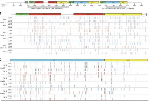
(cross 22), yet being informative for understanding the RIP mechanism in L. maculans. Southern analysis and targeted sequencing of theNPS10silencing construct in the 691+NPS10 parent isolate was used to determine the exact nature of the hairpin insertion (Figure S1). A partial duplication of the silencing construct was identified, whereby two copies of thehphgene, terminator sequence, and promoter sequence were integrated into the isolate (Figure 2A). Inverse PCR was used to identify the insertion site of the T-DNA into supercontig 2: it is unlinked to the endogenous copy of
NPS10, which is located on supercontig 11 (Figure S3).
Octad progeny were collected from seven individual octads and the pairs determined using PCR-based markers that differ between the two parents. Initially, three octads were analyzed (22A, 22B, and 22D). For each of these three octads, four progeny (two pairs) lacked the construct. The endogenous copy of the NPS10gene was sequenced from these progeny and no RIP was detected (data not shown). Since these four progeny did not harbor the construct, they were not analyzed further. For the remaining four progeny (two pairs) that did harbor the construct, a 2894-bp region of the construct encompassing the hairpin repeats and a 1474-bp region encompassing one of the hph repeats (Figure 2B) were sequenced. The number and position of
RIP mutations differed for each progeny, including between progeny pairs from the same octad (Figure 2A and Table 4). In all 12 progeny (representing six pairs from the three octads), there were both G/A and C/T transitions present across the two regions sequenced (Table 4). For some progeny, such as 22D3, the ratio of G/A to C/T transitions was 4:96, while other progeny had almost a 50:50 ratio (progeny 22A8 with 56.9% G/A and 43.1% C/T) (Table 4).
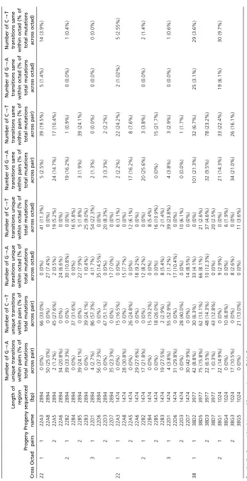
The RIP signatures for each pair within an octad, and all four progeny with duplicated genes from an octad, were analyzed to determine the number of mutations that are common between pairs, unique within pairs, the same within all four progeny from a single octad, or unique across all four progeny from a single octad (Table 5). For all three octads, there were more unique RIP mutations between pairs (16.5– 60%) compared to common RIP mutations (1.7–29.4%). Fur-thermore, when all four progeny from a single octad were analyzed, the frequency of mutations unique across all four progeny was much significantly higher (3.1–25%) than the frequency of mutations common across all four progeny from an octad (0–5.3%) (Student’st-testP,0.001). For 4 of the 12 progeny, both G/A and C/T unique transitions were observed across the sequenced regions.
The frequency of RIP occurring at each nucleotide position across the 2894-bp and 1474-bp regions sequenced from the cross 22A, 22B, and 22D tetrad progeny was determined (Figure S5). Similar to what was seen for the double-hph
construct, the frequency of RIP increased within the hairpin repeat regions and was still present, although at a lower frequency, in the single-copy spacer region between the two hairpin repeats. The frequency of RIP remained constant across all other regions as these were in multiple copies due to the partial tandem insertion of the construct (Figure 2A). As seen for the double-hphprogeny, the dominant RIP mutations were CpA/TpA (average RIP dominance score of 2.06) and CpG/TpG (average RIP dominance score of 0.5) (Figure S6 and Table S4).
A 347-bp region of the hairpin construct was also se-quenced from the progeny from an additional five octads (Figure S7 and Table S3). As seen with the initial three octads of cross 22 (Figure 2B), the pattern and number of RIP mu-tations differed within all progeny pairs. The frequency of RIP mutations ranged from 1.15–4.02% across the 347-bp region and resulted in a decrease in overall GC content of up to 4.2% (Table S3).
For a subset of the progeny, the two repeat regions within a construct (i.e.,hphfor the double-hphconstruct and the hair-pin region of theNPS10silencing construct) were aligned to determine whether the repeats were being targeted by RIP in a similar manner. For cross 57, thehphrepeats were aligned for each of the six progeny of the B octad, and mutations common and unique between each copy of the repeat were determined. The majority of RIP mutations (between 64 and 77%) differed between the two repeats for each of the six progeny, suggesting that the repeats are not subjected to the same RIP mutations (Table S4). Similarly, the hairpin repeat
Table 3 Total number and frequency of RIP mutations across specific regions of the double-hph construct when sequenced from 55L. maculansprogeny
Regiona
Length of region (bp)
Total number of RIP mutations
Average number of RIP mutations
per region
Left border 42 0 0
Spacer 58 11 0.36
hph(copy 1) 1026 2611 4.88
Spacer 49 3 0.14
TrpC promoter 362 350 1.89
Spacer 1 0 0
hph(copy 2) 1026 2388 4.45
TrpC terminator 724 105 0.31
Spacer 58 0 0
RIP, Repeat Induced Point.
regions were aligned for 12 of the cross 22 progeny, and be-tween 74 and 100% of the mutations differed bebe-tween the repeat regions (Table S4).
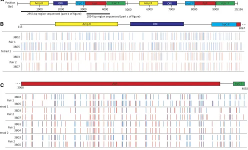
The RIP process wasfirst identified inL. maculanswhen insertional mutants, such as LopC and LopP, which had been generated by restriction enzyme-mediated integration of plasmid DNA, were crossed to wild-type isolates and the hygromycin-resistant phenotype in these two isolates, brought about by the introduction of linearized plasmid pUCATPH, was lost. The plasmid insertions were in tandem, based on Southern blot analysis (Idnurm and Howlett 2003), and confirmed here when the integration sites of the plas-mids into the genome were identified by sequencing ge-nomes of the two isolates (GenBank accession SRP149958; Figure S3). We crossed the original LopC and LopP mutants to wild-type isolate D9, and collected octad progeny. As re-ported previously, all progeny were sensitive to hygromycin, rather than the expected 4:4 ratio of resistant:susceptible. The progeny from the LopP3D9 cross (cross 38) were ex-amined in more detail. Regions of the pUCATPH plasmid
were amplified and sequenced to analyze the RIP signatures. For tetrad 38D, a 3977-bp region was sequenced, while for tetrad 38G, a 1024-bp region was sequenced. As seen for the double-hphand hairpin crosses described above, the RIP sig-natures differed both within and between all the octad pairs (Figure 3, Table 4, and Table 5). Also, as observed in the other crosses, both G/A and C/T transitions were seen in each progeny, and the frequency of unique mutations was much higher than the frequency of mutations that were common across all four progeny from a single octad (Table 5). The frequency of RIP within these progeny was higher than that seen in the other crosses, ranging from 3.87–8.11%, and as a consequence the GC content was also more dramat-ically decreased.
RIP mutations are linked to the sexual cycle
Since mutations differed within octad pairs, RIP may be occurring postmeiosis and could therefore be a mitotic pro-cess. To test if RIP occurs during vegetative growth, two isolates that undergo RIP during mating (LopP and LopC)
were passaged 10 times on media without hygromycin and then, after the 10th passage, spores were plated and colonies were cultured. All 400 remained hygromycin-resistant, sug-gesting that the induction of RIP is linked to the sexual cycle.
Discussion
The genome sequences of many fungal species carry signs of mutations that target repetitive DNA regions, especially trans-posable elements. One mechanism to create these mutations is RIP, with extensive evidence for this or a mechanism like RIP occurring in the fungi (Selker 1990; Clutterbuck 2011; Hane
et al.2015). However widespread these hallmarks are of a
common mutation process, relatively little is known about the mechanisms behind RIP and how conserved they would be in the fungi. In the current study, analyses of a series of crosses and of DNA sequences in L. maculansthat have or have not been affected by RIP indicate that there are both similarities and differences in how this process operates in
different fungi. This mutator phenomenon has consequences for the subsequent fate of DNA duplication in fungi.
In this study, we have shown that inL. maculansRIP occurs at CpA, CpG, and (to a lesser extent) CpT sites, with the CpA sites being the more predominant location. This is consistent with previous studies in N. crassa and F. graminearum, whereby CpA mutations are the predominant mutation, but CpG and CpT mutations are also targeted (Pomraninget al.
2013; Gladyshev 2017). Interestingly, a previous in silico
study using bioinformatic approaches only to analyze repeat regions within theL. maculansgenome sequence suggested that 90% of RIP mutations occur at CpA sites (Amselemet al.
2015). The differences between the two studies might reflect the fact that, in the latter study, transposable elements lo-cated within AT-rich regions of the genome were the focus of the study.
Two commonalities between N. crassa and L. maculans
are that tandem repeats of only 1 kb are enough to trigger RIP, and that RIP leaks into single-copy regions (Selker and
Table 4 Number and frequency of RIP mutations within progeny of crosses betweenL. maculansisolates harboring constructs designed to trigger RIP
Crossa Octad Pair Progenyb
Size of region
sequenced (bp) RIP mutations (%)
G/A transitions (percentage of total
RIP mutations)
C/T transitions (percentage of total RIP mutations)
Decrease in GC content (%)
22 1 1 22A3 2894 108 (3.7) 6 (5.6) 102 (94.4) 3.7
22A8 2894 92 (3.2) 52 (56.5) 40 (43.5) 3.2
2 22A5 2894 88 (3.0) 27 (30.7) 61 (69.3) 3.1
22A6 2894 75 (2.6) 59 (78.7) 16 (21.3) 2.6
2 1 22B2 2894 60 (2.1) 59 (98.3) 1 (1.7) 2.1
22B4 2894 57 (2.0) 19 (33.3) 38 (66.7) 2.0
2 22B3 2894 80 (2.7) 34 (42.5) 46 (57.5) 2.9
22B5 2894 81 (2.8) 8 (9.9) 73 (90.1) 2.9
3 1 22D1 2894 92 (3.1) 6 (6.5) 86 (93.5) 3.2
22D6 2894 58 (2.0) 58 (100.0) 0 (0.0) 2.0
2 22D3 2894 52 (1.8) 3 (5.8) 49 (94.2) 1.8
22D7 2894 40 (1.4) 38 (95.0) 2 (5.0) 1.4
22 1 1 22A3 1474 39 (2.7) 2 (5.1) 37 (94.9) 2.7
22A8 1474 52 (3.5) 30 (57.7) 22 (42.3) 3.5
2 22A5 1474 51 (3.5) 17 (33.3) 34 (66.7) 3.5
22A6 1474 54 (3.7) 46 (85.2) 8 (14.8) 3.7
2 1 22B2 1474 40 (2.7) 37 (92.5) 3 (7.5) 2.7
22B4 1474 38 (2.6) 20 (52.6) 18 (47.4) 2.6
2 22B3 1474 36 (2.4) 19 (52.8) 17 (47.2) 4.2
22B5 1474 33 (2.2) 0 (0.0) 33 (100.0) 1.8
3 1 22D1 1474 66 (4.5) 8 (12.1) 58 (87.9) 4.5
22D6 1474 38 (2.6) 35 (92.1) 3 (7.9) 2.6
2 22D3 1474 29 (2.0) 0 (0.0) 29 (100.0) 2.0
22D7 1474 31 (2.1) 30 (96.8) 1 (3.2) 2.1
38 1 1 38D2 3977 205 (5.2) 144 (70.2) 61 (29.8) 5.1
38D5 3977 270 (6.8) 175 (64.8) 95 (35.2) 6.7
2 38D3 3977 182 (4.6) 52 (28.6) 130 (71.4) 4.5
38D7 3977 154 (3.9) 32 (20.8) 122 (79.2) 3.9
2 1 38G1 1024 79 (7.7) 42 (53.2) 37 (46.8) 7.7
38G4 1024 68 (6.6) 19 (27.9) 49 (72.1) 6.6
2 38G3 1024 79 (7.7) 49 (62.0) 30 (38.0) 7.7
38G5 1024 83 (8.1) 32 (38.6) 51 (61.4) 8.1
RIP, Repeat Induced Point.
aFor details of parental isolates used for crossing, see Table 2.
Garrett 1988; Fudal et al.2009; Van de Wouwet al.2010; Gladyshev and Kleckner 2014, 2017b). Furthermore, it ap-pears that likeN. crassa, both the RID-mediated and DIM-2-mediated RIP pathways are active inL. maculans. InN. crassa, it has been shown that the RID-mediated RIP pathway (in-volving RID) primarily acts on regions with shared homology, while the DIM-2-mediated RIP (involving DIM-5 and DIM-2) pathway leads to mutations that spread significantly into the linked, nonrepetitive regions (Gladyshev and Kleckner 2017a). InN. crassa, when key players in each of these path-ways are mutated, the frequency of RIP-induced mutations changes in the homologous repeats or in the linked, nonrep-eat regions. Although not experimentally defined to be active
inL. maculans, all homologs of both RIP pathways have been
identified in the genome (Rouxel et al.2011), and the fre-quency of RIP-induced mutations reported in the current study are higher in the linked repeat regions compared to the single copy, nonrepeat regions, similar to that reported by Gladyshev and Kleckner (2017a) when both pathways are active. Further work involving mutations of some of the key players in these two RIP pathways would be needed to confirm their role inL. maculans. Unfortunately, the
genera-tion of knockout mutagenera-tions inL. maculansis extremely diffi -cult, with just 10 reported to date for the species. However, clustered regularly interspaced short palindromic repeat/ Cas9 gene editing strategies are currently being developed and could potentially be used for such experiments in the future (Idnurmet al.2017). Conversely, unlike inN. crassa, it appears that unlinked duplications inL. maculansonly trig-ger RIP at a very low frequency. In a single cross whereby a
5.7-kb repeat region was used, RIP was detected at a fre-quency of,0.01% in one progeny from 34 screened (3%). Based on the fact that the frequency of RIP observed in linked duplications in this study (1.4–8.1%) was much lower than in
both N. crassa (30%) and F. graminearum (10–39%)
(Galagan and Selker 2004; Pomraninget al.2013), it is not surprising that the frequency of RIP in unlinked duplications is also much lower inL. maculansthan these other species. It should be noted that the current study on unlinked duplica-tions has limitaduplica-tions. Not all progeny were sequenced, but instead phenotypic screening was used to increase the chan-ces of identifying progeny harboring the RIP mutations. Therefore, the detection of RIP in only 3% of progeny will be biased. Regardless, the frequency of RIP mutations within
those progeny is still extremely low (5 bp within the 5736-bp region).
Perhaps the most significant difference betweenL.
macu-lans andN. crassais the generation of four different
geno-types in the four pairs of progeny harboring the constructs within an octad, compared to only two forN. crassa(Watters
et al.1999). The progeny pairs, generated through a mitotic
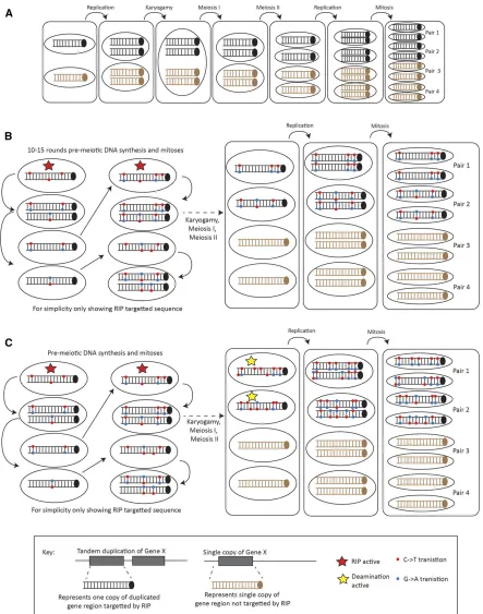
division, have different genotypes inL. maculans. In addition, across the pairs and across the octad, there are both common and unique G/A and C/T unique mutations on a single DNA strand. Several hypotheses could explain thesefindings and one is diagrammatically represented in Figure 4, with a simplified representation of the RIP process inN. crassafor comparison (Figure 4B). InN. crassa, duplications are likely to be detected during the G1 phase of the cell cycle, leading to multiple premeiotic rounds of RIP occurring whereby C/T transitions are generated. It has been hypothesized that up to 10–15 mitoses may occur and it has been shown that the frequency of RIP increases in older ascospores (Singeret al.
1995a). The occurrence of RIP during these premeiotic mi-toses leads to the generation of two different haplotypes in the RIP-targeted progeny, one haplotype for each progeny pair. One possible hypothesis to explain the different haplo-type number observed inL. maculansis that RIP is initially occurring as it does inN. crassa, but that the deamination step is continuing inL. maculanssuch that, during replication after meiosis II, the C/U deamination will be repaired to generate T/As (Figure 4C). The continuation of deamination would then result in unique C/T and G/A transitions on both strands of the four octad progeny. Much of the data generated in the current study supports this hypothesis. First, the occur-rence of RIP during multiple rounds of premeiotic mitoses is supported by the presence of RIP mutations common across all four octad progeny and other mutations common across pairs of progeny. Mutations common across all four octad progeny would represent “older” mutations generated in the earliest rounds of RIP, while those common across pairs would represent RIP mutations generated in later mitoses. The continuation of deamination after karyogamy and the consequent generation of Ts/As is supported by the pres-ence of unique RIP mutations in each of the progeny pairs. Furthermore, all the unique mutations are either G/A or C/T transitions on a single strand (Table 5), and not a combination of both, which supports this hypothesis. An al-ternative but unlikely hypothesis is that a mutation event as late as mitosis occurring postmeiosis is also feasible, although we show that recurrent mitotic passaging of strains was not able to trigger RIP. Further work is needed to fully under-stand differences and similarities in RIP between these species.
Other mechanisms could also contribute to the unusual patterns of RIP detected inL. maculans. These might include differences in mutation rates, methylation events, or the
ef-ficiency and timing of DNA repair between fungal species, which might account, at least in part, for some of the different levels of RIP or genetic haplotypes seen in the octad pairs in
L. maculanscompared toN. crassa. However, currently very
little is known about these processes inL. maculans, making it very difficult to speculate.
Although not explored in the current study, another note-worthy difference betweenL. maculansandN. crassais the lack of association between RIP and cytosine methylation in
L. maculans. InN. crassa, repeats that have been heavily
mu-tated by RIP are then targets for DNA methylation (Selker 1990; Galagan and Selker 2004), and the frequency of RIP correlates with concentrations of S-adenosylmethionine in strains (Rosa et al.2004). However, previous studies using methylation-sensitive restriction enzymes showed no differ-ences in RIP-affected sequdiffer-ences, suggesting that RIP-associated methylation is not occurring in L. maculans (Idnurm and Howlett 2003). Similar studies have been performed in
F. graminearum, with no methylation detected in
associa-tion with RIP (Pomraninget al.2013).
The consequences on gene and genome evolution due to the variability in RIP between fungal species remain to be fully established. It is curious that transposons at some point expanded throughoutL. maculans, in which tandem duplica-tions are required to ensure RIP occurs, before being brought under control by RIP (Rouxelet al.2011), whereas inN. crassa, where the duplications can be on separate chromosomes to trigger RIP, transposons are rare (Galaganet al.2003). Of note, the impact of unlinked duplications escaping RIP provides a mechanism by which duplicated DNA regions can undergo di-vergence in function.
Acknowledgments
We thank Barbara Howlett for her encouragement and comments on the manuscript, as well as two anonymous reviewers and the handling editor, Michael Freitag, for their insightful suggestions. This research was supported by the Australian Grains Research and Development Corporation and the Australian Research Council.
Author contributions: A.V.d.W. conceived and designed the experiments. A.V.d.W., C.E.E., K.M.P., and A.I. performed the experiments. A.V.d.W., C.E.E., K.M.P., and A.I. analyzed the data. A.V.d.W., C.E.E., K.M.P., and A.I. contributed reagents, materials, and analysis tools. A.V.d.W. wrote the paper. C.E.E. and A.I. edited the paper.
Literature Cited
Amselem, J., M. H. Lebrun, and H. Quesneville, 2015 Whole ge-nome comparative analysis of transposable elements provides new insight into mechanisms of their inactivation in fungal ge-nomes. BMC Genomics 16: 141. https://doi.org/10.1186/ s12864-015-1347-1
Clutterbuck, A. J., 2011 Genomic evidence of repeat-induced point mutation (RIP) infilamentous ascomycetes. Fungal Genet. Biol. 48: 306–326.https://doi.org/10.1016/j.fgb.2010.09.002
contribution of supernumerary chromosomes to gene expan-sion. PLoS Genet. 5: e1000618. https://doi.org/10.1371/jour-nal.pgen.1000618
Cozijnsen, A. J., and B. J. Howlett, 2003 Characterisation of the mat-ing-type locus of the plant pathogenic ascomyceteLeptosphaeria mac-ulans. Curr. Genet. 43: 351–357. https://doi.org/10.1007/s00294-003-0391-6
Cozijnsen, A. J., K. M. Popa, A. Purwantara, B. D. Rolls, and B. J. Howlett, 2000 Genome analysis of the plant pathogenic asco-myceteLeptosphaeria maculans; mapping mating type and host specificity loci. Mol. Plant Pathol. 1: 293–302.https://doi.org/ 10.1046/j.1364-3703.2000.00033.x
Cuomo, C. A., U. Guldener, J. R. Xu, F. Trail, B. G. Turgeonet al., 2007 The Fusarium graminearumgenome reveals a link be-tween localized polymorphism and pathoegn specialization. Sci-ence 317: 1400–1402.https://doi.org/10.1126/science.1143708
Elliott, C. E., and B. J. Howlett, 2006 Overexpression of a 3-ketoacyl-CoA thiolase in Leptosphaeria maculans causes re-duced pathogenicity onBrassica napus. Mol. Plant Microbe In-teract. 19: 588–596.https://doi.org/10.1094/MPMI-19-0588
Fitt, B. D. L., H. Brun, M. J. Barbetti, and S. R. Rimmer, 2006 World-wide importance of phoma stem canker (Leptosphaeria maculans
and L. biglobosa) on oilseed rape (Brassica napus). Eur. J. Plant Pathol. 114: 3–15.https://doi.org/10.1007/s10658-005-2233-5
Fox, E. M., D. M. Gardiner, N. P. Keller, and B. J. Howlett, 2008 A Zn(II)2Cys6 DNA binding protein regulates the sirodesmin PL biosynthetic gene cluster in Leptosphaeria maculans. Fungal Genet. Biol. 45: 671–682.
Fudal, I., S. Ross, L. Gout, F. Blaise, M. L. Kuhn et al., 2007 Heterochromatin-like regions as ecological niches for avirulence genes in theLeptosphaeria maculansgenome: map-based cloning ofAvrLm6. Mol. Plant Microbe Interact. 20: 459– 470.https://doi.org/10.1094/MPMI-20-4-0459
Fudal, I., S. Ross, H. Brun, A. L. Besnard, M. Ermel et al., 2009 Repeat-induced point mutation (RIP) as an alternative mechanism of evolution toward virulence inLeptosphaeria mac-ulans. Mol. Plant Microbe Interact. 22: 932–941. https://doi. org/10.1094/MPMI-22-8-0932
Galagan, J. E., and E. U. Selker, 2004 RIP: the evolutionary cost of genome defense. Trends Genet. 20: 417–423.https://doi.org/ 10.1016/j.tig.2004.07.007
Galagan, J. E., S. Calvo, K. A. Borkovich, E. U. Selker, N. D. Read
et al., 2003 The genome sequence of thefilamentous fungus
Neurospora crassa. Nature 422: 859–868. https://doi.org/ 10.1038/nature01554
Gall, C., M. H. Balesdent, P. Robin, and T. Rouxel, 1994 Tetrad analysis of acid phosphates, soluble protein patterns, and mat-ing type inLeptosphaeria maculans. Genetics 84: 1299–1305. Gardiner, D. M., and B. J. Howlett, 2004 Negative selection using
thymidine kinase increases the efficiency of recovery of transform-ants with targeted genes in thefilamentous fungusLeptosphaeria maculans. Curr. Genet. 45: 249–255. https://doi.org/10.1007/ s00294-004-0488-6
Gardiner, D. M., A. J. Cozijnsen, L. M. Wilson, M. S. C. Pedras, and B. J. Howlett, 2004 The sirodesmin biosynthetic gene cluster of the plant pathogenic fungus Leptosphaeria maculans. Mol. Microbiol. 53: 1307–1318. https://doi.org/10.1111/j.1365-2958.2004.04215.x
Gladyshev, E., 2017 Repeat-induced point mutation and other genome defense mechanisms in fungi, pp. 687–699 inThe Fun-gal Kingdom, edited by Heitman, J., B. J. Howlett, P. W. Crous, E. H. Stukenbrock, T. Y. Jameset al.American Society for Mi-crobiology, Washington, DC.
Gladyshev, E., and N. Kleckner, 2014 Direct recognition of homol-ogy between double helices of DNA inNeurospora crassa. Nat. Commun. 5: 3509.https://doi.org/10.1038/ncomms4509
Gladyshev, E., and N. Kleckner, 2017a DNA sequence homology induces cytosine-to-thymine mutation by a heterochromatin-related pathway inNeurospora. Nat. Genet. 49: 887–894.https:// doi.org/10.1038/ng.3857
Gladyshev, E., and N. Kleckner, 2017b Recombination-indepen-dent recognition of DNA homology for repeat-induced point mutation. Curr. Genet. 63: 389–400.https://doi.org/10.1007/ s00294-016-0649-4
Gout, L., I. Fudal, M. L. Kuhn, F. Blaise, M. Eckertet al., 2006 Lost in the middle of nowhere: theAvrLm1avirulence gene of the Dothideomycete Leptosphaeria maculans. Mol. Microbiol. 60: 67–80.https://doi.org/10.1111/j.1365-2958.2006.05076.x
Graïa, F., O. Lespinet, B. Rimbault, M. Dequard-Chablet, E. Coppin
et al., 2001 Genome quality control: RIP (repeat-induced point mutation) comes to Podospora. Mol. Microbiol. 40: 586–595.
https://doi.org/10.1046/j.1365-2958.2001.02367.x
Hane, J. K., and R. P. Oliver, 2008 RIPCAL: a tool for alignment-based analysis of repeat-induced point mutations in fungal ge-nomic sequences. BMC Bioinformatics 9: 478.https://doi.org/ 10.1186/1471-2105-9-478
Hane, J. K., J. P. Anderson, A. H. Williams, J. Sperschneider, and K. B. Singh, 2014 Genome sequencing and comparative geno-mics of the broad host-range pathogenRhizoctonia solaniAG8. PLoS Genet. 10: e1004281. https://doi.org/10.1371/journal. pgen.1004281
Hane, J. K., A. H. Williams, A. P. Taranto, P. S. Solomon, and R. P. Oliver, 2015 Repeat-induced point mutation: a fungal-specific, endogenous mutagenesis process, pp. 55–68 inGenetic Trans-formation Systems in Fungi, Vol. 2, edited by Van den Berg M. A., and K. Maruthachalam. Springer International Publishing, Swit-zerland.https://doi.org/10.1007/978-3-319-10503-1_4
Hayden, H. L., A. J. Cozijnsen, and B. J. Howlett, 2007 Microsatellite and minisatellite analysis of Leptosphaeria maculans in Aus-tralia reveals regional genetic differentiation. Phytopathology 97: 879–887.https://doi.org/10.1094/PHYTO-97-7-0879
Horns, F., E. Petit, R. Yockteng, and M. E. Hood, 2012 Patterns of repeat-induced point mutation in transposable elements of ba-sidiomycete fungi. Genome Biol. Evol. 4: 240–247.https://doi. org/10.1093/gbe/evs005
Idnurm, A., and B. J. Howlett, 2003 Analysis of loss of pathoge-nicity mutants reveals that repeat-induced point mutations can occur in the Dothideomycete Leptosphaeria maculans. Fungal Genet. Biol. 39: 31–37.
Idnurm, A., A. S. Urquhart, D. Vummadi, S. Chang, A. P. Van de Wouw et al., 2017 Spontaneous and CRISPR/Cas9-induced mutation of the osmosensor histidine kinase of the canola path-ogen Leptopshaeria maculans. Fungal Biol. Biotechnol. 4: 12.
https://doi.org/10.1186/s40694-017-0043-0
Ikeda, K. I., H. Nakayashiki, T. Kataoka, H. Tamba, Y. Hashimotoet al., 2002 Repeat-induced point mutation (RIP) inMagnaporthe gri-sea: implications for its sexual cycle in the naturalfield context. Mol. Microbiol. 45: 1355–1364. https://doi.org/10.1046/j.1365-2958.2002.03101.x
Kearse, M., R. Moir, A. Wilson, S. Stones-Havas, M. Cheunget al., 2012 Geneious Basic: an integrated and extendable desktop software platform for the organization and analysis of sequence data. Bioinformatics 28: 1647–1649.https://doi.org/10.1093/ bioinformatics/bts199
Kinsey, J. A., P. W. Garrett-Engele, E. B. Cambareri, and E. U. Selker, 1994 The Neurospora transposon Tad is sensitive to repeat-induced point mutation (RIP). Genetics 138: 657–664. Li, W. C., C. H. Huang, C. L. Chen, Y. C. Chuang, S. Y. Tunget al.,
Marcroft, S. J., V. L. Elliott, A. J. Cozijnsen, P. A. Salisbury, B. J. Howlettet al., 2012 Identifying resistance genes toLeptosphaeria maculansin AustralianBrassica napuscultivars based on reactions to isolates with known avirulence genotypes. Crop Pasture Sci. 63: 338–350.https://doi.org/10.1071/CP11341
Ninomiya, Y., K. Suzuki, C. Ishii, and H. Inoue, 2004 Highly effi -cient gene replacements inNeurosporastrains deficient for non-homologous end-joining. Proc. Natl. Acad. Sci. USA 101: 12248–12253.https://doi.org/10.1073/pnas.0402780101
Ohno, S., 1970 Evolution by Gene Duplication, Springer Berlin Hei-delberg, Heidelberg.https://doi.org/10.1007/978-3-642-86659-3
Pomraning, K. R., L. R. Connolly, J. P. Whalen, K. M. Smith, and M. Freitag, 2013 Repeat-induced point mutation, DNA methyl-ation and heterochromatin in Gibberella zeae (Anamorph:
Fusarium graminearum), pp. 93–109 in Fusarium Genomics,
Molecular and Cellular Biology, edited by Brown D. W., and R. H. Proctor. Caister Academic Press, Great Britain.
Rosa, A. L., H. D. Folco, and M. R. Mautino, 2004 In vivo levels of S-adenosylmethionine modulate C:G to T:A mutations associated with repeat-induced point mutation inNeurospora crassa. Mutat. Res. 548: 85–95.https://doi.org/10.1016/j.mrfmmm.2004.01.001
Rouxel, T., J. Grandaubert, J. K. Hane, C. Hoede, A. P. van de Wouw et al., 2011 Effector diversification within compart-ments of the Leptosphaeria maculans genome affected by Re-peat-Induced Point mutations. Nat. Commun. 2: 202.https:// doi.org/10.1038/ncomms1189
Sambrook, J., and D. Russell, 2001 Molecular Cloning: A Labora-tory Manual, Cold Spring Harbor Labratory Press, Cold Spring Harbor, NY.
Santana, M. F., J. C. F. Silva, E. S. G. Mizubuti, E. F. Araujo, B. J. Condonet al., 2014 Characterization and potential evolution-ary impact of transposable elements in the genome of
Cochliobolus heterostrophus. BMC Genomics 15: 536. https:// doi.org/10.1186/1471-2164-15-536
Selker, E. U., 1990 Premeiotic instability of repeated sequences in Neurospora crassa. Annu. Rev. Genet. 24: 579–613.https://doi. org/10.1146/annurev.ge.24.120190.003051
Selker, E. U., 2002 Repeat-induced gene silencing in fungi. Adv. Genet. 46: 439–450.
Selker, E. U., and P. W. Garrett, 1988 DNA sequence duplications trigger gene inactivation inNeurospora crassa. Proc. Natl. Acad. Sci. USA 85: 6870–6874.https://doi.org/10.1073/pnas.85.18.6870
Selker, E. U., E. B. Cambareri, B. C. Jensen, and K. R. Haack, 1987 Rearrangement of duplicated DNA in specialized cells of Neurospora. Cell 51: 741–752. https://doi.org/10.1016/ 0092-8674(87)90097-3
Singer, M. J., E. A. Kuzminova, A. Tharp, B. S. Margolin, and E. U. Selker, 1995a Different frequencies of RIP among earlyvs.late ascospores ofNeurospora crassa. Fungal Genet. Newsl. 42: 74–75.
Singer, M. J., B. A. Marcotte, and E. U. Selker, 1995b DNA meth-ylation associated with Repeat-Induced Point mutation in
Neurospora crassa. Mol. Cell. Biol. 15: 5586–5597.https://doi. org/10.1128/MCB.15.10.5586
Van de Wouw, A. P., and B. J. Howlett, 2012 Estimating frequen-cies of virulent isolates infield populations of a plant pathogenic fungus,Leptosphaeria maculans, using high-throughput pyrose-quencing. J. Appl. Microbiol. 113: 1145–1153.https://doi.org/ 10.1111/j.1365-2672.2012.05413.x
Van de Wouw, A. P., A. J. Cozijnsen, J. K. Hane, P. C. Brunner, B. A. McDonald et al., 2010 Evolution of linked avirulence ef-fectors inLeptosphaeria maculansis affected by genomic envi-ronment and exposure to resistance genes in host plants. PLoS Pathog. 6: e1001180. https://doi.org/10.1371/journal. ppat.1001180
Van de Wouw, A. P., C. E. Elliott, and B. J. Howlett, 2014a Transformation of fungal isolates with avirulence genes provides tools for identification of corresponding resistance genes in the host plant. Eur. J. Plant Pathol. 140: 875–882.
https://doi.org/10.1007/s10658-014-0505-7
Van de Wouw, A. P., R. G. T. Lowe, C. E. Elliott, D. J. Dubois, and B. J. Howlett, 2014b An avirulence gene, AvrLmJ1, from the blackleg fungus, Leptosphaeria maculans, confers avirulence to
Brassica juncea cultivars. Mol. Plant Pathol. 15: 523–530.
https://doi.org/10.1111/mpp.12105
Van de Wouw, A. P., S. J. Marcroft, A. Ware, K. Lindbeck, R. Khan-guraet al., 2014c Breakdown of resistance to the fungal dis-ease, blackleg, is averted in commercial canola (Brassica napus) crops in Australia. Field Crops Res. 166: 144–151.https://doi. org/10.1016/j.fcr.2014.06.023
Van de Wouw, A. P., S. J. Marcroft, and B. J. Howlett, 2016 Blackleg disease of canola in Australia. Crop Pasture Sci. 67: 273–282.https://doi.org/10.1071/CP15221
Van de Wouw, A. P., B. J. Howlett, and A. Idnurm, 2018 Changes in allele frequencies of avirulence genes in the blackleg fungus,
Leptosphaeria maculans, over two decades in Australia. Crop Pasture Sci. 69: 20–29.
Waalwijk, C., A. Vanheule, K. Audenaert, H. Zhang, S. Warriset al., 2017 Fusariumin the age of genomics. Trop. Plant Pathol. 42: 184–189.https://doi.org/10.1007/s40858-017-0128-6
Watters, M. K., T. A. Randall, B. S. Margolin, E. U. Selker, and D. R. Stadler, 1999 Action of Repeat-Induced Point mutation on both strands of a duplex and on tandem duplications of various sizes in Neurospora. Genetics 153: 705–714.
Zhang, J., 2003 Evolution by gene duplication: an update. Trends Ecol. Evol. 18: 292–298. https://doi.org/10.1016/S0169-5347(03)00033-8