ABSTRACT
FERGUSON, SYLVIA, HOENIG. Interferon-λ Promotes Epithelial Defense and Barrier Function Against Cryptosporidium parvum Infection (Under the direction of Dr. Jody Gookin)
Cryptosporidium spp., is the second leading cause of infectious diarrheal death in children under 5 in developing countries, and a leading cause of diarrheal disease and death in the U.S. dairy industry. The intestinal epithelial cell (IEC) is the primary site of Cryptosporidium parvum (C. parvum) infection. The epithelial response is critical for orchestrating host defense against C. parvum, but is still mechanistically poorly understood. Accordingly, to elucidate the host strategy for defense against C. parvum, we examined the transcriptional response of IECs to C. parvum in a novel large animal model that effectively recapitulates human disease, neonatal piglets. Upregulated genes were dominated by targets of interferon (IFN) with interferon stimulated gene 15 (ISG15) being most highly induced. We next investigated the exact mediator of this IFN-response by performing qRT-PCR for Type I-III IFNs, and identified that a novel interferon, IFN-λ3, was the most upregulated in C. parvum infected piglet ileal mucosa. While IFN-λ has been described as a mediator of epithelial defense against viral pathogens, there is limited knowledge of any role against non-viral
pathogens. We hypothesized that C. parvum induces an IFN-λ mediated immune response which plays a role in epithelial defense. The overall aim of the study was to determine the significance of IFN-λ to epithelial defense and barrier function during C. parvum infection. The significance of C. parvum induced IFN-λ expression was determined using an immunoneutralization approach in neonatal C57BL/6 mice. The ability of the intestinal
of barrier function was examined using polarized, non-transformed porcine intestinal epithelial
cell monolayers (IPEC-J2). Specifically, changes in barrier function were quantified by
measurement of TEER and transepithelial flux studies of creatinine, variably sized dextrans (4
and 70 kDa), and radiolabeled sodium (22Na+).
Immunoneutralization of IFN-λ in C. parvum infected neonatal mice resulted in significantly increased parasite burden, fecal shedding, and villus blunting with crypt
hyperplasia during peak infection. In vitro, C. parvum was sufficient to induce autonomous IFN-λ and ISG15 expression by IECs. Priming of IEC with rHuIFN-λ promoted cellular
defense against C. parvum infection and abrogated C. parvum-induced loss of barrier function by decreasing paracellular permeability to sodium. These studies are the first to
Interferon-λ Promotes Intestinal Epithelial Defense and Barrier Function during Cryptosporidium parvum Infection.
by
Sylvia Hoenig Ferguson
A dissertation submitted to the Graduate Faculty of North Carolina State University
in partial fulfillment of the requirements for the degree of
Doctor of Philosophy
Comparative Biomedical Sciences
Raleigh, North Carolina 2018
APPROVED BY:
_______________________________ _______________________________ Jody Gookin Derek Foster
Committee Chair
ii DEDICATION
iii BIOGRAPHY
Sylvia Hoenig Ferguson was born May 29th, 1985 in Ithaca, NY, and graduated from
Clarke Central High School in 2003. From there, she attended the University of Georgia to study Biology. While completing her undergraduate coursework, Sylvia developed a love of infectious disease, particularly those with zoonotic and public health importance. Inspired by her parents, both of whom are veterinarians and researchers, and her desire to understand the pathogenesis of infectious diseases, Sylvia decided to attend veterinary school. In 2007, she was accepted into the University of Georgia College Of Veterinary Medicine. Through her veterinary school coursework and participation in externship opportunities at Tifton
Diagnostic laboratory in Georgia and Friedrich-Loeffler institute in Germany, she developed a curiosity and love for understanding pathological mechanisms of disease.
Accordingly, following veterinary school, Sylvia completed a residency in anatomic pathology at the University of Tennessee and became board certified by the American
College of Veterinary Pathologists in 2014. Then, as a result of her strong interest in zoonotic infectious disease and immunology kindled during her DVM and pathology training, and a desire to gain a better understanding of translational research and basic laboratory techniques in cellular biology, she began her study at North Carolina State University under the
iv ACKNOWLEDGMENTS
Science and research are a team effort and accordingly, I have many people to thank. Firstly, I must thank Dr. Jody Gookin for giving me a chance by taking me on as a graduate student. I appreciate the time and effort that she put into being my mentor, always pushing me to dig deeper and think outside of the box when it comes to my research. She advised me not only about science, but also my professional development. Secondly, I would also like to thank my committee members Drs. Derek Foster, Barbara Sherry and Scott Magness. They were instrumental in keeping me focused and moving down the right path throughout my research training. I will miss the wonderful discussions that occurred during my committee meetings. Of course, I cannot forget the people that had to put up with me every day in the lab, Stephen Stauffer and Dr. Tory Watson. Stephen was instrumental in teaching me cell culture and most of the molecular techniques contained within this project. Additionally, Stephen’s troubleshooting abilities were invaluable. Tory, as a fellow graduate student and anatomic pathologist, provided great friendship and guidance to me throughout my PhD. Having Tory there to share the highs and lows was immensely helpful.
On a more personal note, I must thank my husband Lou, daughter Ella, my parents, Duncan and Margarethe, and sister Julia, for providing me with unwavering support and
v TABLE OF CONTENTS
LIST OF TABLES ………...vii
LIST OF FIGURES ………viii
Chapter I: Intestinal epithelial responses to Cryptosporidium infection Introduction ..………..1
Infection of enterocyte by Cryptosporidium ..………2
Pathogenesis of cryptosporidial diarrhea ..……….5
Barrier function loss through modulation of tight junctions ..………6
Models of cryptosporidiosis ..………….………8
Host epithelial responses to Cryptosporidium infection ..………11
Toll-like receptors and NF-kB .………12
iNOS, nitric oxide and the nitric oxide derivative, peroxynitrite ………....……14
Tumor necrosis factor-α (TNF-α) ………14
Antimicrobial peptides (AMPs) ..……….15
Interleukin-8 (IL-8) and neutrophils …..………..17
Host-derived microRNAs/parasite derived non-coding RNAs..………..18
Interferon..………19
Type I interferon .……….20
Type II interferon (IFN-γ) .………..20
Type III interferon (IFN-λ) .……….21
Conclusions and future directions ………27
vi Chapter II: Interferon-λ promotes epithelial defense and barrier function against
Cryptosporidium parvum infection ……….……….………..32
Abstract ..………..33
Introduction ..………35
Materials and methods .………37
Results ..………48
Discussion ..………..56
Figures………..62
Supplementary figures ……….71
Table 1 ……….73
Supplementary table………..……….…..77
vii LIST OF TABLES
CHAPTER I
Table 1. Summary of in vitro/ex vivo Cryptosporidium models ……….29 Table 2. Summary of in vivo Cryptosporidium models ………..30 CHAPTER II
Table 1. Differentially expressed genes in C. parvum infected intestinal epithelial cells …..73 Supplementary Table 1. Specific primer sequences and sources used in real-time
viii LIST OF FIGURES
CHAPTER II
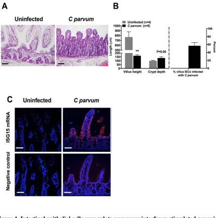
Figure 1. Intestinal epithelial cells upregulate numerous interferon stimulated genes in
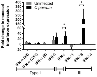
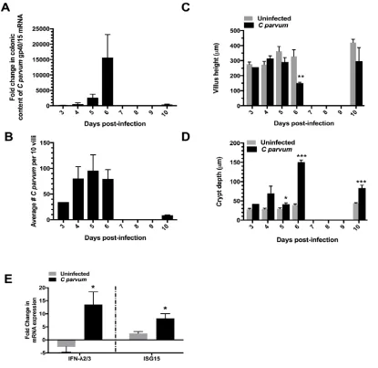
response to C. parvum infection .……….62 Figure 2. C. parvum stimulates ileum mucosal expression of IFN-λ3 and IFN-γ mRNA …..63 Figure 3. A type III interferon response is conserved in neonatal mice infected with
C. parvum………...……….64 Figure 4. Immunoneutralization of IFN-λ exacerbates C. parvum infection.……….65 Figure 5. Non-transformed porcine intestinal epithelial (IPEC-J2) cells express
IFNλR1 and respond to exogenous rHuIFN-λ3 to upregulate ISG15 mRNA expression in a dose and time-dependent manner.………...………...……..66 Figure 6. C. parvum is sufficient to stimulate IEC expression of IFN-λ3 and ISG15
mRNA………..67 Figure 7. Priming of IEC with exogenous IFN- λ decreases the burden of C. parvum
1 CHAPTER I
Intestinal epithelial responses to Cryptosporidium infection Introduction
Cryptosporidium is a protozoal parasite that is responsible for an estimated 8 million cases of diarrhea in young children (<2 years of age) in developing countries every year, leading to 200,000 deaths annually (Kotloff et al., 2013; Sow et al., 2016) with more deaths in young children than malaria and HIV/AIDS combined (Kotloff et al., 2013; Liu et al., 2012; Striepen, 2013). The majority of this diarrheal disease is attributable to two species of the parasite, Cryptosporidium hominis (C. hominis) and parvum (C. parvum) (77% and 10%, respectively) (Sow et al., 2016). Cryptosporidiosis also targets the severely
immunocompromised worldwide (Kotloff et al., 2013; McDonald et al., 2013); conditions leading to immunodeficiency and enhanced cryptosporidial susceptibility include congenital immunodeficiency (e.g.CD40L deficiency) (Cosyns et al., 1998), immunosuppressive drug treatment (Florescu and Sandkovsky, 2016), concurrent infections, and acquired
immunodeficiency syndrome (AIDS) (O'Connor R et al., 2011).
2 Cryptosporidium is a minimally invasive, apicomplexan protozoal parasite that
primarily infects small intestinal epithelial cells causing both a malabsorptive and secretory diarrhea that is self-limiting in healthy, immunocompetent individuals (Farthing, 2000), however, it leads to chronic, debilitating diarrhea, and even death in immunocompromised patients (O'Connor R et al., 2011). Accordingly, a host’s immune response, particularly that orchestrated by the intestinal epithelial cells, plays a critical role in determining the
susceptibility and clinical outcome of cryptosporidiosis (Bouzid et al., 2013; Gookin et al., 2002; McDonald et al., 2013). Therefore, a lot of research effort has gone towards
understanding the epithelial response to C. parvum in hopes that it can provide insight into novel therapeutic targets capable of ameliorating disease, expediting recovery from, and even possibly preventing infection. Our current knowledge of the epithelial cells role in response to Cryptosporidium will be reviewed here.
Infection of enterocyte by Cryptosporidium Excystation and attachment
Transmission of C. parvum is fecal-oral, and outbreaks are typically waterborne (Putignani and Menichella, 2010). Infection occurs following ingestion of environmentally resistant, thick-walled, sporulated oocysts. Upon ingestion, the oocysts pass into the small intestinal tract and excyst in the presence of bile acids and salts to release infective
3 attachment and subsequent invasion (Blackman and Bannister, 2001; Chen et al., 2004b). Although many of these proteins are uncharacterized, we do know that C. parvum expresses several surface proteoglycans. Examples include circumsporozoite-like glycoprotein (CSL), C-type lectins (CpClec), and elongation factor-1α (EF-1α), which can bind lectins (CSL and CpClec) and heparin sulfate (EF-1α) expressed on the epithelial surface (Bhalchandra et al., 2013; Inomata et al., 2015; Langer and Riggs, 1999; Langer et al., 2001; Ludington and Ward, 2016; Matsubayashi et al., 2013). Interestingly, in a study performed in Chinese Hamster Ovary (CHO) cells that varied only in their expression of heparin sulfate, it was determined that efficient invasion of sporozoites requires surface expression of heparin sulfate. The authors hypothesized that differential expression of heparin sulfate and other sulfated polysaccharides in hosts and throughout the length of the intestine might explain variability in C. parvum’s host or tissue tropism (Inomata et al., 2015). In contrast, there was a dose-dependent inhibition of C. parvum invasion in the presence of heparin (Inomata et al., 2015).
Upon sporozoite attachment, the host apical membrane protrudes and envelopes the parasite; this process is necessary for the establishment of C. parvum infection. The steps involved in membrane protrusion involves epithelial cytoskeletal rearrangement through the recruitment of host-actin (Elliott and Clark, 2000; Elliott et al., 2001; O'Hara et al., 2008), as well as redistribution of Na+-glucose and water transport proteins, sodium-dependent-glucose
4 promoted invasion (Chen et al., 2005a). Once internalized, C. parvum resides within a
unique, intracellular yet extracytoplasmic compartment known as the parasitophorous vacuole (PV) which is separated from the host cytoplasm by an electron dense junctional complex predominately composed of host-actin and actin-related proteins (Chen et al., 2004a; Elliott and Clark, 2000; Elliott et al., 2001). Host actin recruitment is dependent on Cdc42 GTPase activation; this process has been shown to be necessary for parasite invasion in a biliary epithelial (H69) model of infection (Chen et al., 2004a).
Intracellular development
Cryptosporidium spp. is an obligate intracellular parasite and therefore depends on the host epithelial cell for survival. For example, once enveloped by the host cell within the PV, C. parvum forms a feeder organelle at the parasite-host interface. The feeder organelle is unique to apicomplexans and, as the name suggests, it is hypothesized that this organelle allows C. parvum to siphon off essential nutrients and energy from the host cytosol (Bouzid et al., 2013; Zhang et al., 2015). This hypothesis was substantiated by a study that
demonstrated that C. parvum localizes important transport proteins that are structurally similar to both cystic fibrosis conductance regulator (CFTR) chloride channel and other multidrug resistance protein family of ATP-binding cassette proteins, to this organelle (Perkins et al., 1999). Additionally, the parasite is equipped with its own L-lactate
5 et al., 2017; Zhang et al., 2015). Lastly, C. parvum actively scavenges low-density
lipoprotein (LDL) cholesterol from the host enterocyte which also is thought to support its intracellular development. (Ehrenman et al., 2013).
Once infection is established, C. parvum undergoes multiple rounds of asexual (merogony) and sexual (gametogony) reproduction within the same host, ultimately resulting in the production of both thin-walled and thick-walled oocysts (Gookin et al., 2002;
Putignani and Menichella, 2010). Thin-walled oocysts are involved in a cycle of
autoinfection of intestinal epithelial cells whereas thick-walled oocysts are environmentally resistant and shed into the environment where they can eventually infect a new host (Gookin et al., 2002).
Pathogenesis of Cryptosporidial Diarrhea
C. parvum is considered a minimally invasive parasite that primarily infects the small intestine resulting in absorptive enterocyte loss and subsequent villus blunting crypt
elongation, and malabsorptive diarrhea (Argenzio et al., 1990). Villous atrophy, in addition to crypt elongation, has been described in animal models during peak infection (day 3-5) in neonatal mice and piglets (Argenzio et al., 1990; Rasmussen and Healey, 1992). However, although epithelial changes are commonly observed, in some patients, diarrhea can result without many histopathologic changes, and it is believed that C. parvum-induced
prostaglandins may be responsible. Upon infection, the intestinal mucosa produces both prostacyclin (PGI2) and prostaglandin E2 (PGE2). In vitro, C. parvum stimulates
prostaglandin G/H synthase 2 production, and PGE2 synthesis by intestinal epithelial cells
6 on intestinal epithelial cells, ultimately enhancing chloride secretion via stimulation of
chloride channels that are enriched in the intestinal crypts (e.g., CFTR) and by concurrently inhibiting NaCl absorption by the villus enterocytes. Supporting this mechanism, in a piglet model of infection, indomethacin, a prostaglandin synthesis inhibitor, restored normal NaCl absorption despite a significant loss of villous surface area (Argenzio et al., 1993). Together, enhanced secretion and decreased absorption results in profuse, watery diarrhea (Clark, 1999).
Barrier Function Loss through Modulation of Tight Junctions
Intestinal epithelial cells (IECs) form a confluent monolayer held together by a linked network of tight junctional proteins that impart both size and charge selectivity to the
intercellular junctions and space. Collectively, tight junctional proteins form an important, selectively permeable barrier that functions to absorb water and nutrients as well as to prevent the translocation of pathogens or commensal bacteria, toxins, and antigens from the intestinal lumen to the systemic circulation. Although minimally invasive, C. parvum does decrease intestinal barrier function; this has been seen in naturally infected humans (Dagci et al., 2002; Goodgame et al., 1995; Zhang et al., 2000), and numerous animal models
7 1972; Tang and Goodenough, 2003), and they facilitate paracellular diffusion of sodium and water. Despite their likely importance in C. parvum pathophysiology, less than a handful of papers have investigated C. parvum’s influence on tight junctional protein expression or distribution (Anbazhagi et al., 2007; Bartelt et al., 2016; de Sablet et al., 2016). While there is a consensus that C. parvum alters tight junctional proteins, there seems to be little
agreement on which proteins are affected, likely due to the wide variety of models used and the inconsistency in which tight junctional proteins are investigated. Furthermore, we still know very little about the functional consequences of C. parvum-induced tight junctional disturbances.
The main groups of tight junctional proteins that have been investigated during C. parvum are claudins, zonula occludens 1 (ZO-1), and occludin. Claudin-2 protein was shown to be upregulated in a murine model of cryptosporidiosis (Bartelt et al., 2016). This finding could have relevance to the pathophysiology and resolution of cryptosporidial diarrhea, given that claudin-2 upregulation has been demonstrated to enhance diarrhea and mediate pathogen clearance during Citrobacter rodentium infection (Anbazhagi et al., 2007). In a separate study, claudin-4 protein expression was decreased in the face of infection (Kumar et al., 2018); alterations in the expression of other claudin proteins have not been identified (de Sablet et al., 2016; Kumar et al., 2018). It is possible that different species of
Cryptosporidium target unique epithelial tight junctional proteins. For example, while studies using C. parvum did not observe changes in zonula occludens-1 (ZO-1) expression or
8 investigated in three different studies, the findings were descriptive and overall contradictory, even when using similar in vitro models (Bartelt et al., 2016; de Sablet et al., 2016; Kumar et al., 2018).
Some intestinal pathogens, like Salmonella typhimurium, non-selectively damage tight junctions by recruiting neutrophils to the site of infection, facilitating their paracellular movement and mucosal invasion and translocation (Kohler et al., 2007). Interestingly, while Cryptosporidium also promotes neutrophil influx to the intestinal mucosa (Gookin et al., 2004; Zadrozny et al., 2006), this influx has been linked to an increase in barrier function without having a significant influence on epithelial parasitism or villus atrophy (Gookin et al., 2004; Zadrozny et al., 2006).
Models of Cryptosporidiosis
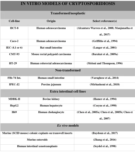
A wide variety of in vivo and in vitro animal models have been employed to study Cryptosporidium. An overview of the most commonly used cell culture and animal models of cryptosporidiosis are summarized in Tables 1 and 2. Although the small intestine is
considered the primary site of natural Cryptosporidium infection in humans and most immunocompetent animal models (Farthing, 2000), extra-intestinal cryptosporidiosis has been reported in immunocompromised humans (HIV/AIDs or CD40 ligand deficiency) and mouse models, with biliary cryptosporidiosis and occasionally colonic cryptosporidiosis being the most common extra-intestinal sites (Hayward et al., 1997; Mead et al., 1991; Reijasse et al., 2001; Stephens et al., 1999; Vakil et al., 1996).
Accordingly, in vitro cell culture models predominately consist of primary or
9 adenocarcinoma cell lines, particularly the HCT-8 line, are most commonly used. They are known to harbor the largest burden of infection, which can be particularly useful in studies aimed at identifying novel therapeutics by performing high-throughput screening
(Manjunatha et al., 2017; Meloni and Thompson, 1996; Upton et al., 1994; Zhang and Zhu, 2015). Overall, despite their frequent use in prior studies, the utility of cancerous cell lines is limited by the fact that they are not derived from the small intestine and are transformed. This fact raises particular concern when these cell lines are used to study host-pathogen interactions, as it is likely that these cancerous epithelial cells respond differently to C. parvum infection. Furthermore, Cryptosporidium undergoes both asexual and sexual development in vivo; however, most cell culture models only support asexual development (Aji et al., 1991; Flanigan et al., 1991).
In an effort to use more biologically relevant models to study C. parvum’s host-pathogen interactions, researchers have increased use of non-transformed or primary cell lines (e.g., FHs 74 Int. and IPEC-J2) or ex-vivo techniques (enteroids) to study
10 studies, long-term propagation of C. parvum was only achieved in large animal models of infection (calves, gnotobiotic pigs).
By far the most commonly utilized models of Cryptosporidium parvum infection are in mice; both immunocompromised and immunocompetent strains have been used. Mice are popular models because they can be genetically or therapeutically manipulated, and a wide range of murine-specific experimental reagents are available. While they are useful for answering certain questions and for investigating key signaling pathways, it is important to remember that they are unnatural hosts, being inherently resistant to Cryptosporidium parvum, and completely resistant to Cryptosporidium hominis, the two most common strains of Cryptosporidium causing human disease. Additionally, mice do not become diarrheic in the face of infection, making them unsuitable models to study mechanisms of or therapy for clinical disease.
Calves and piglets are superior animal models than mice given that they are natural hosts of infection and develop clinical signs of infection. For example, piglets develop clinical disease that is very similar to humans, characterized by villous atrophy, crypt
hyperplasia and impaired sodium-glucose absorption (Argenzio et al., 1990; Farthing, 2000). Despite their relevance as a model, unfortunately, the use of piglets and calves in
Cryptosporidium research is frequently precluded by the fact that they require large animal BSL-2 facilities, are expensive, and cannot be readily genetically or therapeutically
manipulated.
11 2017). Accordingly, as C. parvum is capable of infecting most mammalian species and a wide variety of cell lines, it is used as the infectious agent in the majority of cryptosporidiosis models.
Although the majority of the studies reviewed here used mice to study the immune response to C. parvum, it is important to reiterate that mice do fully recapitulate the disease seen in humans, and therefore are less powerful models than calves or piglets. Adult wild-type mice are resistant to C. parvum infection and, therefore, either neonatal or
immunocompromised mouse models must be used (e.g. severe combined immunodeficiency disease (SCID), interferon-γ, STAT1 knockouts, and protein malnourished) (Bartelt et al., 2016; Benamrouz et al., 2012; Ehigiator et al., 2007; Griffiths et al., 1998; Rasmussen and Healey, 1992).
The literature also contains studies that utilized a variety of uncommon
Cryptosporidium model species, including rats, hamsters, guinea pigs, lambs, primates, and immunocompromised (SCID) foals (Bjorneby et al., 1991; Chrisp et al., 1990; Gibson and Wagner, 1986; Miller et al., 1990; Rehg, 1995; Rossi et al., 1990; Tzipori et al., 1982).
Host epithelial responses to Cryptosporidium parvum infection
12 Toll-like receptors and NF-kB
Unlike the adaptive immune system, the innate immune response is not specific, nor does it have memory. The innate immune system’s strength lies in its ability to recognize and respond quickly to conserved portions of pathogens (pathogen associated molecular patterns or PAMPs). Toll-like receptors (TLR) are extra- and intracellular pathogen sensors that recognize PAMPs, and, when bound, they can quickly trigger an innate immune response (Chen et al., 2005b; Costa et al., 2011; Yang et al., 2015). More specifically, upon
stimulation of most TLRs, a signaling cascade occurs that stimulates the innate adaptor protein MyD88 (TLR-3 is MyD88 independent), resulting in subsequent activation of the transcription factor, NF-κB, as well as other pro-inflammatory pathways. Nuclear
translocation of NF-κB promotes the transcription of pro-inflammatory cytokines (e.g.TNF- α, IL-8), enzymes (e.g. iNOS and cyclooxygenase 2), antimicrobial peptides and miRNAs by the epithelial cells that are ultimately translated and secreted (Abal-Fabeiro et al., 2014; Abreu, 2010; Eckmann et al., 1993; Gookin et al., 2006; Hu et al., 2013; McClure and Massari, 2014; Zhou et al., 2012). Some of these epithelial-derived cytokines will be discussed in greater detail in the following sections.
The exact mechanisms by which TLRs promote epithelial defense against C. parvum are still poorly understood. Two extracellular TLRs, TLR2 and 4, which bind lipopeptides and lipopolysaccharide (LPS), respectively (Fusunyan et al., 2001; Yamamoto and Takeda, 2010), are upregulated during C. parvum infection in mice (Costa et al., 2011) and thought to recognize and initiate an immune response against C. parvum, at least in humans
13 in human enterocytes and cholangiocytes in vitro resulted in secretion of exosomes that contained antimicrobials (Hu et al., 2013). Mice deficient in TLR4, that were infected with C. parvum by direct inoculation of oocysts into the gallbladder, developed exacerbated and more persistent infections compared to wild-type controls (O'Hara et al., 2011).
Interestingly, three studies have demonstrated that intraperitoneal or oral treatment of mice with TLR 3 and 9 agonists, Poly I: C and CpG, respectively, prior to orogastric
infection with C. parvum, resulted in significant reductions in parasite burden (80-95% in one report that used CpG) (Barrier et al., 2006; Bartelt et al., 2016; Lantier et al., 2014). The epithelial defense-promoting effects of these TLR agonists was mainly attributed to
promotion of IL-12, Type I, and II IFN production. The exact origin of these cytokines was not investigated; however immune cells, not epithelial cells, are considered the primary cellular sources. The possibility that these TLR agonists also promoted the newly discovered cytokine, Type III IFN (IFN-λ) was not investigated in these studies. Stimulation of a large number of TLRs on IECs has been demonstrated to promote Type III IFN production
(Odendall et al., 2017). Accordingly, it is likely that IFN- λ played a role in these studies and impacted their results.
Finally, while most studies have demonstrated that TLR/NF-κB activation promotes epithelial clearance of C. parvum, NF-κB activation can also have the exact opposite effect. As a result of C. parvum infection, there is initial, severe loss of infected enterocytes via apoptosis (mediated by executioner caspase 3 activation) which ultimately results in villous atrophy. One study identified that following this initial loss of enterocytes, activated NF-κB promotes the production of the X-linked inhibitor of apoptosis (XIAP). As the name
14 promoting the degradation of activated/cleaved caspase-3 (Foster et al., 2012). This study identified a unique mechanism by which C. parvum can resist its own clearance.
iNOS, nitric oxide and the nitric oxide derivative, peroxynitrite
Inducible nitric oxide synthase (iNOS) is required for the formation of nitric oxide (NO) and promoted by NF-κB activation (Gookin et al., 2006). Intestinal epithelial cells are the primary producers of iNOS during C. parvum infection (Gookin et al., 2004; Leitch and He, 1999). Subsequent increased production of NO by the intestinal mucosa during C. parvum infection results in anti-microbial/anti-parasitic effects (Gookin et al., 2005; Leitch and He, 1999). Two research groups have investigated the in vivo significance of iNOS/NO expression during C. parvum infection (Gookin et al., 2005; Gookin et al., 2006; Gookin et al., 2004; Leitch and He, 1994, 1999). In one study, iNOS was systemically inhibited in C. parvum-infected neonatal BALB/c mice, blocking downstream NO production resulting in exacerbation of C. parvum infection (Leitch and He, 1999). In another study, iNOS was inhibited in neonatal pigs, resulting in enhanced epithelial parasitism and fecal oocyst shedding. These authors demonstrated that iNOS/NO’s anti-C. parvum effects were in part due to localized production of peroxynitrite, a potent oxidant that forms when NO interacts with superoxide (Gookin et al., 2005).
Tumor Necrosis Factor-α (TNF-α)
15 vitro, ex vivo and in vivo models (Kirkpatrick et al., 2006; Lacroix et al., 2001; Lean et al., 2006; Robinson et al., 2001; Seydel et al., 1998).However, there is also a study in humans that demonstrated that TNF-α induction may not always be a consistent feature of infection (Alcantara et al., 2003). In an ex vivo study, TNF-α production significantly increased in both human intestinal xenografts in SCID mice and wild-type infected mice during C. parvum infection (Seydel et al., 1998). The in vivo significance of TNF-α has been demonstrated in two separate studies; SCID mice treated with TNF-α neutralizing antibodies and TNF-α knockout mice were utilized. Both researchers found that depletion of TNF-α did not affect C. parvum susceptibility or disease outcome (Chen et al., 1993; Lean et al., 2006). This is in contrast to another study where IFN-ɣ knockout mice were treated with exogenous TNF-α, and they found that infection severity was ameliorated (Lacroix et al., 2001). While those findings were somewhat conflicting, some in vitro studies have had more consistent findings. For example, one study determined that pretreatment with exogenous TNF-α on HT-29 cell line inhibited C. parvum infection (Pollok et al., 2001).Another study of the human
enterocyte line CMT-93, pre-treated with TNF-α, found that C. parvum invasion was inhibited in a dose-dependent manner (Lean et al., 2006). In summary, conflicting reports exist about the role of TNF-α on C. parvum infection; however, the variation in infection model may explain the disparate conclusions.
Antimicrobial peptides (AMPs)
16 Hu et al., 2013). Stimulation of TLRs and other PRRs is thought to be the primary
mechanism by which epithelial secretion of AMPs is enhanced (Hu et al., 2013). Of the many different AMPs that exist, β-defensin’s role in epithelial defense against C. parvum is best characterized. Several studies have touched on the significance of β-defensins during C. parvum infection in humans, mice as well as in calves (Carryn et al., 2012; Chen et al., 2005b; Tarver et al., 1998; Zaalouk et al., 2004). One study quantified the expression of 3 different human β-defensins (hBD1-3) in a human colonic cell line (HT-29 cell line) and found that C. parvum induced differential expression of each defensin, specifically a partial reduction in hBD-1, an induction of hBD-2, and no effect on hBD-3 gene expression (Zaalouk et al., 2004). Exposure of C. parvum sporozoites to exogenous hBD-1 and hBD-2 prior to infection limited their ability to infect murine colonic cells (CMT-93) in vitro
(Zaalouk et al., 2004). In a separate study, this direct antimicrobial effect of β-defensin 1 and 2 was enhanced when combined with a monoclonal antibody (Mab 3E2) that targets C. parvum’s apical complex and surface glycoprotein ligand CSL (Riggs et al., 1997).
17 IFN-γ dependent decrease in the number of paneth cells in the small intestine (Raetz et al., 2013).
Interleukin-8 (IL-8) and neutrophils
Interleukin-8 is a potent epithelium-derived neutrophil chemotactic agent shown to be significantly up-regulated in humans in vivo in Cryptosporidium-infected children, ex vivo in human intestinal xenograft models in SCID mice, and in vitro in human Caco-2 intestinal epithelial cells (Alcantara et al., 2003; Maillot et al., 2000; Seydel et al., 1998).
Corresponding to this increase in IL-8, neutrophils have been shown to be increased in the intestinal mucosa of infected humans, as well neonatal piglets (Gookin et al., 2005; Seydel et al., 1998).
18 Host-derived MicroRNAs/ Parasite derived non-coding RNAs
Epithelial cells produce miRNAs in response to a wide-variety of pathogens,
including C. parvum (Gong et al., 2011; Hu et al., 2013; Hu et al., 2010; Maudet et al., 2014; Piedade and Azevedo-Pereira, 2016). One study identified that a panel of miRNAs (mir-125b-1, mir-21, mir-30b, and mir-23b-27b-24-1) were up-regulated in infected human cholangiocytes (H69). Functional inhibition of some of these miRNAs resulted in an increased C. parvum burden (Zhou et al., 2009). To date, the predominant mechanisms by which miRNAs influence epithelial defense against C. parvum is by regulating secretion of antimicrobial products or by facilitating leukocyte adhesion and transmigration. For example, miR-27b stabilizes iNOS mRNA, ultimately facilitating NO production. However, in a separate study, downregulation of let-7 miRNAs promoted luminal exosome release from IECs; these exosomes contained antimicrobial peptides, beta-defensin 2, and LL-37 (Abal-Fabeiro et al., 2014). Infected intestinal epithelial cells also down-regulate miRNA-221, resulting in up-regulation of intercellular adhesion molecule 1(ICAM-1) and facilitation of T-lymphocyte adhesion (specifically Jurkat cell line) to infected epithelial cells (Gong et al., 2011). The majority of the work examining the significance of miRNAs during C. parvum infection has been conducted using a cholangiocyte model of infection. Given that intestinal epithelial cells are the most common site of infection, the influence of IEC miRNAs on C. parvum infection deserves to be explored.
Interestingly, although C. parvum does not possess the machinery to make miRNAs (Abrahamsen et al., 2004; Ming et al., 2017), it was recently discovered that during infection, C. parvum injects non-protein coding RNA’s (ncRNAs) into epithelial cells which
19 parvum-derived RNAs have demonstrated ability to suppress the expression of host cadherin 3 (CDH3), also known as P-cadherin, lysyl-oxidase like 4 (LOXL4), β-defensin-1 (DEFB1), and DAZ-interacting zinc finger protein (DZ1P1). The transcriptional suppression of β-defensin-1 by C. parvum is particularly intriguing, given that, as mentioned earlier, β-defensins are thought to have anti-C. parvum potential (Zaalouk et al., 2004).
Interferons
Interferons (IFN) are secreted cytokines that are categorized into Types I-III. They are produced mainly in response to intracellular pathogens and are most well known for their ability to “interfere” with virus infection and replication, playing a critical role in anti-viral defense (Barakat et al., 2009b; Chen et al., 1993; Hayward et al., 2000; Pollok et al., 2001). Each type of IFN mediates their anti-pathogen effects by binding to and stimulating distinct heterodimeric receptors that ultimately lead to activation of the JAK/STAT pathway and upregulate the transcription of interferon-stimulated genes (ISGs) (Platanias, 2005).
20 Type I interferon (Interferon-α/β)
To date, only one study has been published examining the role of IFN α/β during the C. parvum infection. This study examined IFN α/β significance both in vitro using murine rectal polypoid carcinoma cells (CMT-93) and human colonic adenocarcinoma cells (Caco-2) and in vivo in SCID mice treated with or without IFN α/β neutralizing antibodies. In both models, IFN-α/β mRNA was significantly up-regulated within 24 hours of C. parvum infection. When Caco-2 cells were primed with various isotypes of IFN-α and IFN-β before infection, there was significant inhibition of C. parvum parasite burden. In vivo, SCID mice were treated with IFN-α/β neutralizing antibodies before infection and the parasite burden increased drastically. This study was the first to identity an innate host response capable of defending IECs against C. parvum involving IFN-α/β (Barakat et al., 2009b).
Type II interferon (Interferon-γ)
21 one study, researchers infected IFN-γ-KO mice and found that mice either died or developed chronic C. parvum infections. They then went on to compare the cytokine response in both groups and found that wild-type neonatal mice up-regulated IFN-γ, IL-1, and TNF-α. However, in KO mice, TNF-α was not up-regulated. This outcome indicated that TNF-α production seems to be IFN-ɣ-dependent and that a loss in TNF-α production may be linked to increased susceptibility to infection (Lacroix et al., 2001). As discussed previously, studies have ascribed variable importance to TNF-α in C. parvum pathogenesis.
The importance of IFN-γ is further evidenced by the fact that SCID and IFN-γ knockout mice when treated with IFN-γ neutralizing antibodies, became increasingly susceptible to C. parvum infection (Chen et al., 1993; Leav et al., 2005). In another study, pre-treatment with IFN-γ of three different human enterocyte cell lines, each with varied expression of the IFN-γ receptor, induced resistance to C. parvum in a seemingly receptor-dependent fashion. This result indicates that IFN-γ, directly via its receptor, mediates the anti-C. parvum response, rather than having a direct anti-parasitic effect (Pollok et al., 2001). Overall, there seems to be a consensus that IFN- γ is vital for host resistance against and clearance of C. parvum infection.
Type III Interferon (Interferon-λ)
Type III IFN, also known as IFN-λ, is the most recently discovered group of IFNs. There are four main subtypes (λ 1-4). IFN-λ 1-3 genes were first described in 2003 (Kotenko
et al., 2003a; Sheppard et al., 2003), and IFN-λ4 was first described in 2013
(Prokunina-Olsson et al., 2013). Across species, there is great variability in the presence or expression of
22 for IFN λ2, and mice lack an IFN-λ 4 gene (Coordinators, 2018). For humans, IFN-λ4 is
unique in that it can be either a gene or pseudogene, depending on the absence or presence of
a frameshift mutation capable of preventing the expression/secretion of IFN-λ4, respectively.
The prevalence of this mutation in the human population is high, particularly in Asian (93%),
African (63%) and European (23%) populations, indicating that it has likely undergone
positive selection (Prokunina-Olsson et al., 2013). One explanation for this positive selection
is that a failure to secrete IFN-λ4 has been linked to improved clearance of Hepatitis C virus
(HCV). This seems somewhat counter-intuitive given that IFN-λ4 can stimulate weak
anti-viral responses; however, these responses are ineffective at clearing HCV, and additionally,
they decrease the responsiveness of cells to other more powerful Type I and Type III IFNs
that are actually capable of clearing HCV (Hong et al., 2016; Prokunina-Olsson et al., 2013).
In the intestine, IFN-λ is primarily produced by IECs (Hernandez et al., 2015;
Mahlakoiv et al., 2015; Pott et al., 2011), however dendritic cells, eosinophils and mast cells
are also reported sources (Chen et al.; Chiriac et al., 2017; He et al., 2012; Raki et al., 2013).
The production of IFN-λ can be induced in response to mucosal injury (Chiriac et al., 2017),
as well as in response to viral and bacterial pathogens, typically through stimulation of PRRs
(e.g., TLRs and RIG-I) (Odendall et al., 2014; Odendall et al., 2017). All TLRs have been
shown to induce IFN-λ gene transcription in intestinal cells. However, stimulation of some
TLRs (e.g., TLR 2 and 5) have been shown to induce it more strongly than others (Odendall
et al., 2017). Pathogen stimulation of intracellular RIG-I receptors are also linked to Type III
IFN expression; RIG-I activates mitochondrial antiviral signaling protein (MAVS) which is
associated with either mitochondria or peroxisomes, resulting in stimulation of transcription
23 MAVS associated with peroxisomes preferentially induces transcription factors that promote
IFN-λ gene transcription, and peroxisome content in cells increases with differentiation and
is linked to a cell's propensity to respond with a Type III response (Odendall et al., 2014).
Transcription factors primarily responsible for Type III IFN gene transcription are NF-kB,
and interferon regulator factors 1, 3, and 7 (IRF1,3,7) (Syedbasha and Egli, 2017); IRF1, in
particular, has been shown to selectively induce Type III IFN over Type I IFN (Odendall et
al., 2014; Siegel et al., 2011; Ueki et al., 2013).
After transcription, translation, and secretion, all subtypes of IFN-λ signal through the transmembranous, heterodimeric receptor composed of IFNλR1 and IL-10R2 chains and
stimulate the JAK/STAT pathway. The IFNλR1 chain is specific to IFN- λ, whereas IL10R2
is shared with IL-10 (Kotenko et al., 2003a). Interferon-λ binds first to the IFNλR1 chain
leading to recruitment of IL10R2 to the complex. Both receptor chains are directly associated
with Janus tyrosine kinases, specifically Jak1 and Tyk2, which mediate phosphorylation of
the intracellular domain of IFNλR1, allowing for docking and phosphorylation of Signal
Transducer and Activator of Transcription 1 and 2 (STAT1/2) proteins. Subsequently, in the
cytoplasm, STAT1 and 2 binds to IRF 9 to form the IFN-stimulated gene factor 3 (ISGF3)
complex. This complex translocates to the nucleus and binds to specific nucleotide sequences
called interferon-stimulated response elements (ISREs) present in the promoter region of
interferon-stimulated genes (ISGs), ultimately leading to ISG transcription (Kotenko et al.,
2003b; Sheppard et al., 2003; Syedbasha and Egli, 2017).
The intracellular signaling cascade for IFN-λ is identical to that of Type I IFN, and,
as such, both cytokines induce largely overlapping ISG signatures (Witte et al., 2010; Zhou
24 functions were largely redundant to Type I IFN. However, we now know that type III
responses predominately occur at mucosal/barrier sites like the respiratory, gastrointestinal
and urogenital tracts, and the liver. This is primarily due to receptor distribution; the Type I
IFN receptor (IFNAR) is expressed on most nucleated cell types whereas IFNλR is primarily
expressed on epithelial cells at barrier/mucosal sites. Some myeloid cells also express IFNλR
but still seem to have impaired responses to Type III IFN (Witte et al., 2009). Additionally,
in one study, primary intestinal epithelial cells (enteroid culture) infected with an
epitheliotropic virus, upregulated both Type I and Type III IFNs, but preferentially secreted
IFN-λ. This finding provides further evidence that that Type III IFN response is likely the
preferred response of epithelial cells (Pervolaraki et al., 2017).
The importance of IFN-λ for IEC anti-viral defense is now well-recognized and it’s in
vivo significance has been specifically demonstrated in the intestine in response to
epitheliotropic viruses (e.g. norovirus, rotavirus, and reovirus) (Baldridge et al., 2017;
25 To conduct these studies, the majority of researchers have utilized a mouse model first created in 2008 that are genetically modified to lack the IFNλR1 chain (IFNλR1-/) (Ank et al., 2008). One study even specifically depleted IECs of IFNλR1 (Baldridge et al., 2017). As with any immunodeficient mouse model, one of the biggest challenges researchers face when using IFNλR1-/- mice is that they are inherently more susceptible to viral co-infections. One common co-infection that immunodeficient mice often face is the gastrointestinal virus, norovirus (Doom et al., 2009; Henderson, 2008); as such, researchers have to be extremely diligent when screening and housing IFNλR1-/- mice.
While IFN-λ, like other interferons types, are most well-known for their role in anti-viral defense, we are now learning that IFN-λ has a broader role for non-viral pathogen defense, barrier defense in the brain and the intestine, and intestinal mucosal healing. Only a
handful of papers have investigated IFN-λ’s role for non-viral pathogens. For example,
Mycobacterium tuberculosis, Listeria monocytogenes, and Salmonella typhimurium have all
been shown to induce IFN-λ (Bierne et al., 2012; Cohen and Prince, 2013; Lebreton et al.,
2011; Travar et al., 2014). Additionally, IFN-λ signaling has demonstrated importance in
defending against respiratory fungal infection, specifically Aspergillus fumigatus (Espinosa
et al., 2017). To date, there are no published reports of IFN-λ expression or signaling importance in parasitic infection.
26 study identified that Listeria monocytogenes infection induces ISG15 expression and has the capability of limiting infection burden both in vitro and in vivo (Radoshevich et al., 2015). ISG15 proteins are ubiquitin-like and can covalently bind to a wide variety of proteins and influence their expression or cellular distribution through a process called ISGylation (Zhang and Zhang, 2011). Accordingly, to identify the mechanism by which ISG15 limits Listeria infection, these researchers explored ISGylation targets using a proteomics approach. They found that ISG15 targeted endoplasmic reticulum (ER) and Golgi proteins which increased the pro-inflammatory cytokines that are known to counteract Listeria infection, 8, and IL-6 (Radoshevich et al., 2015). The other ISGs mentioned, UBD, GBP1, 2, and TRIM21, all work together to defend against intracellular pathogens that form parasitophorous vacuoles (PV). This mechanism has been best described in the context of the protozoal infection, Toxoplasma gondii (T. gondii). In response to intracellular T. gondii infection, TRIM21, an E3 ubiquitin ligase, is up-regulated, facilitating the ubiquitination of the PV membrane. The presence of ubiquitin then allows the GTPases GBP1 and 2 to bind. This combined process promotes the disruption of the PV, enhancing the exposure of the parasite to the immune mediators within the cytoplasm (Foltz et al., 2017; Haldar et al., 2015).
In addition to promoting pathogen clearance, IFN-λ can promote barrier integrity by
tightening paracellular junctions. This barrier-promoting effect was demonstrated both in the
presence and absence of infection in two separate studies, one at the level of the blood-brain
barrier and the other in the intestine (Lazear et al., 2015; Odendall et al., 2017). In brain
microvascular endothelial cells (BMECs), IFN-λ specifically promoted the relocalization of
the tight junctional protein claudin-5 to the junctional complex. IFN-λ’s influence on other
27 downstream mediators responsible for IFN-λ’s barrier-promoting effect have not been
investigated. The only ISG that has a known barrier promoting effect in the intestine is GBP1
(Schnoor et al., 2009). Upregulation of GBP1 was shown to localize specifically to the IEC
tight junctional protein coxsackie-adenovirus receptor (CAR). Knockdown of GBP1 in IECs
with small interfering RNA (siRNA) specifically decreased transepithelial electrical
resistance (TEER) and enhanced fluorescein isothiocyanate (FITC)-labeled 4 kDa dextran
permeability (Schnoor et al., 2009).
Lastly, IFN-λ upregulation also promotes mucosal defense in response to
non-infectious intestinal diseases, like inflammatory bowel disease (IBD). Expression of IFN-λ
subtypes is up-regulated in IBD patients. Additionally, mice with intact IFN-λ signaling
experience less mucosal damage and colitis when exposed to chemical colitis inducers,
dextran sulfate sodium (DSS) or oxazolone, when compared to IFNλR-/- mice. Additionally,
ex-vivo treatment of murine enteroids with exogenous IFN-λ not only led to increased
epithelial proliferation but also up-regulated several genes known to promote epithelial
defense and wound healing (e.g. USP18, IRF7, APOLP, CXCL10). Overall, these findings
indicate that IFN-λ signaling promotes mucosal healing in the face of non-infectious colitis
(Chiriac et al., 2017).
Conclusions and Future Directions
In conclusion, the epithelial cell is critical for instigating and orchestrating an innate
immune response to Cryptosporidium parvum. That being said, our current understanding of
the epithelial cell’s role in host defense during Cryptosporidium infection is predominately
28 epithelial-derived immune mediators. Additionally, these studies have utilized animal or cell
culture models that do not fully recapitulate human disease (e.g. mice and neoplastic colonic
epithelium, respectively). To date, only two studies, both published by the same research
group, have taken a more global (transcriptomic) approach to investigating IEC responses to
Cryptosporidium, both utilizing in vitro models of C. parvum infection (Mirhashemi et al.,
2018; Yang et al., 2010). Their first study utilized microarray analysis of C. parvum infected
HCT-8 cells and identified through gene ontology (GO) analysis that glycoprotein genes
were among the most significantly upregulated; they then confirmed glycoproteins were
increased and expressed on the surface of HCT-8s with immunofluorescence, however did
not investigate the biological significance of glycoprotein upregulation for infected IECs
(Yang et al., 2010). This research group’s second study, performed 8 years later, took an
almost identical approach however they utilized RNA-sequencing in IPEC-J2 cells. They
found that 813 genes were differentially expressed during infection and identified through
GO analysis that genes related to the spliceosome and cell cycle were among the most
significantly enriched. Unfortunately, while taking a global approach is apt to identifying
novel mechanisms of epithelial defense, these studies failed to perform more mechanistic
experiments that provide more valuable feedback regarding the biological significance of the
identified transcriptomic changes. Accordingly, there is a need for more mechanistic studies
that take an unbiased, global approach to identifying important IEC mechanisms of defense.
Additionally, there is a need for studies to take a global approach utilizing natural models of
infection that effectively recapitulate human disease, for example, calves or piglets. The
added benefit to using these animal models is that IECs would be influenced by signals
29 Table 1. Summary of in vitro/ex vivo Cryptosporidium models
IN VITRO MODELS OF CRYPTOSPORIDIOSIS
Transformed/neoplastic
Cell-line Origin Select reference(s)
HCT-8 Human adenocarcinoma (Alcantara Warren et al., 2008; Manjunatha et
al., 2017)
Caco-2 Human adenocarcinoma (Griffiths et al., 1994)
IEC (4.1 or 6) Rat small intestine (Langer et al., 2001)
CMT-93 Mouse rectal polypoid carcinoma (Barakat et al., 2009a)
HT-29 Human colorectal adenocarcinoma (Meloni and Thompson, 1996)
Non-transformed
FHs 74 Int. Human small intestine (Varughese et al., 2014)
IPEC-J2 Porcine jejunum (Mirhashemi et al., 2018)
Extra intestinal cell lines
MDBK-II Bovine kidney (Hamer et al., 1994)
HepG2 Human hepatocyte (Cosyns et al., 1998)
H69 Human cholangiocyte (Chen et al., 2005a; Chen et al., 2005b; Chen et
al., 2007)
Ex vivo models
Murine (SCID mouse) colonic explants on transwell inserts (Baydoun et al., 2017)
Murine enteroids (Zhang et al., 2016)
30 Table 2. Summary of in vivo Cryptosporidium models
LARGE ANIMAL MODELS
Model Select reference (s)
Calves (Manjunatha et al., 2017; Naciri et al., 1993)
Piglets (Argenzio et al., 1990; Foster et al., 2011; Gookin et al., 2004)
Gnotobiotic piglets (Lee et al., 2017; Pereira et al., 2002)
SMALL ANIMAL MODELS Immunocompetent mice
Adult mice (Garza et al., 2008)
Neonatal mice (Hou et al., 2004; Lantier et al., 2014; Leitch and He, 1999)
Immunocompromised mice
SCID (Barakat et al., 2009b; Benamrouz et al., 2012; Perryman et al., 1993)
T cell receptor deficient (Waters and Harp, 1996)
B-cell deficient (Chen et al., 2003)
RAG -/- (Lukin et al., 2000)
IFN-γ -/- (Griffiths et al., 1998)
TLR4 -/- (O'Hara et al., 2011)
MyD88 -/- (Rogers et al., 2006)
CD40-CD40L (Cosyns et al., 1998)
Athymic nude (Heine et al., 1984)
Germ-free/antibiotic treated (Harp et al., 1988)
Dexamethasone treated (Rasmussen and Healey, 1992)
Protein deficient (Bartelt et al., 2016) (Bartelt et al., 2016)
Uncommon Animal Models
Lamb (Anderson, 1982; Giles et al., 2001)
Gnotobiotic lamb (Snodgrass et al., 1984)
SCID Foal (Bjorneby et al., 1991)
Neonatal rhesus macaques (Miller et al., 1990)
Rabbit (adult/neonatal) (Mosier et al., 1997)
31 Table 2. Summary of in vivo Cryptosporidium models (continued)
Neonatal Rat (Capet et al., 1999; Khaldi et al., 2009; Topouchian et al., 2001)
Immunocompromised Rat:
Cyclophosphamide treated
Steroid treated
(Rehg et al., 1987)
(Brasseur et al., 1994; Rehg, 1995)
32 CHAPTER II
Interferon-λ promotes epithelial defense and barrier function against Cryptosporidium parvum infection
Sylvia H. Ferguson1,4, Derek Foster,2,4, Barbara Sherry3,4, Scott Magness4,7,8, Dahlia M. Nielsen5,6, Jody L. Gookin1,2,4
1Department of Clinical Sciences, College of Veterinary Medicine, North Carolina State
University, Raleigh, North Carolina, USA
2Department of Population Health and Pathobiology, College of Veterinary Medicine, North
Carolina State University, Raleigh, North Carolina, USA
3Department of Molecular Biomedical Sciences, College of Veterinary Medicine, North
Carolina State University, Raleigh, North Carolina, USA
4Comparative Medicine Institute, College of Veterinary Medicine, North Carolina State
University, Raleigh, North Carolina, USA
5Department of Biological Sciences, North Carolina State University, Raleigh, NC, USA 6Bioinformatics Research Center, North Carolina State University, Raleigh, NC, USA
7Department of Medicine, Division of Gastroenterology and Hepatology, University of North
Carolina, Chapel Hill, NC, USA
8Joint Department of Biomedical Engineering, University of North Carolina, Chapel Hill,
33
Abstract
Background and Aims:
The epithelial response is critical for epithelial defense against Cryptosporidium but is poorly understood. To uncover the host strategy for defense against Cryptosporidium, we examined the transcriptional response of intestinal epithelial cells (IECs) to C. parvum in experimentally infected piglets by microarray. Upregulated genes were dominated by targets
of interferon (IFN) and IFN-λ was significantly upregulated in infected piglet mucosa. While
IFN-λ has been described as a mediator of epithelial defense against viral pathogens, there is
limited knowledge of any role against non-viral pathogens. Accordingly, the aim of the study was to determine the significance of IFN-λ to epithelial defense and barrier function
during C. parvum infection. Methods:
The significance of C. parvum induced IFN-λ expression was determined using an immunoneutralization approach in neonatal C57BL/6 mice. Ability of the intestinal
epithelium to upregulate IFN-λ expression in response to C. parvum infection and the influence of IFN-λ on epithelial defense against C. parvum invasion, intracellular
development and loss of barrier function was examined using polarized, non-transformed
porcine intestinal epithelial cell monolayers (IPEC-J2). Specifically, changes in barrier
function were quantified by measurement of TEER and transepithelial flux studies.
Results:
Immunoneutralization of IFN-λ in C. parvum infected neonatal mice resulted in significantly increased parasite burden, fecal shedding, and villus blunting with crypt
34
IFN-λ and ISG15 expression by IECs. Priming of IEC with rHuIFN-λ promoted cellular
defense against C. parvum infection and abrogated C. parvum-induced loss of barrier function by decreasing paracellular permeability to sodium.
Conclusions:
35 Introduction
Cryptosporidiosis causes unrelenting diarrhea in immunocompromised individuals worldwide and is the most common cause of infectious water and foodborne diarrheal outbreaks in the United States, resulting in an estimated 750,000 cases each year (Scallan et al., 2011). In developing countries, Cryptosporidium spp. is the second leading cause of infectious diarrheal death in children under the age of 5 years and results in approximately 200,000 deaths annually (Kotloff et al., 2013; Sow et al., 2016).
Cryptosporidium infection occurs by fecal-oral transmission of environmentally-resistant oocysts. After ingestion, oocysts release infective sporozoites that attach and
become enveloped in the apical membrane of small intestinal villous intestinal epithelial cells (IEC) where they complete a complex and recurring life cycle. When Cryptosporidium infects the villus epithelial cells, they are rapidly shed resulting in profound villus blunting. In this condition, nutrient malabsorption, intestinal secretion, and failure of barrier function result in severe diarrhea, dehydration, starvation, and frequently death. Despite decades of research, there are currently no consistently effective treatments for cryptosporidiosis. Clearance of the pathogen and recovery from infection are reliant on a competent host response and access to supportive care. Use of more insightful therapies to promote recovery of individuals with cryptosporidiosis is hindered by a lack of understanding of how the intestinal epithelium favorably combats the infection.
36 activation and host defense against C. parvum infection. Accordingly, we detected that IECs upregulate numerous interferon-stimulated genes during peak Cryptosporidium infection. By investigating the specific stimulus for this interferon response, we identified that C. parvum infection induces robust mucosal expression of a recently discovered Type III interferon, IFN-λ (Kotenko et al., 2003a; Sheppard et al., 2003).
37 Materials and Methods
Piglet Model of Cryptosporidiosis
One-day-old piglets were infected by orogastric gavage with 108 C. parvum oocysts (Bunchgrass Farms, Deary ID) on day 3 of life and samples were collected at peak infection 3–5 days later. Sections of ileum mucosa were collected for epithelial exfoliation
(microarray), fixation in 10% neutral buffered formalin (histology), or embedded in optimal cutting temperature media and frozen in liquid nitrogen (in situ hybridization). All studies were approved by the Institutional Animal Care and Use Committee of North Carolina State University.
RNA Microarray
The intestinal epithelium was exfoliated from freshly obtained sections of uninfected control (n=4) and C. parvum infected (n=8) porcine ileum mucosa in an oxygenated
chelation buffer containing 2.5 mM glucose and 10 mM EDTA as previously described (Gookin et al., 2006) and frozen at -80°C. Total RNA was isolated and treated with DNase (RNeasy Mini Kit; Qiagen, Germantown, MD) according to manufacturer instructions. RNA quantity and quality were determined by capillary electrophoresis (Agilent 2100 Bioanalyzer; Agilent Technologies, Santa Clara, CA). Fluorescently-labeled cDNA was prepared and hybridized to Affymetrix Porcine GeneChip microarrays by a commercial laboratory
38 expressed genes. A two-fold change in gene expression from the control cells was the
39 images were detected in a GeneChip® Scanner 3000 and expression data was extracted using the GeneChip Command Console Software (AGCC) v 2.0 or later (Affymetrix, Santa Clara, CA).
Quantitative Histopathology
Formalin fixed sections of ileum mucosa were paraffin-embedded, sectioned at a thickness of 5-μm, and stained with hematoxylin and eosin. For each piglet, the average crypt depth and villus height was calculated from measurements of 5 well-oriented villus-crypt units using a brightfield microscope and ocular micrometer. The % infection was determined by dividing the number of infected villus enterocytes by the total number of villus
enterocytes.
Fluorescence In Situ Hybridization
Custom probes designed for hybridization with porcine interferon stimulated gene 15 (ISG15) were purchased (Affymetrix, Santa Clara, CA). Probes were hybridized to formalin-fixed and paraffin embedded (FFPE) 5-µm sections of porcine ileum mucosa (ViewRNA in situ hybridization kit; Affymetrix, Santa Clara, CA) using the manufacturer protocol for 1-plex hybridization. Slides were deparaffinized using xylene (30 minutes) and heat and protease K pre-treated (10 and 30 minutes, respectively). Following initial hybridization, signal amplification was performed using pre-amplifier and amplifier probes using a slide hybridization and denaturation system (ThermoBrite;Leica Microsystem, Wetzlar, Germany). To detect the ISG15 target, slides were incubated with fast-red chromagen (1:30 dilution for 45 minutes), washed and coverslipped using mounting media containing DAPI
40 Leica microsystems, Wetzlar, Germany). Rat ubiquitin (UBC) probes were hybridized to FFPE rat kidney slides as a hybridization control.
Immunoneutralization of IFN-λ2/3 in Neonatal Mouse Pups with Cryptosporidiosis Conventional (specific pathogen-free which includes common gastrointestinal pathogens like norovirus, reovirus, rotavirus, Citrobacter rodentium, Salmonella,
Helicobacter, and Giardia) C57BL/6 pregnant dams (embryonic day 13-15) were purchased (Charles River Laboratories, Durham, NC) and housed in accordance to Biosafety Level 2 guidelines. Litters were assigned to 1 of 4 treatment groups: 1) uninfected control mice treated with isotype (rat IgG) antibody, 2) uninfected control mice treated with rat anti-mouse IFN-λ2/3 immunoneutralizing antibody, 3) C. parvum infected mice treated with isotype (rat IgG) antibody, 4) C. parvum infected mice treated with rat anti-mouse IFN-λ2/3
immunoneutralizing antibody. Each mouse pup was administered either 15 µg of IFNλ2/3 neutralizing antibody (Monoclonal Rat IgG2B Clone #244716) or 15 µg of rat IgG isotype
41 RNA extraction. The ileo-ceco-colic region of the intestine was formalin fixed and paraffin embedded, sectioned at a thickness of 5-μm, and stained with hematoxylin and eosin prior to quantifying the burden of epithelial infection and histomorphometry using light microscopy (Olympus Accu-scope 3025, Corporation, Tokyo, Japan). Digital images of well-oriented individual villus-crypt units were captured (Olympus cellSens software) at a magnification of 400×. The total number of epithelial cell-associated Cryptosporidium parasites were counted for each of 10 well-oriented villus-crypt units. Villus height and crypt depth measurements were obtained from each villus-crypt unit using Image J software.
RNA Extraction and Real-Time PCR Analysis
For RNA extraction from tissue, snap frozen samples of ileum mucosa (piglets) or total distal jejunum/proximal ileum (mice) were placed in ice cold lysis buffer, homogenized, and sonicated to disrupt the tissue. For RNA extraction from cell culture monolayers, media was removed and monolayers were washed 3 times with PBS, lysis buffer was applied directly to monolayers, and cells were scraped off inserts with a sterile cell lifter. Total RNA was extracted using a commercial kit (Ambion PureLink Mini kit; Thermo Fisher Scientific, Raleigh, NC) including DNAse treatment (Turbo DNA free kit, Thermo Fischer Scientific). RNA concentration and purity were quantified by spectrophotometry (Nanodrop ND-1000; Thermo Fisher Scientific). One µg of total RNA was converted to single-stranded cDNA by reverse transcription (high-capacity cDNA reverse transcription kit, ThermoFisher Scientific, Waltham, MA). Following reverse transcription, quantitative PCR was performed in a 15 μL
42 additionally examined following electrophoresis in 1.5% agarose gels stained with GelRed DNA stain (Biotium, Freemont, CA). The identity of amplicons was confirmed by expected size, melting temperature, and commercial sequencing (GeneWiz,South Plainfied, NJ). Comparative threshold cycle analysis was used as described in the LightCycler 480 user manual to calculate relative degree of changes. Expression of the following genes were targeted: C. parvum glycoprotein 40/15 (Cp gp40/15) porcine interferon-alpha (all isoforms, poIFN-α-all), porcine interferon-alpha isoforms 7/11 (poIFN-α-7/11), porcine interferon alpha isoform 9 (poIFN-α9), porcine IFN-beta β), porcine interferon-gamma (poIFN-γ), porcine interferon-lambda 1 (poIFN-λ1), porcine interferon-lambda 3 (poIFN-λ3,) porcine interferon-lambda 4 (poIFN-λ4), porcine interferon-lambda-receptor 1 (poIFN-λR1) porcine interferon stimulated gene 15 (poISG15), murine interferon-lambda2/3 (muIFN-λ2/3), murine interferon-beta (muIFN-β), and murine interferon stimulated gene 15 (muISG15). All primers were purchased from Integrated DNA technologies, Skokie, Illinois.Abundance of specific mRNA transcripts (2-ΔΔCt) were calculated by normalization against expression of a housekeeping gene (porcine: cyclophilin, murine: GAPDH). Primer sequence data and references of origin are shown in Supplementary Table 1). Primers were designed either by the authors or as previously described.
Polarized Intestinal Epithelial Cell Culture Response to Cryptosporidiosis
Non-transformed porcine jejunal epithelial cells (IPEC-J2) were grown in co-culture media which included Dulbecco’s minimal essential medium: Nutrient Mixture F-12
43 µm pore size, either 0.6 cm2 or 4.67cm2; Millipore, Burlington, MA) and cultured until confluent (Transepithelial Electrical Resistance (TEER) ≥ 2,000 Ω × 0.6 cm2or ≥ 1,300 Ω ×
4.67 cm2). TEER was measured using an EVOM2 epithelial voltohmmeter with chopstick electrodes (World Precision Instruments, Sarasota, FL). For immunofluorescence assessment of C. parvum burden, IPEC-J2 cells were seeded onto 8-well chamber slides (Nunc Lab-Tek II; Thermo Fisher, Raleigh, NC) and grown to confluence over a period of 3-4 days prior to use. IPEC-J2 cells were used at passage numbers 38-50. Media was changed every 3-4 days. Infection of IPEC-J2 cells with C. parvum
Cryptosporidium parvum oocysts (Bunchgrass Farms, Deary, ID) were pelleted by centrifugation (13,000 x g for 3 minutes) and treated with 10 mM HCl (Millipore Sigma, St. Louis, MO) in PBS for 60 minutes in a 37° C water bath The oocysts were then pelleted by centrifugation to remove the HCl, reconstituted with 0.2 mM taurodeoxycholate and 22 mM NaHCO3 (Millipore Sigma, Millipore Sigma, St. Louis, MO) in PBS, and incubated for 30 minutes in a 37° C water bath to excyst infective sporozoites. The excysted oocysts were centrifuged and reconstituted in IPEC-J2 culture media. The number of excysted oocysts was counted using a hemacytometer to establish a final concentration of excysted oocysts. A multiplicity of infection of 2:1 of excysted oocysts to IPEC-J2 cells was used to inoculate monolayers. Based on established counts of approximately 5 x 10⁵ IPEC-J2 cells per 30 mm (4.2 cm2) insert at confluence, each monolayer was infected with 1 x 10⁶ excysted oocysts.
Stimulation of IPEC-J2 cells with IFN-λ3