INVESTIGATION
Stringent Analysis of Gene Function
and Protein
–
Protein Interactions Using
Fluorescently Tagged Genes
Ralph A. Neumüller,*,1Frederik Wirtz-Peitz,*,1Stella Lee,†Young Kwon,* Michael Buckner,*,‡ Roger A. Hoskins,§Koen J. T. Venken,** Hugo J. Bellen,**,††Stephanie E. Mohr,† and Norbert Perrimon*,‡,2 *Department of Genetics, Harvard Medical School, Boston, Massachusetts 02115,†Drosophila RNAi Screening Center, Department of Genetics, Harvard Medical School, Massachusetts 02115,‡Howard Hughes Medical Institute, Harvard Medical School, Boston, Massachusetts 02115,§Life Sciences Division, Lawrence Berkeley National Laboratory, Berkeley, California 94702, **Department of Molecular and Human Genetics, Baylor College of Medicine, Houston, Texas 77030, and††Howard Hughes Medical Institute, Baylor College of Medicine, Houston, Texas 77030
ABSTRACTInDrosophilacollections of greenfluorescent protein (GFP) trap lines have been used to probe the endogenous expression patterns of trapped genes or the subcellular localization of their protein products. Here, we describe a method, based on nonover-lapping, highly specific, shRNA transgenes directed against GFP, that extends the utility of these collections to loss-of-function studies. Furthermore, we used a MiMIC transposon to generate GFP traps inDrosophilacell lines with distinct subcellular localization patterns, which will permit high-throughput screens usingfluorescently tagged proteins. Finally, we show thatfluorescent traps, paired with recombinant nanobodies and mass spectrometry, allow the study of endogenous protein complexes inDrosophila.
A
commonly used method for visualizing endogenous proteins without the need for specific antibodies is the “protein trap” approach (Gossler et al. 1989; Morin et al. 2001; Clarket al. 2011). Protein trapping relies on transposons that harbor afluorescent or epitope tagflanked by splice acceptor and donor sites. If such a transposon is inserted into an intron, then its tag is spliced into the open reading frame (ORF) of the trapped gene. This approach has been used successfully in a number of model organisms, including Drosophila, where several collections of green fluorescent protein (GFP)-trapped fly lines have become available over the years (Morin et al. 2001; Clyne et al. 2003; Buszczak et al. 2007; Quiñones-Coello et al. 2007; Rees et al.2011;http://flytrap.med.yale.edu; http://www. flyprot.org).GFP traps have mainly been used to study the endogenous expression patterns of trapped genes or the subcellular localization of their protein products. Here, we show that the GFP tag can also be used to interfere with gene function by RNAi-mediated knockdown of the tagged transcripts. This method, which we refer to as“tag-mediated loss-of-function,” addresses major shortcomings of the classical RNAi approach in which gene-specific sequences are targeted. Furthermore, we show that the GFP tag can be used to efficiently purify endogenous protein complexes for mass spectrometric analy-sis using recombinant nanobodies against GFP. Finally, we screen for mCherry traps inDrosophilacultured cells and de-scribe several lines with mCherry expression in specific sub-cellular patterns for use in high-throughput screening.
Materials and Methods
Fly strains
The following protein traps were described in (Buszczak et al. 2007): dlg1–GFP (CC01936), Spt6–GFP (CA07692), Cp1–GFP (CC01377), Pabp2–GFP (CC00380). Additional protein traps used in this study are listed inSupporting In-formation, Figure S1. nanos-GAL4 was described by Van Copyright © 2012 by the Genetics Society of America
doi: 10.1534/genetics.111.136465
Manuscript received November 3, 2011; accepted for publication December 1, 2011 Supporting information is available online athttp://www.genetics.org/cgi/content/ full/genetics.111.136465/DC1.
1These authors contributed equally to this work.
Doren et al. (1998), and mata4-tub-GAL4 (also known as mat67) is a gift from D. St Johnston. EGFP-shRNA constructs 1, 2 and 3 were generated by annealing the oligos listed in
Table S2 and cloning them into the pVALIUM20 or pVA-LIUM22 vectors (Ni et al. 2011) using NheI and EcoRI. Transgenicflies were established using the attP40 and attP2 landing sites (Grothet al.2004; Marksteinet al.2008). The Spt6-specific shRNA (HMS00364) was obtained from the TRiP at Harvard Medical School.
Fluorescently tagged baits were immunoprecipitated from embryos expressing a GFP-tagged par-6 rescue construct in a par-6 null mutant background (referred to as par-6GFP; Wirtz-Peitz et al. 2008) or from sgg–YFP (CPTI-002603) obtained from the Drosophila Genetic Resource Center, and w2embyros were used as controls.
All tag-mediated loss-of-function experiments in the germ-line were controlled by driving EGFP-shRNAs in the back-ground of the heterozygous GFP trap. The observed phenotypes were invariably wild type, indicating that the targeted genes are neither haplo-insufficient nor subject to transitive RNAi effects; i.e., targeting theGFPexon does not give rise to secondary siRNAs directed against other regions of the transcript (Roignant 2003). Embryos analyzed in Figure 2B were from the following cross: par-6GFP; mat67/UAS–EGFP-shRNA ·
par-6GFP/Y; UAS–EGFP-shRNA. The par-6GFP; mat67 stock served as the control in Figure 2A.
Immunofluorescence
The following antibodies were used: rabbit anti-Vasa (1:500, Santa Cruz, sc-30210), rabbit anti-GM130 (Abcam, ab30637), rabbit Anillin (1:1300, a gift of Tim Mitchison), rat anti-troponin H (1:500), mouse anti-1B1 (1:2, Developmental Studies Hybridoma Bank), rabbit anti-cleaved caspase (1:100, Cell Signaling), rabbit anti-GFP (1:500, Abcam, ab6556), mouse anti-Dlg (1:100, Developmental Studies Hybridoma Bank, 4F3). Immunohistochemistry in ovaries was per-formed as previously described (Neumülleret al.2008). Em-bryos werefixed in 4% (v/v) formaldehyde in PBS/heptane, devitellinized using heptane/methanol, and blocked in 2% (v/v) NGS in PBS, 0.1% (v/v) Triton X-100. Images were acquired on either a Leica TCS SP2 or a Zeiss LSM 710 confocal microscope.
Immunoprecipitation from embryos and mass spectrometry
Overnight collections were extracted with lysis buffer (25 mM Tris–HCl [pH 7.5], 150 mM NaCl, 5 mM EDTA, 1% [v/v] NP-40, 5% [v/v] glycerol, 1 mM DTT, 1·Halt protease and phosphatase inhibitor cocktail [Thermo Scientific]) and de-bris was removed by centrifuging twice at 1200 · g for 5 min. Extracts were cleared by incubation with agarose resin (Thermo Scientific) for 1 hr at 4, followed by centri-fugation at 15,000·gfor 15 min. Immunocomplexes were formed by incubation for 2 hr at 4with the following anti-bodies: anti-GFP nanobodies coupled to agarose beads (10ml of packed beads per IP; ChromoTek, GFP-Trap_A), rabbit
anti-GFP antibodies (5 ml per IP; used in Figure 2, C–E; Invitrogen, A6455) precipitated using Protein A/G agarose (Thermo Scientific), rabbit anti-GFP antibodies (1ml per IP; used inFigure S2, A and B; Abcam, ab6556), rabbit anti-GFP antibodies coupled to sepharose beads (10 ml of packed beads per IP; used in Figure 2F,Figure S2, C and D; Abcam, ab69314). The immunocomplexes were washed four times with lysis buffer, eluted in IgG Elution Buffer (Thermo Sci-entific), and neutralized using 100 mM Tris–HCl (pH 9). The eluates were Western blotted using standard protocols or stained using the PageSilver silver staining kit (Thermo Scientific). The following antibodies were used in Western blotting: rabbit anti-GFP (1:500, Abcam, ab6556), rabbit anti-PKCz(1:500, Santa Cruz, sc-216), mouse anti-a-tubulin (1:1000, Sigma-Aldrich, T6199).
For mass spectrometry the immunocomplexes were washed three times with 10 mM Tris–HCl (pH 7.5) prior to elution to remove detergent. The neutralized eluate was reduced with 5 mM DTT for 30 min at 56and alkylated with 15 mM iodacetamide for 30 min at RT. The alkylation reaction was quenched with 15 mM DTT. The alkylated eluate was digested with 1mg of sequencing grade modified Trypsin (Promega) in 2 mM CaCl2 overnight at 37. The digest was purified on PepClean C-18 spin columns (Thermo Scientific) according to the manufacturer’s instructions, dried in a SpeedVac, and reconstituted in 10ml of 2% (v/v) ace-tonitrile, 0.1% formic acid, of which 4ml was injected into the mass spectrometer. LC-MS/MS analysis was performed essentially as described (Dephoure and Gygi 2011). Peptide spectral matches were filtered to a 2% false discovery rate (FDR).
Immunoprecipitation from cell lines and mass spectrometry
Cells were extracted by incubation in lysis buffer (10 mM Tris–HCl [pH 7.5], 150 mM NaCl, 0.5 mM EDTA, 0.5% [v/v] NP-40, 1 mM PMSF, 1·complete protease inhibitor cocktail [Roche]) for 30 min on ice. The extract was cleared by centrifugation at 20,000 · g for 10 min at 4. mCherry was immunoprecipitated using anti-DsRed nanobodies cou-pled to agarose beads (ChromoTek, RFP-Trap_A) according to the manufacturer’s instructions. The immunoprecipitate was prepared for mass spectrometry and analyzed by LC-MS/MS essentially as described (Sowa et al. 2009; Dephoure and Gygi 2011). Peptide spectral matches were filtered to a 1% FDR.
Generation of mCherry cell lines
repeats and two invertedattP sites forFC31 recombinase-mediated cassette exchange. The resulting plasmid, pGTC, had the following structure: MiL–attP1–SA–mCherry–SD– attP2–MiR.
Drosophila S2R+ cells were transfected with the pCoB plasmid and blasticidin-resistant cells were selected with 25 mg/ml of blasticidin 1 day post-transfection. Resistant cells were cotransfected with the mCherry trap vector de-scribed above (pGTC) and aMinostransposase helper plasmid (Pavlopouloset al.2004) in a 1:1 ratio. Three independently transfected cultures, one for each of the three reading frame constructs, were pooled 2 days post-transfection and cultured for 1 week to allow for maximal expression of the mCherry tag. Approximately 10% of the transfected cells scored positive by FACS and more than 2000 cells, selected from the top 3–5 percentile, were seeded as single cell in 384-well plates. Blasticidin-sensitive feeder cells, 5 · 106, had been added to the plates to promote the survival of these single cells. The cell clones were cultured for 4 weeks in the presence of 12.5 mg/ml of blasticidin to suppress proliferation of the feeder cells. Visible colonies were then isolated, progres-sively expanded to larger culture sizes, and cryopreserved. SeeTable S1for a list of cell lines generated in this study. TheMinosinsertions were mapped by inverse PCR using 0.5mg of genomic DNA, as described (Bellenet al.2011).
Results
Efficient knockdown of protein traps using shRNAs directed against GFP
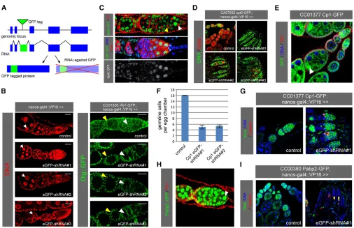
We sought to establish reagents for knocking down GFP-trapped genes using transgenic RNAi (Figure 1A). For this, we opted for small hairpin RNAs (shRNAs) over long dou-ble-stranded RNAs (dsRNAs) because of the consistently high efficacy reported for shRNAs in both soma and germ-line (Haleyet al.2008; Niet al.2011). In addition, despite the small size of the GFP coding sequence, shRNAs allowed us to design multiple nonoverlapping constructs. This pro-vides a choice of constructs if any are found to induce off-target effects in a particular tissue of interest. Consequently, we designed three nonoverlapping shRNAs against the EGFP tag used in the three originalDrosophilaprotein trap screens (Morin et al. 2001; Buszczaket al. 2007; Quiñones-Coello et al. 2007). These shRNAs are predicted to also target the EYFP derivative used in a more recent protein trap screen (Reeset al.2011). We cloned these shRNAs into UAS vectors optimized for either somatic or germline expression (see Materials and Methods).
To validate the efficacy of these constructs, we used nanos-GAL4to drive the shRNAs in the background of a GFP trap inserted in the discs large 1 (dlg) gene (Figure 1B). Germline-specific expression of the EGFP-shRNAs in females resulted in the loss of Dlg–GFP signal in the germline, whereas the cortical signal remained unaffected in the so-matic follicle cells. To extend these observations, we tested a total of 12 additional GFP traps and consistently observed
germline-specific loss of GFP signal upon EGFP-shRNA ex-pression using nanos-GAL4 (Figure S1A; data not shown). Importantly, expression of these EGFP-shRNAs in the germ-line using either nanos-GAL4 or mata4-tub-GAL4 had no adverse effect on germline development as judged from nor-mal differentiation of germaria and egg chambers as well as normal fertility (Figure 1B; data not shown). We conclude that our EGFP-shRNA lines are effective and specific in knocking down GFP-trapped genes in the female germline.
Tag-mediated loss-of-function reveals a role for Spt6 in germline stem cell maintenance
To showcase the utility of these shRNAs in genetic screen-ing, we used them to knock down homozygous viable GFP traps available from the Carnegie collection (Buszczaket al. 2007) that showed strong GFP signal in female germline stem cells (GSCs) (Figure S1A; data not shown). Among these, CA07692 (an insertion inSpt6) was expressed at vari-able levels throughout oogenesis (Figure 1C). The Spt6–GFP signal overlapped with the nuclear DAPI signal, consistent with a recent study implicatingSpt6in transcriptional elon-gation in Drosophila (Ardehali et al. 2009). The 1B1 anti-body, which labels spectrosomes and fusomes, and the Vasa antibody, which labels all germline cells, allowed us to cor-relate Spt6–GFP expression with specific cell types in the germarium. We found that Spt6–GFP was strongly expressed in GSCs adjacent to the stem cell niche (identified as Vasa-positive cells containing a single spectrosome), while lower expression levels were observed in the transit-amplifying cystocytes (distinguished by the presence of a fusome) (Figure 1C).
To assess whether Spt6 is required in GSCs, we used nanos-GAL4 to drive individual EGFP-shRNAs in the back-ground of the Spt6–GFP trap. In contrast to wild-type ova-ries, where Vasa-positive germline cells are detected in egg chambers of all stages, tag-mediated knockdown of Spt6– GFP using three nonoverlapping EGFP-shRNAs resulted in the depletion of Vasa-positive cells from the germline with only somatic cells remaining in the ovaries (Figure 1D). This, together with the finding that knockdown of Spt6 did not trigger apoptosis in the germline (data not shown), suggests thatSpt6is required for GSC maintenance, which is consistent with recent findings that transcriptional elonga-tion is an important regulatory event in stem cell homeosta-sis (Baiet al.2010; Neumülleret al.2011). We corroborated these data using a gene-specific shRNA againstSpt6, which caused a similar phenotype when expressed in the germline (Figure S1C).
Tag-mediated loss-of-function identifies genes required for germ cell survival
in the oocytes of developing egg chambers (Figure 1E). Knockdown of Cp1 using two independent EGFP-shRNAs resulted in egg chambers with fewer Vasa-positive germline cells compared to wild type (Figure 1, F and G;Figure S1D). To identify the underlying cause, we stained for caspase cleavage as a marker for apoptosis. Whereas cleaved cas-pase-positive cells were rarely observed in the wild-type germline, we detected a high frequency of apoptotic germ-line cells upon Cp1 knockdown (Figure 1G). The highest incidence of caspase cleavage was observed in cystocytes (region 2 of the germarium), but apoptotic cells were also detected at later stages of oogenesis. Interestingly, apoptotic cells were not detected in region 1 of the germarium, con-sistent with our observation that ovaries still contained germline cells in 4-day-oldflies.
A third trap identified in our screen is CC00380, an insertion inpolyA-binding protein II(Pabp2). Tag-mediated
knockdown ofPabp2using two independent EGFP-shRNAs resulted in almost complete loss of germline cells (Figure 1I andFigure S1D). The few remaining germline cells showed signs of nuclear fragmentation (data not shown) and stained positive for cleaved caspase (Figure 1I), suggesting that Pabp2is required for cell survival across the germline. Con-sistent with this, Pabp2–GFP was uniformly expressed in the germline (Figure 1H). Ourfindings are also consistent with a study reporting thatflies mutant for a hypomorphic allele ofPabp2are sterile and contain degenerating egg chambers arresting at stage 8 of oogenesis (Benoit et al.2005). The stronger phenotype observed upon tag-mediated knock-down most likely reflects a null or strongly hypomorphic phenotype. Taken together, our tag-mediated knockdown screen revealed a requirement for both Cp1 and Pabp2 in cell survival in the germline, withCp1fulfilling this function mostly at the cystocyte stage of oogenesis.
Tag-mediated loss-of-function in the embryo
Our functional analysis ofSpt6,Cp1, andPabp2in the female germline indicates that tag-mediated knockdown is a power-ful strategy by which to study phenotypes with a high degree of stringency. Besides inducing phenotypes in the germline, shRNAs have been used to deplete the maternal contribution of proteins required in embryogenesis (Niet al.2011). To test if our EGFP-shRNAs are effective in inducing embryonic phe-notypes we turned topar-6, a gene required for the establish-ment of epithelial polarity. While embryos derived frompar-6 homozygous mutant germline clones (par-6GLC) show an em-bryonic lethal defect in cell polarity, the zygotic mutant mostly survives to the larval stage owing to the maternal contribution of Par-6 protein (Petronczki and Knoblich 2001). We used mata4-tub-GAL4 to drive one of our EGFP-shRNAs in par-6 mutant females expressing a GFP-tagged genomic rescue construct (par-6GFP) (Wirtz-Peitz et al. 2008) and analyzed the resulting embryos using antibodies
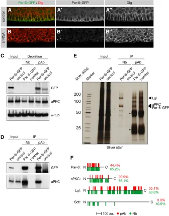
against Dlg and GFP. The ectodermal epithelium of control embryos was organized as a monolayer, and its constituent cells showed a columnar morphology (Figure 2A). In contrast, epithelial cells in RNAi embryos had rounded up and formed an irregular, multilayered, epithelium (Figure 2B), as has been reported for par-6GLC embryos (Petronczki and Knoblich 2001). Furthermore, in control embryos Dlg was confined to the basolateral cell cortex, whereas Par-6–GFP was localized to the subapical region (Figure 2A). In RNAi embryos lacking Par-6–GFP, Dlg was localized all around the cell cortex except in the outermost cells (Figure 2B). Therefore, the EGFP-shRNA directed against par-6–GFP closely phenocopies the epithelial defects inpar-6GLCembryos.
Usingfluorescently tagged genes to isolate endogenous protein complexes
giant larvae (Lgl) (Betschinger et al. 2003). This led us to explore if GFP-tagged fly lines could be used to analyze endogenous protein complexes when combined with re-cently commercialized high-affinity GFP antibodies raised in llamas (Rothbauer et al. 2006, 2008). These so-called nanobodies lack light chains, which allows the heavy chain’s variable domain to be cloned and purified to high titer from a recombinant source. Thus, these antibodies promise to combine the high antigen-binding capacity of polyclonal sera with the specificity of monoclonal antibodies.
To test this we used anti-GFP nanobodies to immuno-precipitate Par-6 frompar-6GFPembryos. Western blotting showed that Par-6–GFP was immunoprecipitated and its binding partner aPKC was co-immunoprecipitated from these embryos by both nanobodies and a polyclonal control serum (Figure 2D). However, in contrast to the control antibodies, the nanobodies completely depleted Par-6 from the extract after as little as 60 min of incubation and even partially de-pleted its binding partner aPKC (Figure 2C andFigure S2, A and B). Although Par-6–GFP is only weakly expressed as inferred from its comparatively faint fluorescence (data not shown), the Par complex containing Par-6, Lgl, and aPKC was clearly detected on a silver-stained gel in immunoprecipitates from200ml of packed embryos (Figure 2E). In addition, these proteins were readily identified by liquid chromatography-tandem mass spectrometry (LC-MS/MS) after in-solution diges-tion of the immunoprecipitate (Figure 2F andFile S1). These proteins were also identified when Par-6–GFP was immunopre-cipitated using agarose-coupled polyclonal control antibodies, albeit with significantly lower peptide coverages. In fact, Star-dust, which so far could be shown to bind Par-6 only when both proteins were overexpressed in cultured cells (Hurd et al. 2003), was specifically identified in the nanobody immunopre-cipitate but not in the polyclonal control immunopreimmunopre-cipitate (Figure 2F andFile S1).
To assess the performance of anti-GFP nanobodies with a different bait we turned to a YFP trap inshaggy(Reeset al. 2011), theDrosophilaortholog for glycogen synthase kinase 3 and a core subunit of the Armadillo destruction complex (Macdonald et al. 2009). Silver staining of the nanobody immunoprecipitate revealed a double band (Figure S2C), which in-solution digestion and LC-MS/MS identified as Shaggy and its well-characterized interactor Axin (Figure S2D). Although both proteins were also identified using
polyclonal control antibodies, the nanobodies recovered a higher amount of bait protein with less background, result-ing in superior peptide coverage. Together, these experi-ments indicate that GFP traps or GFP-tagged genomic rescue constructs can be used in conjunction with anti-GFP nanobodies to interrogate the Drosophilainteractome with high sensitivity and stringency.
Extending the protein trap approach to Drosophila cell lines
Althoughin vivoapproaches are strongly preferred for most Drosophilastudies, cell lines remain a powerful system for
high-throughput RNAi screening. To generate cell lines with fluorescent proteins that label distinct subcellular features for use in such applications, we developed a protein trap approach for DrosophilaS2 cells using the MiMIC transpo-son. MiMIC is aMinos-based protein trap featuring a muta-genic marker cassette containing a splice acceptor site followed by a triple stop (Venkenet al.2011). The cassette is flanked by two inverted FC31 integrase attP sites that allow its replacement with a sequence of interest by recom-binase-mediated cassette exchange (RMCE). As depicted in Figure 3A, we replaced the mutagenic marker cassette with afluorescent protein trap consisting of the mCherry coding sequence in one of three frames flanked by splice acceptor and donor sites.
This modified MiMIC element was mobilized into random positions in S2R+ cells using a transiently trans-fectedMinostransposase (Metaxakiset al.2005). As part of a pilot screen we FACS-sorted more than 2000 mCherry-positive cells and established approximately 50 stable clones from single cells (Figure 3A andTable S1). In a num-ber of these lines mCherry assumed a distinct subcellular distribution, indicative of a successful trapping event (Fig-ure 3, B and C, and Table S1). For example, cell line NPT017 showed a cell-cycle-dependent localization of mCherry, being distributed diffusely in the cytoplasm at interphase and relocalizing to the cleavage furrow at cyto-kinesis. In cell line NPT005, by contrast, mCherry was lo-calized to cytoplasmic puncta, which partially overlapped with BiP, a marker for the endoplasmic reticulum (Otero et al.2010; Figure 3C).
To identify the gene trapped in NPT005 we purified the mCherry-tagged protein using anti-DsRed nanobodies fol-lowed by LC-MS/MS of the in-solution digested immuno-precipitate. This identified chloride intracellular channel (Clic) as the highest-ranked protein hit (Figure 3D), and inverse PCR-based mapping of the MiMIC insertion site con-firmedClicas the trapped gene (data not shown). Using the same strategy we identified atx2 and gap1 as the genes trapped in NPT022 and NPT102, respectively (Table S1; data not shown).
Finally, as proof of principle for RMCE in these cell lines we successfully swapped mCherry for EGFP in NPT101 and NPT102 by cotransfection of an EGFP donor cassette along with FC31 integrase (Figure 3E). Taken together, these data demonstrate that the MiMIC transposon can be used to trap specific genes inDrosophilacell lines and that stable clones of such traps may be established for use in diverse applications ranging from imaging to biochemical studies.
Discussion
Advantages of tag-specific over gene-specific knockdown
method addresses two major challenges in screens based on gene-specific RNAi constructs. First, gene-specific constructs may cause off-target effects, which manifest as false positives in RNAi screens (Ma et al. 2006; Kulkarni et al. 2006; Dietzlet al.2007). Off-target effects can be controlled by rescue of the RNAi phenotype using an RNAi-resistant construct, but such transgenes are not readily available and need to be generated on a gene-by-gene basis (Mohr et al.2010). By contrast, the shRNA used for tag-mediated knockdown can be expressed in the target tissue in the ab-sence of a GFP trap to exclude the possibility of unspecific phenotypes.
Second, the efficacy of gene-specific RNAi constructs varies widely, with ineffective constructs giving rise to false
negatives in RNAi screens (Dietzlet al.2007; Bookeret al. 2011). While the false-negative rate may be lowered by targeting each gene with two or more independent RNAi constructs (Mohret al.2010), the use of a single optimized shRNA in tag-mediated knockdown ensures consistently high efficacy. In addition, the degree of knockdown is readily verified by monitoring GFPfluorescence in the target tissue. GFP-mediated knockdown is therefore a compelling method by which to remove artifacts and ambiguity from RNAi experiments.
Strategies for isolating endogenous protein complexes
typically rely on protein-specific antibody reagents. Because these are time consuming and expensive to raise and recog-nize their protein antigen with widely variable specificity and affinity, an alternative approach is to ectopically express epi-tope-tagged baits, which may then be immunoprecipitated using monoclonal antibodies. However, if such constructs are not expressed at endogenous levels or in the proper spa-tial patterns, then this is predicted to increase the occurrence of false-positive interactors, currently a major challenge of such studies (Gingras et al. 2007). By contrast, the use of GFP traps permits the purification of endogenously expressed protein complexes using a single validated antibody reagent, thereby combining the advantages of both methods.
In addition, our data suggest that the favorable binding properties of GFP-specific nanobodies offset the low endogenous expression levels of many proteins and permit the mass spectrometric detection of even weak interac-tions. Interestingly, neither the interaction between Par-6 and Stardust nor the interaction between Shaggy and Axin were reported in a recent S2 cell-based co-immunoprecip-itation study (Guruharsha et al. 2011), suggesting that many critical interactions are detected only by in vivo assays. Our data, together with another recent study on protein complexes purified from protein traps (Reeset al. 2011), provide an experimental framework for analyzing physical interactions between endogenous proteins in Drosophila.
Perspectives forfluorescent protein trapping in Drosophila
Although it is estimated that approximately 3200 Drosoph-ila genes are permissive to protein trapping by conven-tional P-element and piggyBac transposons, only a few hundred Drosophila genes have so far been tagged with a transgenic GFP trap (Aleksicet al. 2009). Among these, three classes of traps need to be distinguished. First, inser-tions that disrupt protein function and are thus homozy-gous lethal. These traps are not amenable to tag-mediated knockdown, owing to the absence of transitive RNAi in Dro-sophila (see Materials and Methods), and they are similarly unsuitable for biochemical analysis because a tag that disrupts a protein’s activity may also interfere with its physical inter-actions. Second, insertions spliced into only some of a gene’s transcripts (Quiñones-Coello et al. 2007). Such traps have been used to analyze the distribution of cell-type-specific iso-forms (Silies and Klämbt 2010), and we were able to induce germline phenotypes by tag-mediated knockdown of isoform-specific traps. At the same time, we have also encountered a number of lines in which the expression of an unaffected splice form masked the tag-mediated knockdown of the trap-ped transcripts (data not shown). Finally, the third class is composed of homozygous viable insertions that trap all of a gene’s transcripts and which are generally the most useful. While the genome coverage of usable protein traps remains limited, a large-scale effort is currently ongoing to
generate over 6000 insertions using the MiMIC transposon (Venkenet al.2011). Although these traps will need to be individually converted to GFP traps by RMCE, we anticipate that the methods described by us, as well as the traditional utility of the GFP tag in localization studies, will drive the conversion of a large number of these lines. At the same time, improved methods for generation of GFP knockins (Huang et al.2009) and the generation of GFP-tagged ge-nomic rescue constructs by individual investigators or as part of high-throughput projects (Venkenet al.2006; Ejsmontet al. 2009) are expected to increase genome coverage. Such tar-geted methods will also allow tagging of genes refractory to protein trapping (e.g., intronless genes). Finally, we consider the methods described here applicable to other model sys-tems, for example, to haploid cell lines, which appear uniquely suited to gene trapping and tag-mediated knockdown (Debec 1978; Caretteet al.2009).
Note that while our manuscript was under review, Pastor-Pareja and Xu (2011) independently described loss-of-func-tion phenotypes by RNAi-mediated knockdown of GFP in pro-tein trap lines. While our approach is conceptually similar to theirs, the use of shRNA constructs rather than a long dsRNA construct lends added flexibility and stringency to this method. First, shRNAs permit the induction of phenotypes in the female germline and in the embryo, where dsRNAs have proven ineffective (Ni et al.2011). Second, the avail-ability of multiple nonoverlapping constructs allows pheno-types to be validated with an independent shRNA.
Acknowledgments
We thank Allan Spradling, Tim Mitchison, Frank Schnorrer, and the Developmental Studies Hybridoma Bank for fly stocks and antibodies, Quentin Gilly, Christians Villata, and Rich Binari for technical assistance, Noah Dephoure, Robert Everley, and Steven Gygi for technical help with mass spectrometry, and Martha Evans-Holm and Joseph W. Carlson for assistance with mapping of transposon inser-tions. This work was supported by an European Molecular Biology Organization (EMBO) Long-Term Fellowship to R.A.N. and Human Frontier Science Program (HFSP) Long-Term Fellowships to R.A.N. and F.W.P. Y.K. is supported by the Damon Runyon Cancer Research Foundation. This work was supported in part by R01-GM067761 and R01-GM084947 to N.P. S.E.M. is supported by GM067761 with additional support from the Dana-Farber/Harvard Cancer Center. N.P. and H.B. are investigators of the Howard Hughes Medical Institute.
Literature Cited
Ardehali, M. B., J. Yao, K. Adelman, N. J. Fuda, S. J. Peteschet al., 2009 Spt6 enhances the elongation rate of RNA polymerase II in vivo. EMBO J. 28: 1067–1077.
Bai, X., J. Kim, Z. Yang, M. J. Jurynec, T. E. Akie et al., 2010 TIF1gamma controls erythroid cell fate by regulating transcription elongation. Cell 142: 133–143.
Bellen, H. J., R. W. Levis, Y. He, J. W. Carlson, M. Evans-Holmet al., 2011 The Drosophila gene disruption project: progress using transposons with distinctive site specificities. Genetics 188: 731–743.
Benoit, B., G. Mitou, A. Chartier, C. Temme, S. Zaessinger et al., 2005 An essential cytoplasmic function for the nuclear poly (A) binding protein, PABP2, in poly(A) tail length control and early development in Drosophila. Dev. Cell 9: 511–522. Betschinger, J., K. Mechtler, and J. A. Knoblich, 2003 The Par
complex directs asymmetric cell division by phosphorylating the cytoskeletal protein Lgl. Nature 422: 326–330.
Booker, M., A. A. Samsonova, Y. Kwon, I. Flockhart, S. E. Mohr et al., 2011 False negative rates in Drosophila cell-based RNAi screens: a case study. BMC Genomics 12: 50.
Buszczak, M., S. Paterno, D. Lighthouse, J. Bachman, J. Plancket al., 2007 The carnegie protein trap library: a versatile tool for Dro-sophila developmental studies. Genetics 175: 1505–1531. Carette, J. E., C. P. Guimaraes, M. Varadarajan, A. S. Park, I.
Wuethrichet al., 2009 Haploid genetic screens in human cells identify host factors used by pathogens. Science 326: 1231– 1235.
Clark, K. J., D. Balciunas, H.-M. Pogoda, Y. Ding, S. E. Westcotet al., 2011 In vivo protein trapping produces a functional expression codex of the vertebrate proteome. Nat. Methods 8: 506–515. Clyne, P. J., J. S. Brotman, S. T. Sweeney, and G. Davis, 2003 Green
fluorescent protein tagging Drosophila proteins at their native genomic loci with small P elements. Genetics 165: 1433–1441. Debec, A., 1978 Haploid cell cultures ofDrosophila melanogaster.
Nature 274: 255–256.
Dephoure, N., and S. P. Gygi, 2011 A solid phase extraction-based platform for rapid phosphoproteomic analysis. Methods 54: 379–386.
Dietzl, G., D. Chen, F. Schnorrer, K.-C. Su, Y. Barinova et al., 2007 A genome-wide transgenic RNAi library for conditional gene inactivation in Drosophila. Nature 448: 151–156. Ejsmont, R. K., M. Sarov, S. Winkler, K. A. Lipinski, and P. Tomancak,
2009 A toolkit for high-throughput, cross-species gene engineer-ing in Drosophila. Nat. Methods 6: 435–437.
Gingras, A.-C., M. Gstaiger, B. Raught, and R. Aebersold, 2007 Analysis of protein complexes using mass spectrometry. Nat. Rev. Mol. Cell Biol. 8: 645–654.
Gossler, A., A. L. Joyner, J. Rossant, and W. C. Skarnes, 1989 Mouse embryonic stem cells and reporter constructs to detect developmentally regulated genes. Science 244: 463–465.
Groth, A. C., M. Fish, R. Nusse, and M. P. Calos, 2004 Construction of transgenic Drosophila by using the site-specific integrase from phage phiC31. Genetics 166: 1775–1782.
Guruharsha, K. G., J.-F. Rual, B. Zhai, J. Mintseris, P. Vaidyaet al., 2011 A protein complex network ofDrosophila melanogaster. Cell 147: 690–703.
Haley, B., D. Hendrix, V. Trang, and M. Levine, 2008 A simplified miRNA-based gene silencing method for Drosophila mela-nogaster. Dev. Biol. 321: 482–490.
Huang, J., W. Zhou, W. Dong, A. M. Watson, and Y. Hong, 2009 From the cover: directed, efficient, and versatile modifi -cations of the Drosophila genome by genomic engineering. Proc. Natl. Acad. Sci. USA 106: 8284–8289.
Hurd, T. W., L. Gao, M. H. Roh, I. G. Macara, and B. Margolis, 2003 Direct interaction of two polarity complexes implicated
in epithelial tight junction assembly. Nat. Cell Biol. 5: 137– 142.
Kulkarni, M. M., M. Booker, S. J. Silver, A. Friedman, P. Honget al., 2006 Evidence of off-target effects associated with long dsRNAs inDrosophila melanogastercell-based assays. Nat. Meth-ods 3: 833–838.
Ma, Y., A. Creanga, L. Lum, and P. A. Beachy, 2006 Prevalence of off-target effects in Drosophila RNA interference screens. Nature 443: 359–363.
MacDonald, B. T., K. Tamai, and X. He, 2009 Wnt/beta-catenin signaling: components, mechanisms, and diseases. Dev. Cell 17: 9–26.
Markstein, M., C. Pitsouli, C. Villalta, S. E. Celniker, and N. Perrimon, 2008 Exploiting position effects and the gypsy retrovirus insu-lator to engineer precisely expressed transgenes. Nat. Genet. 40: 476–483.
Metaxakis, A., S. Oehler, A. Klinakis, and C. Savakis, 2005 Minos as a genetic and genomic tool inDrosophila melanogaster. Genetics 171: 571–581.
Mohr, S., C. Bakal, and N. Perrimon, 2010 Genomic screening with RNAi: results and challenges. Annu. Rev. Biochem. 79: 37–64.
Morin, X., R. Daneman, M. Zavortink, and W. Chia, 2001 A pro-tein trap strategy to detect GFP-tagged propro-teins expressed from their endogenous loci in Drosophila. Proc. Natl. Acad. Sci. USA 98: 15050–15055.
Neumüller, R. A., J. Betschinger, A. Fischer, N. Bushati, I. Poernbacher et al., 2008 Mei-P26 regulates microRNAs and cell growth in the Drosophila ovarian stem cell lineage. Nature 454: 241–245. Neumüller, R. A., C. Richter, A. Fischer, M. Novatchkova, K. G.
Neumülleret al., 2011 Genome-wide analysis of self-renewal in Drosophila neural stem cells by transgenic RNAi. Cell Stem Cell 8: 580–593.
Ni, J.-Q., R. Zhou, B. Czech, L.-P. Liu, L. Holderbaum et al., 2011 A genome-scale shRNA resource for transgenic RNAi in Drosophila. Nat. Methods 8: 405–407.
Otero, J. H., B. Lizák, and L. M. Hendershot, 2010 Life and death of a BiP substrate. Semin. Cell Dev. Biol. 21: 472–478. Pastor-Pareja, J. C., and T. Xu, 2011 Shaping cells and organs in
Drosophila by opposing roles of fat body-secreted collagen IV and perlecan. Dev. Cell 21: 245–256.
Pavlopoulos, A., A. J. Berghammer, M. Averof, and M. Klingler, 2004 Efficient transformation of the beetle Tribolium casta-neumusing the Minos transposable element: quantitative and qualitative analysis of genomic integration events. Genetics 167: 737–746.
Petronczki, M., and J. A. Knoblich, 2001 DmPAR-6 directs epithe-lial polarity and asymmetric cell division of neuroblasts in Dro-sophila. Nat. Cell Biol. 3: 43–49.
Quiñones-Coello, A. T., L. N. Petrella, K. Ayers, A. Melillo, S. Mazzalupo et al., 2007 Exploring strategies for protein trapping in Drosophila. Genetics 175: 1089–1104.
Rees J. S., N. Lowe, I. M. Armean, J. Roote, G. Johnson, et al., 2011 In vivo analysis of proteomes and interactomes using parallel affinity capture (iPAC) coupled to mass spectrometry. Mol. Cell. Proteom.10: M110.002386.
Roignant, J.-Y., 2003 Absence of transitive and systemic pathways allows cell-specific and isoform-specific RNAi in Drosophila. RNA 9: 299–308.
Rothbauer, U., K. Zolghadr, S. Tillib, D. Nowak, L. Schermelleh et al., 2006 Targeting and tracing antigens in live cells with fluorescent nanobodies. Nat. Methods 3: 887–889.
Silies, M., and C. Klämbt, 2010 APC/C(Fzr/Cdh1)-dependent reg-ulation of cell adhesion controls glial migration in the Drosoph-ila PNS. Nat. Neurosci. 13: 1357–1364.
Sowa, M. E., E. J. Bennett, S. P. Gygi, and J. W. Harper, 2009 Defining the human deubiquitinating enzyme interac-tion landscape. Cell 138: 389–403.
Van Doren M., A. L. Williamson, and R. Lehmann, 1998 Regulation of zygotic gene expression in Drosophila primordial germ cells. Curr. Biol. 8: 243–6.
Venken, K. J. T., Y. He, R. A. Hoskins, and H. J. Bellen, 2006 P [acman]: a BAC transgenic platform for targeted insertion of
large DNA fragments in D. melanogaster. Science 314: 1747– 1751.
Venken, K. J. T., K. L. Schulze, N. A. Haelterman, H. Pan, Y. He et al., 2011 MiMIC: a highly versatile transposon insertion re-source for engineering Drosophila melanogaster genes. Nat. Methods 8(9): 737–743.
Wirtz-Peitz, F., T. Nishimura, and J. A. Knoblich, 2008 Linking cell cycle to asymmetric division: Aurora-A phosphorylates the Par complex to regulate Numb localization. Cell 135: 161–173.
GENETICS
Supporting Information
http://www.genetics.org/cgi/content/full/genetics.111.136465/DC1
Stringent Analysis of Gene Function
and Protein
–
Protein Interactions Using
Fluorescently Tagged Genes
Ralph A. Neumüller, Frederik Wirtz-Peitz, Stella Lee, Young Kwon, Michael Buckner, Roger A. Hoskins, Koen J. T. Venken, Hugo J. Bellen, Stephanie E. Mohr, and Norbert Perrimon
/0 )(#!%" !&"' !!" & !" " " /#!))"-31160-.!!& %" #"!" !#!" .! -"(#"."&!"-/0'!" !""!&"!" #""!&("""(#"."&-% !!"
!&,#!# !!!& 12574-/0&!&" #!.!
!!" #" %( +% !& !" !-.!&!
!"#! "."&/# 20-/0".!!& %( "
#" " !+% !& !" !-"(! !"!
1,2)$"$#" "* #)!"##"/80("%$($$"$/ #12 "$/ ! * $ #1!2 "570564- 9"* # $)!"##"/80("%## $" -12!$ "/80" $)$"$(###**#$" $*##-12#$" $*## $% !"!$$#-1,2
"* #"$"!12("*##%$$ % !"!$$ %#$"$/ #
!#"#)1 0 #$!##(!#""$!*$!7$!7/
!"#"")###)!#2,#1"-,#1)#!#"3
$"(!#/'#!!&!#.!,$!$ $###",&!
#!""1!!#"!2!!3,#$!$ $#"$!#!##<#!/
6"&!'###.00'''/#"/!0##0"$075660670690#"/666/68;9;:/6"(
" #+
"
#
$
%
&
'
!!"
!!#
!!$
!!%
!!& !(*.(('+/$%
!!'
!!(
!!)
!!*
!"!
!""
!"#
!"%
!"&
"!'
!"( #$ %
!")
!"*
!#!
!#"
!## *!(()*-,/*$ %
!#$
!#%
!#&
!#'
!#(
#!)
!#*
!$!
!$"
!$#
!$$
!$%
!$&
!$'
!$(
!$)
!$*
!%!
#
)
.&21//10.* +
#
#
( $#"$
!
%'
%(
%)