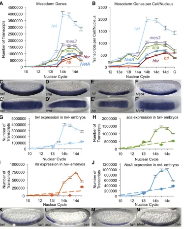
Quantitative Single-Embryo Profile of Drosophila Genome Activation and the Dorsal–Ventral Patterning Network
Full text
Figure


Related documents
The study suggests day-care ureteroscopy in this hospital was effective and safe, in view of careful patient selection, smooth induction, adequate instruments, trained
This showed that prior to treatment, the surgical group was significantly more depressed than the medical and control patients.. Internal consistency for the BDI was high for all
In our study, we showed that TS was inactive in clinical TD- SCVs, that the constructed ⌬ thyA mutant resembled morpholog- ically clinical TD-SCVs, and that the expression of
This study did not aim to study prevalence but aimed to provide a clinical profile as well as the family and social background of children with febrile
In the second part of the study, women in high priority pregnancies (the criteria of the Ministry of Health, Malaysia: Table I) were surveyed at antenatal clinics in all the
Further experiences with the pectoralis myocutaneous flap for the immediate repair of defects from excision of head and
(A) Type I IFN production by SV40T MEFs from matched wild-type and Sting -deficient mice stimulated with 0.1 M CPT for 48 h, measured by bioassay on LL171 cells (data shown as
DCs treated with OMVs containing PSA protect mice from experimental colitis, whereas Gadd45 α -/- DCs are unable to support T cell regulatory response and are defective in
