INVESTIGATION
AKAP9 Is Essential for Spermatogenesis and Sertoli
Cell Maturation in Mice
Kerry J. Schimenti,*,†,1Sky K. Feuer,§,1,2Laurie B. Griffin,§,3Nancy R. Graham,§Claire A. Bovet,§,4
Suzanne Hartford,*,†,5Janice Pendola,‡Carl Lessard,‡,6John C. Schimenti,*,†and Jeremy O. Ward§,7 §Department of Biology, Middlebury College, Middlebury, Vermont 05753, *Department of Biomedical Sciences and
†Department of Molecular Biology and Genetics, Cornell University, Ithaca, New York 14853, and ‡The Jackson Laboratory, Bar Harbor, Maine 04609
ABSTRACTMammalian male fertility relies on complex inter- and intracellular signaling during spermatogenesis. Here we describe three alleles of the widely expressed A-kinase anchoring protein 9 (Akap9) gene, all of which cause gametogenic failure and infertility in the absence of marked somatic phenotypes.Akap9disruption does not affect spindle nucleation or progression of prophase I of meiosis but does inhibit maturation of Sertoli cells, which continue to express the immaturity markers anti-Mullerian hormone and thyroid hormone receptor alpha in adults and fail to express the maturation marker p27Kip1. Furthermore, gap and tight junctions essential for blood–testis barrier (BTB) organization are disrupted. Connexin43 (Cx43) and zona occludens-1 are improperly localized in Akap9 mutant testes, and Cx43 fails to compartmentalize germ cells near the BTB. These results identify and support a novel re-productive tissue-specific role forAkap9in the coordinated regulation of Sertoli cells in the testis.
R
EPRODUCTION in male mammals relies on sex- and age-dependent signal transduction events. Sertoli cells undergo maturation from birth through puberty, with changing gene expression and morphology as they halt pro-liferation (for review see (Sharpe et al. 2003). This differ-entiation includes the formation of the blood–testis barrier (BTB), separating the mitotic/spermatogonial compartment from the meiotic/spermatocyte compartment (Petersen and Soder 2006; Mitalet al.2011). Preleptotene spermatocytes must traverse the BTB, requiring Sertoli cell-regulated dis-assembly and redis-assembly of tight and gap junctions (Griswold 1995, 1998; Cheng and Mruk 2002; Wong and Cheng 2005).Connexin43 (Cx43, also known as GJA1) organizes BTB gap junctions between Sertoli cells and those between Sertoli and germ cells (Juneja et al. 1999; Plum et al. 2000; Roscoe et al. 2001; Mruk and Cheng 2004; Pointis and Segretain 2005; Weideret al.2011). Cx43 is localized to Sertoli cells at the basal region of the seminiferous tubule and participates in germ and Sertoli cell migration, differ-entiation, and survival (Sridharanet al.2007; Gilleronet al. 2009). Basal tight junctions between Sertoli cells form the principal structural component of the BTB by day 16 (Nagano and Suzuki 1976). The expression of the tight-junction protein, zonula occludens 1 (ZO-1, also known as TJP1), in the testis is restricted primarily to the inter-Sertoli cell tight junction interface by puberty (Byerset al.1991). In Sertoli cells, ZO-1 is known to interact with Cx43 to regulate gap junction integrity (Giepmans and Moolenaar 1998; Toyofukuet al.1998; Hunteret al.2005) and proliferation (Rhett et al. 2011) and thus is an important functional marker for Sertoli cell tight junctions and maturation.
Protein kinase A (PKA) signaling cascades in vivo are numerous and insulated from one another via A-kinase an-choring protein (AKAP) scaffolds (Wong and Scott 2004). AKAPs localize protein kinase A and other cAMP-responsive proteins to specific subcellular compartments, thereby orga-nizing combinations of enzymes to respond specifically to
Copyright © 2013 by the Genetics Society of America doi: 10.1534/genetics.113.150789
Manuscript received February 23, 2013; accepted for publication April 8, 2013 Supporting information is available online athttp://www.genetics.org/lookup/suppl/ doi:10.1534/genetics.113.150789/-/DC1.
1These authors contributed equally to this work.
2Present address: University of California, San Francisco, CA 94143. 3Present address: University of Michigan, Ann Arbor, MI 48109.
4Present address: University of Colorado School of Medicine, Aurora, CO 80045. 5Present address: National Institutes of Health, Bethesda, MD 20892.
6Present address: Canadian Animal Genetic Resources Program, Saskatoon,
Canada, S7N 5B4.
7Corresponding author: Department of Biology, MBH 353, Middlebury College,
intracellular second messengers (reviewed in Colledge and Scott 1999; Feliciello et al. 2001; Wong and Scott 2004). AKAPs play critical roles in spermatogenesis, and multiple AKAPs have been identified in varying stages of spermato-genic development and in sperm function (Carreraet al.1994; Turneret al.1999; Vijayaraghavanet al.1999; Reintonet al. 2000; Furusawaet al.2001; Mikiet al.2002).
One major AKAP family member, AKAP9, was first described as an N-methyl-D-aspartic acid (NMDA) receptor anchor in the brain (Linet al.1998) and was further char-acterized in the brain and heart (Feliciello et al. 1999; Kurokawa et al. 2004; Ciampi et al. 2005; Chen et al. 2007; Piggottet al.2008; Chen and Kass 2011; Chopra and Knollmann 2011). Mutations in human AKAP9have been implicated in breast cancer (Milneet al.2011), sporadic pap-illary thyroid carcinomas (Leeet al.2006), and long QT syn-drome (Chen et al. 2007). In rat Sertoli cells, AKAP9 coordinates PKA and phosphodiesterase 4D3 (Taskenet al. 2001). Large isoforms of AKAP9 are associated with centro-somal function and signaling, microtubule organization, and nucleation at thecis-Golgi (Sillibourneet al.2002; Takahashi et al.2002; Keryeret al.2003a; McCahillet al.2005; Rivero et al. 2009). To date, no functional role has been described for AKAP9 in spermatogenesis.
Previously, we conducted forward genetic screens to identify mutant alleles of novel genes necessary for game-togenesis inMus musculus(Ward et al.2003; Lessardet al. 2004). Here, via positional cloning of two such alleles and complementation analyses with a targeted null allele, we report that mutation ofAkap9selectively disrupts male fer-tility and somatic cell-germ cell organization. Thesefindings underscore the importance of highly regulated intracellular and intercellular signaling between these compartments for spermatogenesis in mammals. Furthermore, the results are significant with respect to the major physiological require-ment of the widely studied AKAP9 protein, which hitherto had been demonstrated to be very important for nonrepro-ductive functions.
Materials and Methods
Animal care and use
All animal care and use related to this study was done in strict accordance with procedures and recommendations in the National Research CouncilGuide for the Care and Use of Lab-oratory Animalsand was approved by the institutional animal care and use committees of all affiliated institutions.
Genetic mapping
Linkage was implicated by the association of phenotype with homozygosity for marker loci of the mutagenized parental strains, 129S1/SvImJ and C57BL/6J formei2.5andrepro12, respectively. Heterozygous animals of both strains were crossed to wild-type CAST/Ei animals to take advantage of the higher degree of polymorphism between parental strains
and CAST/Ei. Separate intersubspecific intercrosses were conducted between animals heterozygous for mei2.5 and repro12. Resulting progeny were phenotyped with respect to infertility or testis histology, and genotyped with micro-satellite or SNP markers in the proximal region of Chr 5, as indicated inSupporting Information,Figure S1.
Generation of Akap9 knockout mice
Targeted ES cell clone D01 was acquired from the Knockout Mouse Project (KOMP) repository at the University of California Mouse Biology Program. This clone (from C57BL/ 6N) contains an exon-trapping cassette splice acceptor, beta galactosidase neomycin, poly adenylation (SA-b geo -pA) flanked by Flp-recombinase target (FRT) sites within the intron upstream of exon 8, and loxP sites on either side of exon 8 (see Figure 2). Generated at the Sanger Institute, it has the official allele name ofAkap9tm1a(KOMP)Wtsi (abbrevi-ated Akap9Gt, where“Gt” stands for gene trap). These ES cells were injected into blastocysts of CD1 albino mice and the resultant male chimeras were backcrossed to C57BL/6J females.Akap9Gtmice were crossed to constitutive Cre mice obtained from The Jackson Laboratory (B6.FVB-Tg(EIIa-cre) C5379Lmgd/J) to create the exon-8-deleted allele referred to as Akap92, that still contains the vector insertion and exon trapping features (Table 1).
Genotyping and histology
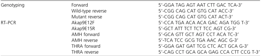
DNA from 2-mm tail tips or ear tags was used for standard and allele-specific PCR reactions. Primer sequences are given below. For histology, testes werefixed in Bouin’s solution for .24 hr, paraffin embedded, and 5-mm sections were cut and stained with hematoxylin and eosin (H&E).
Immunoblotting
Organ protein extracts were prepared with radioimmuno-precipitation assay (RIPA) buffer, separated on a 4–12% NuPAGE Bis-Tris gel (Invitrogen, Carlsbad, CA), transferred to nitrocellulose membrane, and blocked overnight. Immu-noprobing was carried out at room temperature for 1 hr for primary and secondary antibodies followed by washes and chemiluminescent imaging. Antibodies to AKAP9 (a24, rec-ognizes humanAKAP9exons 24–27) were a kind gift of M. Bornens.
RT–PCR
RNA was isolated with the Qiagen RNeasy mini kit (German-town, MD). cDNA was synthesized via reverse transcription of 1 mg of RNA using the Protoscript First-Strand cDNA Synthesis Kit (New England Biolabs, Ipswitch, MA). PCR using primers listed above was performed with Platinum Taq Hi-Fi (Invitrogen).
Immunohistochemistry and immunocytochemistry
in 0.6% hydrogen peroxide in cold methanol and slides were autoclaved for 20 min in 10 mM citric acid (pH 6.0) to retrieve antigenicity. Sections were washed in PBS, blocked, and incubated with primary antibody overnight at 4º. Slides were washed in PBS and incubated 1.5 hr with secondary antibody at 25º, counterstained with Hoechst 33342 or DAPI (0.02mg/mL) in PBS, and mounted with SlowFade Compo-nent A (Molecular Probes, S2828). For immunocytochemistry (ICC), testes from adult males (.60 days postbirth) were harvested, and meiotic chromosome spreads were prepared and immunolabeled as previously described (Peters et al. 1997; Reinholdtet al.2004). For testis squashes, adult testes were harvested, and germ cell squashes were prepared and immunolabeled as previously described (Novelloet al.1996).
Antibodies
All antibodies used and their concentrations are described in Table 2.
Statistical analysis
To determine significance in Wilm’s tumor-1 (WT-1)-positive Sertoli cell per tubule counts,20 tubular cross-sections were counted in each of three animals per genotype (wild type,Akap9mei2.5/mei2.5,Ccnb1IP1mei4/+, andCcnb1IP1mei4/mei4) and a one-tailed Student’st-test was conducted with a level of significance atP,0.01. Levene’s test was performed to verify equal variance using the statistical analysis software SPSS. For analyzing RT–PCR expression of anti-Mullerian hormone (AMH) and thyroid hormone receptor alpha (THRA), net band intensities of each sample were measured using Kodak 1D Image Analysis software (v3.6), and ratios
between each Sertoli cell marker and age-matched b-actin controls were calculated. Assays were conducted in triplicate, and ratio cohorts averaged. Significance between age-matched groups was determined using one-tailed paired-sample Stu-dent’sttests with equal variance.
Results
Mutations in Akap9 disrupt spermatogenesis and alter transcript splicing and protein expression in
multiple tissues
We previously described two male infertility mutations calledmei2.5andrepro12that exhibited abnormal testicular histology (Ward et al. 2003; Lessard et al. 2004, http://
reproductivegenomics.jax.org/mutants/G1-448-151.html).
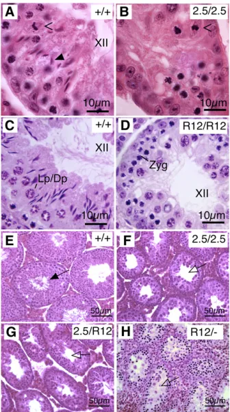
Homozygous males display an incomplete failure of game-togenic progression at approximately, but not uniformly, metaphase I during epithelial stage XII (Figure 1, A vs. B and Cvs.D). Spermatocytes are mislocalized in mutant ani-mals, and round spermatids and luminal spermatozoa are rarely observed.
To identify the mutations responsible for the phenotypes ofmei2.5andrepro12, genetic mapping was performed. The analyses placed these alleles proximal to markersrs13478098 andD5Mit344, respectively, on chromosome 5 (Figure S1A, gray box). Becausemei2.5andrepro12map to the same re-gion and have similar phenotypes, we performed comple-mentation tests. All males doubly heterozygous for the two mutations (Figure 1, F and G) were phenotypically indistin-guishable from mutant homozygotes (Figure 1, B and D), indicating that these mutations are allelic.
Table 1 List of primers used in genotyping and RT–PCR
Genotyping Forward 59-GGA TAG AGT AAT CTT GAC TCA-39
Wild-type reverse 59-CGG CAG CAT GTG CAT ACC-39 Mutant reverse 59-CGG CAG CAT GTG CAT ACT-39
RT–PCR Akap9E12F 59-CCA TGA ACA ACA GAC AGA TGG T-39
Akap9E15R 59-GCT ATT TCT TCT TCC AGT CG-39
AMH forward 59-GCA GTT GCT AGT CCT ACA TC-39
AMH reverse 59-TCA TCC GCG TGA AAC AGC G-39
THRA forward 59-GGA GAT GAT TCG CTC ACT GCA G-39 THRA reverse 59-CAG CCT GCA GCA GAG CCA CTT CCG T-39
Table 2 List of antibodies used in immunohistochemistry analyses
Antigen Host Clonality Concentration Manufacturer Cat. no.
Primary
SUMO-1 Mouse Monoclonal 2.5mg/mL Zymed Laboratories 33-2400
SYCP3 Rabbit Polyclonal 5mg/mL Abcam ab15092
Cx43 (GJA1) Mouse Monoclonal 2.5mg/mL Zymed Laboratories 35-5000
ZO-1 (TJP1) Rabbit Polyclonal 1.25mg/mL Zymed Laboratories 40-2200
p27Kip1(Cdkn1b) Rabbit Monoclonal 4mg/mL Abcam ab7961
SMA (ActaA2) Mouse Monoclonal Unknown, 1:50 Sigma A2547
WT-1 Rabbit Polyclonal 1mg/mL Santa Cruz Biotech. sc-192
Secondary l Cat. no.
Mouse IgG Goat Polyclonal 5mg/mL Molecular Probes 488 A-11001
Mouse IgG Goat Polyclonal 5mg/mL Molecular Probes 594 A-11005
Rabbit IgG Goat Polyclonal 5mg/mL Molecular Probes 594 A-11012
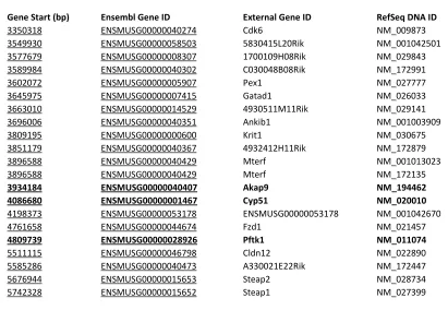
The region proximal tors13478098has 21 genes, (Table S1), but we prioritized Akap9 as a candidate based on its large size and hence a large mutagenesis target. Akap9 is composed of 48 predicted exons that yield a 12-kb transcript encoding 3779 amino acids. Sequencing of themei2.5allele revealed a G-to-A transition in a conserved splice site at the last position of exon 13, causing inclusion of intron 13–14 and a stop codon (Figure 2A). Therepro12allele was found to contain a T-to-A nonsense mutation in exon 14, changing amino acid 1353 from a cysteine to a stop codon (Figure 2A). To verify that theseAkap9mutations underlie the sper-matogenic defect, we generated mice bearing a gene trap vector insertion within intron 7 (Figure 2A, Akap9Gt) and a derivative allele (Akap92) that both retains the gene trap functionality and deletes exon 8, generating a frameshift. The Akap92 allele failed to complement repro12 (Figure
1H), further confirming a novel role forAkap9in spermato-genesis. Heterozygous intercrosses produced homozygotes at Mendelian frequencies (N= 65,X2= 0.415,P= 0.8125),
and theseAkap92/2males were also sterile.
Akap9mei2.5/mei2.5animals demonstrate aberrant splicing and inclusion of intron 13–14 in all tissues examined, as expected due to the nature of the mutation (Figure 2B). Splicing is unaffected in repro12 homozygotes, but these andAkap9Gtmice fail to express AKAP9 protein in testis or pancreas when probed using an AKAP24 (a24) antibody specific to humanAKAP9 exons 24–27 (Riveroet al.2009) that normally detects several 250-kDa protein species (Figure 2B).
Akap9 mutant spermatocytes exhibit normal spindle nucleation and progression through meiosis I
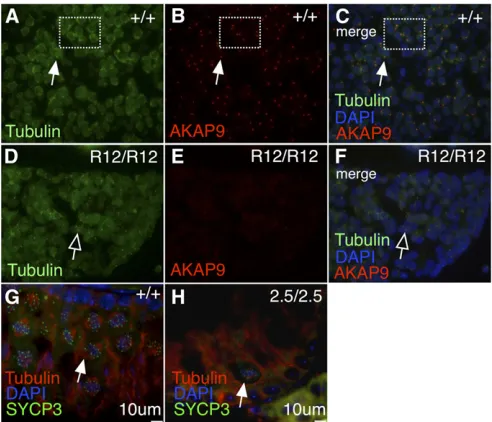
We hypothesized that defects in spindle formation could account for the gametogenic phenotype of Akap9mutants, particularly since isoforms of AKAP9 have been implicated in centrosomal biology and spindle nucleation (Schmidt et al. 1999; Witczak et al. 1999; Keryer et al. 2003b; Kim et al. 2007; Hurtadoet al.2011; Sehrawatet al.2011). Indeed we confirmed that AKAP9 (Figure 3B, red) colocalizes at the centrosome (Figure 3A, green) in wild-type animals (Figure 3C, merge; marked by gamma tubulin), and is strikingly reduced in Akap9repro12/repro12mutants (Figure 3F, merge). Despite this, bipolar meiotic spindle nucleation was ob-served in both wild-type andAkap9mei2.5/mei2.5testis sections (Figure 3, G vs. H, respectively; white arrows). Therefore, the failed spermatogenesis phenotype is not likely to be caused by spindle defects in testis germ cells even though AKAP9 is nearly absent from the centrosome.
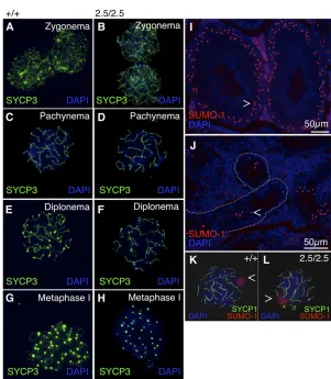
Because the mei2.5 and repro12 alleles primarily affect production of male postmeiotic cells, we next investigated hallmark aspects of spermatocyte development. Immunolab-eling of surface-spread spermatocyte nuclei with the axial element protein SYCP3 revealed that Akap9mei2.5/mei2.5 tes-tes contained all stages of meiotic cells, with the meiotic chromosomes having a normal appearance through diplo-nema and metaphase I (Figure 4, A–H). Immunolabeling for the synaptomenal complex (SC) protein SYCP1 con-firmed thatAkap9mei2.5/mei2.5 autosomes fully pair and are capable of successfully forming the SC central element dur-ing pachynema, indicative of synapsis (Figure 4, K and L). These data indicate that chromosomal defects in meiosis I are not a likely cause of failed spermatogenesis. Finally, we examined formation of the XY body (Figure 4, I and J), the disruption of which is hypothesized to cause meiotic arrest and male infertility in multiple mouse mutants (Cloutier and Turner 2010; Royoet al.2010). Expression of small ubiqui-tin-related modifier 1 (SUMO-1) in Akap9mei2.5/mei2.5 sper-matocytes suggests that the XY body forms normally (Handel 2004) in the few midpachynema cells that are pres-ent (Figure 4, Jvs.I, respectively; open arrowheads). How-ever, XY body-containing cells localize abnormally to the adluminal compartment (Figure 4, J vs. I; dotted lines Figure 1 Mutations inAkap9disrupt spermatogenesis. H&E staining of
indicate tubule boundary), indicating a potential defect in migration or structural support within seminiferous tubules. Mutation of Akap9 leads to a high number of immature Sertoli cells
The absence of obvious meiotic defects coupled with the mislocalization of spermatocytes led us to examine Sertoli cell terminal differentiation at puberty, marked by basal localization and loss of proliferative ability (reviewed in Sharpeet al.2003). Sertoli cell maturation may be charac-terized by the expression of a number of markers, including Wilm’s tumor-1 (WT-1) (Kreidberg et al. 1993; Nordqvist 1995), anti-Mullerian hormone (AMH), thyroid hormone receptor alpha (THRA) (Arambepolaet al.1998; Holsberger et al.2003, 2005), and the cell cycle inhibitor p27Kip1(Beumer
et al. 1999; Tan et al. 2005). Expression of these markers originates from and may initiate additional signal transduction events. Seminiferous tubules of Akap9mei2.5/mei2.5 testes ex-hibit a significantly higher number of WT-1-positive Sertoli cells compared with wild type (Figure 5, A, B, and E; P, 0.01). The relative increase of Sertoli cells inAkap9mutants is not an indirect consequence of germ cell loss: similar analysis of a mutant (Ccnb1ip1mei4/mei4) that undergoes late meiosis I arrest and lacks all germ cell types past meiosis I (Wardet al. 2007) revealed no such increase in Sertoli cells relative to controls (Figure 5, C–E).
To further interrogate Sertoli cell integrity, we examined the expression of the negative cell cycle regulator p27Kip1
and the prepubertal markers AMH and THRA. p27Kip1
ex-pression is typically low in neonatal testes, high in adult testes, and when ablated, leads to abnormally high numbers of Sertoli cells (Beumeret al.1999; Holsbergeret al.2005). Adult Akap9mei2.5/mei2.5 Sertoli cells fail to express p27Kip1
(Figure 6, Avs.B), whereas p27Kip1expression is unaffected
in the Ccnb1ip1mei4meiotic mutant (Figure 6, C vs.D) and hence is not dependent upon postmeiotic cell differentiation. Figure 2 Mutation inAkap9causes failed splicing or protein expression in multiple tissues. (A) mei2.5, re-pro12,AkapGt, andAkap2mutations. Cre-lox recombi-nation was used to remove exon 8 from theAkap9gene (2allele) as shown, with selection byb-galactosidase and neomycin (B gal; neo) in the exon-trapping cassette (SA–Bgeo-pA). Triangles, loxP sites; ovals, FRT sites. (B) RT–PCR ofAkap9exons 12–15 in selected tissues shows a 495-bp amplimer in wild-type (+/+) andAkap9R12/R12 (R12/R12) animals, whereasAkap9mei2.5/mei2.5(2.5/2.5) animals display a unique band (626 bp) corresponding to the inclusion of intron 13–14, indicating thatmei2.5 causes defective splicing at the 39end of exon 13. AKAP9 protein expression in pancreas and testis shows several protein species centered at 250 kDa in wild-type (+/+) animals, which is absent in R12/R12 and Gt/Gt mice.
AMH expression, assayed in triplicate by RT–PCR at post-natal days 10, 20, 60, and 100, declined significantly in wild-type animals (+/+, Figure 6E), but remained high in Akap9 mutants (P,0.05) relative to wild-type controls of the same age. THRA likewise showed continued high expres-sion in Akap9mei2.5/mei2.5 animals relative to age-matched wild-type controls (Figure 6F). Taken together, these results indicate that AKAP9 deficiency causes Sertoli cells to retain a prepubertal character.
AKAP9-deficient mice show irregular tight and gap junctional protein localization
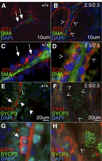
Organization of the blood–testis barrier is an important function of adult Sertoli cells (Smith and Braun 2012). We hypothesized that abnormal Sertoli cell numbers and fertil-ity defects in males reflected a failure to establish junctions characteristic of the BTB, a prediction substantiated by the abnormal localization of germ cells (Figure 4). In prepuber-tal animals, testis ZO-1 is localized apicolaterally (Stevenson et al. 1986; Andersonet al. 1988; Byers et al. 1991), and shifts to associate almost exclusively with Sertoli cell baso-lateral tight junctions in peri- and postpubertal mice (Byers et al.1991). In the wild-type adult testis, we observed ZO-1 proximal to the basement membrane (Figure 7A, red signal, white arrows; higher magnification in Figure 7C), whereas Akap9mei2.5/mei2.5testes exhibit ZO-1 expression towards the
apical surface of Sertoli cells, with weak or no localization along the seminiferous tubule basement membrane (Figure 7B, open arrowheads; higher magnification in Figure 7D).
To investigate gap junctions, we examined the localiza-tion of Cx43 in the seminiferous epithelium (Figure 7, E and F). In wild-type sections, Cx43 is localized at the basal re-gion of seminiferous tubules and forms distinct compart-ments around early meiotic cells identified by SYCP3 labeling (Figure 7E, arrows; higher magnification in Figure 7G). Cx43 expression is dissipated around preleptotene spermatocytes and forms gap junctions in the adluminal compartment, proximal to pachytene spermatocytes (closed arrowheads). In Akap9mei2.5/mei2.5 testis, Cx43 localiza-tion is aberrant throughout the lumen without apparent relation to spermatocytes (Figure 7F, open arrowheads; higher magnification in Figure 7H), and fails to discretely compartmentalize early meiotic cells along the basal compartment. Combined, these data show the lack of appropriate tight and gap junction constituent localiza-tion, particularly at the basal portion of the seminiferous tubule of Akap9mei2.5/mei2 testis.
Discussion
resulting in higher numbers and lack of maturation of Sertoli cells in adult mice. There is prolonged expression of juvenile Sertoli cell markers and a failure to form normal tight and gap junction morphologies, revealing that AKAP9 expression is essential for spermatogenesis but not the process of mei-osisper se.
Surprisingly, despite its widespread expression in many tissues, including the brain and heart (Linet al.1998; Kurokawa et al.2004; Chenet al.2007; Chen and Kass 2011; Chopra and Knollmann 2011; Kendler et al.2011), overall animal viability is not markedly affected in AKAP9-deficient mice. This is even more remarkable considering that AKAP9 has been implicated in proper cell cycle progression via centriole formation and microtubule organizing center (MTOC) func-tion (Keryer et al. 1993, 2003a,b; Takahashi et al. 1999; McCahill et al. 2005; Kim et al. 2007; Rivero et al.2009; Hurtadoet al.2011). Although we have not conducted de-tailed physiological and morphological studies of the pre-sumably null Akap92/2 mice, the lack of drastic somatic phenotypes suggests that additional AKAP family members could perform redundant functions in other tissues, but not those that impact sperm development.
Despite a strong decrease in centrosomal AKAP9 in mutant males, meiotic spindle formation and therefore microtubule organization in AKAP9-deficient mice appear normal, sug-gesting that cell division and spindle formation errors are not responsible for the reproductive phenotype. Proper XY body formation, SYCP1 and SYCP3 chromosome labeling, and rare postmeiotic spermatids indicate functional meiosis. Further, female mice carrying mutations inAkap9have no significant observed reproductive issues. It is unknown whether males homozygous for either mei2.5or repro12 undergo homolo-gous recombination, but since failure to recombine is nor-mally associated with pachytene or metaphase I arrest in males (Morelli and Cohen 2005), it is unlikely that this pathway is affected.Akap9mutations appear instead to dis-rupt homeostasis of the seminiferous tubule by disturbing Sertoli cell function.
The abnormal localization of spermatocytes inAkap9mei2.5/mei2.5 mice suggests that Sertoli cells have lost their proper struc-tural organization along the seminiferous tubule periphery. Additionally, the significantly increased number of Sertoli cells and the absence of p27Kip1 in adult Akap9 mutants
Figure 5 mei2.5homozygosity but not loss of germ cells disrupts Sertoli cell numbers in seminiferous tubules. Sertoli cell nuclei identified by the marker WT-1 (red) are observed adjacent to the basal membrane (smooth muscle actin, SMA; green) in +/+ (A) andmei2.5/mei2.5testes (B), but are significantly more numerous inmei2.5/mei2.5tubules (E, 30.261.6 cells vs. 18.9 61.1 cells) per tubular cross-section (*P ,0.01). Themei4 mutation in mouse geneCcnb1ip1prevents postmeiotic cell differentia-tion (mei4, C and D) (Wardet al.2007). Sections from a heterozygote (mei4/+, C) and homozygote (mei4/mei4, D) were labeled with WT-1 to differentiate between mutation effects (mei2.5) and the consequences of failed meiosis and spermatogenesis (mei4). WT-1-positive Sertoli cell numbers are not affected by lack of postmeiotic germ cell differentiation (E).
Figure 6 mei2.5homozygosity is responsible for Sertoli cell maturation defects. Mature, differentiated, wild-type Sertoli cells stain positively for the negative cell cycle regulator p27Kip1(A, arrow in inset), but this
ex-pression is absent inmei2.5/mei2.5testes (B, open arrowhead in inset). Relative to controls (C),mei4/mei4animals (D) retain p27Kip1expression
signify that these cells may have retained their proliferative capabilities, implying partially failed maturation. The p27Kip1knockout mouse exhibits two-fold larger testes,
in-creased numbers of spermatogonia, and inin-creased Sertoli cell proliferation, indicating that p27Kip1participates in the
halt of Sertoli cell mitogenesis at puberty (Kiyokawa et al. 1996; Beumer et al. 1999; Holsberger et al. 2005). Mice lacking p27Kip1also contain prematurely sloughed off germ
cells in the seminiferous epithelium (Beumer et al.1999); we observed this and multilayering of premeiotic germ cells along the basal lamina ofAkap9 mutant tubules. However, because there are distinct differences between the Akap9 mutants and p27Kip1 knockout phenotypes, the lack of
p27Kip1in Akap9 mutants is likely not the exclusive cause
of the infertile phenotype. Importantly, abnormal numbers of Sertoli cells were not observed inCcnb1ip1mutants, sug-gesting that loss of differentiating germ cells is not the cause of Sertoli cell dysfunction. From these data, we conclude
that AKAP9 deficiency causes Sertoli cell dysfunction either through intracellular signal transduction changes, aberrant intercellular regulation via neighboring cells, or possibly im-pairment of hormonal signaling from other sources in the body.
Dysfunctional and immature Sertoli cells in Akap9 mu-tant mice provide a better explanation for the interrupted support of the later stages of germ cell development and may account for the failed structural management of germ cells with XY bodies. The loss of ZO-1 expression at the basal side of mutant Sertoli cells could indicate a disruption of cell polarity conferred by proper tight junction formation. Irreg-ular tight junctions would impair the compartmentalization of the basal and adluminal environments, compromising Sertoli cell integrity. Similarly, Cx43 no longer distinctly compartmentalizes spermatogonia and preleptotene sper-matocytes in mutants, showing promiscuous expression throughout the adluminal compartment of seminiferous tubules. This patterning is similar to Cx43 expression in immature tubules: during testicular maturation, Cx43 local-ization shifts from punctate and linear arrays throughout the adluminal compartment and near tight junctions to the basal region of seminiferous tubules (Risley et al. 1992; Pointis and Segretain 2005). We note the remarkable simi-larity of phenotype betweenAkap9mutant mice and Sertoli cell-specific Cx43 knockout mice (Brehm et al.2007), sug-gesting a role forAkap9in the negative control of Sertoli cell proliferation and/or terminal differentiation.
We have not yet conducted physiological analyses of the hypothalamic–pituitary–gonadal (HPG) axis in Akap9 mutants. Disruption of the luteinizing and follicle-stimulat-ing hormone pathways causes severe fertility issues in males and females (Wang and Greenwald 1993a,b; O’ Shaugh-nessy et al.2009; Siegel et al.2012) and could contribute to theAkap9mutant phenotype. Interestingly,Akap9mutant females are fertile but a detailed assessment of the HPG axis in these animals will be an important area for future investigation.
Based on the present data, however, we propose a specific role forAkap9in the coordinated regulation of blood–testis barrier dynamics in the testis. It is possible this occurs by a cell type- or junction-specific mechanism, and determining if the requirement for AKAP9 is autonomous to Sertoli cells or germ cells may be addressed in the future using methods such as spermatogonial transplantation. Importantly, the Akap9mei2.5/mei2.5phenotypes of multiple layers of spermato-gonia along the basal lamina, sloughed off germ cells, and adluminal spermatocytes do not reflect the impairment of one specific junction. Binding of ZO-1 targets Cx43 to the plasma membrane, controlling gap junction size and distri-bution, and may shepherd signal molecules or actin fi la-ments to Cx43 to integrate gap junction formation with the actin cytoskeleton and modulate intracellular signaling (Giepmans and Moolenaar 1998; Toyofukuet al.1998; Wei et al.2004). ZO-1 is additionally required for PKC-controlled disassembly of Cx43 from gap junctional plaques (Wuet al. Figure 7 Junctional complexes characteristic of mature Sertoli cells are
2003; Hunteret al.2005; Akoyev and Takemoto 2007). The AKAP350 splice variant of AKAP9 associates with PKA, PP1, and PKC in a multiprotein complex and interacts with ZO-1 in cultured mammalian cells (Schmidt et al.1999; Diviani and Scott 2001; Berryman and Goldenring 2003). Loss of a major regulator of Cx43 activity would impede proper gap junction formation and function in the testis, impairing sper-matogenesis. However, these associations have not yet been investigated in the testis.
In conclusion, we find that AKAP9 is critical for male fertility but is not essential for basic cellular function or mouse development, contrary to prior evidence. Our phe-notypic studies implicate a key role for this protein in the establishment of proper somatic cell–germ cell interactions in the seminiferous tubules, and formation of the blood– testis barrier.
Acknowledgments
The authors thank Martine Stewart and Charlie Dunning (Middlebury College) for critical reading of the manuscript and editorial assistance. They also thank Bill Buaas (The Jackson Laboratory) for critical reading of the manuscript, Vicki Major for animal care, and Michel Bornens (Institut Curie) for the kind gift of the a24 AKAP9 antibody. This work was supported by grants to Middlebury College [National Science Foundation (NSF) Course, Curriculum, and Language Improvement 0088412 and Major Research Instrumentation, Division of Biological Infrastructure 0215782], The Jackson Laboratory (NCI CA34196), and to S.K.F. (SigmaXi GAR), J.O.W. [National Research Service Award GM64275-01A, National Institutes of Health (NIH) P20 RR16462, NIH Academic Research Enhancement Award R15 GM078183–01, and NSF CAREER 0844941], and to J.C.S. (NIH GM45415 and NIH HD42137).
Literature Cited
Akoyev, V., and D. J. Takemoto, 2007 ZO-1 is required for protein kinase C gamma-driven disassembly of connexin 43. Cell. Sig-nal. 19: 958–967.
Anderson, J. M., B. R. Stevenson, L. A. Jesaitis, D. A. Goodenough, and M. S. Mooseker, 1988 Characterization of ZO-1, a protein component of the tight junction from mouse liver and Madin-Darby canine kidney cells. J. Cell Biol. 106: 1141–1149. Arambepola, N. K., D. Bunick, and P. S. Cooke, 1998 Thyroid
hor-mone effects on androgen receptor messenger RNA expression in rat Sertoli and peritubular cells. J. Endocrinol. 156: 43–50. Berryman, M. A., and J. R. Goldenring, 2003 CLIC4 is enriched at
cell-cell junctions and colocalizes with AKAP350 at the centro-some and midbody of cultured mammalian cells. Cell Motil. Cytoskeleton 56: 159–172.
Beumer, T. L., H. Kiyokawa, H. L. Roepers-Gajadien, L. A. van den Bos, T. M. Locket al., 1999 Regulatory role of p27kip1 in the mouse and human testis. Endocrinology 140: 1834–1840. Brehm, R., M. Zeiler, C. Ruttinger, K. Herde, M. Kibschull et al.,
2007 A sertoli cell-specific knockout of connexin43 prevents initiation of spermatogenesis. Am. J. Pathol. 171: 19–31.
Byers, S., R. Graham, H. N. Dai, and B. Hoxter, 1991 Development of Sertoli cell junctional specializations and the distribution of the tight-junction-associated protein ZO-1 in the mouse testis. Am. J. Anat. 191: 35–47.
Carrera, A., G. L. Gerton, and S. B. Moss, 1994 The majorfibrous sheath polypeptide of mouse sperm: structural and functional similarities to the A-kinase anchoring proteins. Dev. Biol. 165: 272–284.
Chen, L., and R. S. Kass, 2011 A-kinase anchoring protein 9 and IKs channel regulation. J. Cardiovasc. Pharmacol. 58: 413-459. Chen, L., M. L. Marquardt, D. J. Tester, K. J. Sampson, M. J. Ackerman
et al., 2007 Mutation of an A-kinase-anchoring protein causes long-QT syndrome. Proc. Natl. Acad. Sci. USA 104: 20990–20995.
Cheng, C. Y., and D. D. Mruk, 2002 Cell junction dynamics in the testis: Sertoli-germ cell interactions and male contraceptive de-velopment. Physiol. Rev. 82: 825–874.
Chopra, N., and B. C. Knollmann, 2011 Genetics of sudden car-diac death syndromes. Curr. Opin. Cardiol. 26: 196–203. Ciampi, R., J. A. Knauf, R. Kerler, M. Gandhi, Z. Zhu et al.,
2005 Oncogenic AKAP9-BRAF fusion is a novel mechanism of MAPK pathway activation in thyroid cancer. J. Clin. Invest. 115: 94–101.
Cloutier, J. M., and J. M. Turner, 2010 Meiotic sex chromosome inactivation. Curr. Biol. 20: R962–R963.
Colledge, M., and J. D. Scott, 1999 AKAPs: from structure to function. Trends Cell Biol. 9: 216–221.
Diviani, D., and J. D. Scott, 2001 AKAP signaling complexes at the cytoskeleton. J. Cell Sci. 114: 1431–1437.
Feliciello, A., L. Cardone, C. Garbi, M. D. Ginsberg, S. Varroneet al., 1999 Yotiao protein, a ligand for the NMDA receptor, binds and targets cAMP-dependent protein kinase II(1). FEBS Lett. 464: 174–178.
Feliciello, A., M. E. Gottesman, and E. V. Avvedimento, 2001 The biological functions of A-kinase anchor proteins. J. Mol. Biol. 308: 99–114.
Furusawa, M., T. Ohnishi, T. Taira, S. M. Iguchi-Ariga, and H. Ariga, 2001 AMY-1, a c-Myc-binding protein, is localized in the mitochondria of sperm by association with S-AKAP84, an anchor protein of cAMP-dependent protein kinase. J. Biol. Chem. 276: 36647–36651.
Giepmans, B. N., and W. H. Moolenaar, 1998 The gap junction protein connexin43 interacts with the second PDZ domain of the zona occludens-1 protein. Curr. Biol. 8: 931–934.
Gilleron, J., D. Carette, P. Durand, G. Pointis, and D. Segretain, 2009 Connexin 43 a potential regulator of cell proliferation and apoptosis within the seminiferous epithelium. Int. J. Bio-chem. Cell Biol. 41: 1381–1390.
Griswold, M. D., 1995 Interactions between germ cells and Sertoli cells in the testis. Biol. Reprod. 52: 211–216.
Griswold, M. D., 1998 The central role of Sertoli cells in sper-matogenesis. Semin. Cell Dev. Biol. 9: 411–416.
Handel, M. A., 2004 The XY body: a specialized meiotic chroma-tin domain. Exp. Cell Res. 296: 57–63.
Holsberger, D. R., S. Jirawatnotai, H. Kiyokawa, and P. S. Cooke, 2003 Thyroid hormone regulates the cell cycle inhibitor p27Kip1 in postnatal murine Sertoli cells. Endocrinology 144: 3732–3738.
Holsberger, D. R., G. M. Buchold, M. C. Leal, S. E. Kiesewetter, D. A. O’Brienet al., 2005 Cell-cycle inhibitors p27Kip1 and p21Cip1 regulate murine Sertoli cell proliferation. Biol. Reprod. 72: 1429–1436.
Hurtado, L., C. Caballero, M. P. Gavilan, J. Cardenas, M. Bornens
et al., 2011 Disconnecting the Golgi ribbon from the centro-some prevents directional cell migration and ciliogenesis. J. Cell Biol. 193: 917–933.
Juneja, S. C., K. J. Barr, G. C. Enders, and G. M. Kidder, 1999 Defects in the germ line and gonads of mice lacking connexin43. Biol. Reprod. 60: 1263–1270.
Kendler, K. S., G. Kalsi, P. A. Holmans, A. R. Sanders, S. H. Aggen
et al., 2011 Genomewide association analysis of symptoms of alcohol dependence in the molecular genetics of schizophrenia (MGS2) control sample. Alcohol. Clin. Exp. Res. 35: 963–975. Keryer, G., R. M. Rios, B. F. Landmark, B. Skalhegg, S. M. Lohmann
et al., 1993 A high-affinity binding protein for the regulatory subunit of cAMP-dependent protein kinase II in the centrosome of human cells. Exp. Cell Res. 204: 230–240.
Keryer, G., B. Di Fiore, C. Celati, K. F. Lechtreck, M. Mogensenet al., 2003a Part of Ran is associated with AKAP450 at the centro-some: involvement in microtubule-organizing activity. Mol. Biol. Cell 14: 4260–4271.
Keryer, G., O. Witczak, A. Delouvee, W. A. Kemmner, D. Rouillard
et al., 2003b Dissociating the centrosomal matrix protein AKAP450 from centrioles impairs centriole duplication and cell cycle progression. Mol. Biol. Cell 14: 2436–2446.
Kim, H. S., M. Takahashi, K. Matsuo, and Y. Ono, 2007 Recruitment of CG-NAP to the Golgi apparatus through interaction with dynein-dynactin complex. Genes Cells 12: 421–434.
Kiyokawa, H., R. D. Kineman, K. O. Manova-Todorova, V. C. Soares, E. S. Hoffmanet al., 1996 Enhanced growth of mice lacking the cyclin-dependent kinase inhibitor function of p27(Kip1). Cell 85: 721–732.
Kreidberg, J. A., H. Sariola, J. M. Loring, M. Maeda, J. Pelletier
et al., 1993 WT-1 is required for early kidney development. Cell 74: 679–691.
Kurokawa, J., H. K. Motoike, J. Rao, and R. S. Kass, 2004 Regulatory actions of the A-kinase anchoring protein Yotiao on a heart potas-sium channel downstream of PKA phosphorylation. Proc. Natl. Acad. Sci. USA 101: 16374–16378.
Lee, J. H., E. S. Lee, Y. S. Kim, N. H. Won, and Y. S. Chae, 2006 BRAF mutation and AKAP9 expression in sporadic pap-illary thyroid carcinomas. Pathology 38: 201–204.
Lessard, C., J. K. Pendola, S. A. Hartford, J. C. Schimenti, M. A. Handel et al., 2004 New mouse genetic models for human contraceptive development. Cytogenet. Genome Res. 105: 222–227.
Lin, J. W., M. Wyszynski, R. Madhavan, R. Sealock, J. U. Kimet al., 1998 Yotiao, a novel protein of neuromuscular junction and brain that interacts with specific splice variants of NMDA recep-tor subunit NR1. J. Neurosci. 18: 2017–2027.
McCahill, A., T. McSorley, E. Huston, E. V. Hill, M. J. Lynchet al., 2005 In resting COS1 cells a dominant negative approach shows that specific, anchored PDE4 cAMP phosphodiesterase isoforms gate the activation, by basal cyclic AMP production, of AKAP-tethered protein kinase A type II located in the centro-somal region. Cell. Signal. 17: 1158–1173.
Miki, K., W. D. Willis, P. R. Brown, E. H. Goulding, K. D. Fulcher
et al., 2002 Targeted disruption of the Akap4 gene causes defects in sperm flagellum and motility. Dev. Biol. 248: 331– 342.
Milne, R. L., J. Lorenzo-Bermejo, B. Burwinkel, N. Malats, J. I. Arias
et al., 2011 7q21-rs6964587 and breast cancer risk: an ex-tended case-control study by the Breast Cancer Association Con-sortium. J. Med. Genet. 48: 698–702.
Mital, P., B. T. Hinton, and J. M. Dufour, 2011 The blood-testis and blood-epididymis barriers are more than just their tight junctions. Biol. Reprod. 84: 851–858.
Morelli, M. A., and P. E. Cohen, 2005 Not all germ cells are cre-ated equal: aspects of sexual dimorphism in mammalian meio-sis. Reproduction 130: 761–781.
Mruk, D. D., and C. Y. Cheng, 2004 Sertoli and Sertoli-germ cell interactions and their significance in germ cell move-ment in the seminiferous epithelium during spermatogenesis. Endocr. Rev. 25: 747–806.
Nagano, T., and F. Suzuki, 1976 Freeze-fracture observations on the intercellular junctions of Sertoli cells and of Leydig cells in the human testis. Cell Tissue Res. 166: 37–48.
Nordqvist, K., 1995 Sex differentiation – gonadogenesis and novel genes. Int. J. Dev. Biol. 39: 727–736.
Novello, A., M. Kralewski, and R. Benavente, 1996 Immuno-cytochemistry of soluble and non-soluble nuclear proteins on squash preparations of mammalian meiotic cells. Chromosome Res. 4: 622–623.
O’Shaughnessy, P. J., I. D. Morris, I. Huhtaniemi, P. J. Baker, and M. H. Abel, 2009 Role of androgen and gonadotrophins in the development and function of the Sertoli cells and Leydig cells: data from mutant and genetically modified mice. Mol. Cell. Endocrinol. 306: 2–8.
Peters, A. H., A. W. Plug, M. J. van Vugt, and P. de Boer, 1997 A drying-down technique for the spreading of mammalian meio-cytes from the male and female germline. Chromosome Res. 5: 66–68.
Petersen, C., and O. Soder, 2006 The sertoli cell–a hormonal tar-get and’super’nurse for germ cells that determines testicular size. Horm. Res. 66: 153–161.
Piggott, L. A., A. L. Bauman, J. D. Scott, and C. W. Dessauer, 2008 The A-kinase anchoring protein Yotiao binds and regu-lates adenylyl cyclase in brain. Proc. Natl. Acad. Sci. USA 105: 13835–13840.
Plum, A., G. Hallas, T. Magin, F. Dombrowski, A. Hagendorffet al., 2000 Unique and shared functions of different connexins in mice. Curr. Biol. 10: 1083–1091.
Pointis, G., and D. Segretain, 2005 Role of connexin-based gap junc-tion channels in testis. Trends Endocrinol. Metab. 16: 300–306. Reinholdt, L., T. Ashley, J. Schimenti, and N. Shima, 2004 Forward
genetic screens for meiotic and mitotic recombination-defective mutants in mice. Methods Mol. Biol. 262: 87–107.
Reinton, N., P. Collas, T. B. Haugen, B. S. Skalhegg, V. Hansson
et al., 2000 Localization of a novel human A-kinase-anchoring protein, hAKAP220, during spermatogenesis. Dev. Biol. 223: 194–204.
Rhett, J. M., J. Jourdan, and R. G. Gourdie, 2011 Connexin 43 connexon to gap junction transition is regulated by zonula oc-cludens-1. Mol. Biol. Cell 22: 1516–1528.
Risley, M. S., I. P. Tan, C. Roy, and J. C. Saez, 1992 Cell-, age- and stage-dependent distribution of connexin43 gap junctions in testes. J. Cell Sci. 103(Pt 1): 81–96.
Rivero, S., J. Cardenas, M. Bornens, and R. M. Rios, 2009 Microtubule nucleation at the cis-side of the Golgi apparatus requires AKAP450 and GM130. EMBO J. 28: 1016–1028.
Roscoe, W. A., K. J. Barr, A. A. Mhawi, D. K. Pomerantz, and G. M. Kidder, 2001 Failure of spermatogenesis in mice lacking con-nexin43. Biol. Reprod. 65: 829–838.
Royo, H., G. Polikiewicz, S. K. Mahadevaiah, H. Prosser, M. Mitchell
et al., 2010 Evidence that meiotic sex chromosome inactiva-tion is essential for male fertility. Curr. Biol. 20: 2117–2123. Schmidt, P. H., D. T. Dransfield, J. O. Claudio, R. G. Hawley, K. W.
Trotteret al., 1999 AKAP350, a multiply spliced protein kinase A-anchoring protein associated with centrosomes. J. Biol. Chem. 274: 3055–3066.
Sharpe, R. M., C. McKinnell, C. Kivlin, and J. S. Fisher, 2003 Proliferation and functional maturation of Sertoli cells, and their relevance to disorders of testis function in adulthood. Reproduction 125: 769–784.
Siegel, E. T., H. G. Kim, H. K. Nishimoto, and L. C. Layman, 2012 The molecular basis of impaired follicle-stimulating hor-mone action: evidence from human mutations and mouse mod-els. Reprod. Sci. 20: 211–233.
Sillibourne, J. E., D. M. Milne, M. Takahashi, Y. Ono, and D. W. Meek, 2002 Centrosomal anchoring of the protein kinase CK1delta mediated by attachment to the large, coiled-coil scaf-folding protein CG-NAP/AKAP450. J. Mol. Biol. 322: 785–797. Smith, B. E., and R. E. Braun, 2012 Germ cell migration across
Sertoli cell tight junctions. Science 338: 798–802.
Sridharan, S., L. Simon, D. D. Meling, D. G. Cyr, D. E. Gutsteinet al., 2007 Proliferation of adult sertoli cells following conditional knockout of the Gap junctional protein GJA1 (connexin 43) in mice. Biol. Reprod. 76: 804–812.
Stevenson, B. R., J. D. Siliciano, M. S. Mooseker, and D. A. Goodenough, 1986 Identification of ZO-1: a high molecular weight polypep-tide associated with the tight junction (zonula occludens) in a va-riety of epithelia. J. Cell Biol. 103: 755–766.
Takahashi, M., H. Shibata, M. Shimakawa, M. Miyamoto, H. Mukai
et al., 1999 Characterization of a novel giant scaffolding protein, CG-NAP, that anchors multiple signaling enzymes to centrosome and the golgi apparatus. J. Biol. Chem. 274: 17267–17274.
Takahashi, M., A. Yamagiwa, T. Nishimura, H. Mukai, and Y. Ono, 2002 Centrosomal proteins CG-NAP and kendrin provide mi-crotubule nucleation sites by anchoring gamma-tubulin ring complex. Mol. Biol. Cell 13: 3235–3245.
Tan, K. A., K. De Gendt, N. Atanassova, M. Walker, R. M. Sharpe
et al., 2005 The role of androgens in sertoli cell proliferation and functional maturation: studies in mice with total or Sertoli cell-selective ablation of the androgen receptor. Endocrinology 146: 2674–2683.
Tasken, K. A., P. Collas, W. A. Kemmner, O. Witczak, M. Contiet al., 2001 Phosphodiesterase 4D and protein kinase a type II con-stitute a signaling unit in the centrosomal area. J. Biol. Chem. 276: 21999–22002.
Toyofuku, T., M. Yabuki, K. Otsu, T. Kuzuya, M. Hori et al., 1998 Direct association of the gap junction protein connexin-43 with ZO-1 in cardiac myocytes. J. Biol. Chem. 273: 12725– 12731.
Turner, R. M., R. L. Eriksson, G. L. Gerton, and S. B. Moss, 1999 Relationship between sperm motility and the processing and tyrosine phosphorylation of two human sperm fibrous sheath proteins, pro-hAKAP82 and hAKAP82. Mol. Hum. Re-prod. 5: 816–824.
Vijayaraghavan, S., G. A. Liberty, J. Mohan, V. P. Winfrey, G. E. Olsonet al., 1999 Isolation and molecular characterization of AKAP110, a novel, sperm-specific protein kinase A-anchoring protein. Mol. Endocrinol. 13: 705–717.
Wang, X. N., and G. S. Greenwald, 1993a Hypophysectomy of the cyclic mouse. I. Effects on folliculogenesis, oocyte growth, and follicle-stimulating hormone and human chorionic gonadotro-pin receptors. Biol. Reprod. 48: 585–594.
Wang, X. N., and G. S. Greenwald, 1993b Hypophysectomy of the cyclic mouse. II. Effects of follicle-stimulating hormone (FSH) and luteinizing hormone on folliculogenesis, FSH and human chorionic gonadotropin receptors, and steroidogenesis. Biol. Re-prod. 48: 595–605.
Ward, J. O., L. G. Reinholdt, S. A. Hartford, L. A. Wilson, R. J. Munroe et al., 2003 Toward the genetics of mammalian re-production: induction and mapping of gametogenesis mutants in mice. Biol. Reprod. 69: 1615–1625.
Ward, J. O., L. G. Reinholdt, W. W. Motley, L. M. Niswander, D. C. Deaconet al., 2007 Mutation in mouse hei10, an e3 ubiquitin ligase, disrupts meiotic crossing over. PLoS Genet. 3: e139. Wei, C. J., X. Xu, and C. W. Lo, 2004 Connexins and cell signaling
in development and disease. Annu. Rev. Cell Dev. Biol. 20: 811– 838.
Weider, K., M. Bergmann, and R. Brehm, 2011 Connexin 43: its regulatory role in testicular junction dynamics and spermato-genesis. Histol. Histopathol. 26: 1343–1352.
Witczak, O., B. S. Skalhegg, G. Keryer, M. Bornens, K. Taskenet al., 1999 Cloning and characterization of a cDNA encoding an A-kinase anchoring protein located in the centrosome, AKAP450. EMBO J. 18: 1858–1868.
Wong, C. H., and C. Y. Cheng, 2005 The blood-testis barrier: its biology, regulation, and physiological role in spermatogenesis. Curr. Top. Dev. Biol. 71: 263–296.
Wong, W., and J. D. Scott, 2004 AKAP signalling complexes: focal points in space and time. Nat. Rev. Mol. Cell Biol. 5: 959–970. Wu, J. C., R. Y. Tsai, and T. H. Chung, 2003 Role of catenins in the development of gap junctions in rat cardiomyocytes. J. Cell. Biochem. 88: 823–835.
GENETICS
Supporting Information http://www.genetics.org/lookup/suppl/doi:10.1534/genetics.113.150789/-/DC1
AKAP9 Is Essential for Spermatogenesis and Sertoli
Cell Maturation in Mice
Kerry J. Schimenti, Sky K. Feuer, Laurie B. Griffin, Nancy R. Graham, Claire A. Bovet, Suzanne Hartford, Janice Pendola, Carl Lessard, John C. Schimenti, and Jeremy O. Ward
Table S1 (NCBI m37)
Gene Start (bp) Ensembl Gene ID External Gene ID RefSeq DNA ID
Cdk6 NM_009873
5830415L20Rik NM_001042501
1700109H08Rik NM_029843
C030048B08Rik NM_172991
Pex1 NM_027777
Gatad1 NM_026033
4930511M11Rik NM_029141
Ankib1 NM_001003909
Krit1 NM_030675
4932412H11Rik NM_172879
Mterf NM_001013023
Mterf NM_172135
Akap9 NM_194462
Cyp51 NM_020010
ENSMUSG00000053178 NM_001042670
Fzd1 NM_021457
Pftk1 NM_011074
Cldn12 NM_022890
A330021E22Rik NM_172447
Steap2 NM_028734