Chronobiology Revisited in Psychiatric Disorders: From a Translational Perspective
Simge Seren Kirlioglu, MD, Yasin Hasan Balcioglu, MD
S.S.K. ORCID ID: 0000-0001-9778-6617
Y.H.B. ORCID ID: 0000-0002-1336-1724
Department of Psychiatry, Bakirkoy Prof Mazhar Osman Training and Research Hospital for
Psychiatry, Neurology and Neurosurgery, 34147, Istanbul, Turkey
Author Note:
Please address correspondence and full-text requests to Yasin Hasan Balcioglu, MD,
Department of Psychiatry, Bakirkoy Prof Mazhar Osman Training and Research Hospital for
Psychiatry, Neurology, and Neurosurgery, 34147, Istanbul, Turkey.
E-mail: [email protected]
Phone: 0090 212 409 1515
Submitted to:
Psychiatry Investigation
Abstract 241 words, text 7351 words, 1 figure, 3 tables, 282 references
The authors declared no potential conflicts of interest with respect to the research, authorship,
and/or publication of this article.
The authors received no financial support for the research, authorship, and/or publication of
Abstract
Objective: Several lines of evidence support a relationship between circadian rhythms disruption in the
onset, course, and maintenance of mental disorders. Despite the study of circadian phenotypes promising
a decent understanding of the pathophysiologic or etiologic mechanisms of psychiatric entities, several
questions still need to be addressed. In this review, we aimed to synthesize the literature investigating
chronobiologic theories and their associations with psychiatric entities.
Methods: The Medline, Embase, PsycInfo, and Scopus databases were comprehensively and
systematically searched and articles published between January 1990 and October 2019 were reviewed.
Different combinations of the relevant keywords were polled. We first introduced molecular elements
and mechanisms of the circadian system to promote a better understanding of the chronobiologic
implications of mental disorders. Then, we comprehensively and systematically reviewed circadian
system studies in mood disorders, schizophrenia, and anxiety disorders.
Results: Although subject characteristics and study designs vary across studies, current research has
demonstrated that circadian pathologies, including genetic and neurohumoral alterations, represent the
neural substrates of the pathophysiology of many psychiatric disorders. Impaired HPA-axis
function-related glucocorticoid rhythm and disrupted melatonin homeostasis have been prominently demonstrated
in schizophrenia and other psychotic disorders, while alterations of molecular expressions of circadian
rhythm genes including CLOCK, PER, and CRY have been reported to be involved in the pathogenesis of mood disorders.
Discussion: Further translational work is needed to identify the causal relationship between circadian physiology abnormalities and mental disorders and related psychopathology, and to develop sound
pharmacologic interventions.
Keywords: biological clocks; circadian rhythm disorders; mental disorders; melatonin; HPA-axis
Highlights
• Sleep and circadian biorhythms are major physiologic functions responsible for emotional,
cognitive, and somatic responses of the living organism.
• Mental disorders are often associated with disruptions in circadian rhythm functions.
• Molecular elements and expressions of genes including CLOCK, PER, and CRY, which are
directly involved in the circadian system, are reported altered in many psychiatric disorders,
particularly in mood disorders.
• Glucocorticoid rhythm supported by the hypothalamus–pituitary–adrenal (HPA) axis and
melatonergic activity have a crucial role in the regulation of biorhythm, and oscillations of
tissue and organ systems including the central nervous system, and both systems have been
demonstrated impaired in major mental illnesses including schizophrenia and other psychotic
“There is a time for many words, and there is also a time for sleep.”
Homer, 850 BC
Introduction
Rhythmicity is a fundamental characteristic of the nature of life. Time as a dynamic and complex
phenomenon, plays a pivotal role to sustain rhythmicity for the biologic essentials and needs of living
organisms. Chronobiology aims to define basic principles of vital reactions that occur nearly 24 hours
per day through circadian rhythms and biologic processes in anything from single cells to human beings.
The first scientific awareness of circadian rhythms started with observations of the mimosa plant
(Mimosa pudica) folding independent of daylight by the French astronomer Jean Jacques d'Ortous de Mairan, in 1729 1. In the 1930s, the German biologist Erwin Bünning subsequently noticed that the
movement of the bean plant had an intrinsic period that did not change under constant light conditions
and inferred that such periodic alterations were arranged with an endogenous clock 1.
The term ‘circadian’ was first used by Franz Halberg in 1959. It means ‘about a day’ and an endogenous
day slightly shorter or longer than 24 hours (from the Latin term circa: about and diem: day) depending
on constant conditions, preserved from environmental factors 2. Uncovering interactions between
molecules and cells within an endogenous day was a major advancement in the discovery of the essential
mechanism of circadian rhythm, which was a remarkable scientific milestone in chronobiology. It had
been eagerly attempted to explain the further molecular mechanisms of circadian rhythm; however, the
oscillation process could not be unraveled until 1971. Konopka and Benzer first determined a gene by
observing the differences of circadian period lengths among three mutant flies 3. They demonstrated three
mutants, one was arrhythmic, another had a shorter period of 19 h, and the third had a longer period of
28 h; flies with neither the short-period gene nor the long-period gene or the arrhythmic gene would not
produce a normal rhythm. They concluded that the same functional gene with a point mutation appeared
to be affected in all cases. This work inspired Jeffery C. Hall, Michael Rosbash, and Michael Young,
independently. They cloned and rescued the Drosophila Period gene, which was recognized as the first clock gene, found in 1984 4,5. They defined the transcriptional translational feedback loop (TTFL) model
with the analysis of Per gene expression and they demonstrated additional genes and proteins in further work. The simple genetic model they postulated revealed the generation of an autonomous oscillator,
including transcription-translation cycles from interacting positive and negative feedback loops that
depend on ribonucleic acid (RNA) and protein levels, which is still used to understand circadian rhythms.
Consequently, they were awarded the Nobel Prize in Physiology and Medicine in 2017 for their
explanatory findings of molecular mechanisms controlling the circadian rhythm 6.
Despite the fact that the understanding of the neural basis of rhythmicity and central nervous system
(CNS) involvement in circadian mechanisms is not long-standing knowledge, the discovery of the
suprachiasmatic nucleus (SCN) of the anterior hypothalamus, which was later described as the master
circadian pacemaker in mammals, is actually not very recent. The SCN was first defined as a cluster of
brains through comparative studies of the hypothalamus by Crosby andWoodburne 7,8. However, the
discovery of its regulatory function on circadian rhythm occurred nearly 100 years later. The SCN
contains a complex neurochemical organization and its functional organization had been revealed with
comprehensive experimental studies regarding the function of localization, the neuronal mini-network it
contains, and its role in the circadian system. Consequently, the SCN is recognized as a coordinator of
biologic processes regulating numerous cellular clocks of the brain and other organ systems.
The findings of considerable studies revealing that a broad range of cell types in the body and brain have
biologic clocks raised questions regarding the specific function of circadian rhythm and its contribution
to illnesses. Circadian rhyhthms in peripheral organ systems and their impeccable relationship with the
SCN and other physiologic and metabolic mechanisms are essential for physical and mental health. The
internal desynchronization between the central and peripheral clocks which may be a result of shiftwork
or diversity of clock genotypes or circadian rhythm sleep disorders including delayed sleep phase
disorder, advanced sleep phase disorder, non-entrained type, irregular sleep-wake rhythm, shift work
sleep disorder and jet lag disorder have been associated with many illnesses including metabolic
dysfunctions, obesity, cancer, and mental disorders 9,10.
Circadian rhyhtms disruption refers to a range nosological penumbra that includes changes in phase and
amplitude of circadian rhythms, circadian misalignment, altered phase relationship between the
sleep-wake cycles and endogenous circadian rhythms. Therefore, we would like to provide brief information
about descriptions of circadian rhyhtms disruption types. The amplitude of a function is the distance
between the mean value and the peak. Therefore, the amplitude resembles half the range of oscillation.
Amplitude of the circadian rhythm is involved in the sleep-wake cycle and the changes in amplitude lead
to the changes of timing and consolidation of sleep and wakefulness 11. Phase is also one of the
parameters that characterize a circadian rhythm. The circadian pacemaker is known to drive a number of
physiologic variables including body temperature and the rhythms of melatonin and cortisol through
suprachiasmatic nuclei 12. The phase of the circadian rhythms is mostly determined by both genetic and
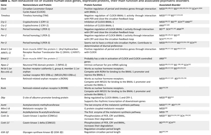
environmental factors such as routine daily activities and sunlight. The internal phase advance of
biological rhythms which reflects any disturbance of either sleep-wake cycle or endogenous circadian
rhythms is related to illnesses and aging11. The term “circadian misalignment” describes a range of
circumstances, such as inappropriately timed sleep and wake, misalignment of sleep/wake with feeding
rhythms, or misaligned central and peripheral clocks 13. Another subtype of circadian misalignment
includes the misalignment of body rhythms with environmental cycles that is usually found in night-shift
workers characterizes a condition of chronic desynchronization similar to that produced by persistent jet
lag. Different types of circadian misalignment have been associated with increased risk for both physical
and psychiatric disorders 13.
Circadian rhythms abnormality, a common manifestation of nearly all psychiatric disorders, is not a
surprising predisposing factor for mental disorders, because sleep is considered as a cardinal
common disrupted mechanisms related to the circadian rhythm in psychiatric disorders could be
determined as the melatonergic system, its effects of sleep pattern, and the hypothalamus–pituitary–
adrenal (HPA) axis. Besides, studies of human circadian rhythm genes revealed that genetic
polymorphisms of these genes predisposed to psychiatric disorders 14–16. Therefore, circadian
disturbances seem to be the common thread to all these possible underlying mechanisms that contribute
to illness onset, maintenance, and even the response to treatment. Special attention ought to be paid
toward the physiology and pathology of circadian rhythm to understand the etiology of psychiatric
disorders, and to develop appropriate treatment strategies because chronobiology is an essential field of
work in mental disorders. Related literature provides information on circadian rhythm disturbances for
certain psychiatric diagnoses such as schizophrenia, mood and anxiety disorders. However, we are aware
of a lack of a comprehensive perspective of molecular and neural substrates of common disrupted
circadian mechanisms to clinical manifestations in psychiatric disorders. There have been recent reviews
relevant to the subject. For instance, Jones and Benca reviewed circadian disruptions in psychiatric
disorders, however, they only focused on schizophrenia and mood disorders 17. Wulff and colleagues
comprehensively reviewed sleep and circadian rhythms disruption in psychiatric disorders 18.
Nevertheless, their review was lack of findings regarding current genetic, molecular and neurohumoral
models of circadian pathologies. Therefore, we aimed to present a comprehensive review from a
translational perspective regarding the reciprocal relationship between neurobiologic underpinnings of
circadian rhythm pathologies and psychiatric disorders in this article.
Searching strategy and selection criteria of reviewed studies
An electronic database search was performed by the authors in the MEDLINE, Embase, PsycInfo, and
Scopus databases for relevant articles published between January 1990 and October 2019. We searched
reference lists of relevant reviews. Different combinations of the keywords psychiatric disorder, mental disorder, mood disorder, bipolar disorder, depression, unipolar depression, major depressive disorder, schizophrenia, psychotic disorders, anxiety disorders, circadian rhythms, circadian markers, chronotype, chronobiology, circadian gene, clock gene, melatonin, and HPA axis were polled. Articles published only in English were reviewed. Unpublished studies, case reports, theses, and conference papers were
excluded. Several highly cited and regarded comprehensive review articles and meta-analyses are cited
due to space considerations. Eligible open-access and institutional-access articles were recruited. The
articles were filtered through an inspection of the abstracts in order to select the most suitable articles
related to the topic. In addition to database searches, the reference lists of the relevant articles were also
evaluated manually for additional publications matching the scope of our review. The authors avoided
incorporating duplicated samples of the key papers; however, studies with similar methodology were
included if they provide essential findings to the literature (Figure 1).
Molecular regulation of the circadian rhythm
We believe that it is noteworthy to briefly summarize the molecular underpinnings of circadian science
that gave input to the research into neural substrates of rhythmicity. Although the aforementioned
biological clock, it did not mean comprehension of all circadian molecular mechanisms. The circadian
rhythm started to be more understandable with the determination of alterations in PER protein and period
mRNA levels during a day. Hall and Rosbash ascertained that levels of period mRNA peaked in the early night, several hours earlier than the peak PER protein abundance 19. The TTFL model emerged with the
discovery of further circadian rhythm genes found in subsequent studies. According to this model, PER
and TIM (a protein encoded by the timeless gene) proteins transformed into a heterodimer form in the cytoplasm in order to translocate into the nucleus. TIM protein allows nuclear entry of PER 20. Besides
CLOCK and CYCLE [orthologues of mammalian CLOCK and BMAL-1 (a protein encoded by the brain
muscle ARNT-like protein-1 (Bmal-1) gene), respectively] constitute a protein couple that supports the
transcription of period and timeless genes [the equivalent of period 1-3 and cryptochrome 1-2(Cry)) in
mammalian cells] in the nucleus 21,22. When the PER-TIM heterodimer binds to the CLOCK-CYCLE
couple, CLOCK-CYCLE segregates from DNA and the transcription of downstream genes related to
PER and TIM conclude. In other words, the PER and TIM heterodimer terminate their transcription.
However, in the event of a decrement in PER and TIM protein levels, the CLOCK and CYCLE couple
activates their transcription once again, and TTFL starts over. All of these biochemical reactions include
transcription and translation processes that occur rapidly. However, a near 24-h period needs a delay
period and timeless gene transcriptions. The explanation about the regulation of the needed delay comes from the discovery of the doubletime gene, another member of the clock genes 23,24. The doubletime
gene’s product casein kinase-1 (CSNK-Iε; casein kinase 1 epsilon in mammals) phosphorylates PER for
degradation. Thus, activity of the doubletime gene reduces the stability and accumulation of PER, thereby promoting a delay between PER-TIM transcription and PER-TIM nuclear function 6,25. This molecular
mechanism occurs both in the SCN and nearly all peripheral cells.
The maestro of chronophysiologic rhythms including body temperature, sleep-wake cycle motor activity,
and neuroendocrine functions, is located in the SCN of the hypothalamus. The clock genes in the
peripheral cells such as hepatocytes, adipocytes or epidermal and dermal cells have their own rhythmicity;
however, cyclic processes in which the SCN is involved provide an integrative organization of the
physiologic functions and behavioral outputs of the body 26,27. The circadian system sustains an
endogenous rhythmic activity in spite of environmental cues. Regardless of the presence of light, the
neuronal activity in the SCN occurs at a higher frequency during the day compared with the night. The
neurons of the SCN tend to be excitable in the day to maintain spontaneous activity through persistent
Na++ currents, oscillations in chloride pumps, K+ channels, and Ca++ pools in the morning. Conversely,
hyperpolarized neurons are inhibited and keep the silence in the SCN at night 28. CRY and PER proteins
gather in the cytoplasm before translocating into the nucleus where they inhibit CLOCK-BMAL-1
activity during the night. In other words, CRY and PER proteins terminate their own transcription when
they inhibit CLOCK-BMAL-1 complex activity. After that, degradation of PER and CRY manages the
inhibition of CLOCK-BMAL1 toward the morning, followed by resumed transcription of
period/cryptochrome and other clock genes 29.
The master clock synchronizes the endogenous rhythm to the external world, mainly in the presence of
major environmental input – light 30–32. A specialized tract, called the retino-hypothalamic tract, which
starts from the retinal ganglion cells that include the essential photoreceptor pigment melanopsin, and
terminating at the SCN. This tract aids upregulation of clock gene expression and increases neuronal
activity in the SCN 33,34. Nevertheless, functions of the SCN, such as synchronization by the light/dark
cycle, do not only depend on this molecular mechanism. Many inputs of the SCN have been determined
including melatonin, food intake, blood pressure, and physical activity 35–38. In addition, the SCN receives
non-photic timing inputs from the raphe nucleus, which means the serotoninergic system plays a
substantial role in the regulation of circadian rhythm 39. Furthermore, the SCN serves in the excretion of
numerous neurotransmitters that interact with other hypothalamic structures, hence neuropeptidergic
signaling maintains circadian rhythm of the SCN. Consequently, the biologic interactions between the
brain and body are modulated by the SCN, which is critically involved in the organism’s adjustment to
the environment through the impact of internal signals, which are mediated by hormonal rhythms, the
autonomic nervous system, and external time indicators such as light and food intake 10. The master clock
regulates the endogenous rhythm in response to environmental inputs and dysfunction of the master clock
could contribute to a wide range of illnesses including obesity, diabetes mellitus, autoimmune disorders,
and particularly mental disorders 40–44. Disruption that arises due to a misalignment between inner
physiology and the external world or a clock gene polymorphism may facilitate the emergence of
diseases, increased disease severity and worsened prognosis, and heightened risk for poor treatment
outcomes 45,46.
(Table 1 to be inserted here)
Neurohumoral and hormonal regulation of circadian rhythm
The SCN collects information about the endogenous clocks through nervous projections and peripheral
hormones. The SCN’s monosynaptic outputs mainly target the pre-autonomic neurons of the
paraventricular nucleus (PVN) in the hypothalamus. The SCN is directly involved in the hypothalamic
output to the preganglionic parasympathetic regions of the brainstem and to sympathetic preganglionic
motor neurons of the spinal cord 47–49. These projections allow the SCN to command the rhythmic control
of hormone release and metabolism of all visceral structures through parasympathetic and sympathetic
outputs. It has been determined that the SCN could increase glucose production from the liver through
the sympathetic output to the liver with its projections that reach to the PVN 50. Similarly, the SCN could
increase corticosterone secretion in the adrenal or support glucose uptake into the muscle cells via
sympathetic activation 51–53. Besides, hormonal signals predominantly controlled by the SCN have a
critical role in the regulation of internal synchronization 27. Internal synchronization is supplied by
adrenal glucocorticoids, pineal melatonin, adipocyte-derived leptin, pancreatic insulin or stomach ghrelin
induced by the SCN. Internal synchronization included many multi-synaptic neuronal pathways that
modulate behavior. For example, leptin increases during food intake in rats, ghrelin increases following
Glucocorticoids are produced in the adrenal glands from cholesterol and rhythmically released at
ultradian (pulsatile) and circadian (daily) scales. Glucocorticoid release peaks typically prior to the onset
of physical activity and depends on the fluctuations of corticotropin (adrenocorticotropic hormone,
ACTH), a polypeptide secreted from the anterior pituitary under the control of corticotropin-releasing
hormone (CRH), during the day. Glucocorticoid levels are regulated by a complex interaction between
the adrenal clock and sympathetic outputs from the PVN and SCN 56. Furthermore, the daily variation of
glucocorticoids is influenced by stressful life events that activate the HPA axis and the autonomous
nervous system. Glucocorticoid rhythm has a crucial role in the regulation of other hormonal rhythms
and peripheral oscillations of metabolic gene expressions in the cells of tissues such as liver and white
adipose tissue 56.
On the other hand, adrenal glucocorticoids can modulate the synchronization of the master clock to light
via serotonergic projections from the raphe nucleus 57. Serotonergic neurons release serotonin in the
presence of glucocorticoid and locomotor activity. Such neuronal activity ensures transmitting feedback
to the SCN in order to sustain the functioning of the clock itself 58. In other words, serotonergic
projections stimulated by locomotor activity provide a re-synchronization of the SCN 59. Furthermore,
brain serotonin synthesis and catabolism have their own circadian rhythm, closely related to the SCN.
Neuronal serotonin release in the SCN is provided in the absence of photic stimulation, and serotonin
levels increase in the raphe nucleus after the beginning of the dark phase 60. Tryptophan hydroxylase
(TpH), the rate-limiting enzyme in the synthesis of serotonin, is one of the regulators of circadian rhythm
in the raphe nucleus. It is known that TpH peaks during the dark phase, helping the interaction between
the serotoninergic system and the SCN through the increment of serotonin levels 60. Also, serotonergic
neurotransmission alterations could cause phase shifts and changes in SCN activity affecting the
phosphorylation of CLOCK proteins 61.
Melatonin, a member of the class of acetamides, is another hormone related to biologic rhythm. It is
primarily released by the pineal gland, particularly at night. Melatonin release is adjusted by the length
of night time and melatonin per se regulates the seasonality of energy metabolism and reproduction in photoperiodic species 62. Melatonin secretion peaks a few hours before sleep or at the time of minimal
vigilance propensity, and decreases as wakefulness approaches under normal conditions 63. In contrast,
core body temperature reaches the highest degree during the day and has a nocturnal decline related to
the melatonin peak 64. This inverse relationship between melatonin and core body temperature is
organized by the SCN. The nocturnal release of melatonin is induced by the SCN input to the PVN
noradrenergic (sympathetic) afferents to the pineal gland 53. Melatonin accumulates sleep both by setting
the SCN and inhibiting neural centers such as the locus coeruleus (LC) and raphe nuclei, which mediate
arousal through the ventrolateral preoptic nucleus of the hypothalamus (VLPO). It has been determined
that melatonin receptor agonists increase monoaminergic neuronal activity and contribute to the
regulation of dopamine and 5-HT neurotransmission 65. In other words, melatonin has a modulatory role
on the monoaminergic activity by linking the circadian and monoamine systems. The SCN modulates
the release of melatonin mainly through γ-aminobutyric acid (GABA) neurons that project from the SCN
that inhibit the same PVN neurons through GABAergic projections and cease the secretion melatonin 32.
The daily rhythm of melatonin has remarkable effects on the molecular clockworks of both the brain and
body alongside regulating the sleep/wake cycle 67,68. Melatonin receptors (MT1 and MT2) are mainly
localized in the CNS but also have been detected beyond the CNS in a wide range of somatic cells 69.
This diversity could be interpreted as melatonin having an integrative role in the light-induced circadian
rhythms controlled by the SCN in the whole organism.
Circadian rhythm and its implications on psychiatric disorders
At the core of any psychiatric disorder is an abnormality in neurotransmitter signaling. It is well known
that the disruption of circadian physiology has widespread effects on all aspects of neural and
neuroendocrine function, which leads to psychiatric disorders. The aforementioned information
regarding neural substrates of biologic rhythm is frequently reported impaired in many mental disorders.
Following the comprehensive conceptual framework of neural substrates of chronobiologic processes
mentioned above, we will next discuss the reciprocal associations between circadian rhythm disturbances
and psychiatric disorders, and draw a clinical picture for common diagnoses.
(Table 2 to be inserted here)
Mood disorders
In 1681, Robert Burton defined the autumn as the most melancholic season in his best-known classic,
The Anatomy of Melancholia 70. Circadian rhythm abnormalities in mood disorders have been pointed
towards by the observers of melancholia for sixty years 71–73. A wide range of body functions such as
core body temperature, blood pressure, pulse rate, and hormones such as plasma cortisol levels,
thyroid-stimulating hormone, and melatonin have been found disturbed in patients with manic depression and
depression compared with people without a mental disorders 72,73. Moreover, mood and other symptoms
of the disorder have been previously reported to show diurnal variation in depression 74. Disordered
sleep/wake cycle is considered as another clue for physicians in patients with bipolar disorder (BD) and
major depressive disorder (MDD) 74. In addition, it was recognized that disrupted rhythms were
re-synchronized after antidepressant or mood-stabilizing treatment 75. Another significant feature is that
mood episodes recur seasonally and previous studies showed that there could be an association between
light and the emergence of mood states 76–80. Thus, all of these findings suggested the possibility of
circadian rhythm disturbance in mood disorders. Consequently, the earliest mention of seasonality took
place in the Diagnostic and Statistical Manual of Mental Disorders Third Edition, Revised Version
(DSM-III-R), and seasonal pattern was defined as a specifier in the affective disorders section 81.
Chronotype is another concept associated with mental disorders, particularly with affective disorders,
and resembles individual physiologic functions and activities such as sleeping, eating, or hormone release.
Chronotype has usually been used to denote sleep habits: morning and evening types. The relationship
chronotype has been related to a vulnerability to depression and increased alcohol and stimulant drug
use 82.
Although sleep/wake cycle alteration, which is considered as a consequence of circadian system
disruption, had been the best-known contributor to the pathophysiology of mood disorders for years,
today, it is well-recognized that circadian rhythm is entangled with a wide range of molecular and cellular
processes that are hypothesized to lead to mood disorders 83. Accordingly, below we discuss in detail
internal and external factors that may play a role in the emergence of mood disorders through various
psychophysiological mechanisms within the circadian rhythm processes.
Major depressive disorder
As a cardinal element of chronobiologic processes, sleep behavior and its disturbances have received the
strongest spotlight regarding research into their undisputed etiologic and prognostic association with
mood disorders. The concomitance of sleep disruption and depression had been the main focus of
research into the contribution of circadian rhythms disruption to depression development since the 1970s
84–86. The relationship between sleep and mood could easily be observed even in healthy individuals
exposed to jet lag or shiftwork 87. The presence of sleep disruption may cause negative effects, irritability,
and fatigue. Sleep behavior changes, such as difficulties in initiating/maintaining sleep or early morning
awakening have been determined in 90% of patients with MDD 18. Sleep-wake disruptions are among
the criteria for the diagnosis of depression, and comorbid parasomnias are associated with poor treatment
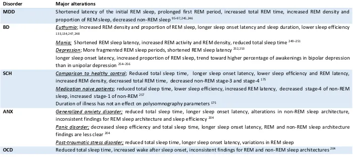
outcomes, increased suicidality, and greater relapse risk in depression 82,88–90. Sleep architecture
alterations including shortened latency of the initial rapid eye movement (REM) sleep, prolonged first
REM period, increased total REM time, increased REM density and proportion of REM sleep, and
decreased non-REM sleep have been demonstrated in depression 91–97. It has been suggested that there is
a reciprocal relationship between sleep-wake cycle variables such as wakefulness and sleep latency and
sleep architecture features likes of REM sleep latency 98. For instance, preclinical studies have
consistently demonstrated that prolonged REM sleep was associated with decreased wakefulness in
depressive subjects 99. In addition, an endogenous circadian rhythm abnormality, the phase advance, is
related to decreased REM latency after falling asleep among individuals with depression 100. These
findings suggest that sleep itself has multiple and complex regulators related with homeostatic
mechanisms along with the endogenous circadian rhythm.
Melatonin output and the timing of its release have been found closely associated with other rhythms as
mentioned above. Numerous studies have been conducted to show alterations of melatonin release and
its phase to determine circadian misalignment between internal and external clocks in patients with mood
disorders 101. To date, the most consistent results suggested lower nocturnal melatonin levels, delayed
melatonin secretion onset, and offset in patients with depression 101. Besides, the length of the interval
between melatonin secretion and sleep onset has been found related to depression severity 102. However,
a few studies demonstrated increased nocturnal melatonin levels in depressive patients 103. Such
patients with psychotic symptoms. Abnormal body temperature variations including the absence of the
nocturnal decline of core body temperature and daily mean temperature degrees are also observed in
patients with depression and these higher values normalized with antidepressant treatment 82,104. This is
probably due to the impaired control of the melatonergic system over thermoregulatory processes in
depressive patients. 104,105.
There is an irrefutable association between circadian genes and mood regulation. Even though mood
disorders are not directly related to clock gene mutations, findings suggest that individual genetic
polymorphisms of clock genes may influence the clinical features of the disorder, such as age at disease
onset and treatment response 106,107. Genetic studies have implicated clock, timeless, cryptochrome-1
(Cry-1), period-2,3 (Per-2,3), Bmal-1,2, neuronal pas domain protein 2 (Npas-2), nuclear receptor subfamily-1, group d, member 1 (Nr1d-1), retinoid-related orphan receptor a (Rora), CSNK-Iε, D site of albumin promoter binding protein (Dbp), acetylserotonin methyltransferase (Asmt), melatonin receptor 1b (Mtnr1-B), arylalkylamine n-acetyltransferase (Aanat) genes in unipolar depression 108–112.
However, most of these studies have small sample sizes and need to be replicated in larger groups.
Glucocorticoids are adrenal steroid hormones and have multifunctional roles in the body and brain such
as metabolism, immunity, arousal, neuronal survival, and neurogenesis 113. Glucocorticoids have their
own circadian rhythm and an important role in synchronizing peripheral clocks and the SCN. In addition,
they have anti-inflammatory properties and regulate the immune system response 114. Since Carroll
defined the resistance of the dexamethasone suppression test in patients with depression in 1968 115,
hypothalamic-pituitary-adrenal (HPA) axis dysregulation has been one of the most consistent findings
in mental disorders, particularly in depression 83,115. Hypercortisolemia-flattened HPA axis circadian
rhythm and disrupted response of the HPA axis to glucocorticoid feedback are commonly observed in
patients with depression 116,117. Dehydroepiandrosterone (DHEA), is another adrenal steroid that has a
neuroprotective role and modulates corticosteroid-induced cell death. An increased cortisol/DHEA ratio,
which assesses the degree of ‘functional’ hypercortisolemia, is seen in adults and adolescents with
depression 118–120. Glucocorticoid receptor hypofunction has also been found in peripheral tissue cells
including mononuclear cells and skin cells 121. Furthermore, findings support that antidepressant
treatment repairs the impaired HPA axis dysfunction in depression 122.
Depression and inflammatory disorders such as rheumatoid arthritis, inflammatory bowel disease, and
asthma have been found coexisting, and such common comorbidities point to the neuroinflammatory
background and immune-associated contributions in the etiopathogenesis of depression 123,124. Studies
have also shown that pro-inflammatory cytokines could induce a depression-like symptom cluster
including anhedonia, fatigue, increased sleep, and decreased locomotor activity 125. Inflammatory
markers such as interleukin (IL)-1β, IL-2, IL-6, tumor necrosis factor (TNF)-α, C-reactive protein (CRP),
and prostaglandin E2 (PGE2) have been reported increased in patients with depression 126. Sleep-wake
cycle changes and circadian misalignment between the internal and external clocks may be other
contributors to increased pro-inflammatory cytokine levels in depression. The arrhythmic clock system
regulators of inflammation in the body and activates the inflammatory response 127,128. Besides, sleep
disturbances and long sleep duration were found related with the increased cytokines levels and the risk
for depression 129. We may interpret the aforementioned findings as the circadian system’s involvement
in the pathophysiology of MDD being not limited to sleep/wake cycle disruption, it is also related to
complex associations between biologic rhythm, environment-gene interactions, HPA axis dysfunction,
and immune system alterations.
Bipolar disorder
Sleep disturbances have been the core common characteristic feature in bipolar mood episodes, both
mania and depression, since the first definition of Kraepelin 130. In turn, insomnia or hypersomnia and
decreased need for sleep are typical for manic and depressive episodes. Studies showed that sleep
architecture was characterized by increased REM density and reduced REM latency in bipolar manic
episodes 131. Sleep disturbances are also frequently observed in euthymic patients with BD. Increased
REM density and the proportion of REM sleep have been shown in remitted patients with BD 132.
Moreover, findings revealed that remitted patients with BD have longer sleep latency and sleep duration
and lower sleep efficiency 133,134. Bipolar depression has similar polysomnographic findings including a
tendency for more early awakenings and more fragmented REM sleep periods. However, total REM
density was found greater in bipolar depression than in unipolar depression 132 (See table 2 for detailed
information). Although abnormalities of sleep architecture are seen in episodes and inter-episodes, sleep
disturbances worsen before relapses. Sleep loss and reduced sleep duration were defined as reliable
predictors of hypomania and mania 132. In addition, hypersomnia in euthymia is found associated with
the development of upcoming depressive symptoms 135. On the other hand, a large amount of euthymic
patients describe symptoms that meet the diagnostic criteria for insomnia 134,136. Sleep-wake disturbances
have been found as one of the reasons for a worse course of illness, relapses, increased symptom severity,
and poor treatment outcomes 137–140. These findings may explain the reason for the treatment need in
remitted patients with BD 89.
Involvement of the melatonergic system in the pathogenesis of BD through circadian dysregulations such
as changes in the release timing, melatonin rhythm, and the sleep-wake cycle 132. In terms of the phase
relationship between the sleep-wake cycle and melatonin rhythm, it has been demonstrated that the
melatonin secretion in the hours surrounding habitual sleep onset which is considered as a crucial
circadian moment for sleep regulation was increased in patients with BD compared to those with unipolar
depression 141. Although findings of melatonin function in patients with BD are inconsistent, circadian
system characteristics generally vary depending on the current episode; mania or depression 82.
Melatonin levels were found higher in the daytime in manic patients than in healthy controls and patients
with depressive episode 142. Findings about nocturnal melatonin levels among BD phases are not
consistent 72,143–145. It remains unclear as to whether these alterations derive from a primary dysfunction
some studies supported the beneficial effect of exogenous melatonin administration, which provides
sleep and mood improvement 146.
Some of the clock genes have been found intimately associated with both the onset of BD and illness
course. Studies revealed that circadian gene polymorphisms may increase the predisposition to BD and
indirectly affect recurrences and symptoms across all BD phases 147. Genetic linkage and gene expression
studies implicated the variant genes related to BD as clock, timeless, Cry-1, Npas-2, Bmal-1,2, Dbp,
Nr1d-1, Per-2,3, Rora, Rorb, Asmt,Csnk-1ε, Csnk-1, and glycogen synthase kinase-3(GSK-3) 111,147– 151. It has been demonstrated that ClockD19, the mutant gene that occurs with the deletion of exon 19 in
the Clock gene, produces a dominant negative CLOCK protein capable of DNA binding but deficient in transcriptional activity. This gene induces dopamine synthesis and increased dopaminergic activity,
which result in an increase in tyrosine hydroxylase (TH) expression in the ventral tegmental area (VTA)
and manic-like behavior in animal models 152–154. Moreover, ClockD19- related higher dopaminergic
activity in the VTA normalized after lithium treatment, which suggests increased dopaminergic activity
may be the main reason for the manic-like behavior of mice 154. Recently, several lines of evidence have
emphasized the importance of the molecular and synaptic mechanisms of monoaminergic systems and
circadian gene interactions, which are closely related to molecular alterations associated with the
ClockD19 model in the VTA and nucleus accumbens.155 On the other hand, lithium, a potent inhibitor of
the GSK-3 enzyme, regulates the clock gene Nr1d-1 and BMAL-1 through GSK-3 156. Some
polymorphisms including Clockrs3805148, Clockrs534654, Timelessrs11171856, and
Timelessrs2291739 are associated with suicidal behavior in BD 157.
A dysfunctional HPA axis is suggested to play an important role in the pathophysiology of BD, although
the mechanism needs to be elucidated. Increased levels of cortisol and ACTH are the most replicated
findings in BD 158,159. However, CRH levels are not determined to increase in BD.158 Depressive
symptoms and cognitive deficits are thought to be associated with the higher levels of cortisol, and ACTH
and cortisol seem to be related to manic episodes 159. A meta-analysis suggested that abnormalities of
stress-related pathways including increased morning cortisol levels were mainly prominent in manic
episodes. Such abnormalities are even observed in remitted patients, which means that the long-term
pathology of the HPA axis is related to clinical states of BD and contributes to the stress-vulnerability
models of illness development and progression 160.
Immune abnormalities have received increased attention due to their possible role in the pathophysiology
of BD, as well as MDD. Systematic reviews on cytokine levels in patients with BD revealed that IL-4,
IL-6, IL-10, soluble IL-2 receptor, soluble IL-6 receptor, and TNF-α levels were increased in patients
compared with healthy controls, whereas IL-2, IL-8, IFN-gamma, and C-C motif ligand were not
different from controls 161. Moreover, a comparison of cytokine levels in another study determined that
proinflammatory cytokines including IL-2, IL-4, IL-6 were higher during manic episodes, and IL-6 levels
were higher in depressive state than in healthy controls 162. It was also demonstrated that mood symptoms
had a positive correlation with IL-6 and IL-2 levels 162. When bipolar depression and unipolar depression
at higher levels than in unipolar depression 163. In conclusion, sleep disturbances may be a reliable
indicator of an upcoming mood episode in BD.
Schizophrenia
Although the relationship between mood disorders and circadian abnormalities has become clearer in
recent times, the links between schizophrenia and disrupted circadian rhythms have yet to be elucidated
fully. However, sleep disorders and sleep-wake cycle alterations have been known as common and
consistent features of schizophrenia and other psychotic disorders since the first definition of Kraepelin
in 1883 164. Schizophrenia has been associated with abnormalities in sleep including delayed and
advanced sleep onset, altered resting activity patterns, and irregular sleep-wake cycle 165. Research into
circadian abnormalities and sleep disruption in schizophrenia has attempted to explain the causal
relationship in a reciprocal context. Hyperdopaminergia is a well-known phenomenon in psychosis
syndromes and striatal hyperdopaminergic activity may be a result of sleep disruption and circadian
abnormalities, and increased dopamine levels may induce sleep disruptions 166–168. For instance, the
Clock T3111C polymorphism, which is associated with increased dopamine levels in the SCN, has been determined in a population of Japanese patients with schizophrenia 14. Furthermore, the blind-drunk
mutant mouse, which carries a mutation in the gene encoding an exocytotic synaptic protein,
synaptosomal-associated protein-25 (Snap-25), exhibits schizophrenia-like symptoms 169,170. This mouse
model of schizophrenia has been shown to display phase advance and fragmentation of the circadian
cycle 171. Most consistent findings of the circadian genetics studies have been associations between
CLOCK, PERIOD1, PERIOD3, and TIMELESS genes and schizophrenia 172. Circadian rhythms
disruption has been reported in approximately 80% of patients with schizophrenia 173. Abnormal sleep
patterns in schizophrenia have been described in both unmedicated patients and patients currently
receiving antipsychotic treatment 18. The major findings in sleep architecture could be aligned, such as
long sleep-onset latency, increased intermittent-awakenings, decreased total sleep time, and poor sleep
efficiency 174. Moreover, reductions in REM latency, REM density, and duration of non-REM Stage 4
are other alterations in micro-sleep architecture 17,18,175–177. Sleep disturbances are also important to
predict increased suicide attempts in patients with schizophrenia 178.
Melatonin is a versatile neurohormone that plays an important role in the pathophysiology of
schizophrenia. 5-HT synthesis regulation, sleep-wake cycle, and anti-oxidant effects against
neuroinflammation are impaired due to melatonin dysfunction in schizophrenia 167,179. It has been shown
that melatonin increases endogenous antioxidants by increasing phosphorylated glycogen synthase
kinase-3 (GSK-3) levels and provides an anti-inflammatory effect 179,180. Galván-Arrieta et al. reported a
reduction in axogenesis associated with lower levels of phosphorylated GSK-3 subtype β and less
expression of melatonergic receptors in patients with schizophrenia compared with healthy controls.
These findings may indicate a melatonin-derived neurodevelopmental deficit at a cellular level 181. A
lack of normal melatonin rhythmicity, decreased nocturnal secretion of melatonin, and phase advance in
melatonin circadian rhythms have also been described in patients with schizophrenia 167,179,182.
schizophrenia, and this structural change has been found associated with cortical atrophy 183. Mainly, the
clinical importance of the relationship schizophrenia and melatonergic dysfunction might come from the
fact that impairment of sleep-wake cycles which are in close relationship with melatonin phase are
associated with schizophrenia symptoms such as somatic complaints, anxiety, depression, and paranoia.
Further support has shown that sleep-deprived schizophrenia patients might exhibit increased psychotic
symptoms 167. Moreover, the circadian rhythm of dopamine is dependent on melatonin 184. A combination
of sleep disruption and circadian rhythms disturbance may lead to the elevation of dopamine activity in
the brain, both directly and through phase advance in melatonin circadian rhythm 167. Preclinical studies
have been also demonstrated that melatonergic receptor agonism may prevent the increase of glutamate
release in prefrontal cortex 185, which has been suggested in the pathophysiology of schizophrenia,
particularly of cognitive symptoms of the disorder 186. Because of its significance in the pathogenesis of
schizophrenia, melatonin has become a therapeutic target for researchers. It has been shown that
melatonin agonists are efficacious agents for schizophrenia-associated sleep disorders and drug-related
tardive dyskinesia 187,188. Moreover, its improving effects on behavioral deficits via reducing brain
oxidative stress have been shown in an animal model of schizophrenia 189.
The relationship between clock genes and schizophrenia is another undiscovered area for scientists. Few
studies have been conducted to show linking circadian clock gene polymorphisms in schizophrenia to
date. Takao et al. identified the Clock 311C/T polymorphism, which is associated with higher
dopaminergic neurotransmission in the SCN in patients with schizophrenia 14. These results were
confirmed in another study conducted in a Chinese schizophrenic population 190. Period-1 mRNA
expression in the temporal lobe of post-mortem subjects with schizophrenia was found down-regulated
when compared with healthy controls 191. In addition, disrupted diurnal rhythms of the 1, 2,
Per-3, Npas-2 and phase delay in the expression of Per-2 have been reported in white blood cells of patients with schizophrenia 192. More recently, the absence of rhythmic expression of Cry-1 and Per-2 was
determined in the fibroblasts of patients with schizophrenia compared with cells obtained from healthy
controls.193 Pinacho et al. reported decreased levels of CSNK1 protein levels in the prefrontal cortex of
patients with schizophrenia 194. However, due to the small sample sizes of the available studies, the
association between schizophrenia and clock genes still needs to be clarified with further studies with
larger populations.
The stress-vulnerability model for schizophrenia was first proposed in the 1970s and has been further
developed since that time 195,196. Thus, the HPA axis has been one of the most attractive research targets
to understand the pathophysiology of schizophrenia for decades. Increased cortisol levels have been
determined in patients with schizophrenia and even in individuals at high risk for schizophrenia
compared with controls 197–199. However, mean baseline cortisol level measurements in schizophrenia
are not consistent in the literature 200. Nevertheless, blunted cortisol levels in response to stressors are
much more consistent findings, regardless of disease stage, chronicity, and treatment condition 201. To
conclude, despite it being widely accepted that sleep and circadian disorders have an important role in
the etiopathogenesis of schizophrenia, well-designed and comprehensive clinical studies are still needed
Other Psychiatric Disorders
Anxiety disorders are seen as the most frequent type of psychiatric disorders with a lifetime prevalence
of 29% in the general population 202. Sleep disturbance is a common feature of anxiety disorders and is
included in the symptom criteria for several anxiety disorders such as post-traumatic stress disorder and
generalized anxiety disorder 203. The presence of sleep disturbances has been reported as 74% in patients
with anxiety disorders 132. However, MDD as a frequent comorbid condition in anxiety disorders is a
confounder in understanding the relationship of sleep disturbances and anxiety disorders. Studies related
to generalized anxiety disorder have reported decreased total sleep time, increased sleep-onset latency,
and alterations in non-REM sleep architecture, whereas findings of REM sleep and sleep efficiency are
inconsistent 204. Patients with panic disorder frequently have both sleep disorder and/or another anxiety
disorder because they could have nocturnal panic attacks, which usually occur in Stage-2 or Stage-3 of
non-REM sleep, as well as decreased sleep efficiency, total sleep time, and increased sleep onset latency
132,204. Although sleep disturbances, including REM sleep-related nightmares, have been investigated in
post-traumatic stress disorder, conclusions are not consistent 132. There is no significant difference in
sleep architecture in social anxiety disorder 205,206. In an animal model, Cry-1 and Cry-2 gene protein
deficiencies led to behavioral alterations characterized by an abnormally high level of anxiety 207.
Akiyama et al. suggested that period-1 mRNA levels reduced after anti-anxiety treatment in the mouse cerebellum 208. Cry-2 expression was determined reduced in the hippocampus in another animal study 209. Furthermore, a polymorphism in BMAL-2rs2306073 has been found associated with social phobia 210.
Obsessive-compulsive disorder (OCD) is another debilitating disorder that is segregated from the anxiety
disorders category in the DSM-5 211. Although sleep disturbances have been reported including decreased
total sleep time, alterations in REM and non-REM sleep architecture are less clear 204. Certain
chronotypes have been found as predictors of OCD symptoms in adults, and circadian rhythm disorders
have been found as predictors of treatment outcomes 212. To the best of our knowledge, the role of
circadian rhythms disruption in all anxiety disorders, including OCD, has yet to go beyond showing sleep
disturbance; comprehensive research is warranted in the context of chronobiologic mechanisms of
anxiety disorder pathology.
(Table 3 to be inserted here)
Conclusion
The circadian system is responsible for the temporal organization of physiologic functions, and
disruptions can have marked functional influences on the living organism. As the role of chronobiologic
systems in both physical and mental health have become better understood, research into neurobiologic
mechanisms of circadian rhythms has been expanded. Mood, cognition, and behavior have complex
with impaired circadian biology. Extensive research has shown that impaired circadian mechanisms
could lead to psychiatric entities, whereas they may be an outcome of mental disturbances. Impaired
HPA axis function and melatonin homeostasis are the most consistent findings in mental disorders.
Independent from sleep disorders, the circadian system has a distinct role in homeostatic processes,
whose impairment has an impact in emotion regulation, cognition, behavior, and, most importantly,
neural plasticity, all of which are often disrupted in psychiatric phenotypes. There is some evidence
suggesting that circadian rhythm genes are associated with psychiatric disorders; however, the specificity
and causality of these associations have yet to be made clear. In our opinion, we are a long way from
establishing a robust causative link between circadian rhythms disruption and phenotypic complexity of
psychiatric disorders. A decent translational approach to the findings of animal models would likely
result in a clearer understanding of pathophysiologic implications of the circadian system. Further
support from continued and integrated investigations of these issues may promote a deeper appreciation
of the contribution of circadian disturbances to the pathophysiology of psychiatric illnesses and related
psychopathology, and will hopefully yield improved therapeutic strategies for their treatment.
Conflicts of interest
The authors declared no potential conflicts of interest with respect to the research, authorship, and/or
publication of this article.
Funding
The authors received no specific grant from any funding agency, commercial or not-for-profit sectors for
the research, authorship, and/ or publication of this article.
Author contributions
Dr. Simge Seren Kirlioglu (SSK) and Dr. Yasin Hasan Balcioglu (YHB) conceived the presented idea
and contributed equally to concept development and designing of the article. The screening of the
literature was performed by SSK. Overall, while SSK drafted the article, both authors were involved in
reviewing and revising the article and contributed to the intellectual content. SSK generated the tables,
while YHB did the figure. YHB also performed a critical review and made necessary corrections. Both
References
1. Foster RG, Kreitzman L. Rhythms of Life: The Biological Clocks That Control the Daily Lives of Every Living Thing. New Haven (Connecticut): Yale University Press; 2005.
2. Halberg F, Cornélissen G, Katinas G, Syutkina E V, Sothern RB, Zaslavskaya R, et al. Transdisciplinary unifying implications of circadian findings in the 1950s. J Circadian Rhythms. 2003;1:2.
3. Konopka RJ, Benzer S. Clock mutants of Drosophila melanogaster. Proc Natl Acad Sci U S A. 1971;68:2112-2116.
4. Bargiello TA, Jackson FR, Young MW. Restoration of circadian behavioural rhythms by gene
transfer in Drosophila. Nature. 1984;312:752-754.
5. Reddy P, Zehring WA, Wheeler DA, Pirrotta V, Hadfield C, Hall JC, et al. Molecular analysis of the period locus in Drosophila melanogaster and identification of a transcript involved in biological rhythms. Cell. 1984;38:701-710.
6. Huang R-C. The discoveries of molecular mechanisms for the circadian rhythm: The 2017 Nobel Prize in Physiology or Medicine. Biomed J. 2018;41:5-8.
7. Crosby EC, Woodburne RT. The mammalian midbrain and isthmus regions. Part II. The fiber
connections. C. The hypothalamo-tegmental pathways. J Comp Neurol. 1951;94:1-32.
8. Sollars PJ, Pickard GE. The Neurobiology of Circadian Rhythms. Psychiatr Clin North Am. 2015;38:645-665.
9. Zhu L, Zee PC. Circadian Rhythm Sleep Disorders. Neurol Clin. 2012;30:1167-1191. 10. Gillette M. Chronobiology: Biological Timing in Health and Disease. Prog Mol Biol Transl
Sci. 2013:376.
11. Dijk DJ, Duffy JF, Czeisler CA. Contribution of circadian physiology and sleep homeostasis to age- related changes in human sleep. Chronobiol Int. 2000;17:285-311.
12. Panda S. Circadian physiology of metabolism. Science (80- ). 2016;354:1008-1015.
13. Baron KG, Reid KJ. Circadian misalignment and health. Int Rev Psychiatry. 2014;26:139-154.
14. Takao T, Tachikawa H, Kawanishi Y, Mizukami K, Asada T. CLOCK gene T3111C
polymorphism is associated with Japanese schizophrenics: A preliminary study. Eur Neuropsychopharmacol. 2007;17:273-276.
15. Benedetti F, Serretti A, Colombo C, Barbini B, Lorenzi C, Campori E, et al. Influence ofCLOCK gene polymorphism on circadian mood fluctuation and illness recurrence in bipolar depression. Am J Med Genet. 2003;123B:23-26.
3111T/C and preferred circadian phase in Korean patients with bipolar disorder. Prog Neuro-Psychopharmacology Biol Psychiatry. 2010;34:1196-1201.
17. Jones SG, Benca RM. Circadian disruption in psychiatric disorders. Sleep Med Clin. 2015;10:481-493.
18. Wulff K, Gatti S, Wettstein JG, Foster RG. Sleep and circadian rhythm disruption in psychiatric and neurodegenerative disease. Nat Rev Neurosci. 2010;11:589-599.
19. Hardin PE, Hall JC, Rosbash M. Feedback of the Drosophila period gene product on circadian cycling of its messenger RNA levels. Nature. 1990;343:536-540.
20. Gekakis N, Saez L, Delahaye-Brown AM, Myers MP, Sehgal A, Young MW, et al. Isolation of
timeless by PER protein interaction: defective interaction between timeless protein and long-period mutant PERL. Science. 1995;270:811-815.
21. Allada R, White NE, So WV, Hall JC, Rosbash M. A Mutant Drosophila Homolog of
Mammalian Clock Disrupts Circadian Rhythms and Transcription of period and timeless. Cell. 1998;93:791-804.
22. Rutila JE, Suri V, Le M, So WV, Rosbash M, Hall JC. CYCLE Is a Second bHLH-PAS Clock
Protein Essential for Circadian Rhythmicity and Transcription of Drosophila period and timeless. Cell. 1998;93:805-814.
23. Price JL, Blau J, Rothenfluh A, Abodeely M, Kloss B, Young MW. double-time Is a Novel Drosophila Clock Gene that Regulates PERIOD Protein Accumulation. Cell. 1998;94:83-95.
24. Kloss B, Price JL, Saez L, Blau J, Rothenfluh A, Wesley CS, et al. The Drosophila Clock Gene double-time Encodes a Protein Closely Related to Human Casein Kinase Iε. Cell. 1998;94:97-107.
25. Lowrey PL, Shimomura K, Antoch MP, Yamazaki S, Zemenides PD, Ralph MR, et al.
Positional syntenic cloning and functional characterization of the mammalian circadian mutation tau. Science. 2000;288:483-492.
26. Mohawk JA, Green CB, Takahashi JS. Central and Peripheral Circadian Clocks in Mammals.
Annu Rev Neurosci. 2012;35:445-462.
27. Challet E. Keeping circadian time with hormones. Diabetes, Obes Metab. 2015;17:76-83. 28. Colwell CS. Linking neural activity and molecular oscillations in the SCN. Nat Rev Neurosci.
2011;12:553-569.
29. Tsang AH, Astiz M, Friedrichs M, Oster H. Endocrine regulation of circadian physiology. J Endocrinol. 2016;230:R1-R11.
30. Mrosovsky N, Hattar S. Impaired Masking Responses to Light in Melanopsin‐Knockout Mice.
Chronobiol Int. 2003;20:989-999.
31. Dibner C, Schibler U, Albrecht U. The Mammalian Circadian Timing System: Organization
and Coordination of Central and Peripheral Clocks. Annu Rev Physiol. 2010;72:517-549. 32. Pevet P, Challet E. Melatonin: Both master clock output and internal time-giver in the
circadian clocks network. J Physiol. 2011;105:170-182.
33. Hankins MW, Peirson SN, Foster RG. Melanopsin: an exciting photopigment. Trends Neurosci. 2008;31:27-36.
34. Amaral FG do, Cipolla-Neto J, Amaral FG do, Cipolla-Neto J. A brief review about melatonin, a pineal hormone. Arch Endocrinol Metab. 2018;62:472-479.
35. Asher G, Sassone-Corsi P. Time for Food: The Intimate Interplay between Nutrition, Metabolism, and the Circadian Clock. Cell. 2015;161:84-92.
36. Buijs FN, Cazarez F, Basualdo MC, Scheer FAJL, Perusquía M, Centurion D, et al. The suprachiasmatic nucleus is part of a neural feedback circuit adapting blood pressure response.
Neuroscience. 2014;266:197-207.
38. Sabbar M, Dkhissi-Benyahya O, Benazzouz A, Lakhdar-Ghazal N. Circadian Clock Protein Content and Daily Rhythm of Locomotor Activity Are Altered after Chronic Exposure to Lead in Rat. Front Behav Neurosci. 2017;11:178.
39. Zhang T, Huang L, Zhang L, Tan M, Pu M, Pickard GE, et al. ON and OFF retinal ganglion cells differentially regulate serotonergic and GABAergic activity in the dorsal raphe nucleus.
Sci Rep. 2016;6:26060.
40. Robillard R, Carpenter JS, Feilds K-L, Hermens DF, White D, Naismith SL, et al. Parallel Changes in Mood and Melatonin Rhythm Following an Adjunctive Multimodal
Chronobiological Intervention With Agomelatine in People With Depression: A Proof of Concept Open Label Study. Front Psychiatry. 2018;9:624.
41. Duval F, Mokrani M-C, Erb A, Gonzalez opera F, Calleja C, Paris V. Relationship between chronobiological thyrotropin and prolactin responses to protirelin (TRH) and suicidal behavior in depressed patients. Psychoneuroendocrinology. 2017;85:100-109.
42. Robillard R, Carpenter JS, Rogers NL, Fares S, Grierson AB, Hermens DF, et al. Circadian rhythms and psychiatric profiles in young adults with unipolar depressive disorders. Transl Psychiatry. 2018;8:213.
43. Buttgereit F, Smolen JS, Coogan AN, Cajochen C. Clocking in: chronobiology in rheumatoid arthritis. Nat Rev Rheumatol. 2015;11:349-356.
44. Saetung S, Nimitphong H, Siwasaranond N, Manodpitipong A, Crowley SJ, Hood MM, et al.
Eveningness Is Associated With Greater Depressive Symptoms in Type 2 Diabetes Patients: A Study in Two Different Ethnic Cohorts. Behav Sleep Med. 2019;17:291-301.
45. Charrier A, Olliac B, Roubertoux P, Tordjman S, Charrier A, Olliac B, et al. Clock Genes and Altered Sleep–Wake Rhythms: Their Role in the Development of Psychiatric Disorders. Int J Mol Sci. 2017;18:938.
46. Barandas R, Landgraf D, McCarthy MJ, Welsh DK. Circadian Clocks as Modulators of
Metabolic Comorbidity in Psychiatric Disorders. Curr Psychiatry Rep. 2015;17:98.
47. Guilding C, Piggins HD. Challenging the omnipotence of the suprachiasmatic timekeeper: are circadian oscillators present throughout the mammalian brain? Eur J Neurosci. 2007;25:3195-3216.
48. Kalsbeek A, Palm IF, La Fleur SE, Scheer FAJL, Perreau-Lenz S, Ruiter M, et al. SCN Outputs and the Hypothalamic Balance of Life. J Biol Rhythms. 2006;21:458-469.
49. Ono T, Nishino H, Sasaka K, Muramoto K, Yano I, Simpson A. Paraventricular nucleus
connections to spinal cord and pituitary. Neurosci Lett. 1978;10:141-146.
50. la Fleur SE, Kalsbeek A, Wortel J, Buijs RM. Polysynaptic neural pathways between the hypothalamus, including the suprachiasmatic nucleus, and the liver. Brain Res. 2000;871:50-56.
51. Shimazu T, Minokoshi Y. Systemic Glucoregulation by Glucose-Sensing Neurons in the
Ventromedial Hypothalamic Nucleus (VMH). J Endocr Soc. 2017;1:449-459.
52. la Fleur SE, Kalsbeek A, Wortel J, Fekkes ML, Buijs RM. A daily rhythm in glucose tolerance: a role for the suprachiasmatic nucleus. Diabetes. 2001;50:1237-1243.
53. Buijs RM, Guzmán Ruiz MA, Méndez Hernández R, Rodríguez Cortés B. The suprachiasmatic
nucleus; a responsive clock regulating homeostasis by daily changing the setpoints of physiological parameters. Auton Neurosci. 2019;218:43-50.
54. Shiiya T, Nakazato M, Mizuta M, Date Y, Mondal MS, Tanaka M, et al. Plasma Ghrelin
Levels in Lean and Obese Humans and the Effect of Glucose on Ghrelin Secretion. J Clin Endocrinol Metab. 2002;87:240-244.
55. Kalsbeek A, Fliers E, Romijn JA, la Fleur SE, Wortel J, Bakker O, et al. The Suprachiasmatic Nucleus Generates the Diurnal Changes in Plasma Leptin Levels. Endocrinology.
2001;142:2677-2685.