ABBS – Lab Note
Elevated serum interleukin-5 levels in severe chronic obstructive
pulmonary disease
Jennifer Perret
1,2, Christine McDonald
1,2,4, Vasso Apostolopoulos
1,3,4*
1
Institute of Breathing and Sleep (IBAS), Victoria, Australia
2
Department of Respiratory and Sleep Medicine, Austin Health, Victoria, Australia
3Immunology of Chronic Diseases Program, Centre for Chronic Disease, College of Health and
Biomedicine, Victoria University, Melbourne, Victoria, Australia
4
These authors contributed equally
*
Correspondence address: Tel: +61-3-99192025; E-mail: vasso.apostolopoulos@vu.edu.auRunning Head: IL-5 and COPD
Chronic obstructive pulmonary disease (COPD) is a progressive inflammatory lung disorder, which
is now ranked equal third as a leading cause of death [1]. Fixed airflow obstruction that is
measured by spirometry following inhaled bronchodilator is essential to its diagnosis, and in its
advanced stages, systemic manifestations such as inflammation and cachexia are also
characteristic. In the lung, pathological features include increased numbers of airway neutrophils,
a cytotoxic (Tc) predominant lymphocytic response associated with lymphoid follicles containing B
and T cells, and proinflammatory cytokines including interleukin (IL)-6 and IL-8. Lung fibrosis and
parenchymal destruction, otherwise known as emphysema can also feature in addition to small
airways obstruction, and some evidence suggest these processes are linked to the proteolytic
enzymes, matrix metalloproteinases (MMP).
While the predominant immune response in allergic asthma resembles the TH2 pattern, a TH1
lymphocytic response appears to predominate in COPD. Interferon gamma (IFN-γ) attracts TH1
and Tc1 cells into the lung and this results in persistent inflammatory activation, with possible
Tc1-related lung damage via type 1 pneumocyte apoptosis [2]. However, the distinction between
asthma and COPD in terms of inflammatory and immune responses is often not clear [2]. In a
study that examined bronchoalveolar lavage (BAL) specimens from COPD patients, IL-4 and IL-13
expression were increased largely by pulmonary Tc2 lymphocytes, and levels were inversely
correlated with the degree of fixed airflow obstruction or COPD severity [3]. Up to 40% of patients
with stable COPD have been shown to exhibit sputum eosinophilia [4, 5] that is most apparent
during acute COPD exacerbations, thus resembling the immunological profile which is typical of
acute asthma exacerbations. This may be primarily mediated by the cytokines IL-5, eotaxin and
RANTES [6], via increased eosinophil production and release from bone marrow [5].
Systemic inflammation in COPD has been best demonstrated by the presence of raised
high-sensitive C-reactive protein levels, which have been shown to correlate with disease severity, but
associated with increasing plasma concentrations of IL-13, CCL2 (MCP-1), CCL4 (MIP-1β), CCL11
(eotaxin) but not with IL-1β in stable COPD subjects. In a study that measured multiple serum
cytokines in 21 stable COPD patients, serum CCL11 (eotaxin) and IL-6 were elevated compared
with controls. This contrasted cytokines from the TH2 and TH17 lineage which did not show
significant differences.
A distinct CD4+ T-cell subset, TH17 cells, produce IL-17A that regulates neutrophil emigration and
systemic granulopoietic responses to both pulmonary bacterial and allergen challenges. While this
TH17 pathway is involved in host defence against extracellular bacteria and autoimmune disease
and has been linked to allergic asthma, bacterial pneumonia and acute rejection in lung
transplantation, its involvement in COPD is unclear.
COPD-related inflammation continues to progress, even despite the cessation of smoking. It is
unclear if this is due to persistent microbial colonisation, to persistent toxic stimuli in a susceptible
individual, or possibly due to autoantigens that develop in repetitively damaged tissue. In this
context, we aimed to measure serum cytokine levels representative of the TH1, TH2 and TH17
inflammatory pathways by multiplex immunobead-based assay in those with severe COPD,
mild-moderate COPD and those without COPD.
Here, we studied participants with and without physician-diagnosed COPD who were either
ex-smokers or current ex-smokers with at least a 10 pack-year history of smoking. Non-ex-smokers with
fixed airflow obstruction and emphysema documented by high-resolution computerised tomography
chest scanning were also included in the study as comparison. Fixed airflow obstruction was
defined as a ratio of post-bronchodilator forced expiratory volume in one second/ forced vital
capacity (FEV1/FVC) below the lower limit of the predicted normal range. As per the Global
Initiative for Chronic Obstructive Lung Disease (GOLD) Strategy Document, severe COPD was
defined as a FEV1 less than 50% predicted, and mild-moderate COPD was defined by a FEV1
match as much as possible, being either current smokers, ex-smokers or non-smokers with a ratio
of FEV1/FVC within the normal predicted range. Exclusion criteria included a self-reported or
physician-diagnosis of asthma, a respiratory infection within the preceding 4 weeks, current use of
oral corticosteroids, known bronchiectasis or alternative systemic inflammatory disorder, and
significant co-morbidity. The study protocol was approved by Austin Health Human Research
Ethics Committee, and all participants provided written, informed consent. Venous blood was
collected, serum isolated and tested using bead-based multiplex immunoassay for cytokines (MIA,
9 Bio-Plex Panel B, BioRad Laboratories Inc.) - IL-12p70, IL-5, IL-13, IL-17A, IL-7, G-CSF, IL-1β,
CCL2 and CCL4. Thawed serum samples were diluted in a ratio of 1:3 with the human serum
specific diluent. High PMT standard settings were prepared with “blank” negative controls in
duplicate. Ninety-six-well plates were coated with beads, followed by the addition of samples and
standards and detection antibodies, then streptavidin-phytoerythrin as per manufacturer’s
instructions. The beads were resuspended and then the fluorescence output was read and
calculated on the Bio-Plex array reader; all relevant controls were included to validate the cytokine
analyis. Non-parametric analysis of variance (Kruskal-Wallis test) was used to compare the means
between the three groups. Spearman rank correlations were used to compare values between
individual cytokines. A conventional cut-off of p < 0.05 was used to determine statistical
significance.
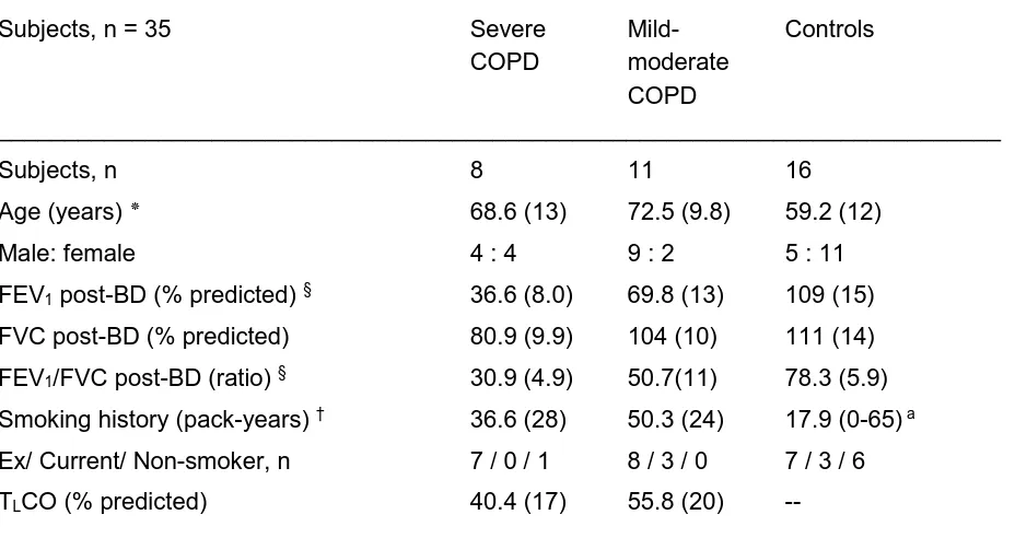
The demographic and lung function data of the 35 participants are summarised in Table 1. There
was a male predominance in the combined COPD group (n = 13, 68%), and a female
predominance in the control group (n = 11, 69%). Those with COPD were on average between 11
and 12 years older than the control participants (p = 0.01). Significant differences in lung function
were seen between participant groups, where the mean FEV1 was 37% of predicted for those with
severe COPD, 70% of predicted for those with mild-to-moderate COPD and 109% of predicted for
control subjects. Gas transfer factor measurements were significantly lower for COPD patients
[40.4% of predicted (95% confidence interval (CI) 26-54) versus 55.8% of predicted (95%CI
43-69)].
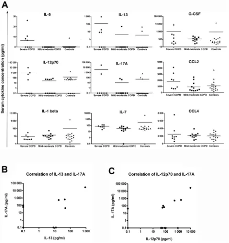
Individual levels of each cytokine are illustrated in Figure 1a, where there were no differences
between control and COPD groups, and between COPD severity categories, for most of the
cytokines tested. A significant difference in serum IL-5 levels was noted between those who had
severe COPD to those who had mild-to-moderate COPD or controls (Figure 1a, p = 0.005).
Although most participants had undetectable levels of IL-5, half the individuals in the severe COPD
group (n = 4) had detectable levels with a mean (SEM) level of 6.4 (3.2) pg/ml. Similarly, half with
severe COPD had detectable IL-13 levels, although the trend in levels between groups did not
quite reach statistical significance (p = 0.079) (Figure 1a). A modest correlation was noted
between the TH2 type cytokines, IL-5 and IL-13 (Spearman r = 0.429, p = 0.0102). However,
moderate correlations were noted between lineages, namely IL-17A and IL-12p70 (Spearman r =
0.52, p = 0.0015), as well as between IL-17A and IL-13 (Spearman r = 0.55, p = 0.0006) (Figure
1b, 1c).
This study has documented levels of serum cytokine that are representative of the TH1, TH2 and
TH17 pathways in COPD patients with mild-to-moderate and severe airflow obstruction. Although a
T-lymphocyte type 1 response that primarily involves IFN-γ predominates in COPD [2], our
detection of serum IL-5 levels in those with severe COPD and a lower gas transfer factor supports
the body of evidence that suggests type 2 responses co-exist, especially during exacerbations.
Oral corticosteroid therapy has been shown to reduce sputum eosinophilic cationic protein and
blood eosinophil count in patients with stable COPD in whom asthma was excluded, and an anti-
IL-5 receptor-α monoclonal antibody might be useful in the subgroup of COPD patients with sputum
eosinophilia.
The overlap between asthma and COPD has now been formally recognized to be a separate
this immunological response being typically associated with asthma [5]. Mean systemic IL-5 levels
up to 80 pg/mL have been measured by ELISA in individuals with childhood persistent asthma,
although much lower levels are generally reported [9]. In a study of stable COPD patients with
bronchial asthma excluded by a pulmonologist, the eosinophilic chemotactic protein, serum
eotaxin, was found to be higher when compared with controls, but this study did not report
corresponding serum IL-5 values. When compared with smokers with no or minimal lung disease
as defined by FEV1/FVC > 0.7, FEV1 ≥ 80% and low attenuation areas (LAA) ≤ 2.4, plasma IL-5
levels were statistically higher for those with radiological evidence of emphysema (LAA ≥ 8), airflow
obstruction (FEV1/FVC > 0.7, FEV1 ≤ 60%) or both. While a history of childhood asthma is often
inaccurate when it is recalled in adulthood, this prospective history is generally unavailable in the
clinical setting. As our study participants did not self-report a history of lifetime asthma, it is
therefore still possible that those with severe COPD may have had end-stage, insidious, allergic
asthma that was otherwise not detectable.
Elevated serum cytokine levels might be due to a “spill-over” effect of the pathological process in
the lung into the systemic circulation [10], and a linear and predictable nature cannot be presumed.
In a key study of eleven subjects with COPD of moderate severity of whom four underwent a
peripheral blood study, increased numbers of Tc2-like cytokine-expressing cells were isolated from
BAL fluid, and this was consistent with other data supporting increased IL-4 and IL-13 expression
by pulmonary Tc2 lymphocytes [3]. In comparison with the increased numbers of (CD8+) Tc1-like
cells detectable in blood, only around one-sixth of (CD8+) Tc2-like, (CD4+) TH1-like, and (CD4+)
TH2-like cells were detectable in the blood compared with BAL fluid. This suggests that expression
of IL-5 by pulmonary Tc2 lymphocytes is likely to have been increased, especially for some of our
participants who had severe COPD.
Our correlation data suggests moderate interrelationships between serum IL-17A levels and the
cytokine IL-12p70, a promoter of TH1 differentiation, and the cytokine IL-13 that is inhibitory to TH1
addition to T-regulatory cells. The interrelationships are consistent with naïve precursor TH17 cells
being inhibited by TH1 and TH2 cytokines, although the interpretation of our finding was limited
without IFN-γ and IL-4 levels.
Over-expression of IL-13 in the lungs of adult mice has been shown to induce many features
associated with COPD. These include emphysema, mucus goblet cell hyperplasia, and airway
inflammation associated with macrophages, lymphocytes and eosinophils and increased matrix
metalloproteinases. Low or undetectable levels of tissue IL-13 have been reported in lung
specimens of human subjects with severe emphysema, whereas plasma levels of IL-13 have been
shown to be modestly elevated in phenotypes associated with emphysema in another clinical
study, with and without airflow obstruction.
In terms of limitations, we acknowledge our study had modest numbers of participants (n=35),
although numbers were comparable to the similar study by Bade and colleagues (n=30). As the
measurement of cytokines in the systemic compartment lacks sensitivity when compared with
either BAL fluid or surgical lung biopsy, this might have attenuated or effectively eliminated
associations between serum cytokines and the severity categories of COPD. Other tests that
would have been useful to be included, including the measurement of IFN-γ, IL-8 and IL-4 for the
interpretation of serum IL-17A and its relationship to TH1 and TH2 lymphocytic pathways; and
objective measures of allergic and asthma such as blood eosinophil count, bronchial provocation
and/or skin prick testing. Further studies are needed to clarify whether serum IL-5 might be a
biomarker for severe COPD.
This study aimed to document the systemic immune responses in participants with COPD of
varying severity, and appears to show an enhanced systemic TH2 expression of IL-5 levels for
participants who had severe airflow obstruction and a lower mean gas transfer factor. Although the
mechanisms of COPD and asthma are classically different, increasing disease severity and clinical
overlap of these two obstructive lung diseases can blur the distinction.
Acknowledgements
We thank Dr Owen Proudfoot and Dr Dodie Pouniotis, Austin Research Institute for assisting in
conducting some of the Bioplex experiments and assays. The authors would like to thank the
financial support of the Institute of Breathing and Sleep (IBAS), Melbourne, Victoria, Australia. The
experiments were done in the laboratory of VA (Immunology and Vaccine Laboratory), at the Austin
Research Institute (now known as Burnet Institute), and all authors thank the Austin Research
Institute for financial support. At the time of the study VA was supported by NHMRC RD Wright
Fellowship (223316) and NHMRC project grant (223310).
References
1 WHO. World Health Organization. The top 10 causes of death. Fact sheet N°310. .
http://wwwwhoint/mediacentre/factsheets/fs310/en/ Updated May 2014,
2 Barnes PJ. Immunology of asthma and chronic obstructive pulmonary disease. Nat Rev Immunol 2008, 8: 183-192
3 Barcelo B, Pons J, Fuster A, Sauleda J, Noguera A, Ferrer JM, Agusti AG. Intracellular cytokine profile of T lymphocytes in patients with chronic obstructive pulmonary disease. Clin Exp Immunol 2006, 145: 474-479
4 Brightling CE, Monteiro W, Ward R, Parker D, Morgan MD, Wardlaw AJ, Pavord ID. Sputum eosinophilia and short-term response to prednisolone in chronic obstructive pulmonary disease: a randomised controlled trial. Lancet 2000, 356: 1480-1485
5 Saha S, Brightling CE. Eosinophilic airway inflammation in COPD. International journal of chronic obstructive pulmonary disease 2006, 1: 39-47
6 Zhu J, Qiu YS, Majumdar S, Gamble E, Matin D, Turato G, Fabbri LM, et al.
Exacerbations of Bronchitis: bronchial eosinophilia and gene expression for interleukin-4, interleukin-5, and eosinophil chemoattractants. American journal of respiratory and critical care medicine 2001, 164: 109-116
7 Gibson PG, Simpson JL. The overlap syndrome of asthma and COPD: what are its features and how important is it? Thorax 2009, 64: 728-735
8 Global Strategy for Asthma Management and Prevention, 2014. Diagnosis of Diseases of Chronic Airflow Obstruction: Asthma, COPD and Asthma-COPD Overlap Syndrome (ACOS). http://wwwginasthmaorg 2014,
9 Huang CS, Chen SJ, Chung RL, Tang RB. Serum interleukin-5 measurements for monitoring acute asthma in children. J Asthma 2005, 42: 297-300
Table 1 Baseline spirometry and smoking characteristics
___________________________________________________________________________
Subjects, n = 35 Severe Mild- Controls COPD moderate
COPD
___________________________________________________________________________
Subjects, n 8 11 16
Age (years) ٭ 68.6 (13) 72.5 (9.8) 59.2 (12)
Male: female 4 : 4 9 : 2 5 : 11
FEV1 post-BD (% predicted) § 36.6 (8.0) 69.8 (13) 109 (15)
FVC post-BD (% predicted) 80.9 (9.9) 104 (10) 111 (14)
FEV1/FVC post-BD (ratio) § 30.9 (4.9) 50.7(11) 78.3 (5.9)
Smoking history (pack-years) † 36.6 (28) 50.3 (24) 17.9 (0-65) a
Ex/ Current/ Non-smoker, n 7 / 0 / 1 8 / 3 / 0 7 / 3 / 6
TLCO (% predicted) 40.4 (17) 55.8 (20) --
___________________________________________________________________________
Abbreviations: BD, bronchodilator; FEV1, forced expiratory volume in one second; FVC, forced
vital capacity; TLCO, transfer factor of the lung for carbon monoxide
Results are expressed as mean (SD) except where otherwise indicated
a range § p < 0.0001, † p = 0.0041, ٭ p = 0.01
Figure 1 (A) Serum cytokine levels between control and COPD groups. Only serum IL-5 levels
showed statistical significance between severe COPD and controls (p = 0.005) and between severe COPD to mild-moderate COPD (p = 0.005). There was no statistical significance for all other serum cytokines, IL-13 (p = 0.079), G-CSF (p = 0.47), IL-12p70 ((p = 0.41), IL-17A (p = 0.815), CCL2 (p = 0.18), IL-1β (p = 0.345), IL-7 (p = 0.063) and CCL4 (p = 0.89). (B, C)