Clinical and Experimental Gastroenterology
Development of a simplified diagnostic indicators
scoring system and validation for peptic ulcer
perforation in a developing country
Chutikarn Suriya1
Nongyao Kasatpibal2
Wipada Kunaviktikul2
Toranee Kayee3
1Clinical Epidemiology Program,
Faculty of Medicine, Chiang Mai University, 2Faculty of Nursing,
Chiang Mai University, 3Department
of Surgery, Nakornping Hospital, Chiang Mai, Thailand
Correspondence: Chutikarn Suriya Clinical Epidemiology, Faculty of Medicine, Chiang Mai University, Suthep Road, Muang District, Chiang Mai 50200, Thailand Tel +66 53 121 122-4 ext 117 Fax +66 53 121 125
Email [email protected]
Objective: To perform and confirm a simplified diagnostic indicators scoring system for predicting peptic ulcer perforation (PUP).
Methods: A case–control study was conducted including 812 consecutive patients with PUP from retrospective medical records. Each diagnostic indicator measurable at the time of admittance was analyzed by a multiple regression. Stepwise logistic regression was applied with backward elimination of statistically significant predictors from the full model, with P $ 0.05 for exclusion. The item scores were transformed from regression coefficients and computed to a total score. The risk of PUP was interpreted using total scores as a simple predictor. This system was internally validated in 218 consecutive patients and compared to existing systems.
Results: A PUP risk score was determined from the diagnostic indicators associated with PUP: gender, age, nonsteroidal antiinflammatory drugs used, history of peptic ulcer, intense abdominal pain, guarding, X-ray free air positive, and referral from other hospitals. Item scores ranged from 0–6.0 and the total score ranged from 0–34.0. The area under the receiver operating characteristic curve shows that there was 91.73% accuracy in the total scores predicting the likelihood of PUP. The likelihood of PUP among low risk (scores ,10.5), moderate risk (scores 11–21), and high risk (scores $ 21.5) patients was 0.13, 11.44, and 1.95, respectively.
Conclusion: This scoring system is an effective diagnostic indicator for identifying the complex cases of PUP. It is a simple system and can help guide clinicians, providing them with a more efficient way to accurately subgroup patients while also reducing potential biases.
Keywords: peptic ulcer perforation, risk scoring, prediction
Introduction
Patient safety is attracting worldwide attention in all disciplines of medicine. Peptic ulcer perforation (PUP) is a major crucial complication of peptic ulcer.1,2,4 PUP
accounts for about 10%–15% of peptic ulcer patients and accounts for more than 70% of deaths.1 The mortality rate after PUP is approximately 10%–15% upon delayed
diagnosis.2,3,5
Severe upper abdominal pain is the most prominent symptom of PUP. About half of patients with PUP usually present with nausea, vomiting reflex, and vomiting – mostly food debris and gastric juice mixed with blood or coffee-like liquid, and can lead to intestinal paralysis. Approximately 1–5 hours after perforation, a patient’s stomach content can spill into the abdominal cavity due to an increase in peritoneal exudates. This may provide varying degrees of relief from abdominal pain and abdominal tension, but the tenderness is still significant. At this point, it would be easy to misdiagnose. In the first 10–12 hours after perforation, with peritoneal exudates absorption and
Dove
press
O R i G i N A L R E S E A R C H
open access to scientific and medical research
Open Access Full Text Article
Clinical and Experimental Gastroenterology downloaded from https://www.dovepress.com/ by 118.70.13.36 on 20-Aug-2020
For personal use only.
Number of times this article has been viewed
This article was published in the following Dove Press journal: Clinical and Experimental Gastroenterology
secondary bacterial infection, late diagnosis and treatment of patients infected with severe peritonitis can lead to chills, fever, or even toxic intestinal sepsis, and physicians must be highly vigilant to avoid death due to toxic shock.6
The literature suggests that the diagnosis of PUP should rarely be made on the basis of physical examination,5,7
whereas in clinical practice, the diagnosis of PUP is mainly established by patient history, signs and symptoms, physical examination, radiology investigation, and laboratory findings.1
A major limitation of scoring systems is their dependence on sophisticated methods, which may not be available in developing countries. A simplified diagnostic indicators scoring system for predicting PUP – which can be easily used in developing countries – is needed.
Materials and methods
Definition
The definition of PUP according to the International Statisti-cal Classification of Diseases and Related Health Problems, 10th Revision includes gastric ulcer (coded K25, subcatego-ries 25.1, 25.2, 25.5, 25.6) and duodenal ulcer (coded K26, subcategories 26.1, 26.2, 26.5, 26.6).8 The subcategories are
defined as acute with perforation, acute with both hemorrhage and perforation, chronic or unspecified with perforation, and chronic or unspecified with both hemorrhage and perfora-tion, respectively.
Development of the scoring system
A case–control study was conducted using medical records of PUP patients (cases and controls) – enrolled between January 1, 2005 and December 31, 2008 – that were reviewed retrospectively. PUP patients aged $15 years who were hospitalized in Nakornping Hospital (Chiang Mai, Thailand) were included in the cases group of the study, while nonperforation patients were included in the control group. Invariably, the diagnosis for PUP was obtained during the postoperation diagnosis by surgeons. The PUP patients with malignant ulcers or trauma were excluded.
It was important to estimate a sufficient sample size to achieve adequate statistical power. To yield a significant finding at α= 0.05, β= 0.20, and minimum detectable odds ratio = 4.9, the best sample size required 812 cases. There were 406 cases in the medical records; therefore 406 controls were analytically selected from nonperforated peptic ulcer patients immediately before and after the cases. Patient characteristics of cases and controls were compared using Student t-tests
and exact probability tests to determine their suitability for possible risk indicators. The diagnostic indicators comput-able at the time of admittance were designated and analyzed by a stepwise logistic regression to determine a set of statis-tically significant predictors. Mathematical predictors were divided into three levels; the cutoff points of which were clarified from the values that produced all statistically sig-nificant regression coefficients and the utmost area under the receiver operating characteristic (ROC) curve of the logistic regression model. Regression coefficients of all diagnostic indicator predictors were changed into item assigned scores, which were then built into a total score (diagnostic indicators score) for each item. The total scores signified the summary measure of risk for PUP. Total scores were then evaluated as the sole predictor in the logistic regression model.
Validation of the scoring system
The medical records of 218 patients aged $15 years who underwent PUP surgery at the same setting (Nakornping Hospital) between January 1, 2009 and December 31, 2009 were reviewed retrospectively. All patient profiles for the PUP operation were assessed. The patients in whom histological diagnosis confirmed a malignant lesion in the peptic ulcer area were excluded.
The research protocol was accepted by the Chiang Mai University’s Faculty of Medicine Research in Human Subjects Ethical Review Committee and the Director of Nakornping Hospital. Information was analyzed by using Stata® statistical software package version 11.0 for Windows
(StataCorp LP, College Station, TX).
Results
Patient characteristics
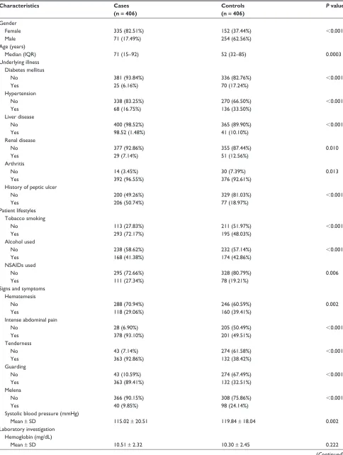
There were statistically significant differences between cases and controls in female gender, median age, underlying illness (diabetes mellitus P , 0.001, hypertension P , 0.001, liver disease P , 0.001, renal disease P , 0.01, arthritis P = 0.013, and history of peptic ulcer P , 0.001), patient lifestyles (tobacco smoking P , 0.001, alcohol used P , 0.001, and nonsteroidal antiinflammatory drugs used P = 0.006), signs and symptoms (hematemesis P = 0.002, intense abdominal pain P , 0.001, tenderness P , 0.001, guarding P , 0.001, melena P , 0.001, and systolic blood pressure P , 0.001), laboratory investigations (blood urea nitrogen/creatinine ratio P , 0.001), radiological finding (X-ray with free air P , 0.001), and treatment role (refer-ral from other hospitals P , 0.001) (Table 1). The backward stepwise logistic regression analysis was used to develop a
Dovepress
Suriya et al
Clinical and Experimental Gastroenterology downloaded from https://www.dovepress.com/ by 118.70.13.36 on 20-Aug-2020
Table 1 Characteristics of patients with peptic ulcer perforation (cases) and nonperforation (controls)
Characteristics Cases (n = 406)
Controls (n = 406)
P value
Gender Female Male Age (years) Median (iQR) Underlying illness Diabetes mellitus No
Yes Hypertension No Yes Liver disease No Yes Renal disease No Yes Arthritis No Yes
History of peptic ulcer No
Yes Patient lifestyles Tobacco smoking No
Yes Alcohol used No Yes NSAiDs used No Yes
Signs and symptoms Hematemesis No Yes
intense abdominal pain No
Yes Tenderness No Yes Guarding No Yes Melena No Yes
Systolic blood pressure (mmHg) Mean ± SD
Laboratory investigation Hemoglobin (mg/dL) Mean ± SD
335 (82.51%) 71 (17.49%)
71 (15–92)
381 (93.84%) 25 (6.16%)
338 (83.25%) 68 (16.75%)
400 (98.52%) 98.52 (1.48%)
377 (92.86%) 29 (7.14%)
14 (3.45%) 392 (96.55%)
200 (49.26%) 206 (50.74%)
113 (27.83%) 293 (72.17%)
238 (58.62%) 168 (41.38%)
295 (72.66%) 111 (27.34%)
288 (70.94%) 118 (29.06%)
28 (6.90%) 378 (93.10%)
43 (7.14%) 363 (92.86%)
43 (10.59%) 363 (89.41%)
366 (90.15%) 40 (9.85%)
115.02 ± 20.51
10.51 ± 2.32
152 (37.44%) 254 (62.56%)
52 (32–85)
336 (82.76%) 70 (17.24%)
270 (66.50%) 136 (33.50%)
365 (89.90%) 41 (10.10%)
355 (87.44%) 51 (12.56%)
30 (7.39%) 376 (92.61%)
329 (81.03%) 77 (18.97%)
211 (51.97%) 195 (48.03%)
232 (57.14%) 174 (42.86%)
328 (80.79%) 78 (19.21%)
246 (60.59%) 160 (39.41%)
205 (50.49%) 201 (49.51%)
274 (61.58%) 132 (38.42%)
274 (67.49%) 132 (32.51%)
308 (75.86%) 98 (24.14%)
119.84 ± 18.04
10.30 ± 2.45
,0.001
0.0003
,0.001
,0.001
,0.001
0.010
0.013
,0.001
,0.001
,0.001
0.006
0.002
,0.001
,0.001
,0.001
,0.001
0.002
0.222
(Continued)
Dovepress Diagnostic indicators score for predicting PUP
Clinical and Experimental Gastroenterology downloaded from https://www.dovepress.com/ by 118.70.13.36 on 20-Aug-2020
Table 1 (Continued)
Characteristics Cases (n = 406)
Controls (n = 406)
P value
Hematocrit (5) Mean ± SD BUN/Cr ratio Mean ± SD Radiological finding X-ray free air positive No
Yes Treatment role
Referral from other hospitals No
Yes
30.71 ± 6.91
19.00 ± 18
167 (41.13%) 239 (58.87%)
34 (8.37%) 372 (91.63%)
30.56 ± 7.56
10.00 ± 7.50
356 (87.68%) 50 (12.32%) 160 (39.41%) 246 (60.59%) 0.777 ,0.001 ,0.001 ,0.001
Abbreviations: BUN, blood urea nitrogen; Cr, creatinine; IQR, interquartile range; NSAIDs, nonsteroidal antiinflammatory drugs; SD, standard deviation.
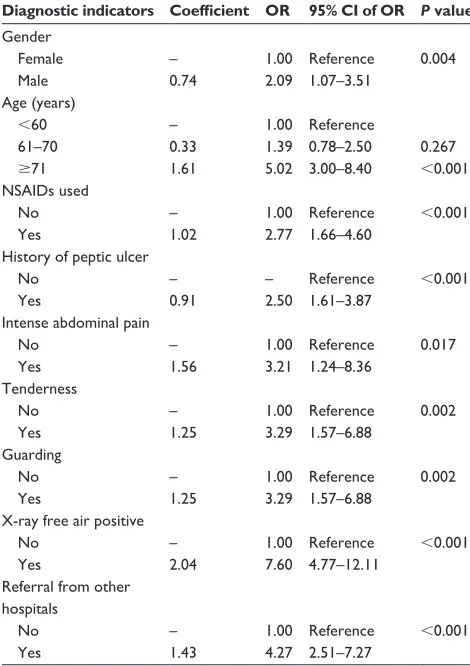
logistic regression model. The eight risk predictors were gender, age, nonsteroidal antiinflammatory drugs used, history of peptic ulcer, intense abdominal pain, guarding, X-ray free air positive, and referral from other hospitals (Table 2).
Table 2 Regression coefficient, risk ratio, and 95% confidence interval of diagnostic indicators for peptic ulcer perforation from logistic regression
Diagnostic indicators Coefficient OR 95% CI of OR P value Gender Female Male Age (years) ,60 61–70 $71 NSAiDs used No Yes
History of peptic ulcer No
Yes
intense abdominal pain No Yes Tenderness No Yes Guarding No Yes
X-ray free air positive No
Yes
Referral from other hospitals No Yes – 0.74 – 0.33 1.61 – 1.02 – 0.91 – 1.56 – 1.25 – 1.25 – 2.04 – 1.43 1.00 2.09 1.00 1.39 5.02 1.00 2.77 – 2.50 1.00 3.21 1.00 3.29 1.00 3.29 1.00 7.60 1.00 4.27 Reference 1.07–3.51 Reference 0.78–2.50 3.00–8.40 Reference 1.66–4.60 Reference 1.61–3.87 Reference 1.24–8.36 Reference 1.57–6.88 Reference 1.57–6.88 Reference 4.77–12.11 Reference 2.51–7.27 0.004 0.267 ,0.001 ,0.001 ,0.001 0.017 0.002 0.002 ,0.001 ,0.001
Note: Area under the receiver operating characteristic curve = 94.46%. Abbreviations: CI, confidence interval; NSAIDs, nonsteroidal antiinflammatory drugs;
OR, odds ratio.
Development of the risk score
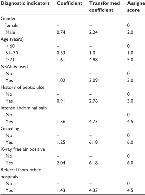
A risk score was determined from these eight predictors to pre-dict the risk for PUP. Numerical prepre-dictors were categorized into three levels. Substantially better outcomes were confirmed when a greater area under the ROC curve was obtained (94.46%). The regression coefficients were transformed by dividing with the smallest coefficient in the model (0.33) and rounded up to the nearest 0.5 to obtain item scores. Every item score ranged from zero up to 2.0 or 6.0. After item scores were added to get the total scores, they ranged from zero to 34 (Table 3). The area under the ROC curve shows that there was 91.73% accuracy in the total scores predicting the likelihood of PUP (Figure 1).
Validation of the scoring system
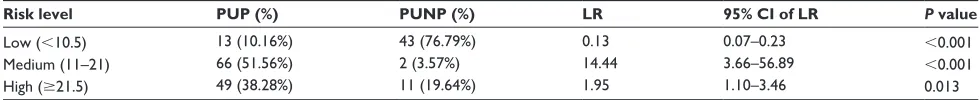
A graph mapping the proportion of PUP against total scores shows that the actual risk for higher total scores correlates well with the predicted risk from logistic estimation (Figure 2). Classification of the validation cohort risk score resulted in three levels: low risk (scores , 10.5), moderate risk (scores 11–21), and high risk (scores $ 21.5). Of the cases that were classified as low risk, 10.16% had PUP, compared with 51.56% in the moderate-risk group and 38.28% in the high-risk group. Of the controls, 76.79% were categorized as low risk, 3.57% as moderate risk, and 19.64% as high risk. The intended likelihood ratio shows that low-risk cases were only 0.13 times more likely to result in PUP, while moderate-risk cases were 14.44 times, and high-risk cases were 1.95 times more likely to result in PUP. Even supposing that PUP occurs, the relationship between scores and treatment show that the higher scores have a higher likelihood of surgery (Table 4).
Discussion
The development of a simplified diagnostic indicators score was based on the philosophy of clinical prediction rules
Dovepress
Suriya et al
Clinical and Experimental Gastroenterology downloaded from https://www.dovepress.com/ by 118.70.13.36 on 20-Aug-2020
Table 3 item scoring scheme for predictors of peptic ulcer perforation derived from coefficients of select diagnostic indicators
Diagnostic indicators Coefficient Transformed coefficient
Assigned score Gender
Female Male Age (years)
,60 61–70
$71 NSAiDs used No Yes
History of peptic ulcer No
Yes
intense abdominal pain No
Yes Guarding No Yes
X-ray free air positive No
Yes
Referral from other hospitals
No Yes
– 0.74
– 0.33 1.61
– 1.02
– 0.91
– 1.56
– 1.25
– 2.04
– 1.43
– 2.24
– 1.0 4.88
– 3.09
– 2.76
– 4.73
– 6.18
– 6.18
– 4.33
0 2.0
0 1.0 5.0
0 3.0
0 3.0
0 4.5
0 6.0
0 6.0
0 4.5
Abbreviations: BUN, blood urea nitrogen; Cr, creatinine; NSAiDs, nonsteroidal
antiinflammatory drugs.
Estimated risk
Total score
Predicted risk
40 30
20 10
0 0 20 40 60 80 100
Figure 2 Score predicted risk (dots) and logistic estimated risk (solid line) of peptic ulcer perforation (%) for each total score.
of a variety of clinical outcomes.10–16 However, not one of
these methods was relevant for estimating the risk of PUP in routine clinical practice. The process currently used to appraise the risk of PUP is very complicated. In the field, clinicians have been undergoing a paradigm shift recently as the importance of evidence is realized in practice.1 Effective
use of current evidence requires the clinician to draw on clinical experience and assess patient values as well as to collect, analyze, and implement the research into practice. The easy-to-use and proficient risk score for PUP might enable prompt identification of patients at high risk for PUP.
This report demonstrated a method for multivariable logistic regression analysis dividing the continuous variables into a dichotomous score yielding arithmetically identical estimates of risk. The benefit of the methodology over the typical appearance of logistic regression is that essentially anyone is able to estimate the risk of an event associated with given predictors. The risk score for predicting PUP was explained by an area under the ROC curve of 91.73%. The proportion of PUP increased sharply when the score increased from low risk to high risk.
An easy and realistic risk score was determined to predict the risk of PUP, which gained high prediction precision. To encourage the risk score to be successful in its forecasting, only the variables identifiable at the time of hospitalization were chosen.
This study demonstrated a simple and realistic method of computing the risk score to enable categorization of peptic ulcer patients into levels of PUP risk. The area under the ROC curve for PUP risk score was 91.73%. High prediction accuracy observed in this study was a result of the rigorous diagnostic indicators definition for PUP from a previous study to avoid misclassification.1 It was previously known that
when diagnostic indicators are transformed to a risk score, the prediction accuracy is reasonably reduced. The current applied to some areas in PUP,9 such as a score to predict
postoperative morbidity and mortality,10–13,15 poor outcome,14
and to compare with a validated risk score for PUP.15
Clinicians and their team prefer to be able to precisely predict the presence or nonappearance of PUP. Statistical methods using logistic regression have estimated the risk
0.
00
0.
25
0.
50
0.
75
1.
00
S
en
si
ti
vi
ty
0.00 0.25 0.50 0.75 1.00
1 – specificity
Figure 1 Receiver operating characteristic curve of risk for peptic ulcer perforation predicted by risk scoring (curved line) and a 50% chance prediction (diagonal line). Note: Area under receiver operating characteristic curve = 0.9173.
Dovepress Diagnostic indicators score for predicting PUP
Clinical and Experimental Gastroenterology downloaded from https://www.dovepress.com/ by 118.70.13.36 on 20-Aug-2020
study, using total scores to calculate risk, predicted PUP with an accuracy of 91.73% – a slight reduction from the unscored regression analysis which yielded an accuracy of 94.46%.
The combined results of studies suggest that the diag-nostic indicators of patients are related to the multifactorial pathogenesis of peptic ulcer complications.6 Older age groups
and males were more likely to experience PUP last century, but that is now changing. In England, PUP most commonly affects young women.18 In addition, the incidence of PUP
across many countries has shown that a marked prognostic factor was patient history of peptic ulcer,10,11,13,16,19 and the
literature suggests a continuing increase in the use of non-steroidal antiinflammatory drugs.1,20
Delayed treatment of peptic ulcer patients who are referred from other hospitals with severe signs and symp-toms may progress to gastrointestinal perforation. PUP at the anterior surface of the stomach is indicated by sudden intense abdominal pain. Posterior wall penetration leads to tenderness and guarding, which regularly radiates abdominal pain to the back. These variables reduce survival, increase poor clinical outcomes, and lengthen hospitalization.6
It is appreciated that complications arising from perfora-tion (eg, peritonitis) and mortality rates increase with delayed diagnosis. Higher rates of lethal outcomes in PUP emergency operations have been observed.17
The diagnostic indicators score in the current study may also be used to classify patients with an increased likelihood of experiencing PUP in the future. The diagnostic indicators score might be useful for clinicians and nurses in develop-ing countries in prepardevelop-ing for surgery. However, external validation of the scoring scheme will need to be applied to a different setting of PUP patients. This validation will be essential before any real use in applications such as clinical decision making and evaluation reviews of clinicians.
Additionally, the prediction rule is implemented as part of a critical pathway so that a hospital or clinic has procedures and policies established for how to manage patients identified as high or low risk for disease, which impact clinical outcomes. Therefore, the more intensively the prediction rule is implemented, the more benefits will occur. As a result, the risk of PUP may be predicted by a simplified score using
eight predictors. Because of this, the risk score can accurately identify patients with low, moderate, and high risk.
Based on routine and intensive care in high-risk groups, the simplified prediction may bring some benefit, even though several studies have suggested that Helicobacter pylori colonizes the stomach and induces chronic gastritis and a long-lasting inflammation of the stomach.19–29 Additionally, H. pylori bacterium was first reported in the stomach and,
along with acid secretion, can damage the tissue of the stomach and duodenum.22 H. pylori causes inflammation
and can develop into gastric and duodenal ulcer.22,23 H. pylori
infection mutates host glycosylation and influences the pattern of putative colonization that is associated with lifetime risk of gastric perforation.22 Studies have shown that H. pylori is
etiologically linked to stomach cancer,24–27 and a study from
India has shown that H. pylori infection is less common in more developed Asian nations.24 Gastric cancer is common
among ethnic Chinese with East Asian genotype.25 H. pylori
plays an important role in the pathogenesis of peptic ulcer disease, distal gastric adenocarcinoma, and gastric lymphoma in the United Kingdom and Hong Kong.26,27 Importantly, the
exact role that H. pylori plays could not be established in the present study for two reasons. Firstly, during the 4-year study period (2005–2008), the H. pylori test was not well known in Thailand and was rarely ordered by doctors in cases of perforation. Secondly, about 70% of the patients required urgent or emergent surgery. Therefore, H. pylori was not detected in these patients.
A study from the 1950s found that the ABO blood group was associated with gastric diseases, and gastric cancer was found to be associated with blood group A in 1969.29 Villalobos et al demonstrated that blood type O
was associated with peptic ulcer in 1990.30 The correlation
between blood type A and gastric cancer has been confirmed and researchers have also confirmed that blood group O has a higher risk of peptic ulcers than those with other blood groups.31 However, the ABO blood group variable was not
used in the current analysis, as testing for blood type is not part of routine laboratory investigation and is therefore rarely noted in the medical records. Only subgroups who receive a blood transfusion are tested for blood type.
Table 4 Distribution of risk for perforation among patients with peptic ulcer perforation and nonperforation
Risk level PUP (%) PUNP (%) LR 95% CI of LR P value
Low (,10.5) Medium (11–21) High ($21.5)
13 (10.16%) 66 (51.56%) 49 (38.28%)
43 (76.79%) 2 (3.57%) 11 (19.64%)
0.13 14.44 1.95
0.07–0.23 3.66–56.89 1.10–3.46
,0.001
,0.001 0.013
Abbreviations: CI, confidence interval; LR, likelihood ratio; PUNP, peptic ulcer nonperforation; PUP, peptic ulcer perforation.
Dovepress
Suriya et al
Clinical and Experimental Gastroenterology downloaded from https://www.dovepress.com/ by 118.70.13.36 on 20-Aug-2020
Study strengths and weaknesses
A strength of this study was that the simplified clinical pre-diction included predictor variables obtained from patient history, examination, and simple diagnostic tests, which can assist in making diagnosis appropriate management strategies.
A weakness of this study was that the retrospectively reviewed medical records were sometimes incomplete in regard to the patient’s lifestyle (eg, stress) and laboratory investigation (eg, glucocorticoids and ABO blood groups). These variables are major contributing factors to peptic ulcers and are interrelated to perforation.
Conclusion
This risk score might be relevant for clinicians and nurses to support them in early detection and treatment of patients who are at high risk for PUP. Consequently, the scoring scheme needs to be externally validated in independent patients who are undergoing PUP.
Acknowledgments
The authors would like to acknowledge the Nakornping Hospital health care team who granted access to patients’ information and for their contribution to the data collection.
Disclosures
The authors report no conflicts of interest in this work. This study was a part of Chutikarn Suriya’s PhD dissertation project in clinical epidemiology.
References
1. Koo J, Ngan YK, Lam SK. Trends in hospital admissions, perforation and mortality of peptic ulcer in Hong Kong from 1970 to 1980. Gastroenterology. 1983;84(6):1558–1562.
2. Morris A, Midwinter MJ. Perforated peptic ulcer. In: Brooks A, Cotton BA, Tai N, Mahoney PF, editors. Emergency Surgery. Oxford: Wiley-Blackwell; 2010:43–45.
3. Noguiera C, Silva AS, Santos JN, et al. Perforated peptic ulcer: main factors of morbidity and mortality. World J Surg. 2003;27(7):782–787. 4. Kocer B, Surmeli S, Solak C, et al. Factors affecting mortality and
morbidity in patients with peptic ulcer perforation. J Gastroenterol Hepatol. 2007;22(4):565–570.
5. Way LW. Stomach and duodenum. In: Way LW, editor. Current Surgical Diagnosis and Treatment. 10th ed. Norwalk, CT: Appleton and Lange; 1994:437–459.
6. Silen W. Cope’s Early Diagnosis of the Acute Abdomen. 20th ed. New York, NY: Oxford University Press; 2000.
7. Flasar MH, Goldberg E. Acute abdominal pain. Med Clin North Am. 2006;90(3):481–503.
8. World Health Organization. International Statistical Classification of Diseases and Related Health Problems, 10th Revision (ICD-10). Geneva: World Health Organization; 1994.
9. Glynn PE, Weisbach PC. Clinical Prediction Rules: A Physical Therapy Reference Manual. Sudbury, MA: Jones and Bartlett Publishers; 2011.
10. Boey J, Wong J, Ong GB. A prospective study of operative risk factors in perforated duodenal ulcers. Ann Surg. 1982;195(3):265–269. 11. Lohsiriwat V, Prapasrivorakul S, Lohsiriwat D. Perforated peptic ulcer:
clinical presentation, surgical outcomes, and the accuracy of the Boey scoring system in predicting postoperative morbidity and mortality. World J Surg. 2009;33(1):80–85.
12. Lee FY, Leung KL, Lai BS, Ng SS, Dexter S, Lau WY. Predicting mortality and morbidity of patients operated on for perforated peptic ulcers. Arch Surg. 2001;136(1):90–93.
13. Arici C, Mesci A, Dincer D, Dinckan A, Colak T. Analysis of risk factors predicting (affecting) mortality and morbidity of peptic ulcer perforations. Int Surg. 2007;92(3):147–154.
14. Evans JP, Smith R. Predicting poor outcome in perforated peptic ulcer disease. Aust N Z J Surg. 1997;67(11):792–795.
15. Koc M, Yoldas O, Kilic YA, et al. Comparison and validation of scoring systems in a cohort of patients treated for perforated peptic ulcer. Langenbecks Arch Surg. 2007;392(5):581–585.
16. Rajesh V, Chandra SS, Smile SR. Risk factors predicting operative mortality in perforated peptic ulcer disease. Trop Gastroenterol. 2003; 24(3):148–150.
17. Testini M, Portincasa P, Piccinni G, Lissidini G, Pellegrini F, Greco L. Significant factors associated with fatal outcome in emergency open surgery for perforated peptic ulcer. World J Gastroenterol. 2003;9(10): 2338–2340.
18. Crisp E. Cases of perforation of the stomach with deductions there from relative to the character and treatment of that lesion. Lancet. 1843;40(1040):639–649.
19. Lanas A, Garcia-Rodriguez LA, Polo-Tomas M, et al. Time trends and impact of upper and lower gastrointestinal bleeding and perforation in clinical practice. Am J Gastroenterol. 2009;104(7):1633–1641. 20. Rosenstock S, Jorgensen T, Bonnevie O, Andersen L. Risk factors
for peptic ulcer disease: a population based prospective cohort study comprising 2416 Danish adults. Gut. 2003;52(2):186–193.
21. Christensen S, Riis A, Norgaard M, Sorensen HT, Thomsen RW. Short-term mortality after perforated or bleeding peptic ulcer among elderly patients: a population-based cohort study. BMC Geriatr. 2007;7:8. 22. Kusters JG, van Vliet AH, Kuipers EJ. Pathogenesis of Helicobacter
pylori infection. Clin Microbiol Rev. 2006;19(3):449–490.
23. Ng CY, Squires TJ, Busuttil A. Acute abdomen as a cause of death in sudden, unexpected deaths in the elderly. Scott Med J. 2007;52(1): 20–23.
24. Singh K, Ghoshal UC. Causal role of Helicobacter pylori infection in gastric cancer: an Asian enigma. World J Gastroenterol. 2006;12(9): 1346–1351.
25. Vilaichone RK, Mahachai V, Tumwasorn S, Wu JY, Graham DY, Yamaoka Y. Molecular epidemiology and outcome of Helicobacter pylori infection in Thailand: a cultural cross roads. Helicobacter. 2004; 9(5):453–459.
26. McNamara D, El-Omar E. Helicobacter pylori infection and the pathogenesis of gastric cancer: a paradigm for host-bacterial interactions. Dig Liver Dis. 2008;40(7):504–509.
27. Xia B, Xia HH, Ma CW, et al. Trends in the prevalence of peptic ulcer disease and Helicobacter pylori infection in family physician-referred uninvestigated dyspeptic patients in Hong Kong. Aliment Pharmacol Ther. 2005;22(3):243–249.
28. Matsuda R, Morizane T, Tsunematsu S, Kawana I, Tomiyama M. Helicobacter pylori prevalence in dentists in Japan: a seroepidemiological study. J Gastroenterol. 2002;37(4):255–259.
29. Havlik RJ, Feinleib M, Garrison RJ, Kannel WB. Blood-groups and coronary heart-disease. Lancet. 1969;2(7614):269–270.
30. Villalobos JJ, Vargas F, Villareal HA, et al. A 10-year prolective study on cancer of the digestive system. Rev Gastroenterol Mex. 1990;55(1): 17–24. Spanish.
31. Edgren G, Hjalgrim H, Rostgaard K, et al. Risk of gastric cancer and peptic ulcers in relation to ABO blood type: a cohort study. Am J Epidemiol. 2010;172(11):1280–1285.
Dovepress Diagnostic indicators score for predicting PUP
Clinical and Experimental Gastroenterology downloaded from https://www.dovepress.com/ by 118.70.13.36 on 20-Aug-2020
Clinical and Experimental Gastroenterology
Publish your work in this journal
Submit your manuscript here: http://www.dovepress.com/clinical-and-experimental-gastroenterology-journal Clinical and Experimental Gastroenterology is an international,
peer-reviewed, open access journal, publishing all aspects of gastroenterology in the clinic and laboratory, including: Pathology, pathophysiology of gastrointestinal disease; Investigation and treatment of gastointes-tinal disease; Pharmacology of drugs used in the alimentary tract;
Immunology/genetics/genomics related to gastrointestinal disease. This journal is indexed on CAS. The manuscript management system is completely online and includes a very quick and fair peer-review system. Visit http://www.dovepress.com/testimonials.php to read real quotes from published authors.
Dovepress
Dove
press
Suriya et al
Clinical and Experimental Gastroenterology downloaded from https://www.dovepress.com/ by 118.70.13.36 on 20-Aug-2020