Int. J. Mol. Sci. 2020, 21, 8795; doi:10.3390/ijms21228795 www.mdpi.com/journal/ijms Article
Identification
of
Recurrent
Mutations
in
the
microRNA
‐
Binding
Sites
of
B
‐
Cell
Lymphoma
‐
Associated
Genes
in
Follicular
Lymphoma
Erika Larrea 1,2,3, Marta Fernandez‐Mercado 1,4, José Afonso Guerra‐Assunção 5, Jun Wang 6,
Ibai Goicoechea 1,7, Ayman Gaafar 8, Izaskun Ceberio 9, Carmen Lobo 10, Jessica Okosun 6,
Anton J. Enright 11, Jude Fitzgibbon 6 and Charles H. Lawrie 1,12,13,*
1 Molecular Oncology Group, Biodonostia Research Institute, 20014 San Sebastián, Spain; erika.larrea@gmail.com (E.L.); marfermer@yahoo.es (M.F.‐M.); ibai.goicoechea@gmail.com (I.G.) 2 Chinese Institute for Brain Research (CIBR), Beijing 102206, China 3 School of Life Sciences, Tsinghua University, Beijing 100084, China 4 Biomedical Engineering, School of Engineering, University of Navarra, 20014 San Sebastian, Spain 5 Great Ormond Street Institute of Child Health, University College London (UCL), London WC1N 1EH, UK; a.guerra@ucl.ac.uk 6 Barts Cancer Institute, Queen Mary University of London, London EC1M 6BE, UK; j.a.wang@qmul.ac.uk (J.W.); j.okosun@qmul.ac.uk (J.O.); j.fitzgibbon@qmul.ac.uk (J.F.) 7 Multiple Myeloma Group, Centro de Investigación Médica Aplicada (CIMA), Pamplona, 31008 Navarra, Spain 8 Department of Pathology, Cruces Hospital, 48903 Bilbao, Spain; ayman.gaafareleraky@osakidetza.eus 9 Hematology Department, Hospital Universitario Donostia, 20014 San Sebastián, Spain; izaskun.zeberioetxetxipia@osakidetza.eus 10 Department of Pathology, Hospital Universitario Donostia, 20014 San Sebastián, Spain; mariacarmen.lobomoran@osakidetza.eus 11 Department of Pathology, University of Cambridge, Cambridge CB2 1QP, UK; aje39@cam.ac.uk 12 IKERBASQUE, Basque Foundation for Science, 48009 Bilbao, Spain 13 Radcliffe Department of Medicine, University of Oxford, Oxford OX4 3DU, UK * Correspondence: charles.lawrie@biodonostia.org; Tel.: +34‐943‐006138 Received: 22 October 2020; Accepted: 18 November 2020; Published: 20 November 2020
Abstract: Follicular lymphoma (FL) is a common indolent B‐cell lymphoma that can transform into
the more aggressive transformed FL (tFL). However, the molecular process driving this
transformation is uncertain. In this work, we aimed to identify microRNA (miRNA)‐binding sites
recurrently mutated in follicular lymphoma patients, as well as in transformed FL patients. Using
whole‐genome sequencing data from FL tumors, we discovered 544 mutations located in
bioinformatically predicted microRNA‐binding sites. We then studied these specific regions using
targeted sequencing in a cohort of 55 FL patients, found 16 recurrent mutations, and identified a
further 69 variants. After filtering for QC, we identified 21 genes with mutated miRNA‐binding sites
that were also enriched for B‐cell‐associated genes by Gene Ontology. Over 40% of mutations
identified in these genes were present exclusively in tFL patients. We validated the predicted
miRNA‐binding sites of five of the genes by luciferase assay and demonstrated that the identified
mutations in BCL2 and EZH2 genes impaired the binding efficiency of miR‐5008 and miR‐144 and
regulated the endogenous levels of messenger RNA (mRNA).
Keywords: follicular lymphoma (FL); diffuse large B‐cell lymphoma (DLBCL); microRNA; mutation
1. Introduction
Follicular lymphoma (FL) is the most common form of low‐grade B‐cell lymphoma accounting
for approximately 20% of all diagnosed lymphomas [1]. Due to its slow progressive nature, FL is
often considered an incurable malignancy, with a median life expectancy of approximately 15–20
years [2,3]. However, 5–10% of FL patients undergo high‐grade histological transformation into a
much more aggressive form of lymphoma with a poorer outcome, generally termed transformed FL
(tFL) [4–6]. Despite the high frequency of this event, the transformation process is only poorly
understood at the molecular level, and no predictive biomarkers currently exist for patients at risk
from this phenomenon.
The dysfunctional expression of microRNAs (miRNAs) in B‐cell lymphoma is now well
established [7]. Our group and others have shown that the pattern of dysfunctional miRNAs in FL
differs from that of other B‐cell lymphomas including diffuse large B‐cell lymphoma (DLBCL) and
Burkitt’s lymphoma, as well as between transformed and nontransformed FL cases [8–14]. Indeed,
several studies have proposed a major functional role for miRNAs in the transformation process
[8,14]. In addition to changes in the expression of miRNAs in FL, recurrent mutations in the mature
sequence of miRNAs have also been associated with FL (and DLBCL) cases [15]. However, the
presence of mutations in the miRNA‐binding sites of target genes have not yet been explored in FL
or indeed any lymphoma.
To address this question, we used whole‐genome sequencing (WGS) data to investigate the
presence of somatic mutations in miRNA‐binding sites from matched FL tissue obtained before and
after transformation. Functional testing showed that mutations in BCL2 and EZH2, key genes for
lymphomagenesis, impair the binding efficiency of miR‐5008 and miR‐144 and the regulation of
endogenous messenger RNA (mRNA) expression.
2. Results
2.1. Identification of Somatic Mutations in miRNA‐Binding Sites of FL
We used whole‐genome sequencing data from tumor samples from six FL patients who
underwent transformation to high‐grade disease to identify somatic mutations located in microRNA
binding sites. This discovery cohort consisted on paired samples before and after transformation.
From the 2056 somatic mutations identified, approximately half of them were located in coding
regions of the genes (47%), whereas 38% of mutations were located in 3′ untranslated regions (UTRs)
and 15% were located in 5′ UTRs [16].
Using a bespoke bioinformatic pipeline, we identified 544 mutations (Table S1, Supplementary
Materials) located in predicted miRNA‐binding sites of 490 different genes (26.5% of total mutations).
Nearly all these mutations (92%) were located in 3′ UTRs. Mutations in coding regions of these 490
genes appeared to be mutually exclusive, with only 5% of genes (n = 27) having mutations in both
coding and 3′ UTR regions. Interestingly, from all the somatic mutations located in 3’ UTR sequences
(n = 788), 70% arose in predicted miRNA‐binding sites (Figure S1, Supplementary Materials).
Ontology analysis of the miRNA‐binding site mutated genes showed a significant enrichment for
pathways of hematological disease (p = 2.18 × 10⁻⁴) and B‐cell receptor pathways (p = 1.16 × 10⁻³)
(Figure S2 and Table S1, Supplementary Materials). Consequently, we designed a panel for targeted
sequencing to assess the frequency of the miRNA‐binding site mutations in a larger cohort of FL
cases.
2.2. Genes Showing Recurrent Mutations in miRNA‐Binding Sites Are Highly Enriched for Germinal‐ Center‐Like B‐Cell Lymphoma Genes
A custom sequencing panel covering >90% of the identified mutations (503/544) was used to
sequence a cohort of 55 FL patient samples together with 34 healthy control samples pooled in five
samples. From these 55 patients, 34 underwent a histological transformation (tFL) and 21 were
2270× (Figure S3, Supplementary Materials). Eighty percent of the mutations identified by WGS were
also identified and validated by targeted sequencing in the original discovery cohort (401/503) (Figure
1A).
Figure 1. Targeted sequencing results: (A) comparison of variants detected by whole‐genome sequencing (WGS) and targeted resequencing (TR); (B) number of nontransformed (ntFL) and transformed FL cases (tFL) harboring any of the 23 identified recurrent variants after filtering in microRNA (miRNA)‐binding sites.
From the 503 mutations analyzed, 16 were identified recurrently in validation cohort samples
(Table 1). In addition, to the originally identified mutations, a further 69 variants were identified by
targeted next‐generation sequencing (NGS) as being within predicted miRNA‐binding sites. Forty‐
six of these variants (67%) were identified in more than one sample (Table S2, Supplementary
Materials). From the total of 85 identified variants (i.e., 16 recurrent discovery and 69 new validation
variants), 36 were removed from further analysis as they were also identified in the pooled healthy
control samples (n = 34 pooled in five samples). A further 26 were removed as they were present in
homopolymer or repetitive regions, which are frequently artefactual [17,18]. The remaining 3 variants
consisting of seven of the original identified mutations and 16 of the newly discovered variants were
located in 21 different genes (Table S1, Supplementary Materials). Ontology analysis of these 21 genes
demonstrated that they were highly enriched for germinal‐center‐like (GC‐like) B‐cell lymphoma
genes (p = 4.39 × 10⁻⁵). We observed that these variants occurred more commonly in tFL (n = 73
variants in 34 cases) compared to non‐tFL cases (n = 46 variants in 21 cases), and that 10 out of the 23
variants (43%) occurred exclusively in tFL cases (Figure 1B).
Table 1. Mutations from the original discovery cohort recurrently identified in the validation cohort.
Gene Mutated Position Mutation Location miRNA Cases (n = 55)
EZH2 chr7:148508727 T/A Exon hsa‐mir‐144 8
ARMC10 chr7:102739179 A/G 3′ UTR hsa‐mir‐222 18
TUBB chr6:30692754 C/CTT 3′ UTR hsa‐mir‐1302 7
MEF2B chr19:19260045 T/A Exon hsa‐mir‐1265 3
ZNF195 chr11:3380000 t/TC 3′ UTR hsa‐mir‐1915 16
BCL2 chr18:60793447 G/A 3′ UTR hsa‐mir‐5008 2
THOC3 chr5:175386586 A/G 3′ UTR hsa‐mir‐371a 15
TXNDC2 chr18:9887493 T/C Exon hsa‐mir‐2110 3
PCDH7 chr4:30732983 GTA/G Intron hsa‐mir‐329 4
RC3H1 chr1:173901940 A/AAAT 3′ UTR hsa‐mir‐548an 2
AQP3 chr9:33441702 C/A 3′ UTR hsa‐mir‐146b 8
DPY19L2 chr12:63953768 T/C 3′ UTR hsa‐mir‐1303 11
MYO5B chr18:47352742 T/G 3′ UTR hsa‐mir‐216b 55
MYO5B chr18:47352754 A/G 3′ UTR hsa‐mir‐2681 55
YY2 chrX:21876221 A/G 3′ UTR hsa‐mir‐448 36
The table shows the mutated position in the genome, the mutation identified, the location in the gene, the microRNA targeting this region, and the number of cases identified. UTR, untranslated region.
2.3. In Vitro Luciferase Assays Demonstrated That Mutations in miRNA Binding Sites Interfere with the miRNA Activity
We selected five candidate genes from the 21 genes for further analysis. These genes were
selected on the basis that they had mutations in multiple samples (n = 11), which were not previously
identified as a single‐nucleotide polymorphism in the dbSNP database (n = 7) [19] (Table S1,
Supplementary Materials) and which we could confirm by Sanger sequencing (n = 5) (Table 2).
Table 2. Selected candidate genes for functional testing. Gene Predicted miRNA Binding
Site miRNA Mutation
Total Patients
(n = 55) (%) tFL/ntFL
ARMC10 chr7:102739177–102739198 hsa‐mir‐222 c.1320A > G 18 (33) 13/7
BCL2 chr18:60793436–60793458 hsa‐mir‐5008 c.3623C > T 2 (4) 2/0
METTL15 chr11:28353429–28353448 hsa‐mir‐4313 c.2588G > A 12 (22) 1/11
EZH2 chr7:148508722–148508742 hsa‐mir‐144 c.2115A > T 8 (15) 11/2
EZH2 chr7:148508722–148508742 hsa‐mir‐144 c.2114T > A 5 (9) 4/2
MEF2B chr19:19260038–19260055 hsa‐mir‐1265 c.336A > T 3 (6) 3/1
These genes were prioritized from the 21 genes on the basis of their frequency (present in more than one patient), while not being reported as common single‐nucleotide polymorphisms (SNPs) in the dbSNP database (153) and able to be validated by Sanger sequencing. Therefore, a total of five genes
(ARMC10, BCL2, METTL15, EZH2, and MEF2B) were tested using the luciferase reporter system.
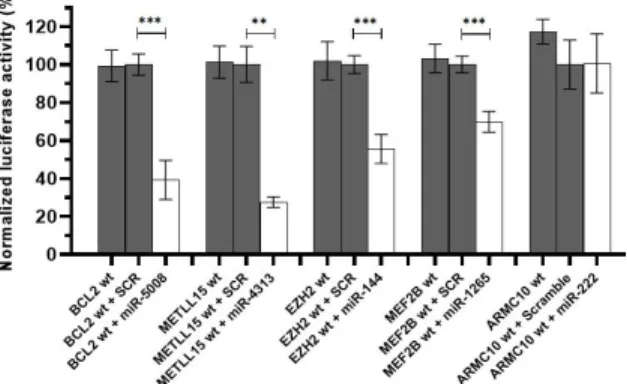
Next, we tested whether or not the predicted miRNA‐binding sites where mutations were found
were actually functional using a luciferase assay system. We cloned the wildtype sequence of the
microRNA‐binding sites, flanked by 150 bp upstream and downstream, into the psiCHECK dual‐
luciferase vector (Figure S4, Supplementary Materials). These constructs were co‐transfected into
HeK293 cells along with the respective miRNA or a control scramble sequence. The sequences of
BCL2, METTL15, EZH2, and MEF2B displayed a significant reduction in luciferase output in the
presence of their respective cognate miRNA when compared to a scramble control sequence (Figure
2). There was no significant reduction in the ARMC10/miR‐222 combination suggesting that this
Figure 2. Luciferase results for the wildtype target sequences. Luciferase data showed that the binding sites in BCL2, METTL15, EZH2 and MEF2B for miR‐5008, miR‐431, miR‐144, and miR‐1265, respectively, were functional. A downregulation in the luciferase activity can be appreciated under the effect of the miRNA compared to the control conditions. This was not the case for the predicted binding site in ARMC10 gene. SCR refers to a scramble miRNA sequence used as a negative control. *** p < 0.0001, ** p < 0.01.
Next, we assessed the effect of the identified mutations on the binding affinity of the miRNAs.
To do this we compared the luciferase activity between cells transfected with the wild‐type target
sequence versus cells transfected with the mutated target sequences in the presence of the respective
miRNA or scramble sequence. The luciferase output of both METLL15 and MEF2B variants did not
significantly change between the mutated sequence and the wild‐type sequence (Figure 3).
Figure 3. Luciferase results for the mutated target sequences: Luciferase data showed that the mutations identified in BCL2 (C) and EZH2 (D) genes in FL patients negatively interfere with miR‐
5008 and miR‐144 binding, respectively, as a significant increase in the luciferase activity could be
detected compared to the wildtype condition. In contrast, this was not observed in METTL15 (A) and
MEF2B (B). SCR refers to a scramble miRNA sequence used as a negative control. For EZH2, mut 1
However, mutations in BCL2 and EZH2 genes promoted a significant increase in luciferase
activity compared to the wildtype sequence, indicating an impediment in microRNA binding and,
subsequently, a less effective regulation of the luciferase gene. We observed this for the c.3623C > T
mutation in the BCL2 gene and c.2115A > T and c.2114T > A mutations in the EZH2 gene (Figure 3).
We additionally tested the c.2114T > C EZH2 mutation which was previously reported in FL patients
[20]. This mutation also resulted in a significant increase in luciferase output when compared to the
wildtype sequence.
2.4. Testing the effect of miR‐5008 and miR‐144 on Basal Target Gene Expression
In order to test the effect of varying the levels of miR‐5008 on BCL2 expression, we transfected
the lymphoma cell lines FL18 and U2932 with either a mimic of miR‐5008 or a scrambled control
sequence. As can be seen from Figure 4A, although there was an increase in BCL2 expression, this
was not significant.
We tested the effect of the addition of miR‐144 to endogenous levels of EZH2 in the HT (wildtype
EZH2 sequence) and WSU‐DLCL2 (mutated EZH2 sequence) cell lines. As can be seen from Figure
4B, increasing the levels of miR‐144 resulted in a significant decrease in the levels of endogenous
EZH2 mRNA in the HT cell line, which has a wildtype EZH2‐binding site sequence, but had no effect
on the WSU‐DLCL2 cell line, which harbors the c.2115A > T mutation in the miR‐144‐binding site in
EZH2 gene [21]. This suggests that mutations affecting this binding site promote that EZH2 is no
longer regulated by miR‐144 in lymphoma cells.
Figure 4. Effect of adding selected miRNAs to endogenous expression levels of target genes. (A) BCL2 expression in lymphoma cells transfected with miR‐5008. BCL2 expression levels were measured using RT‐qPCR in FL‐18 and U2932 lymphoma cell lines. (B) EZH2 expression in lymphoma transfected with miR‐144. SCR refers to a scramble miRNA sequence used as a negative control. 3. Discussion
It was previously reported that mutations in the binding sites of miRNA‐target genes are
associated with several cancer types from in silico studies [22,23], as well as experimentally in
colorectal cancer [24] and acute myeloid leukaemia (AML) [25]. However, outside of these studies,
little has been researched. In this work, we identified mutations in BCL2 and EZH2 in FL patients in
the miR‐5008‐ and miR‐144‐binding sites, respectively, and experimentally demonstrated that these
mutations have the potential to impair these miRNAs binding.
BCL2 and EZH2 genes are both key genes in the pathogenesis of FL [20,26,27]. The t(14;18)
translocation, present in >85% of FL cases, is considered the molecular hallmark of this disease and
results in constitutive expression of the antiapoptotic protein BCL2 [28–30]. We observed that
mutations in the BCL2 3´ UTR binding site for miR‐5008 decreased the downregulation of this gene,
phenomenon in FL18 or U2932 cells, probably due to high basal levels of BCL2 expression in these
t(14;18) cell lines. As the vast majority of FL cases also contain the t(14;18) translocation, the regulatory
relevance of this mutation remains speculative. However, it remains possible that this mechanism
could be relevant in t(14;18) negative FL, as the mechanism for BCL2 overexpression in these cases
currently remains unresolved [31]. Interestingly, the two FL cases that we identified as containing the
c.3623C > T(BCL2) mutation both underwent high‐grade transformation, consistent with the finding
that BCL2 mutations have been reported to have an increased risk increased of transformation [32].
EZH2 is a well‐characterized histone methyltransferase in lymphoma that plays a major role in
the pathogenesis of FL, and mutations in this gene have been established as an early event in FL
lymphomagenesis with activating somatic mutations in the catalytic SET domain of EZH2 in more
than 25% of FL patients [20]. Mutations of the EZH2 gene increase repression via enhanced H3K27
trimethylation, and this transcriptional profile favors proliferation of lymphoma cells and results in
the repression of plasma cell differentiation signatures [20,27,33–35]. We identified mutations in the
Y641 codon (c.2115A > T, Y641F and c.2114T > A, Y641N) in our study cohort. Interestingly, these
activating mutations have previously been reported to prevent Jak2/β‐TrCP‐mediated degradation
of the protein [36]. On the basis of our findings, we propose an additional novel mechanism for these
mutations which is to alter the binding affinity of the miRNA miR‐144. Indeed, EZH2 was previously
reported as a direct target of miR‐144 in cancer [37–39]. Some of these studies demonstrated an inverse
correlation between miR‐144 and EZH2 expression in samples of patients, with miR‐144 usually
downregulated and EZH2 upregulated [38,39], as well as a strong correlation with the aggressiveness
of the associated tumor [38]. Furthermore, a significant role for miR‐144 in B‐cell lymphoma was
previously demonstrated [40]. Importantly, EZH2 expression was found to be significantly higher in
FL patients carrying these activating mutations in EZH2 compared to nonmutated FL patients [41].
In conclusion, in this work, we identified novel recurrent mutations in FL cases that occur in a
nonrandom manner in the miRNA‐binding sites of genes that are key drivers of FL pathogenesis.
This includes the demonstration of a novel regulatory mechanism for arguably two of the most
important genes in FL, BCL2 and EZH2. This work suggests that noncoding mutations and
specifically mutations in miRNA‐binding sites, although often over‐looked in genetic studies, are
likely to be important factors in lymphomagenesis and cancer in general that surely warrant further
investigation.
4. Materials and Methods
4.1. Patient Selection
A total of 85 retrospectively collected tumor samples (77 frozen and eight formalin‐fixed and
paraffin‐embedded (FFPE)) were obtained from 55 patients diagnosed with FL attending the Barts
Cancer Institute, London (n = 25), Cruces University Hospital, Bizkaia, Spain (n = 4), or Donostia
University Hospital, Gipuzkoa, Spain (n = 26). Thirty‐four of these samples were sampled
longitudinally both prior to transformation (i.e., antecedent FL (antFL) and after histological
transformation (i.e., tFL). The other 21 FL cases were classified as nontransforming (ntFL) cases on
the basis of not having had a recorded transformation event with a minimum follow‐up time of 5
years. Pretransformation samples were obtained prior to treatment. Individual data on these patients
are given in Table S3 (Supplementary Materials). Written informed consent was obtained from all
patients for the inclusion of their samples in this study, and samples were collected in accordance
with the Declaration of Helsinki and approved by the local ethics committees (CEIC‐Euskadi
approval number PI2013038, 2 May2013).
4.2. Bioinformatic Identification of miRNA‐Binding Site Mutations
Whole‐genome sequencing (WGS) data obtained from tumor tissue of six FL patients that
underwent transformation and were longitudinally sampled, resulting in 14 samples in total, were
generated as previously described [16]. Somatic mutation data from this study were interrogated to
mature sequences of all human miRNAs retrieved from miRbase (version 19) [42], along with the
genomic coordinates of human UTRs taken from Ensembl bioMart (version 68) [43], were used to
produce FASTA files containing UTR sequences without mutations and separate files containing the
identified WGS mutations. The TargetScan and miRanda standalone algorithms were then used to
predict target sites in the generated sequence files [44]. Only mutations located among the 75% most
confident target sites for each miRNA were considered. The miRanda target prediction algorithm
was then employed on these targets to assess the thermodynamic stability and the effects of the
mutations on potential miRNA binding to the target [45]. Ontology analysis of mutations identified
in predicted miRNA‐binding sites was carried out using Ingenuity Pathway Analysis tool [46].
4.3. Ampliseq (Ion Torrent) Panel NGS
The Ion Ampliseq Designer v3.4 tool was used to design a custom panel containing the
mutations identified by WGS (n = 544). The panel consisted of a single pool of 482 separate amplicons
(125–175 bp in length), covering a total of 57 kb, and included 503 of the 544 (92%) of the originally
identified mutations. The panel was used to sequence 85 samples obtained from 55 patients consisting
of 21 ntFL cases, 27 paired tFL, and matched antecedent FL samples, one longitudinally sampled tFL
case that was sampled at time of diagnosis, 1 year post diagnosis and 2 years post diagnosis and post
transformation, antecedent samples from four tFL cases, and two samples from tFL cases without the
corresponding antecedent sample. In addition, five pools of DNA obtained from the peripheral blood
of 34 healthy controls were sequenced. Samples were sequenced either using an Ion Torrent Personal
Genome Machine (PGM) (n = 70) or an Ion Proton machine (n = 15). Signal processing and base calling
were performed within the Torrent Server using default parameters. After demultiplexing, reads
were aligned against the hg19 version of the human genome, and variants were identified using the
Ion Reporter v5.2 online tool using somatic low stringency parameters from the Torrent Variant
Caller 4.4 plugin. Only variants with a coverage >30× and a VAF >5% were considered in this analysis.
4.4. Luciferase Assays
Luciferase assays were performed on the predicted miRNA‐binding sites of five genes
(ARMC10, BCL2, METTL15, EZH2, and MEF2B) to assess the functional consequences of identified
mutations on miRNA‐binding affinity. The respective predicted miRNA‐binding site sequences were
cloned, with and without the identified mutations, along with 150 bp of flanking sequence,
immediately downstream of the luciferase reporter gene of the psiCHECK‐2 dual‐luciferase vector
(Promega) (Figure S4, Supplementary Materials).
In brief, approximately 105 HEK293 cells, a kind gift from Prof. Adrian Harris (University of
Oxford,UK) were transfected with individual psiCHECK‐2 constructs (200 ng/mL transfection
media) along with 50 nM of either the respective miRNA mimic or a scramble miRNA control
sequence (miRIDIAN miRNA mimics, Dharmacon, Lafayette, CO, USA). Transfections were
performed in biological triplicate and 48 h post‐transfection cells were harvested and lysed with
Dual‐Luciferase Reporter Assay buffer according to the manufacturer’s protocol (Promega, Madison,
WI, USA). Measurements were carried out in triplicate using a PHERAstar microplate reader (BMG
LABTECH, Ortenberg, Germany). Renilla luciferase activity was normalized to the firefly
luminescence measurement for each well. Results were compared using Mann–Whitney U test
carried out in GraphPad Prism software (version 5.01).
4.5. Cell Culture and Transfection
WSU‐DLCL2 is a germinal center (GC)‐type DLBCL cell line containing the t(14; 18)(q32; q21)
translocation and the Y641F mutation in the EZH2 gene. The HT cell line is also GC‐type DLBCL but
contains the wildtype sequence of the EZH2 gene. Both cell lines were obtained from the Barts Cancer
Institute. FL‐18 is a follicular lymphoma cell line carrying the t(14;18)(q32;q21) translocation [47], and
U2932 is a DLBCL cell line that overexpresses BCL2 [48]. All the cell lines were maintained in RPMI
medium with 10% of FBS, 1% L‐glutamine, and 1% penicillin–streptomycin (Gibco™, Thermo Fisher
Scientific, DE, USA).
Cells were transfected with either 1 μM hsa‐miR‐144‐3p (WSU‐DLCL2 and HT) or hsa‐miR‐5008‐ 5p (FL‐18 and U2932) mimic or a scramble control (miRIDIAN miRNA mimics, Dharmacon).
Transfections were carried out in biological triplicate via electroporation using a 4D‐Nucleofector
machine (Amaxa Biosystems, Cologne, Germany). Cells were harvested 24 h after transfection, and
total RNA was extracted using TRIzol reagent (Invitrogen,Carlsbad, CA, USA).
4.6. qRT‐PCR
Total RNA was used as a template for reverse transcription using the High‐Capacity Reverse
Transcription Kit (Applied Biosystems, Foster City, CA, USA). PCR was carried out in triplicate using
a specific Taqman probe for EZH2 (Hs00544830_m1) and BCL2 (Hs00608023_m1) according to the
manufacturer´s protocol (ThermoFisher Scientific, Waltham, MA, USA) on a CFX Connect Real‐Time
PCR Detection System (Bio‐Rad, Hercules, CA, USA). Hypoxanthine phosphoribosyl transferase 1
(HPRT1) and TATA‐binding protein (TBP) were used individually as housekeeping controls, as the
Ct values for these genes were similar to basal EZH2 expression levels. Relative expression was
calculated using the 2−ΔCt method using the median value of both housekeeping controls. For the
statistical analysis, the paired t‐test was applied using GraphPad Prism software (version 5.01).
Supplementary Materials: The following are available online at www.mdpi.com/1422‐0067/21/22/8795/s1:
Figure S1: Distribution of the frequency of the mutations (n = 2056) identified using WGS in 6 FL cases in protein coding genes; Figure S2. B‐cell receptor signalling pathway; Figure S3: Sequencing coverage archived using the custom targeted sequencing panel; Figure S4. Schematic diagram of the construct for luciferase assays in psiCHECK‐2 vector; Table S1. Mutations identified in predicted miRNA‐binding sites in the WGS datatitle; Table S2. Recurrent mutations identified by targeted sequencing in the validation cohort and additional variants within predicted miRNA‐binding sitestitle; Table S3. Sample detailed information
Author Contributions: Conceptualization, M.F.‐M., J.F. and C.H.L.; methodology, M.F.‐M., E.L., J.F. and C.H.L.; formal analysis, J.A.G.‐A., J.W., A.J.E. and I.G.; resources, A.G., I.C., C.L., J.O. and J.F.; writing—original draft preparation, E.L., M.F.‐M. and C.H.L.; writing—review and editing, all authors. All authors have read and agreed to the published version of the manuscript.
Funding: C.H.L. and his research are supported by grants from the IKERBASQUE foundation for science, the
Starmer‐Smith memorial fund, Ministerio de Economía y Competitividad (MINECO) of the Spanish Central Government, the ISCIII and FEDER funds (PI12/00663, PIE13/00048, DTS14/00109, PI15/00275, PI18/01710), Departamento de Sanidad of the Basque government, Asociación Española Contra el Cancer (AECC), the Diputación Foral de Guipuzcoa (DFG), and Gobierno Vasco, Departamento de Desarrollo Económico e Infraestructuras (ELKARTEK Project code: KK‐2019 00015). This work was funded by an ISCIII PFIS fellowship (FI14/00426).
Conflicts of Interest: The authors declare no conflicts of interest. References
1. Smith, A.; Crouch, S.; Lax, S.; Li, J.; Painter, D.; Howell, D.; Patmore, R.; Jack, A.; Roman, E. Lymphoma incidence, survival and prevalence 2004–2014: Sub‐type analyses from the UK’s Haematological Malignancy Research Network. Br. J. Cancer 2015, 112, 1575–1584, doi:10.1038/bjc.2015.94.
2. Kahl, B.S.; Yang, D.T. Follicular lymphoma: Evolving therapeutic strategies. Blood 2016, 127, 2055–2063, doi:10.1182/blood‐2015‐11‐624288.
3. Provencio, M.; Sabin, P.; Gomez‐Codina, J.; Torrente, M.; Calvo, V.; Llanos, M.; Guma, J.; Quero, C.; Blasco, A.; Cruz, M.A.; et al. Impact of treatment in long‐term survival patients with follicular lymphoma: A Spanish Lymphoma Oncology Group registry. PLoS ONE 2017, 12, e0177204, doi:10.1371/journal.pone.0177204.
4. Lossos, I.S.; Gascoyne, R.D. Transformation of follicular lymphoma. Best Pract. Res. Clin. Haematol. 2011, 24, 147–163, doi:10.1016/j.beha.2011.02.006.
5. Wagner‐Johnston, N.D.; Link, B.K.; Byrtek, M.; Dawson, K.L.; Hainsworth, J.; Flowers, C.R.; Friedberg, J.W.; Bartlett, N.L. Outcomes of transformed follicular lymphoma in the modern era: A report from the National LymphoCare Study (NLCS). Blood 2015, 126, 851–857, doi:10.1182/blood‐2015‐01‐621375.
6. Federico, M.; Caballero Barrigon, M.D.; Marcheselli, L.; Tarantino, V.; Manni, M.; Sarkozy, C.; Alonso‐ Alvarez, S.; Wondergem, M.; Cartron, G.; Lopez‐Guillermo, A.; et al. Rituximab and the risk of transformation of follicular lymphoma: A retrospective pooled analysis. Lancet Haematol. 2018, 5, e359–e367, doi:10.1016/S2352‐3026(18)30090‐5.
7. Sole, C.; Larrea, E.; Di Pinto, G.; Tellaetxe, M.; Lawrie, C.H. miRNAs in B‐cell lymphoma: Molecular mechanisms and biomarker potential. Cancer Lett. 2017, 405, 79–89, doi:10.1016/j.canlet.2017.07.020. 8. Musilova, K.; Devan, J.; Cerna, K.; Seda, V.; Pavlasova, G.; Sharma, S.; Oppelt, J.; Pytlik, R.; Prochazka, V.;
Prouzova, Z.; et al. miR‐150 downregulation contributes to the high‐grade transformation of follicular lymphoma by upregulating FOXP1 levels. Blood 2018, 132, 2389–2400, doi:10.1182/blood‐2018‐06‐855502. 9. Lawrie, C.H.; Chi, J.; Taylor, S.; Tramonti, D.; Ballabio, E.; Palazzo, S.; Saunders, N.J.; Pezzella, F.;
Boultwood, J.; Wainscoat, J.S.; et al. Expression of microRNAs in diffuse large B cell lymphoma is associated with immunophenotype, survival and transformation from follicular lymphoma. J. Cell. Mol. Med. 2009, 13, 1248–1260, doi:10.1111/j.1582‐4934.2008.00628.x.
10. Lawrie, C.H.; Soneji, S.; Marafioti, T.; Cooper, C.D.; Palazzo, S.; Paterson, J.C.; Cattan, H.; Enver, T.; Mager, R.; Boultwood, J.; et al. MicroRNA expression distinguishes between germinal center B cell‐like and activated B cell‐like subtypes of diffuse large B cell lymphoma. Int. J. Cancer 2007, 121, 1156–1161, doi:10.1002/ijc.22800.
11. Wang, W.; Corrigan‐Cummins, M.; Hudson, J.; Maric, I.; Simakova, O.; Neelapu, S.S.; Kwak, L.W.; Janik, J.E.; Gause, B.; Jaffe, E.S.; et al. MicroRNA profiling of follicular lymphoma identifies microRNAs related to cell proliferation and tumor response. Haematologica 2012, 97, 586–594, doi:10.3324/haematol.2011.048132. 12. Gebauer, N.; Gollub, W.; Stassek, B.; Bernard, V.; Rades, D.; Feller, A.C.; Thorns, C. MicroRNA signatures
in subtypes of follicular lymphoma. Anticancer Res. 2014, 34, 2105–2111.
13. Malpeli, G.; Barbi, S.; Greco, C.; Zupo, S.; Bertolaso, A.; Scupoli, M.T.; Krampera, M.; Kamga, P.T.; Croce, C.M.; Scarpa, A.; et al. MicroRNA signatures and Foxp3(+) cell count correlate with relapse occurrence in follicular lymphoma. Oncotarget 2018, 9, 19961–19979, doi:10.18632/oncotarget.24987.
14. Thompson, M.A.; Edmonds, M.D.; Liang, S.; McClintock‐Treep, S.; Wang, X.; Li, S.; Eischen, C.M. miR‐31 and miR‐17‐5p levels change during transformation of follicular lymphoma. Hum. Pathol. 2016, 50, 118–126, doi:10.1016/j.humpath.2015.11.011.
15. Hezaveh, K.; Kloetgen, A.; Bernhart, S.H.; Mahapatra, K.D.; Lenze, D.; Richter, J.; Haake, A.; Bergmann, A.K.; Brors, B.; Burkhardt, B.; et al. Alterations of microRNA and microRNA‐regulated messenger RNA expression in germinal center B‐cell lymphomas determined by integrative sequencing analysis.
Haematologica 2016, 101, 1380–1389, doi:10.3324/haematol.2016.143891.
16. Okosun, J.; Bodor, C.; Wang, J.; Araf, S.; Yang, C.Y.; Pan, C.; Boller, S.; Cittaro, D.; Bozek, M.; Iqbal, S.; et al. Integrated genomic analysis identifies recurrent mutations and evolution patterns driving the initiation and progression of follicular lymphoma. Nat. Genet. 2014, 46, 176–181, doi:10.1038/ng.2856.
17. Marine, R. L.; Magana L. C.; Castro C. J.; Zhao K.; Montmayeur A. M.; Schmidt A.; Diez‐Valcarce M.; Fan Ng T. F.; Vinje J.; Burns C. C.; Allan Nix W.; Rota P. A.; Steven Oberste M. Comparison of Illumina MiSeq and the Ion Torrent PGM and S5 platforms for whole‐genome sequencing of picornaviruses and caliciviruses. J Virol Methods 2020. doi:10.1016/j.jviromet.2020.113865
18. Song, L.; Huang, W.; Kang, J.; Huang, Y.; Ren, H.; Ding, K. Comparison of error correction algorithms for Ion Torrent PGM data: application to hepatitis B virus. Sci Rep 2017 doi:10.1038/s41598‐017‐08139‐y 19. Sherry, S.T.; Ward, M.H.; Kholodov, M.; Baker, J.; Phan, L.; Smigielski, E.M.; Sirotkin, K. dbSNP: The NCBI
20. Bodor, C.; Grossmann, V.; Popov, N.; Okosun, J.; O’Riain, C.; Tan, K.; Marzec, J.; Araf, S.; Wang, J.; Lee, A.M.; et al. EZH2 mutations are frequent and represent an early event in follicular lymphoma. Blood 2013,
122, 3165–3168, doi:10.1182/blood‐2013‐04‐496893.
21. Donaldson‐Collier, M.C.; Sungalee, S.; Zufferey, M.; Tavernari, D.; Katanayeva, N.; Battistello, E.; Mina, M.; Douglass, K.M.; Rey, T.; Raynaud, F.; et al. EZH2 oncogenic mutations drive epigenetic, transcriptional, and structural changes within chromatin domains. Nat. Genet. 2019, 51, 517–528, doi:10.1038/s41588‐018‐ 0338‐y.
22. Wu, W.; Wu, L.; Zhu, M.; Wang, Z.; Wu, M.; Li, P.; Nie, Y.; Lin, X.; Hu, J.; Eskilsson, E.; et al. miRNA Mediated Noise Making of 3′UTR Mutations in Cancer. Genes 2018, 9, 545, doi:10.3390/genes9110545. 23. Ziebarth, J.D.; Bhattacharya, A.; Cui, Y. Integrative analysis of somatic mutations altering microRNA
targeting in cancer genomes. PLoS ONE 2012, 7, e47137, doi:10.1371/journal.pone.0047137.
24. Lopes‐Ramos, C.M.; Barros, B.P.; Koyama, F.C.; Carpinetti, P.A.; Pezuk, J.; Doimo, N.T.S.; Habr‐Gama, A.; Perez, R.O.; Parmigiani, R.B. E2F1 somatic mutation within miRNA target site impairs gene regulation in colorectal cancer. PLoS ONE 2017, 12, e0181153, doi:10.1371/journal.pone.0181153.
25. Ramsingh, G.; Koboldt, D.C.; Trissal, M.; Chiappinelli, K.B.; Wylie, T.; Koul, S.; Chang, L.W.; Nagarajan, R.; Fehniger, T.A.; Goodfellow, P.; et al. Complete characterization of the microRNAome in a patient with acute myeloid leukemia. Blood 2010, 116, 5316–5326, doi:10.1182/blood‐2010‐05‐285395.
26. Kridel, R.; Sehn, L.H.; Gascoyne, R.D. Pathogenesis of follicular lymphoma. J. Clin. Investig. 2012, 122, 3424– 3431, doi:10.1172/jci63186.
27. Beguelin, W.; Popovic, R.; Teater, M.; Jiang, Y.; Bunting, K.L.; Rosen, M.; Shen, H.; Yang, S.N.; Wang, L.; Ezponda, T.; et al. EZH2 is required for germinal center formation and somatic EZH2 mutations promote lymphoid transformation. Cancer Cell 2013, 23, 677–692, doi:10.1016/j.ccr.2013.04.011.
28. Sungalee, S.; Mamessier, E.; Morgado, E.; Gregoire, E.; Brohawn, P.Z.; Morehouse, C.A.; Jouve, N.; Monvoisin, C.; Menard, C.; Debroas, G.; et al. Germinal center reentries of BCL2‐overexpressing B cells drive follicular lymphoma progression. J. Clin. Investig. 2014, 124, 5337–5351, doi:10.1172/jci72415. 29. Roulland, S.; Faroudi, M.; Mamessier, E.; Sungalee, S.; Salles, G.; Nadel, B. Early steps of follicular
lymphoma pathogenesis. Adv. Immunol. 2011, 111, 1–46, doi:10.1016/b978‐0‐12‐385991‐4.00001‐5.
30. Castellino, A.; Santambrogio, E.; Nicolosi, M.; Botto, B.; Boccomini, C.; Vitolo, U. Follicular Lymphoma: The Management of Elderly Patient. Mediterr. J. Hematol. Infect. Dis. 2017, 9, e2017009, doi:10.4084/mjhid.2017.009.
31. Leich, E.; Hoster, E.; Wartenberg, M.; Unterhalt, M.; Siebert, R.; Koch, K.; Klapper, W.; Engelhard, M.; Puppe, B.; Horn, H.; et al. Similar clinical features in follicular lymphomas with and without breaks in the BCL2 locus. Leukemia 2016, 30, 854–860, doi:10.1038/leu.2015.330.
32. Correia, C.; Schneider, P.A.; Dai, H.; Dogan, A.; Maurer, M.J.; Church, A.K.; Novak, A.J.; Feldman, A.L.; Wu, X.; Ding, H.; et al. BCL2 mutations are associated with increased risk of transformation and shortened survival in follicular lymphoma. Blood 2015, 125, 658–667, doi:10.1182/blood‐2014‐04‐571786.
33. Sneeringer, C.J.; Scott, M.P.; Kuntz, K.W.; Knutson, S.K.; Pollock, R.M.; Richon, V.M.; Copeland, R.A. Coordinated activities of wild‐type plus mutant EZH2 drive tumor‐associated hypertrimethylation of lysine 27 on histone H3 (H3K27) in human B‐cell lymphomas. Proc. Natl. Acad. Sci. USA 2010, 107, 20980– 20985, doi:10.1073/pnas.1012525107.
34. Yap, D.B.; Chu, J.; Berg, T.; Schapira, M.; Cheng, S.W.; Moradian, A.; Morin, R.D.; Mungall, A.J.; Meissner, B.; Boyle, M.; et al. Somatic mutations at EZH2 Y641 act dominantly through a mechanism of selectively altered PRC2 catalytic activity, to increase H3K27 trimethylation. Blood 2011, 117, 2451–2459, doi:10.1182/blood‐2010‐11‐321208.
35. Morin, R.D.; Johnson, N.A.; Severson, T.M.; Mungall, A.J.; An, J.; Goya, R.; Paul, J.E.; Boyle, M.; Woolcock, B.W.; Kuchenbauer, F.; et al. Somatic mutations altering EZH2 (Tyr641) in follicular and diffuse large B‐cell lymphomas of germinal‐center origin. Nat. Genet. 2010, 42, 181–185, doi:10.1038/ng.518.
36. Sahasrabuddhe, A.A.; Chen, X.; Chung, F.; Velusamy, T.; Lim, M.S.; Elenitoba‐Johnson, K.S. Oncogenic Y641 mutations in EZH2 prevent Jak2/beta‐TrCP‐mediated degradation. Oncogene 2015, 34, 445–454, doi:10.1038/onc.2013.571.
37. Guo, Y.; Ying, L.; Tian, Y.; Yang, P.; Zhu, Y.; Wang, Z.; Qiu, F.; Lin, J. miR‐144 downregulation increases bladder cancer cell proliferation by targeting EZH2 and regulating Wnt signaling. FEBS J. 2013, 280, 4531– 4538, doi:10.1111/febs.12417.
38. Cao, J.; Han, X.; Qi, X.; Jin, X.; Li, X. TUG1 promotes osteosarcoma tumorigenesis by upregulating EZH2 expression via miR‐144‐3p. Int. J. Oncol. 2017, 51, 1115–1123, doi:10.3892/ijo.2017.4110.
39. Lin, L.; Zheng, Y.; Tu, Y.; Wang, Z.; Liu, H.; Lu, X.; Xu, L.; Yuan, J. MicroRNA‐144 suppresses tumorigenesis and tumor progression of astrocytoma by targeting EZH2. Hum. Pathol. 2015, 46, 971–980, doi:10.1016/j.humpath.2015.01.023.
40. Wang, H.; Wang, A.; Hu, Z.; Xu, X.; Liu, Z.; Wang, Z. A Critical Role of miR‐144 in Diffuse Large B‐cell Lymphoma Proliferation and Invasion. Cancer Immunol. Res. 2016, 4, 337–344, doi:10.1158/2326‐6066.cir‐15‐ 0161.
41. Huet, S.; Xerri, L.; Tesson, B.; Mareschal, S.; Taix, S.; Mescam‐Mancini, L.; Sohier, E.; Carrere, M.; Lazarovici, J.; Casasnovas, O.; et al. EZH2 alterations in follicular lymphoma: Biological and clinical correlations. Blood
Cancer J. 2017, 7, e555, doi:10.1038/bcj.2017.32.
42. Kozomara, A.; Griffiths‐Jones, S. miRBase: Integrating microRNA annotation and deep‐sequencing data.
Nucleic Acids Res. 2011, 39, D152–D157, doi:10.1093/nar/gkq1027.
43. Kinsella, R.J.; Kahari, A.; Haider, S.; Zamora, J.; Proctor, G.; Spudich, G.; Almeida‐King, J.; Staines, D.; Derwent, P.; Kerhornou, A.; et al. Ensembl BioMarts: A hub for data retrieval across taxonomic space.
Database J. Biol. Databases Curation 2011, 2011, bar030, doi:10.1093/database/bar030.
44. Friedman, R.C.; Farh, K.K.; Burge, C.B.; Bartel, D.P. Most mammalian mRNAs are conserved targets of microRNAs. Genome Res. 2009, 19, 92–105, doi:10.1101/gr.082701.108.
45. Enright, A.J.; John, B.; Gaul, U.; Tuschl, T.; Sander, C.; Marks, D.S. MicroRNA targets in Drosophila. Genome
Biol. 2003, 5, R1, doi:10.1186/gb‐2003‐5‐1‐r1.
46. Kramer, A.; Green, J.; Pollard, J., Jr.; Tugendreich, S. Causal analysis approaches in Ingenuity Pathway Analysis. Bioinformatics 2014, 30, 523–530, doi:10.1093/bioinformatics/btt703.
47. Ohno, H.; Doi, S.; Fukuhara, S.; Nishikori, M.; Uchino, H.; Fujii, H. A newly established human lymphoma cell line, FL‐18, carrying a 14;18 translocation. Jpn. J. Cancer Res. 1985, 76, 563–566.
48. Quentmeier, H.; Amini, R.M.; Berglund, M.; Dirks, W.G.; Ehrentraut, S.; Geffers, R.; Macleod, R.A.; Nagel, S.; Romani, J.; Scherr, M.; et al. U‐2932: Two clones in one cell line, a tool for the study of clonal evolution.
Leukemia 2013, 27, 1155–1164, doi:10.1038/leu.2012.358.
Publisher’s Note: MDPI stays neutral with regard to jurisdictional claims in published maps and institutional affiliations. © 2020 by the authors. Licensee MDPI, Basel, Switzerland. This article is an open access article distributed under the terms and conditions of the Creative Commons Attribution (CC BY) license (http://creativecommons.org/licenses/by/4.0/).