https://doi.org/10.1007/s12094-019-02134-7
SPECIAL ARTICLE
Multidisciplinary consensus statement on the clinical management
of patients with stage III non‑small cell lung cancer
M. Majem
1· J. Hernández‑Hernández
2· F. Hernando‑Trancho
3· N. Rodríguez de Dios
4· A. Sotoca
5·
J. C. Trujillo‑Reyes
1· I. Vollmer
6· R. Delgado‑Bolton
7· M. Provencio
8 Received: 26 March 2019 / Accepted: 11 May 2019 / Published online: 6 June 2019 © The Author(s) 2019Abstract
Stage III non-small cell lung cancer (NSCLC) is a very heterogeneous disease that encompasses patients with resected,
potentially resectable and unresectable tumours. To improve the prognostic capacity of the TNM classification, it has been
agreed to divide stage III into sub-stages IIIA, IIIB and IIIC that have very different 5-year survival rates (36, 26 and 13%,
respectively). Currently, it is considered that both staging and optimal treatment of stage III NSCLC requires the joint work
of a multidisciplinary team of expert physicians within the tumour committee. To improve the care of patients with stage III
NSCLC, different scientific societies involved in the diagnosis and treatment of this disease have agreed to issue a series of
recommendations that can contribute to homogenise the management of this disease, and ultimately to improve patient care.
Keywords
Lung cancer · Multimodal management · Staging · Multidisciplinary team · Induction therapy · Chemotherapy ·
Radiotherapy · Surgery
Introduction
In the 8th edition of the TNM classification proposed by
the International Association for the Study of Lung Cancer
(IASLC), accepted by the Union for International Cancer
Control (UICC) and the American Joint Committee on
Can-cer (AJCC), stage III non-small cell lung canCan-cer (NSCLC)
encompasses patients who, in the absence of metastatic
disease (M0), present N2 or N3 disease, a tumour with T4
characteristics or one classified as T3 N1 [
2
] (Table
1
). It is,
therefore, a very heterogeneous definition, which includes
patients with resected, potentially resectable and
unresect-able tumours.
To improve the prognostic capacity of the 8th edition of
the TNM classification compared to the previous one, certain
modifications have been carried out, focusing mainly on: (a)
defining the T category that has been regrouped based on the
tumour diameter, with 1-cm increment in size between T1a,
T1b, T1c, T2a and T2b; T3 for 5
‒
7-cm tumours, and T4 for
tumours larger than 7 cm. A tumour is considered T2 when
there is main bronchial involvement that does not reach the
main carina or partial/total atelectasis/pneumonitis, and
T4 when there is invasion of the diaphragm [
1
,
2
]; and (b)
dividing stage III into sub-stages IIIA, IIIB and IIIC, since
survival rates between stages are significantly different, with
5-year survival of 36, 26 and 13%, respectively [
2
,
3
].
Staging and treatment of stage III NSCLC requires
mul-tidisciplinary management by expert physicians, and
evalu-ation by cancer committees is essential. Given the
hetero-geneity of stage III NSCLC, the scientific societies involved
in this work (
Grupo Español de Cáncer de Pulmón
[GECP],
Sociedad Española de Cirugía Torácica
[SECT],
Sociedad
Española de Medicina Nuclear e Imagen Molecular
[SEM-NIM];
Sociedad Española de Oncología Médica
[SEOM];
Sociedad Española de Oncología Radioterápica
[SEOR];
This is a collaborative project of: Mariano Provencio on behalf of Grupo Español de Cáncer de Pulmón (GECP); Florentino Hernando-Trancho on behalf of Sociedad Española de Cirugía Torácica (SECT); Roberto Delgado-Bolton on behalf of Sociedad Española de Medicina Nuclear e Imagen Molecular (SEMNIM); Margarita Majem on behalf of Sociedad Española de Oncología Médica (SEOM); Nuria Rodríguez de Dios and Amalia Sotoca on behalf of Sociedad Española de Oncología Radioterápica
(SEOR); Jesús Hernández-Hernández and Juan Carlos Trujillo-Reyes on behalf of Sociedad Española de Neumología y Cirugía Torácica (SEPAR) and Ivan Vollmer on behalf of Sociedad Española de Radiología Médica (SERAM).
* M. Majem
MMajem@santpau.cat
Sociedad Española de Neumología y Cirugía Torácica
[SEPAR] and
Sociedad Española de Radiología Médica
[SERAM]) have developed this consensus statement to
homogenise its treatment and, ultimately, improve the care
of patients with stage III NSCLC.
Staging of stage III NSCLC
Non‑invasive staging
Correct clinical staging is essential to manage patients with
lung cancer. The first steps in the study of a possible thoracic
neoplasm are the clinical history and a chest X-ray [
4
].
Fur-ther examinations should then be carried out to determine
the local and distant involvement of the neoplasm.
Com-puted tomography (CT) with intravenous contrast is the
preferred technique in the study of lung cancer [
2
,
5
], and
it should include the entire thorax and upper abdomen. It is
not necessary to cover a larger area of the abdomen, since it
does not significantly increase the accuracy of staging [
6
].
Positron-emission tomography (PET) with the glucose
ana-logue
18F-FDG and especially, PET/CT with
18F-FDG, have
revolutionised the staging of lung cancer.
T staging by CT will be indicated by the size of the main
tumour, and this is one of the prognostic factors [
2
].
How-ever, the degree of invasion of the mediastinal structures or
the chest wall modifies the value of the T descriptor, as it
impacts prognosis [
7
]. CT allows assessing the invasion of
mediastinal vascular structures, although other techniques
such as ultrasound or magnetic resonance imaging (MRI)
have better results than CT when assessing the infiltration
of the parietal pleura and the chest wall [
1
,
8
]. In the
pre-operative assessment of Pancoast tumours, MRI plays a
fun-damental role, with better results than CT scans [
9
].
When assessing mediastinal lymph node involvement,
PET/CT with
18F-FDG also plays a key role, with better
results than CT [
10
–
14
]. However, its sensitivity is
dimin-ished in lymph nodes that are smaller than 10 mm in its
short axis [
15
].
Initially, the presence of metastasis will be ruled out by
cytohistological confirmation of suspicious lesions and
pos-sible extrathoracic lymph nodes that can classify the tumour
as N3. A fine-needle aspiration (FNA) or an
ultrasound-guided core-needle biopsy (CNB) can also be used [
16
,
17
].
The initial scans should include the organs with the
great-est potential of lung cancer metastases. One of the major
contributions of PET/CT with
18F-FDG in the initial
diag-nosis of lung cancer is the detection of previously unknown
metastases, with the consequent change in staging [
18
].
Brain MRI is indicated in patients with lung tumours who
are going to be treated with curative intent, to screen for
brain M1 [
19
]. Brain MRI is superior to CT [
20
] and to
PET/CT [
21
].
Non‑surgical intrathoracic invasive staging
In the case of already diagnosed intrathoracic tumours, stage
III (N2 or N3) will be established without requiring
patho-logical confirmation when there is an extensive mediastinal
infiltration (bulky disease) [
22
].
In central tumours or those with enlarged hilar and
medi-astinal lymph nodes, the tumoral nature of the lymph nodes
should be confirmed. An endobronchial ultrasound
(EBUS)-guided puncture will be performed since the positive and
negative predictive values (PPV and NPV, respectively) of
CT or PET are insufficient. EBUS would provide access to
enlarged paratracheal, posterior tracheal, subcarinal, hilar,
interlobar and lobar lymph nodes; and/or an endoscopic
ultrasound (EUS) with access to paratracheal, subcarinal,
paraesophageal and pulmonary ligament lymph nodes. Both
techniques have a sensitivity close to 90% and a specificity
of 100% [
17
,
22
–
25
]. However, if the result is negative, not
assessable or not sufficiently reliable (NPV: < 90%), staging
must be completed with surgical techniques [
24
,
26
,
27
].
Peripheral thoracic tumours without nodal disease require
mediastinal invasive staging if not subsolid and with a
diam-eter greater than 3 cm [
28
], since in these cases the
possi-bility of finding occult N2 nodes exceeds 10% [
22
–
24
,
28
]
(Fig.
1
).
Table 1 Stage IIIA in the 8th TNM classification of lung cancer
See the definition of the T, N and M descriptors in Goldstraw et al. [3] T N M Stage IIIA T1a N2 M0 T1b N2 M0 T1c N2 M0 T2a N2 M0 T2b N2 M0 T3 N1 M0 T4 N0 M0 T4 N1 M0 Stage IIIB T1a N3 M0 T1b N3 M0 T1c N3 M0 T2a N3 M0 T2b N3 M0 T3 N2 M0 T4 N2 M0 Stage IIIC T3 N3 M0 T4 N3 M0
Invasive surgical staging
Invasive surgical mediastinal staging should be performed
when the result of non-surgical invasive techniques is
nega-tive or non-assessable. Despite the greater morbidity and
mortality, these methods are the standard of excellence of
mediastinal staging, having a higher NPV (Table
2
).
Transcervical mediastinoscopy
In the transcervical mediastinoscopy, a biopsy should be
performed at a minimum number of nodal stations (#4R,
#4L and #7), as well as at stations #2R and #2L if they can
be identified. Complications are scarce, with most being
mild, and mortality is practically non-existent [
29
].
Extended cervical mediastinoscopy
Extended cervical mediastinoscopy is performed through the
same incision of conventional mediastinoscopy and allows
exploring the paraaortic and aortopulmonary window (#5
and #6) in the tumours of the left upper lobe, which are not
accessible through conventional mediastinoscopy.
Left parasternal mediastinotomy
In the left parasternal mediastinotomy, stations #5 and #6
are explored through a second incision in the second left
parasternal intercostal space.
Fig. 1 Performance algorithm for the staging of NSCLC* [17]. *The pathways leading to the diagnosis of stage III are highlighted in red.
CT computed tomography, NSCLC non-small cell lung cancer, EBUS
endobronchial ultrasound, EUS endoscopic ultrasound, PET
positron-emission tomography, PET/CT with 18F-FDG PET/CT with 18F
fluorodeoxyglucose, NMR nuclear magnetic resonance, X-ray–CT
Mediastinum pleuroscopy
Mediastinum pleuroscopy is indicated when lymph node (N)
and pleural (M) dissemination should be ruled out. Unlike
all the other explorations, this technique should be
per-formed with single-lung ventilation [
30
,
31
].
Videothoracoscopy
The main advantage of videothoracoscopy is that it allows
the exploration of the lower stations (#8, #9), but requires
single-lung ventilation. It is also useful in the pre-operative
staging of the T descriptor, as it can identify unresectable
tumours that are not detected with imaging tests [
32
].
Video‑assisted mediastinal lymphadenectomy (VAMLA)
The objective of VAMLA is the lymphadenectomy of those
stations that can be accessed through mediastinoscopy (#4R,
#4L, #7, #2R, #2L).
Transcervical‑extended mediastinal lymphadenectomy
(TEMLA)
TEMLA provides the opportunity for a much wider
lym-phadenectomy to the lower stations except for station #9.
Morbidity and mortality are higher than in a conventional
mediastinoscopy.
Methods of restaging after induction
Imaging studies
Although the usefulness of CT in restaging is uncertain, it
has been observed that the response to neoadjuvant treatment
by CT scan predicts a higher survival rate [
33
]. CT-based
complete response has a high predictive value for complete
pathological response, although it tends to underestimate it
[
34
]. PET with
18F-FDG offers good results when assessing
the treatment response of the primary tumour and metastases,
although it is less accurate in the assessment of the mediastinal
response, with a false negative rate of 20% and a false positive
rate of 25% [
35
]. As a prognostic factor, the degree of
reduc-tion of the standardized uptake value (SUV) in the primary
tumour may be predictive of survival and of the pathological
response to treatment [
36
–
38
].
Cytohistological confirmation studies
Re-evaluation usually starts with the same techniques used
for initial staging.
Non‑invasive or minimally invasive techniques
Bronchoscopy is reserved to confirm local tumour progression.
Transbronchial needle aspiration (TBNA) achieves a correct
diagnosis in 71% of patients and avoids other invasive
proce-dures in 35% of cases [
39
]. The use of FNA by EBUS or EUS
in restaging has a sensitivity lower than 80%, with an NPV
lower than this value [
40
–
42
]. Using both EBUS and EUS
combined does not improve these results [
43
,
44
]. When the
results of EBUS and/or EUS do not show malignancy, it is
recommended to use a surgical technique to reduce the
propor-tion of false negatives [
26
,
45
].
Surgical techniques
The first mediastinoscopy can be reserved for restaging
when N2 is initially confirmed by TBNA, EBUS or EUS
during the initial staging.
Re-mediastinoscopy is technically more complex. It
shows a sensitivity higher than 60% (range 60–74%), a
specificity of 100%, an accuracy greater than 80% (range
80–92%), a PPV of 100% and a NPV of 73% (range 73–86%)
[
46
–
48
].
There are no restaging cases using VAMLA. As for
TEMLA, authors present a restaging series with a sensitivity
Table 2 Main invasive surgical techniques for stage III NSCLC staging
NSCLC Non-small cell lung cancer, TEMLA transcervical-extended mediastinal lymphadenectomy,
VAMLA video-assisted mediastinal lymphadenectomy, NPV negative predictive value, PPV positive predic-tive value
Technique Patients Sensitivity (%) Specificity (%) NPV (%) PPV (%) Transcervical mediastinoscopy [130] 1362 86 100 > 94 100 Extended cervical mediastinoscopy [131] 221 67 100 94 100 Left parasternal mediastinotomy [132] 45 86 100 89 100 Videothoracoscopy [133] 55 100 100 100 100 VAMLA [134] 144 94 100 – 100 TEMLA [135] 698 96 100 99 100
of 95%, an NPV of 97%, an accuracy of 98% and a
specific-ity and PPV of 100% [
49
]. Finally, there is only one study
that uses videothoracoscopy as a restaging method [
50
]
with a sensitivity of 83%, a NPV of 64% and a specificity
of 100%.
Pre‑treatment functional assessment
Pre‑operative functional assessment
Before surgery, it is necessary to check patient’s heart
func-tion with patient’s history and heart medicafunc-tion revision.
It is also necessary to check if they have a thoracic revised
cardiac risk index value that does not exceed 1.5 points [
51
].
The patient must be referred for a cardiology consultation
if necessary.
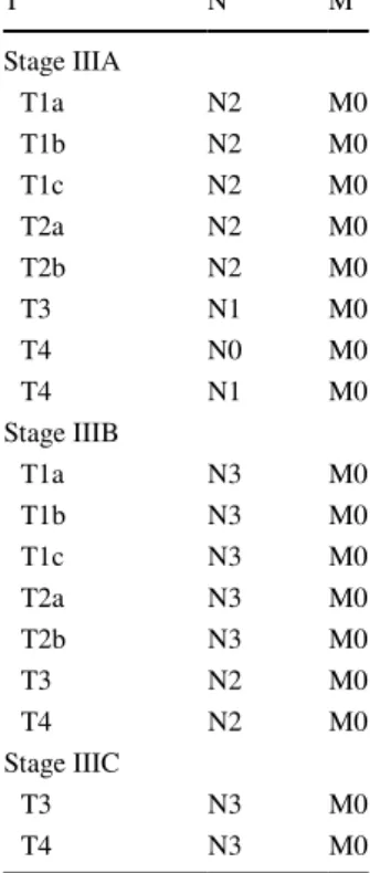
To assess the risk derived from pulmonary resection, a
pulmonary function study should be performed. This study
should calculate the maximum expiratory volume in the first
second of forced expiration (FEV1) and the diffusion
capac-ity of the lung for carbon monoxide (DLCO) planned in the
post-operative period (ppoFEV1 and ppoDLCO) [
51
–
53
].
When both indexes are greater than 60% of their theoretical
values, the patient presents low risk and does not require
further studies [
51
]. When the ppoFEV1 or the ppoDLCO
is less than 30%, a cardiopulmonary exercise testing (CPET)
will be indicated to quantify maximum oxygen consumption
(VO
2max). If this is lower than 10 mL/kg/min (or less than
35% of its theoretical value), the surgical risk is high. If the
value is between 10 and 20 mL/kg/min (between 35 and
75% of the theoretical value) the risk is intermediate, and
if they are above the latter values the risk is low. When the
ppoFEV1 or the ppoDLCO is less than 60% and both exceed
30%, a CPET may be indicated, or stair climb/shuttle walk
test may be used before CPET (Fig.
2
) [
51
].
Pre‑radiotherapy functional assessment
Although there are no clearly defined FEV1 or DLCO limits
for radiotherapy, the same criteria of surgical case series are
used in most chemotherapy/radiotherapy clinical trials. Both
the dosimetric parameters, the mean lung dose (MLD) and
the percentage of healthy lung volume that receives at least
20 Gy (V20) are effective tools to assess the risk of
pulmo-nary toxicity, although some studies support the importance
of the clinical characteristics of patients in the estimation of
lung damage secondary to radiation [
54
].
Multimodal management of stage III NSCLC
Incidental stage IIIA (N2)
The need for adjuvant treatment has been evidenced by
the poor results of local control and overall survival (OS)
after surgery in patients with stage IIIA NSCLC. Several
randomised clinical trials and meta-analyses have shown a
Fig. 2 Pre-operative functional assessment [51]. DLCO diffusing capacity of the lung for carbon monoxide, ppoDLCO planned post-operatively DLCO, FEV1 forced expiratory volume in the first
sec-ond, ppoFEV1 planned post-operatively FEV1, CPET cardiopulmo-nary exercise test, VO2max maximum oxygen consumption
5% increase in OS at 5 years when administering adjuvant
platinum-based chemotherapy [
55
,
56
].
The role of post-operative radiotherapy (PORT) remains
controversial. A first meta-analysis conducted in 1998
showed a relative increase in the risk of death (21%), with a
lower survival rate in patients receiving PORT [
57
].
How-ever, a subsequent subgroup analysis established that this
negative impact occurred in N0–N1 patients, although it was
not clearly demonstrated in N2 patients. In several
meta-analyses and subsequent retrospective studies, PORT in N2
patients reduced the risk of local relapse, without showing
significant differences in OS [
58
–
62
]. However, since most
of these studies were not performed with advanced
radio-therapy techniques, the validity of their results could be
questioned. It is, therefore, a priority to obtain information
from randomised trials with modern techniques to establish
its real impact on OS [
63
].
Regarding the sequence of treatments, it is recommended
to administer sequential treatment starting with
chemother-apy and to reserve concomitant treatment for patients with
unresectable residual tumours, since adjuvant
chemother-apy–radiotherapy has not shown an increase in OS and there
was a greater toxicity.
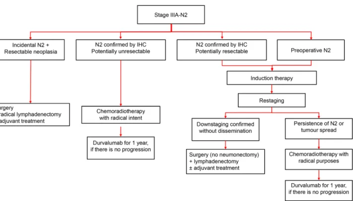
Potentially resectable stage IIIA (N2)
The group of patients with stage IIIA N2 NSCLC is
hetero-geneous and their treatment should be discussed in a
mul-tidisciplinary committee. For this purpose, the individual
characteristics of the patient, such as age, lung function,
comorbidity and functional status, must be considered before
and after the induction therapy (Fig.
3
).
The main objectives of induction therapy are: (a) to
eradi-cate subclinical metastases and mediastinal lymph node
disease; (b) to improve local control of the disease; (c) to
increase resectability; and (d) to reduce the magnitude of
surgical resection.
The factors associated with a better prognosis in patients
who undergo surgery are: confirmation of a complete
response of the mediastinal (ypN0), achieving a complete
resection and confirmation of a complete pathological
response.
Induction chemotherapy
Several phase III studies have shown that platinum-based
induction chemotherapy increases OS [
64
–
71
]. In stage IIIA
(cN2) patients, induction chemotherapy increases OS
com-pared to surgery alone [
67
,
69
,
72
,
73
]. These results have
been confirmed in a subsequent meta-analysis [
41
].
Fig. 3 Algorithm for the clinical management of patients with stage IIIA-N2 NSCLC. NSCLC Non-small cell lung cancer, IHC immunohisto-chemistry
Induction chemoradiotherapy
The trials that analyse the role of treatment with
induc-tion chemoradiotherapy followed by surgery failed to show
a survival benefit except in certain subgroups of patients
[
74
–
76
]. A clinical trial randomised patients with
resect-able N2 disease (75% a single station affected) to receive a
concomitant chemoradiotherapy treatment (radiation dose of
45 Gy + cisplatin–etoposide) followed by surgery or radical
radiotherapy (61 Gy). This trial showed a significant
ben-efit in progression-free survival (PFS) in favour of trimodal
treatment, without significant differences in OS [
74
]. The
lack of benefit in the surgery arm may be a consequence of a
higher early mortality, especially in patients who underwent
a pneumonectomy. A study in patients with resectable stage
III NSCLC showed a benefit for induction chemotherapy
in terms of the pathological response, and also an
improve-ment of the mediastinal stage, with no differences in survival
rates [
77
].
A meta-analysis showed that the addition of radiotherapy
to induction therapy does not increase survival [
78
], which
raises questions about the need to add radiotherapy. It should
be noted that several randomised studies have shown that
mediastinal downstaging is associated with a better
prog-nosis [
79
].
Surgical treatment after induction
Surgical resection after induction therapy is indicated when
imaging tests rule out extrathoracic disease progression,
functional assessment after induction therapy indicates that
the patient can tolerate resection, restaging techniques
con-firm an improvement of the mediastinal status and the type
of resection ensures a complete resection, but avoids a
pneu-monectomy [
74
,
75
].
The goal of surgical treatment after induction therapy
is to achieve surgery with complete resection (R0). The
R0 criteria defined by the IASLC include: (a) tumour-free
resection margins confirmed microscopically; (b)
system-atic mediastinal lymph node dissection; (c) no
extracapsu-lar invasion of affected nodes; and (d) the most distal node
resected should be free of disease [
80
].
There is some consensus that a minimum of six lymph
nodes from three N2 stations should be analysed (always
including station 7) [
81
–
83
]. An adequate
lymphadenec-tomy is considered a criterion for surgical quality [
84
,
85
].
Surgical resection is not recommended if R0 surgery is not
feasible, radical radiotherapy could be administered if not
previously done [
86
].
Based on the results obtained in patients who underwent
surgery after induction therapy and still had mediastinal
lymph node involvement (ypN2), surgery can be an option
despite the persistence of N2 involvement in very specific
cases, such as initial disease confined to a station that is not
enlarged (resectable stage IIIA-N2), with PET/CT with
18F-FDG results showing minimal residual disease, with
resec-tion less than a pneumonectomy, and when R0 is feasible.
Adjuvant treatment with chemotherapy
Although adjuvant chemotherapy improves disease-free
survival in R0 patients [
87
], no specific data support
adju-vant chemotherapy after induction therapy and surgery in
initial stage IIIA-N2 NSCLC. Therefore, administration of
adjuvant chemotherapy should be tailored for each patient,
according to the pathological response and the pathology
findings.
Adjuvant treatment with radiation therapy
Adjuvant radiotherapy therapy to the mediastinum is not
recommended in pN0 or pN1 stage. When there is multiple
hilar involvement, in extracapsular invasion or in pN2 stage,
adjuvant radiotherapy may be considered if it has not been
administered during induction therapy [
88
].
Administration of adjuvant radiotherapy to the T is not
recommended in patients with surgery R0 and in the case of
R1 or R2 surgery, adjuvant radiotherapy if not administered
previously [
88
].
Unresectable stages IIIA (N2), IIIB and IIIC
Determination of unresectability in patients with stage
III NSCLC must be determined by a multidisciplinary
committee.
Treatment with concomitant chemotherapy/radiotherapy
Concomitant chemotherapy/radiotherapy is the treatment of
choice for patients with a good general condition (ECOG
0–1) and a weight loss of less than 5% in the previous
3 months [
89
]. This is a radical treatment that aims to cure
the disease. A platinum-based chemotherapy regimen is
recommended [
90
]. Gemcitabine regimens are not
mended because of higher pulmonary toxicity. The
recom-mended radiotherapy dose is 60–66 Gy [
91
]. Concurrent
chemoradiotherapy provides a median OS of 22–25 months
and a 5-year OS of 20% [
92
], with a grade 3 toxicity or
higher consisting of oesophagitis (7–21%) and pneumonitis
(3–7%) [
93
].
Treatment with sequential chemoradiotherapy
In patients with ECOG > 1, weight loss greater than 5% and
a large volume to be irradiated with an unacceptable risk of
pneumonitis, the recommendation is to administer induction
chemotherapy followed by radical radiotherapy.
Systemic treatment of stage III NSCLC
Adjuvant chemotherapy after stage IIIA (N2)
incidental disease
Patients with N2 disease documented during surgery are
candidates for adjuvant chemotherapy (level of evidence:
I, recommendation grade: A). The recommended regimen
in patients without contraindications is cisplatin doublet
chemotherapy, since it has shown to improve OS in
com-plete resected patients. The recommended number of cycles
is four (level of evidence: I, recommendation grade: A) [
14
].
Cisplatin–etoposide and cisplatin–vinorelbine are the
plati-num doublets with the greatest evidence, as shown in the
LACE meta-analysis [
56
]. Carboplatin can be administered
if there are contraindications for treatment with cisplatin.
Induction chemotherapy
Patients with potentially resectable IIIA-N2 disease can
receive pre-operative treatment with chemotherapy, with
or without radiotherapy, followed by surgery. The
recom-mended induction chemotherapy regimen in patients without
contraindications is to administer three to four cycles of a
cisplatin doublet, based on complementary chemotherapy
studies (level of evidence: II, recommendation grade: B).
Neoadjuvant chemotherapy has been shown to improve
OS. In a meta-analysis of 15 randomised studies, a
signifi-cant 5-year overall survival benefit of 5% was observed in
patients at stage IB–IIIA (HR: 0.87; 95% CI 0.78–0.96;
p
= 0.007) [
73
].
Chemotherapy combined with radiotherapy
In patients with unresectable stage III NSCLC, the
admin-istration of chemotherapy concomitant with radiotherapy is
recommended (level of evidence: I, recommendation grade:
A) [
73
]. In patients in whom concomitant treatment is not
possible, the alternative is a sequential administration.
The recommended chemotherapy regimen is a cisplatin
regimen with vinorelbine or etoposide [
89
,
94
]. Most
ran-domised studies comparing sequential versus concomitant
treatment use cisplatin with etoposide or cisplatin with
vinorelbine [
92
,
95
]. It is recommended to administer two
to four cycles of chemotherapy concurrent with radiotherapy
(level of evidence I, recommendation grade: A) [
89
].
Cisplatin can be replaced by carboplatin in patients
with comorbidities that contraindicate treatment with
cis-platin (carbocis-platin and paclitaxel is one of the most used
regimens). In unresectable patients, cisplatin and
peme-trexed regimens have not shown better results than standard
treatment with cisplatin and etoposide concomitant with
radiotherapy [
96
].
Age does not justify administering a suboptimal treatment
in elderly patients. Therefore, the most convenient treatment
should be administered according to the patient’s illness and
comorbidities (level of evidence: I, recommendation grade:
A).
Radiotherapeutic treatment of stage III
NSCLC
Radiotherapy volumes
Gross tumour volume (GTV) includes the primary tumour
and the affected lymph nodes, when there is lymph node
involvement. The clinical target volume (CTV) includes the
GTV with a three-dimensional margin that incorporates the
microscopic extension of the disease. The planning target
volume (PTV) includes the CTV with a three-dimensional
margin that considers the tumour movement and the
uncer-tainties in the patient’s daily positioning. Improving the
immobilisation systems, respiratory movement control and
image-guided radiation therapy (IGRT) can reduce the PTV
margin [
97
].
Prophylactic irradiation of non-affected lymph node areas
is not recommended, especially if PET/CT with
18F-FDG
has been performed during staging, since it does not increase
survival and causes more toxicity [
98
,
99
].
When post-operative radiotherapy is administered, the
CTV should include the bronchial stump and the ipsilateral,
subcarinal and contralateral hilar and paratracheal nodal
areas [
60
,
100
].
A correct definition of the healthy organs is a priority
to minimise side effects, especially respiratory and cardiac
[
101
,
102
]. A useful source is the RTOG volume contouring
atlas [
103
].
New technological advances such as 4D radiotherapy, the
use of PET/CT with
18F-FDG for the simulation,
intensity-modulated radiotherapy (IMRT) or volumetric-intensity-modulated
arc therapy (VMAT), IGRT and the control of respiratory
movement permit the administration of radical doses and
reduce the impact on healthy organs, and should be used
whenever available [
104
].
Radiotherapy dose
As part of a radical treatment, doses of 60–66 Gy at 2.0 Gy/
day are recommended. Higher doses do not improve results
and increase side effects [
91
]. In the case of sequential
radiation therapy, accelerated radiotherapy schemes should
be evaluated [
105
]. In post-operative irradiation, doses of
50–54 Gy at 1.8–2.0 Gy/day are usually indicated, although
higher doses may be administered when there is
extracap-sular involvement or involvement of resection margins [
60
].
If radiation therapy is administered as part of an induction
therapy, doses of 45–54 Gy at 1.8–2.0 Gy/day are usually
used. A team of experienced thoracic surgeons is required
when administering higher doses as induction therapy to
avoid possible post-operative complications [
106
,
107
].
Surgical treatment of stage III NSCLC
Surgery in patients with stage IIIA and IIIB non-N3 NSCLC
should be considered in a multidisciplinary approach,
espe-cially if the R0 is feasible without pneumonectomy [
19
,
81
,
82
,
108
–
111
]. The best results are obtained after an adequate
selection of the optimal therapeutic scheme and when the
surgery is performed at hospitals that have trained
surgi-cal teams with anaesthesiology, thoracic surgery, nursing,
rehabilitation, etc. [
81
–
83
,
108
,
109
,
111
].
It is important to assess the resectability of the lesion and
the operability of the patient. As mentioned previously, there
are updated guidelines for this purpose in which predictable
ppoFEV1 and ppoDLCO values and oxygen consumption
have a central role in decision-making. There is not a single
value or scale (Thoracoscore, ThRCRI, etc.) that indicates
operability. The determination of the patient’s general
condi-tion using the ECOG scale or the Karnofsky index as well as
their expectations and socio-family environment will help to
make individualised decisions regarding the surgical options
[
112
,
113
].
The standard anatomical resection is lobectomy or
bilobectomy (level of evidence: I, recommendation grade:
A). Anatomical segmental resection is considered
in situa-tions of impairment of the cardiopulmonary reserve.
Pneu-monectomy can be avoided, as far as possible, with
bron-choplastic and angioplastic resections, since this procedure
(especially if it is right pneumonectomy) has a significant
impact on the results [
114
,
115
].
The setting for the surgical approach would be the least
invasive one, such as muscle-sparing thoracotomies,
video-assisted thoracic surgery (VATS) or robot-video-assisted thoracic
surgery (RATS). The purpose of this approach is to be the
least aggressive possible and to manage these patients, who
must necessarily receive neo- or coadjuvant treatments,
safely. Anaesthetic and peri-operative care are crucial in
the future for these patients [
115
]. In this context, there are
multimodal rehabilitation protocols established, such as
fast-track or enhanced recovery after surgery (ERAS) [
116
].
Consolidation treatment in unresectable
stage III NSCLC
Until recently, consolidation treatment with chemotherapy
[
92
,
117
], targeted therapy or some types of immunotherapy
[
118
] failed to increase survival in patients with stage III
NSCLC [
119
].
Recently, the administration of the anti-PD-L1 drug
dur-valumab as consolidation treatment for 1 year in patients
with unresectable stage III NSCLC treated with radical
chemoradiotherapy and with no progressive disease has
been established as a new standard (level of evidence: I,
recommendation grade: A). This is based on the results of a
phase III clinical trial, in which durvalumab compared with
placebo increased both the PFS (17.2 months vs. 5.6 months,
respectively, HR, 0.51, 95% CI 0.41–0.63) and the 2-year
overall survival of 66.3% (95% CI 61.7–70.4) with
dur-valumab to 55.6% (95% CI 48.9–61.8) with placebo (HR,
0,68; 99.73% CI 0,47–0,997;
p
= 0.0025). The benefit was
consistent in all the pre-specified subgroups. Durvalumab
was well tolerated, with a 15.4% of treatment
discontinua-tion due to toxicity, and with grades 3–4 toxicity of 30.5%
in patients treated with durvalumab and 26% of patients in
the placebo group [
120
]. The European Medicines Agency
(EMA) has approved durvalumab as a consolidation
treat-ment in patients with locally advanced or unresectable
NSCLC with expression of PD-L1 ≥ 1% of tumour cells
and whose disease has not progressed after platinum-based
chemoradiotherapy, based on an unplanned post hoc analysis
of PD-L1 expression.
Follow‑up of patients with stage III NSCLC
A key point in the follow-up of the patients diagnosed
with a pulmonary neoplasm is quitting smoking, since this
improves the prognosis of the disease [
121
]. Of note, it
has been shown that a causal relationship may be inferred
between smoking and increased mortality from any cause
and mortality associated with cancer, as well as regarding
the appearance of second lung neoplasms [
121
]. There is
also a relationship, although it cannot be considered a cause,
between smoking and an increased risk of recurrence, worse
response to treatment and greater probability of suffering
treatment-related toxicity [
121
].
The follow-up frequency of patients with lung cancer is
controversial. SEOM and SERAM recommend that patients
treated with surgery undergo follow-up with CT scan every
6–12 months during the first 2 years, and annually thereafter
[
5
,
122
]. Some authors suggest that performing a PET/CT
with
18F-FDG 1 year after surgery is more sensitive than CT
Table 3 Recommendations for the clinical management of patients with stage III NSCLC
CT Computed tomography, DLCO diffusing capacity of the lung for carbon monoxide, EBUS endobronchial ultrasound, ECOG Eastern Coop-erative Oncology Group, EUS endoscopic ultrasound, FEV1 forced expiratory volume in the first second, MRI magnetic resonance imaging,
NSCLC non-small cell lung cancer, PET positron-emission tomography, PET/CT with 18F-FDG, OS overall survival, R0 complete resection
Non-invasive staging of stage III NSCLC:
CT scan of the thorax and upper abdomen with intravenous contrast is the preferred technique in the initial assessment when lung cancer is suspected
PET/CT with 18F-FDG is the most sensitive technique to explore mediastinal lymphadenopathy
Brain MRI is indicated in patients with lung neoplasm who are going to be treated with curative intent
Invasive staging of stage III NSCLC:
Non-surgical invasive staging methods are the first choice due to their lower associated morbidity and mortality.
In cases in which the result of invasive non-surgical techniques is negative or non-assessable, surgical staging methods should be recommended, as they remain the standard of excellence in stage III NSCLC staging
Pre-treatment functional assessment:
Before conducting surgery on the patient, it is necessary to ensure that their cardiac function is adequate and to estimate the risk derived from pulmonary resection by means of a pulmonary function study
The FEV1 and the DLCO predicted after the scheduled surgery will be calculated
Functional assessment prior to radiotherapy is less defined, but the lung and heart doses should be minimised
Multimodal management of stage III NSCLC Incidental Stage IIIA (N2):
In stage IIIA with incidental N2 involvement, the recommended treatment is surgical resection and adjuvant chemotherapy (level of evidence: I, recommendation grade: A)
The administration of sequential post-operative radiotherapy after completing chemotherapy has shown to increase local control of the disease, without being clear whether it provides an OS benefit (level of evidence: II, recommendation grade: C)
Potentially resectable stage IIIA (N2):
In potentially resectable stage IIIA-N2, initial surgical resection is not recommended (level of evidence: I, recommendation grade: A) In potentially resectable stage IIIA-N2, the recommended treatment is induction chemotherapy, followed by surgery if there is improvement of
the stage (level of evidence: I, recommendation grade: A), followed by adjuvant radiotherapy according to the findings after surgery (level of evidence: IV, recommendation grade: C)
Neoadjuvant chemotherapy/radiotherapy achieves a greater downstaging of mediastinal disease volume with respect to induction chemotherapy, without an impact on OS (level of evidence: I, recommendation grade: C)
Unresectable stages IIIA (N2); IIIB and IIIC:
In patients with ECOG 0–1, without weight loss > 5% and with irradiation volumes that do not compromise cardiopulmonary functionality, the administration of concomitant platinum-based chemotherapy/radiotherapy is recommended (level of evidence: I, recommendation grade: A), followed by consolidation treatment with durvalumab, if there is no disease progression
The administration of radiotherapy at doses higher than 66 Gy is not recommended (level of evidence: I, recommendation grade: A)
Systemic treatment of stage III NSCLC
The recommended chemotherapy regimens, either adjuvant, induction or radical combined with radiotherapy, are cisplatin doublets
Radiotherapeutic treatment of stage III NSCLC:
The recommended radiotherapy dose in radical chemoradiotherapy is 60–66 Gy at 2 Gy/day. If administered sequentially, accelerated schemes that reduce the total duration of treatment are recommended
In post-operative irradiation, doses of 50–54 Gy at 1.8–2.0 Gy/day are recommended. In case of extracapsular involvement or resection margins, higher doses may be administered
In case of induction radiotherapy, doses of 45–54 Gy are usually used
Surgical treatment of stage III NSCLC:
The standard anatomical resection is lobectomy or bilobectomy (level of evidence: I, recommendation grade: A).
Surgery in patients with stage III NSCLC is considered in a multidisciplinary and personalised treatment environment, especially if R0 is feasi-ble without pneumonectomy
This surgery should be carried out at hospitals that have trained surgical teams
Stage III NSCLC consolidation treatment:
Durvalumab is recommended as consolidation treatment for 1 year in patients with unresectable stage III NSCLC with expression of PDL1 ≥ 1% in tumour cells and without progression after radical chemoradiotherapy (level of evidence: I, recommendation grade: A)
Follow-up of patients with stage III NSCLC:
Quitting smoking should be advised to patients undergoing treatment for lung cancer
It is recommended to carry out a CT scan every 6–12 months during the first 2 years and later annually PET/CT with 18F-FDG shows better results in the assessment of patients treated with chemoradiotherapy
In patients treated with radiotherapy or
chemoradiother-apy, an initial PET/CT with
18F-FDG and during follow-up
is useful for predicting areas with greater potential for
recur-rence or treatment failure [
124
]. On the other hand,
treat-ment response can be assessed early with a post-treattreat-ment
PET/CT with
18F-FDG [
125
–
127
].
The response assessment criteria will be established
based on the systemic treatment received. In most cases, the
RECIST 1.1 criteria will apply [
128
], but for patients who
have received immunotherapy, iRECIST criteria should be
used [
129
]. In these patients, successive controls should be
carried out in the event of tumour growths, given the
pos-sibility of pseudoprogression. However, to verify that these
are true progressions and not peritumoural inflammatory
reactions, controls should not be performed before 4 weeks
after the last assessment.
Conclusions
Stage III NSCLC is a very heterogeneous disease in which
multidisciplinary management is essential. A multimodal
approach is necessary when establishing treatment for stage
III NSCLC. This will depend on the stage III subtype
(inci-dental, potentially resectable or unresectable), and may
include surgery, chemotherapy and radiotherapy, as detailed
in Table
3
. Recently, consolidation therapy with durvalumab
has become the new standard treatment for unresectable
NSCLC after radical chemoradiotherapy and without
dis-ease progression.
Acknowledgements The authors are grateful for the editorial
assis-tance of Beatriz Gil-Alberdi of HealthCo (Madrid, Spain) in the pro-duction of this manuscript.
Funding GECP, SECT, SEMNIM, SEOM, SEOR, SEPAR and
SERAM are grateful for financial support for this project in the form of unrestricted grants from AstraZeneca.
Compliance with ethical standards
Conflict of interest M Majem reports personal fees from AstraZeneca,
Roche, Eli Lilly, Bristol-Myers Squibb, Pfizer, Boehringer Ingelheim, Novartis, Tesaro, Takeda, Vifor Pharma, AbbVie, outside the submit-ted work. M Provencio has received honoraria from Roche, Eli Lilly, Boehringer Ingelheim, AstraZeneca, and Bristol-Myers Squibb, out-side the submitted work. The rest of the authors declare that they have not conflicts of interest.
Ethical statement The study has been performed in accordance with
the ethical standards of the Declaration of Helsinki and its later amend-ments. This article does not contain any studies with human partici-pants or animals performed by any of the authors.
Informed consent For this type of study formal consent is not required.
Open Access This article is distributed under the terms of the Crea-tive Commons Attribution 4.0 International License (http://creat iveco mmons .org/licen ses/by/4.0/), which permits unrestricted use, distribu-tion, and reproduction in any medium, provided you give appropriate credit to the original author(s) and the source, provide a link to the Creative Commons license, and indicate if changes were made.
References
1. Rami-Porta R, Bolejack V, Crowley J, Ball D, Kim J, Lyons G. The IASLC lung cancer staging project: proposals for the revisions of the T descriptors in the forthcoming eighth edi-tion of the TNM classificaedi-tion for lung cancer. J Thorac Oncol. 2015;10:990–1003.
2. Rami-Porta R, Call S, Dooms C, Obiols C, Sanchez M, Travis WD, et al. Lung cancer staging: a concise update. Eur Respir J. 2018;51(5):1800190.
3. Goldstraw P, Chansky K, Crowley J, Rami-Porta R, Asamura H, Eberhardt WE, et al. The IASLC lung cancer staging pro-ject: proposals for revision of the TNM stage groupings in the forthcoming (eighth) edition of the TNM classification for lung cancer. J Thorac Oncol. 2016;11:39–51.
4. Kirsch J, Ramirez J, Mohammed TL, Amorosa JK, Brown K, Dyer DS, et al. ACR Appropriateness Criteria(R) acute respira-tory illness in immunocompetent patients. J Thorac Imaging. 2011;26:W42–4.
5. de Castro J, Cobo M, Isla D, Puente J, Reguart N, Cabeza B, et al. Recommendations for radiological diagnosis and assess-ment of treatassess-ment response in lung cancer: a national consen-sus statement by the Spanish Society of Medical Radiology and the Spanish Society of Medical Oncology. Clin Transl Oncol. 2015;17:11–23.
6. Gross BH, Brown RK, Kalemkerian GP. Optimal anatomic cover-age for CT in staging lung cancer: lessons from PET-CT correla-tion. Lung Cancer. 2011;73:59–62.
7. Kajiwara N, Akata S, Uchida O, Usuda J, Ohira T, Kawate N, et al. Cine MRI enables better therapeutic planning than CT in cases of possible lung cancer chest wall invasion. Lung Cancer. 2010;69:203–8.
8. Bandi V, Lunn W, Ernst A, Eberhardt R, Hoffmann H, Herth FJ. Ultrasound vs. CT in detecting chest wall invasion by tumor: a prospective study. Chest. 2008;133:881–6.
9. Bruzzi JF, Komaki R, Walsh GL, Truong MT, Gladish GW, Munden RF, et al. Imaging of non-small cell lung cancer of the superior sulcus: part 2: initial staging and assessment of resecta-bility and therapeutic response. Radiographics. 2008;28:561–72. 10. Gelberg J, Grondin S, Tremblay A. Mediastinal staging for lung
cancer. Can Respir J. 2014;21:159–61.
11. Gould MK, Kuschner WG, Rydzak CE, Maclean CC, Demas AN, Shigemitsu H, et al. Test performance of positron emission tomography and computed tomography for mediastinal staging in patients with non-small-cell lung cancer: a meta-analysis. Ann Intern Med. 2003;139:879–92.
12. Prenzel KL, Monig SP, Sinning JM, Baldus SE, Brochhagen HG, Schneider PM, et al. Lymph node size and metastatic infiltration in non-small cell lung cancer. Chest. 2003;123:463–7.
13. Shim SS, Lee KS, Kim BT, Chung MJ, Lee EJ, Han J, et al. Non-small cell lung cancer: prospective comparison of integrated FDG PET/CT and CT alone for preoperative staging. Radiology. 2005;236:1011–9.
14. Zhao L, He ZY, Zhong XN, Cui ML. (18)FDG-PET/CT for detection of mediastinal nodal metastasis in non-small cell lung cancer: a meta-analysis. Surg Oncol. 2012;21:230–6.
15. Lv YL, Yuan DM, Wang K, Miao XH, Qian Q, Wei SZ, et al. Diagnostic performance of integrated positron emission tomography/computed tomography for mediastinal lymph node staging in non-small cell lung cancer: a bivariate systematic review and meta-analysis. J Thorac Oncol. 2011;6:1350–8. 16. Stigt JA, Boers JE, Boomsma MF. Ultrasound-guided tissue
core biopsies in supraclavicular lymph nodes in patients with suspected thoracic malignancies. Respiration. 2015;90:412–5. 17. Hernández Hernández JR, Cordovilla-Pérez R, Novoa-Valentín
NM, Gómez-Hernández MT, Sánchez de Cos Escuín J. Car-cinoma broncogénico: diagnóstico y estadificación. Manual de la Sociedad Española de Neumología y Cirugía Torácica (SEPAR). vol Capítulo 67. 2018. https ://www.separ .es. Accessed 10 Dec 2018.
18. Harders SW, Balyasnikowa S, Fischer BM. Functional imaging in lung cancer. Clin Physiol Funct Imaging. 2014;34:340–55. 19. Postmus PE, Kerr KM, Oudkerk M, Senan S, Waller DA, Van-steenkiste J, et al. Early and locally advanced non-small-cell lung cancer (NSCLC): ESMO clinical practice guidelines for diagnosis, treatment and follow-up. Ann Oncol. 2017;28:1–21. 20. Millon D, Byl D, Collard P, Cambier SE, Van Maanen AG,
Vlassenbroek A, et al. Could new reconstruction CT techniques challenge MRI for the detection of brain metastases in the con-text of initial lung cancer staging? Eur Radiol. 2018;28:770–9. 21. Lee HY, Lee KS, Kim BT, Cho YS, Lee EJ, Yi CA, et al. Diag-nostic efficacy of PET/CT plus brain MR imaging for detection of extrathoracic metastases in patients with lung adenocarci-noma. J Korean Med Sci. 2009;24:1132–8.
22. Silvestri GA, Gonzalez AV, Jantz MA, Margolis ML, Gould MK, Tanoue LT, et al. Methods for staging non-small cell lung cancer: diagnosis and management of lung cancer, 3rd ed: American College of Chest Physicians evidence-based clini-cal practice guidelines. Chest. 2013;143:e211S–50S.
23. Ettinger DS, Wood DE, Aisner DL, Akerley W, Bauman J, Chang JY. Non-Small Cell Lung Cancer, Version 5.2018, NCCN clinical practice guidelines in oncology. 2018. https ://www.nccn.org/profe ssion als/physi cian_gls/pdf/nscl.pdf. Accessed 10 Dec 2018.
24. Murgu SD. Diagnosing and staging lung cancer involving the mediastinum. Chest. 2015;147:1401–12.
25. Vilmann P, Clementsen PF, Colella S, Siemsen M, De Leyn P, Dumonceau JM, et al. Combined endobronchial and oesopha-geal endosonography for the diagnosis and staging of lung can-cer. European Society of Gastrointestinal Endoscopy (ESGE) guideline, in cooperation with the European Respiratory Soci-ety (ERS) and the European SociSoci-ety of Thoracic Surgeons (ESTS). Eur Respir J. 2015;46:40–60.
26. De Leyn P, Dooms C, Kuzdzal J, Lardinois D, Passlick B, Rami-Porta R, et al. Revised ESTS guidelines for preoperative mediastinal lymph node staging for non-small-cell lung cancer. Eur J Cardiothorac Surg. 2014;45:787–98.
27. Decaluwe H, Dooms C, D’Journo XB, Call S, Sanchez D, Haa-ger B, et al. Mediastinal staging by videomediastinoscopy in clinical N1 non-small cell lung cancer: a prospective multicen-tre study. Eur Respir J. 2017;50(6):1701493.
28. Gao SJ, Kim AW, Puchalski JT, Bramley K, Detterbeck FC, Boffa DJ, et al. Indications for invasive mediastinal staging in patients with early non-small cell lung cancer staged with PET-CT. Lung Cancer. 2017;109:36–41.
29. Hammoud ZT, Anderson RC, Meyers BF, Guthrie TJ, Roper CL, Cooper JD, et al. The current role of mediastinoscopy in the evaluation of thoracic disease. J Thorac Cardiovasc Surg. 1999;118:894–9.
30. Deslauriers J, Beaulieu M, Dufour C, Michaud P, Despres JP, Lemieux M. Mediastinopleuroscopy: a new approach to
the diagnosis of intrathoracic diseases. Ann Thorac Surg. 1976;22:265–9.
31. Trujillo-Reyes JC, Martinez-Tellez E, Rami-Porta R, Obi-ols C, Call S, Belda-Sanchis J. Combination video-assisted mediastinoscopic lymphadenectomy and transcervical thora-coscopy. Multimed Man Cardiothorac Surg. 2018. https ://doi. org/10.1510/mmcts .2018.004
32. Loscertales J, Jimenez-Merchan R, Congregado M, Ayarra FJ, Gallardo G, Trivino A. Video-assisted surgery for lung cancer. State of the art and personal experience. Asian Cardiovasc Thorac Ann. 2009;17:313–26.
33. Tanvetyanon T, Eikman EA, Sommers E, Robinson L, Boul-ware D, Bepler G. Computed tomography response, but not positron emission tomography scan response, predicts survival after neoadjuvant chemotherapy for resectable non-small-cell lung cancer. J Clin Oncol. 2008;26:4610–6.
34. Milleron B, Westeel V, Quoix E, Moro-Sibilot D, Braun D, Lebeau B, et al. Complete response following preoperative chemotherapy for resectable non-small cell lung cancer: accu-racy of clinical assessment using the French trial database. Chest. 2005;128:1442–7.
35. Cerfolio RJ, Bryant AS. When is it best to repeat a 2-fluoro-2-deoxy-d-glucose positron emission tomography/computed
tomography scan on patients with non-small cell lung can-cer who have received neoadjuvant chemoradiotherapy? Ann Thorac Surg. 2007;84:1092–7.
36. Dooms C, Verbeken E, Stroobants S, Nackaerts K, De Leyn P, Vansteenkiste J. Prognostic stratification of stage IIIA-N2 non-small-cell lung cancer after induction chemotherapy: a model based on the combination of morphometric-pathologic response in mediastinal nodes and primary tumor response on serial 18-fluoro-2-deoxy-glucose positron emission tomogra-phy. J Clin Oncol. 2008;26:1128–34.
37. Eschmann SM, Friedel G, Paulsen F, Reimold M, Hehr T, Budach W, et al. 18F-FDG PET for assessment of therapy response and preoperative re-evaluation after neoadjuvant radio-chemotherapy in stage III non-small cell lung cancer. Eur J Nucl Med Mol Imaging. 2007;34:463–71.
38. Lardinois D, Weder W, Hany TF, Kamel EM, Korom S, Seifert B, et al. Staging of non-small-cell lung cancer with integrated positron-emission tomography and computed tomography. N Engl J Med. 2003;348:2500–7.
39. Kunst PW, Lee P, Paul MA, Senan S, Smit EF. Restaging of mediastinal nodes with transbronchial needle aspiration after induction chemoradiation for locally advanced non-small cell lung cancer. J Thorac Oncol. 2007;2:912–5.
40. Herth FJ, Annema JT, Eberhardt R, Yasufuku K, Ernst A, Kras-nik M, et al. Endobronchial ultrasound with transbronchial nee-dle aspiration for restaging the mediastinum in lung cancer. J Clin Oncol. 2008;26:3346–50.
41. Szlubowski A, Herth FJ, Soja J, Kolodziej M, Figura J, Cmiel A, et al. Endobronchial ultrasound-guided needle aspiration in non-small-cell lung cancer restaging verified by the tran-scervical bilateral extended mediastinal lymphadenectomy–a prospective study. Eur J Cardiothorac Surg. 2010;37:1180–4. 42. Zielinski M, Szlubowski A, Kolodziej M, Orzechowski S,
Laczynska E, Pankowski J, et al. Comparison of endobron-chial ultrasound and/or endoesophageal ultrasound with tran-scervical extended mediastinal lymphadenectomy for staging and restaging of non-small-cell lung cancer. J Thorac Oncol. 2013;8:630–6.
43. Szlubowski A, Zielinski M, Soja J, Filarecka A, Orzechowski S, Pankowski J, et al. Accurate and safe mediastinal restaging by combined endobronchial and endoscopic ultrasound-guided needle aspiration performed by single ultrasound broncho-scope. Eur J Cardiothorac Surg. 2014;46:262–6.
44. Zielinski M, Hauer L, Hauer J, Nabialek T, Szlubowski A, Pankowski J. Non-small-cell lung cancer restaging with tran-scervical extended mediastinal lymphadenectomy. Eur J Car-diothorac Surg. 2010;37:776–80.
45. Rami-Porta R, Call S. Invasive staging of mediastinal lymph nodes: mediastinoscopy and remediastinoscopy. Thorac Surg Clin. 2012;22:177–89.
46. De Waele M, Serra-Mitjans M, Hendriks J, Lauwers P, Belda-Sanchis J, Van Schil P, et al. Accuracy and survival of repeat mediastinoscopy after induction therapy for non-small cell lung cancer in a combined series of 104 patients. Eur J Cardiothorac Surg. 2008;33:824–8.
47. Marra A, Hillejan L, Fechner S, Stamatis G. Remediastinoscopy in restaging of lung cancer after induction therapy. J Thorac Car-diovasc Surg. 2008;135:843–9.
48. Stamatis G, Fechner S, Hillejan L, Hinterthaner M, Krbek T. Repeat mediastinoscopy as a restaging procedure. Pneumologie. 2005;59:862–6.
49. Kuzdzal J, Zielinski M, Papla B, Szlubowski A, Hauer L, Nabi-alek T, et al. Transcervical extended mediastinal lymphadenec-tomy—the new operative technique and early results in lung cancer staging. Eur J Cardiothorac Surg. 2005;27:384–90 (dis-cussion 90).
50. Jaklitsch MT, Gu L, Demmy T, Harpole DH, D’Amico TA, McKenna RJ, et al. Prospective phase II trial of preresection thoracoscopic mediastinal restaging after neoadjuvant therapy for IIIA (N2) non-small cell lung cancer: results of CALGB Protocol 39803. J Thorac Cardiovasc Surg. 2013;146:9–16.
51. Brunelli A, Kim AW, Berger KI, Addrizzo-Harris DJ. Physi-ologic evaluation of the patient with lung cancer being consid-ered for resectional surgery: diagnosis and management of lung cancer, 3rd ed: American College of Chest Physicians evidence-based clinical practice guidelines. Chest. 2013;143:e166S–90S. 52. Lim E, Baldwin D, Beckles M, Duffy J, Entwisle J, Faivre-Finn
C, et al. Guidelines on the radical management of patients with lung cancer. Thorax. 2010;65(Suppl 3):1–27.
53. Sawabata N, Nagayasu T, Kadota Y, Goto T, Horio H, Mori T, et al. Risk assessment of lung resection for lung cancer accord-ing to pulmonary function: republication of systematic review and proposals by guideline committee of the Japanese asso-ciation for chest surgery 2014. Gen Thorac Cardiovasc Surg. 2015;63:14–21.
54. Dehing-Oberije C, De Ruysscher D, van Baardwijk A, Yu S, Rao B, Lambin P. The importance of patient characteristics for the prediction of radiation-induced lung toxicity. Radiother Oncol. 2009;91:421–6.
55. Douillard JY, Rosell R, De Lena M, Carpagnano F, Ramlau R, Gonzales-Larriba JL, et al. Adjuvant vinorelbine plus cisplatin versus observation in patients with completely resected stage IB-IIIA non-small-cell lung cancer (Adjuvant Navelbine Inter-national Trialist Association [ANITA]): a randomised controlled trial. Lancet Oncol. 2006;7:719–27.
56. Pignon JP, Tribodet H, Scagliotti GV, Douillard JY, Shepherd FA, Stephens RJ, et al. Lung adjuvant cisplatin evaluation: a pooled analysis by the LACE Collaborative Group. J Clin Oncol. 2008;26:3552–9.
57. Okawara G, Ung YC, Markman BR, Mackay JA, Evans WK, Group TL, PORT Meta-analysis Trialists Group. Postoperative radiotherapy in non-small-cell lung cancer: systematic review and meta-analysis of individual patient data from nine ran-domised controlled trials. Lancet. 1998;352:257–63.
58. Burdett S, Rydzewska L, Tierney J, Fisher D, Parmar MK, Arria-gada R et al. Postoperative radiotherapy for non-small cell lung cancer. Cochrane Database Syst Rev. 2016;9:CD002142.
59. Burdett S, Stewart L, Group PM-a. Postoperative radiotherapy in non-small-cell lung cancer: update of an individual patient data meta-analysis. Lung Cancer. 2005;47:81–3.
60. Gomez A, Gonzalez JA, Counago F, Vallejo C, Casas F, de Dios NR. Evidence-based recommendations of postoperative radiotherapy in lung cancer from Oncologic Group for the Study of Lung Cancer (Spanish Radiation Oncology Society). Clin Transl Oncol. 2016;18:331–41.
61. Lally BE, Zelterman D, Colasanto JM, Haffty BG, Det-terbeck FC, Wilson LD. Postoperative radiotherapy for stage II or III non-small-cell lung cancer using the surveil-lance, epidemiology, and end results database. J Clin Oncol. 2006;24:2998–3006.
62. Robinson CG, Patel AP, Bradley JD, DeWees T, Waqar SN, Morgensztern D, et al. Postoperative radiotherapy for pathologic N2 non-small-cell lung cancer treated with adjuvant chemother-apy: a review of the National Cancer Data Base. J Clin Oncol. 2015;33:870–6.
63. Billiet C, Decaluwe H, Peeters S, Vansteenkiste J, Dooms C, Haustermans K, et al. Modern post-operative radiotherapy for stage III non-small cell lung cancer may improve local control and survival: a meta-analysis. Radiother Oncol. 2014;110:3–8. 64. Burdett S, Stewart LA, Rydzewska L. A systematic review and
meta-analysis of the literature: chemotherapy and surgery versus surgery alone in non-small cell lung cancer. J Thorac Oncol. 2006;1:611–21.
65. Nagai K, Tsuchiya R, Mori T, Tada H, Ichinose Y, Koike T, et al. A randomized trial comparing induction chemotherapy followed by surgery with surgery alone for patients with stage IIIA N2 non-small cell lung cancer (JCOG 9209). J Thorac Cardiovasc Surg. 2003;125:254–60.
66. Rosell R, Gomez-Codina J, Camps C, Sanchez JJ, Maestre J, Padilla J, et al. Preresectional chemotherapy in stage IIIA non-small-cell lung cancer: a 7-year assessment of a randomized controlled trial. Lung Cancer. 1999;26:7–14.
67. Rosell R, Gomez-Codina J, Camps C, Maestre J, Padille J, Canto A, et al. A randomized trial comparing preoperative chemother-apy plus surgery with surgery alone in patients with non-small-cell lung cancer. N Engl J Med. 1994;330:153–8.
68. Roth JA, Atkinson EN, Fossella F, Komaki R, Bernadette Ryan M, Putnam JB Jr, et al. Long-term follow-up of patients enrolled in a randomized trial comparing perioperative chemotherapy and surgery with surgery alone in resectable stage IIIA non-small-cell lung cancer. Lung Cancer. 1998;21:1–6.
69. Roth JA, Fossella F, Komaki R, Ryan MB, Putnam JB Jr, Lee JS, et al. A randomized trial comparing perioperative chemo-therapy and surgery with surgery alone in resectable stage IIIA non-small-cell lung cancer. J Natl Cancer Inst. 1994;86:673–80. 70. Song WA, Zhou NK, Wang W, Chu XY, Liang CY, Tian XD,
et al. Survival benefit of neoadjuvant chemotherapy in non-small cell lung cancer: an updated meta-analysis of 13 randomized control trials. J Thorac Oncol. 2010;5:510–6.
71. Varlotto JM, Yao AN, DeCamp MM, Ramakrishna S, Recht A, Flickinger J, et al. Nodal stage of surgically resected non-small cell lung cancer and its effect on recurrence patterns and overall survival. Int J Radiat Oncol Biol Phys. 2015;91:765–73. 72. Andre F, Grunenwald D, Pignon JP, Dujon A, Pujol JL, Brichon
PY, et al. Survival of patients with resected N2 non-small-cell lung cancer: evidence for a subclassification and implications. J Clin Oncol. 2000;18:2981–9.
73. Group NM-aC. Preoperative chemotherapy for non-small-cell lung cancer: a systematic review and meta-analysis of individual participant data. Lancet. 2014;383:1561–71.
74. Albain KS, Swann RS, Rusch VW, Turrisi AT 3rd, Shepherd FA, Smith C, et al. Radiotherapy plus chemotherapy with or without
![Fig. 1 Performance algorithm for the staging of NSCLC* [17]. *The pathways leading to the diagnosis of stage III are highlighted in red](https://thumb-us.123doks.com/thumbv2/123dok_us/9673564.2848902/3.892.75.805.390.994/performance-algorithm-staging-nsclc-pathways-leading-diagnosis-highlighted.webp)

![Fig. 2 Pre-operative functional assessment [51]. DLCO diffusing capacity of the lung for carbon monoxide, ppoDLCO planned post-operatively DLCO, FEV1 forced expiratory volume in the first](https://thumb-us.123doks.com/thumbv2/123dok_us/9673564.2848902/5.892.78.817.657.1009/operative-functional-assessment-diffusing-capacity-monoxide-operatively-expiratory.webp)