Wayne State University Dissertations
1-1-2015
Drosophila Cyclin J And The Somatic Pirna
Pathway Cooperate To Regulate Germline Stem
Cells
Paul Michael Albosta
Wayne State University,
Follow this and additional works at:http://digitalcommons.wayne.edu/oa_dissertations
This Open Access Dissertation is brought to you for free and open access by DigitalCommons@WayneState. It has been accepted for inclusion in Wayne State University Dissertations by an authorized administrator of DigitalCommons@WayneState.
Recommended Citation
Albosta, Paul Michael, "Drosophila Cyclin J And The Somatic Pirna Pathway Cooperate To Regulate Germline Stem Cells" (2015).
by
PAUL MICHAEL ALBOSTA DISSERTATION
Submitted to the Graduate School of Wayne State University,
Detroit, Michigan
in partial fulfillment of the requirements for the degree of
DOCTOR OF PHILOSOPHY 2015
MAJOR: MOLECULAR MEDICINE AND GENETICS
Approved by:
COYPRIGHT
by
PAUL MICHAEL ALBOSTA 2015
DEDICATION
This work is dedicated to my mother Janet, father Robert, sister Kristina, and brother Philip Albosta. Most importantly, this dedication goes out to my wife Merra and son Gabriel Albosta, who encouraged me to be the best scientist I can be. The love and support of my family will keep me strong for many years to come.
ACKNOWLEDGMENTS
First and foremost, I would like to thank my advisor, Dr. Russell L. Finley, Jr. Russ has guided me on my journey, provided the right balance of encouragement and constructive criticism, and most importantly, taught me how to think. I am also grateful for my committee; Dr. Michael Tainsky, Dr. George Brush, and Dr. Victoria (Vicky) Meller. They have been wonderful, insightful, flexible, and have contributed to my graduate experience being somewhat pleasant. I want to thank the graduate director, Dr. Gregory Kapatos. None of this would be possible without the Center for Molecular Medicine and Genetics, namely the chair, Dr. Lawrence (Larry) Grossman, the best secretary in the world, Suzanne Shaw, and our fearless financial administrator, David Wissbrun. I must also thank god, through whom all things are possible.
I would like a moment of silence for the millions of flies who gave their lives for this study… And a huge thanks to the fantastic fly community here at WSU starting with the fly food technician, Charles Hogan, for making my job easier. Some of my best presentations and feedback came from fly club, organized by Dr. Vicky Meller. I would say more, but the first rule of fly club is you don’t talk about fly club (just kidding, that is a movie reference). Finally, I would like to thank the former members of the Finley lab, namely Nermin Gerges, Phillip Selman M.S., Dr. Thilakam Murali, Dr. Dumrong Mairiang, Dr. Dongmei Liu, Dr. George Roberts III, Dr. Steven Guest, and Huamei (Heidi) Zhang, all of who provided constructive conversations and/or reagents for my project and results. Above all, I must thank Dr. Govindaraja (Raj) Atikukke. His work with Cyclin J provided me with a running start on my project. His findings and interpretations have been absolutely essential to this study.
TABLE OF CONTENTS
DEDICATION...ii
ACKNOWLEDGMENTS ... iii
LIST OF TABLES ... viii
LIST OF FIGURES ...ix
CHAPTER 1: INTRODUCTION ... 1
1.1 Summary... 1
1.2 Cyclins are highly conserved serine/threonine kinase regulators ... 2
1.3 Cyclin J is a poorly characterized and highly conserved putative cell cycle regulator specifically expressed in Drosophila ovaries ... 3
1.4 Oogenesis: The making of an oocyte ... 8
1.5 The piRNA pathways are conserved protectors of germline genome integrity and regulators of oogenesis... 12
1.6 Overview ... 17
CHAPTER 2: A ROLE FOR DROSOPHILA CYCLIN J IN OOGENESIS REVEALED BY GENETIC INTERACTIONS WITH THE piRNA PATHWAYS... 19
2.1 Introduction ... 19
2.2 Materials and Methods... 20
2.2.1 Drosophila strains ... 20
2.2.2 Creation of Df(3L)armi-J and transgenic constructs for rescue experiments... 21
2.2.3 Ovary dissection and staining ... 23
2.2.4 Hatch rates... 24
2.2.5 Whole fly DNA extraction, PCR, and agarose gel electrophoresis . 25 2.2.6 PCR verification of Df(3L)armi-J... 26
2.2.8 Tissue specific expression with Gal4>UAS... 28
2.2.9 Oogenesis phenotype quantification ... 30
2.3 Results ... 30
2.3.1 Loss of both armi and CycJ results in oogenesis defects characterized by severely disorganized ovarioles containing mispackaged egg chambers ... 30
2.3.2 CycJ transgenes suppress oogenesis defects in the armi-CycJ double mutant ... 37
2.3.3 CycJ is not essential for oogenesis... 44
2.3.4 CycJ genetically interacts with multiple members of the piRNA pathways ... 46
2.3.5 armi null defects, but not armi-CycJ null, were suppressed by a checkpoint pathway mutation... 48
2.4 Discussion and Summary ... 49
CHAPTER 3: CYCLIN J COOPERATES WITH THE SOMATIC piRNA PATHWAY TO CONTROL GERMLINE CONTENT AND PACKAGING OF EGG CHAMBERS ... 54
3.1 Introduction ... 54
3.2 Materials and Methods... 56
3.2.1 Drosophila strains ... 56
3.2.2 Ovary dissection and staining ... 56
3.2.3 Phenotype quantification... 58
3.2.4 Identification of p{HZ14-CycJ} transgene genomic location with inverse PCR ... 58
3.2.5 Generation of armi null on a Df(3L)armi-J chromosome with meiotic recombination... 62
3.2.6 Tissue specific expression with Gal4>UAS... 65
3.3 Results ... 65
3.3.2 Somatic armi and piwi along with CycJ limit accumulation of
GSC-like cells and promote egg chamber packaging ... 67
3.3.3 Somatic armi and piwi limit GSC accumulation by limiting BMP signaling ... 71
3.3.4 CycJ promotes follicle cell proliferation by limiting BMP signaling in the absence of armi... 81
3.4 Discussion and Summary ... 85
CHAPTER 4: CONCLUSIONS AND FUTURE DIRECTIONS... 88
4.1 Conclusions ... 88
4.1.1 CycJ genetically interacts with the piRNA pathways to regulate egg chamber packaging and maturation... 88
4.1.2 CycJ and the somatic piRNA pathway cooperate to limit GSC accumulation ... 90
4.1.3 The somatic piRNA pathway cooperates with CycJ topermit egg chamber packaging by limiting BMP signaling... 91
4.1.4 The somatic piRNA pathway and CycJ regulate oogenesis from ovarian somatic cells and promote proliferation of follicle cells... 93
4.1.5 A working model for the role of CycJ during oogenesis ... 93
4.2 Future Directions... 95
4.2.1 Under what condition is CycJ required for oogenesis? ... 95
4.2.2 How do CycJ and the somatic piRNA pathway regulate BMP signaling? ... 98
4.2.3 In what cells is CycJ required?... 99
4.2.4 What is the mechanism for production of mispackaged egg chambers in these mutants? ... 103
APPENDICES... 109
Appendix A: List of flies ... 109
Appendix B: List of primers ... 111
ABSTRACT... 125 AUTOBIOGRAPHICAL STATEMENT ... 127
LIST OF TABLES
Table 1: Embryogenesis defects ... 33
Table 2: CycJ differentially regulates egg chamber formation, packaging, and
maturation in the absence of the piRNA pathways... 42
Table 3: Insertion site of HZ14-CycJ transgenes based on iPCR sequence data... 59
Table 4: Driving the expression of shRNAs targeting both CycJ and piRNA pathway
LIST OF FIGURES
Figure 1: CycJ is expressed almost exclusively in Drosophila ovaries, a pattern that is
unique among the superfamily of cyclins... 5
Figure 2: Drosophila oogenesis ... 9
Figure 3: BMP signaling represses GSC differentiation ... 11
Figure 4: The piRNA pathways are conserved RNA silencing mechanisms that function to maintain and regulate germline development... 13
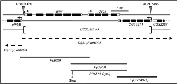
Figure 5: Genomic tools used for analysis of CycJ... 22
Figure 6: The Df(3L)armi-J chromosome contains a 2,100 bp transposon remnant left
behind from transposon-mediated deletion ... 29
Figure 7: Diagnostic Df(3L)armi-J PCR using cDNA as template verifies that transcripts
of the three genes are absent in the deletion ... 31
Figure 8: A deficiency removing eIF5B (Df(3L)Exel6094) is fully complemented by both
Df(3L)armi-J and a deficiency also removing these three genes
(Df(3L)Exel6095) ... 34
Figure 9: Deletion of both armi and CycJ results in accumulation of egg chambers with
excess germline cells... 36
Figure 10: Multiple cystoblasts divide and differentiate within a single egg chamber in
armi-CycJ mutants... 38
Figure 11: CycJ genetically interacts with armi, piwi,and aub... 40
Figure 12: CycJ expression suppresses mispackaged egg chamber production in
armi-CycJ, piwi-armi-CycJ, and aub-CycJ... 41
Figure 13: Expression of CycJ in germline and somatic cells in an armi-CycJ null
background resulted in a significant reduction of egg chambers with too many germline cells... 43
Figure 14: Cystoblast division, differentiation, and packaging are relatively normal in
CycJ null, armi null, and aub mutants, but not in aub-CycJ double mutants, which often have multiple cysts in one egg chamber ... 45
Figure 15: Production of a new chromosome 3 containing Df(3L)armi-J and
p{HZ14-CycJ, mW}72-12 using meiotic recombination ... 63
Figure 16: Mating scheme for meiotic recombination used to produce a new "armi null" chromosome 3... 64
Figure 17: CycJ cooperates with somatic armi to promote egg chamber packaging .... 66
Figure 18: Somatic armi and piwi along with CycJ limit accumulation of GSC-like cells
... 69-70
Figure 19: Somatic armi and piwi along with CycJ permit egg chamber packaging by
limiting BMP signaling ... 72-73
Figure 20: CycJ and the somatic piRNA pathway cooperate to limit BMP signaling 75-76
Figure 21: Bam expression is reduced in mutants of piwi, armi, piwi-CycJ, and armi-CycJ... 77-78
Figure 22: Engrailed expression in terminal filament and cap cells is not altered by
CycJ, armi, piwi, or the dpp allele... 80
Figure 23: CycJ promotes proliferation of follicle cells in ovarioles by limiting BMP
signaling in the absence of armi... 82-83
Figure 24: Somatic expression of CycJ increases follicle cell proliferation in the absence
of armi... 84
Figure 25: Model – CycJ cooperates with the somatic piRNA pathway to limit GSC accumulation ... 94
Figure 26: Double RNAi in the germline resulted in an increase of egg chambers with too many germline cells... 101
Figure 27: CycJ and the somatic piRNA pathway cooperate to limit Notch signaling . 105
Figure 28: Mutant cell populations can be produced in mitotically active cells of the ovary using FLP/FRT recombination ... 106
CHAPTER 1: INTRODUCTION 1.1 Summary
My goal for this project was to determine and characterize a role for Cyclin J (CycJ) in oogenesis. CycJ is conserved in all metazoans and is homologous to well-characterized cell cycle regulators, but its function is unknown. In Drosophila, it is expressed exclusively in ovaries and early embryos, suggesting a possible role in one or both of these tissues. Initial analyses in Drosophila revealed that CycJ might regulate embryogenesis, but is dispensable during oogenesis under normal conditions (Kolonin and Finley 2000; Althoff, Viktorinova et al. 2009; Atikukke 2009). Further
characterization showed that CycJ genetically interacted with two piRNA pathway
members, armitage (armi) and aubergine (aub); a CycJ null enhanced the oogenesis
defects of armi or aub single mutants (Atikukke 2009). I extended these analyses by
demonstrating a genetic interaction between CycJ and a third piRNA pathway member,
piwi, as well as quantifying the resulting oogenesis defects of all CycJ-piRNA pathway double and single mutants. The key findings are that piwi and armi are required for
packaging of germline cells into egg chambers and CycJ contributes to this process as
discussed in Chapter 2 (Atikukke, Albosta et al. 2014). I went on to demonstrate that
piwi and armi function from somatic cells to regulate bone morphogenetic protein (BMP)
signaling and the number of germline stem cells (GSCs), and that CycJ interacts with
this somatic piRNA pathway as described in Chapter 3 [Albosta 2015 in prep]. These data suggest that the piRNA pathway in ovarian somatic cells controls GSCs via BMP
signaling and this leads to a requirement for CycJ limiting these excess GSCs,
1.2 Cyclins are highly conserved serine/threonine kinase regulators
Cyclins are a superfamily of highly conserved eukaryotic regulatory proteins. The defining feature of cyclins is a cyclin box, which is a domain that interacts with cyclin-dependent kinases (Cdks) (Hadwiger, Wittenberg et al. 1989; Jeffrey, Russo et al. 1995). Cyclin binding is required for the activation of kinase activity and contributes to the substrate specificity of Cdks (Kobayashi, Stewart et al. 1992; Lees and Harlow 1993; Horton and Templeton 1997). The founding member of this protein superfamily, Cyclin B (CycB), was originally identified as a protein that oscillated with the cell division cycles of sea urchin embryos and was aptly named Cyclin (Evans, Rosenthal et al. 1983). This protein was later identified as CycB after cloning and sequencing, and was
found to be the only sea urchin mRNA capable of inducing frog (Xenopus) oocyte
maturation upon injection, suggesting a conserved specific role for this molecule in
driving the cell cycle (Pines and Hunt 1987). Further studies in Xenopus demonstrated
that CycB was required for transition from the Gap 2 cell cycle phase to Mitosis (i.e., the
G2M transition) (Minshull, Blow et al. 1989). Finally, CycB destruction was shown to
be required for the exit of mitosis (Murray, Solomon et al. 1989). CycB is a conserved
cell cycle regulator in most eukaryotic organisms including unicellular Dictyostelium
(Cao, Chen et al. 2014). In metazoans, cyclins are named with letters and multiple cyclins in addition to Cyclin B (e.g., Cyclin A, D, E) are known to have conserved roles in regulating the cell cycle by activating Cdks and phosphorylating specific substrates that are required for cell cycle transitions (reviewed in (Hochegger, Takeda et al. 2008)). Other cyclins (e.g., Cyclin C, H, T) have conserved roles in regulating Cdks involved in transcription or other cellular processes, and some have multiple functions, like Cyclins
D and E that function in both cell cycle and transcription regulation (reviewed in (Lim
and Kaldis 2013)). In Drosophila, there are 16 highly conserved unique cyclins and only
a few of them, including CycY and CycJ, have poorly or uncharacterized functions. 1.3 Cyclin J is a poorly characterized and highly conserved putative cell cycle regulator specifically expressed in Drosophila ovaries
CycJ is a poorly characterized highly conserved cyclin specifically expressed in
ovaries and early embryos. It was identified originally in Drosophila as a Cdk interacting
protein in yeast-two-hybrid screens (Finley and Brent 1994; Finley, Thomas et al. 1996).
CycJ has been shown to physically interact with both Cdk1 and Cdk2 in vivo, and the
kinase activity of CycJ immunoprecipitates has been verified in vitro with an H1 kinase
assay (Finley, Thomas et al. 1996; Kolonin and Finley 2000; Althoff, Viktorinova et al. 2009). Based on amino acid sequence, CycJ is more closely related to the A and B type cyclins, which are known cell cycle regulators, but a definitive role for CycJ in regulating the cell cycle has not been established. Sequence alignment of CycJ amino acid
sequences reveals high conservation between Drosophila, human, mouse, and frog,
indicating that CycJ likely has an important function (Atikukke 2009). In Drosophila,
CycJ mRNA is present in ovaries and very early embryos suggesting a function in one or both of these tissues (Finley, Thomas et al. 1996; Kolonin and Finley 2000). Two
studies looked for a potential role for CycJin regulating the Drosophila early embryo cell
cycles, but came to different conclusions. In one study, injection of CycJ antibodies or peptide aptamers into early embryos caused chromatin bridges during the rapid nuclear divisions of the early syncytial embryo suggesting that CycJ is required for cell cycle progression and chromosome segregation (Kolonin and Finley 2000). Contrary to these
results, the Lehner lab showed that in early embryos, CycJ-EGFP fusion proteins were not expressed and the syncytial cell cycles in early embryos of CycJ null were not
disrupted (Althoff, Viktorinova et al. 2009). Both groups agree that CycJ null exhibited
reduced egg-hatching rates compared to controls (Althoff, Viktorinova et al. 2009; Atikukke 2009). Considering that the CycJ null embryos that fail to hatch are fragile, making them difficult to isolate and examine, it is possible that the Lehner group only examined the good embryos rather than those that were defective. Despite the differing results, these studies suggest that CycJ is conserved and specifically expressed in
ovaries and/or early embryos of Drosophila, but its function remains elusive.
The expression of Drosophila CycJ is unique and somewhat conserved between
organisms. Figure 1 shows mRNA expression from all 16 Drosophila cyclins across 55
stages and tissues from two high-throughput sources, modENCODE and Fly Atlas (Arbeitman, Furlong et al. 2002; Graveley, Brooks et al. 2011; Chintapalli, Wang et al.
2013), demonstrating that CycJ is specifically expressed in ovaries and early embryos,
a pattern that is unique among the cyclins and suggests a possible function in one or both of these tissues. The unique mRNA expression pattern is also conserved in the
mosquito (Aedes aegypti) ortholog of CycJ (Cyclin A3 in mosquito, AAEL008256), which
is only expressed in early embryos and ovaries after a blood meal when oogenesis becomes active (Akbari, Antoshechkin et al. 2013). In Drosophila, CycJ is also in ovaries and early embryos, and also demonstrates that it is up-regulated in ovaries when germline stem cell (GSC) number is increased by mutations that inhibit
Figure 1: CycJ is expressed almost exclusively in Drosophila ovaries, a pattern that is unique among the superfamily of cyclins. This is a heatmap of cyclin
expression in 55 stages and tissues of Drosophila as a percentage of each gene’s
maximal expression (% max). Expression data were acquired from two high throughput
repositories, FlyAtlas for tissue expression (Chintapalli, Wang et al. 2013) and modENCODE for stage expression (Graveley, Brooks et al. 2011). Raw values were obtained from each source, the highest value for each gene was determined, and that gene’s expression was scaled as a percent of this maximal value as described (Murali, Pacifico et al. 2014). Percent max was determined for FlyAtlas and modENCODE data
independently from one another. CycJ is maximally expressed only in early 0-2 hour old
embryos (column 1) and ovaries (column 2), and unlike all other cyclins, it is not expressed above ~20% of its maximum at any other time or in any other tissue. Columns labeled 3 and 4 represent adult females 5 and 30 days post eclosion (dpe),
respectively, where CycJ is expressed above 50% max. Since all parts of the fly have
been dissected, analyzed individually, and most demonstrate little to no CycJ
expression, this increase in adult females is due to high ovary expression. ND = no data for Koko. This figure has been published in (Atikukke, Albosta et al. 2014) and is
normally detected in testis, but is expressed in testis of benign gonial cell neoplasm (bgcn) mutants and bgcn mutants over-expressing the Jak/STAT ligand outstretched, which inhibits germline differentiation and promotes accumulation of GSCs, respectively (Terry, Tulina et al. 2006). These data suggest that CycJ is expressed in stem cell populations and expression can be induced when stem cell populations are expanded. Interestingly, the expression pattern of human CycJ (CCNJ) is also associated with stem cells. Meta analysis of genome-wide expression studies found in the NCBI GEO
database demonstrate that CCNJ is expressed at low levels in many tissues, including
oocytes and early embryos, as well as embryonic stem cells. Strikingly, CCNJ is
expressed in induced pluripotent stem cells from multiple sources, whereas expression is significantly lower in the cells of origin (Ohi, Qin et al. 2011). Together, these data
demonstrate conserved expression of CycJ in oocyte, early embryo, and stem cells,
both endogenous and induced. These unique conserved expression profiles suggest that CycJ may play a role in oogenesis, early embryogenesis, and/or stem cell regulation.
Although virtually nothing is known about CycJ function in any organism, multiple
studies suggest that aberrant CycJ regulation may be associated with human cancers.
Human CCNJ is up-regulated in three cancers, and one study suggests that it may
contribute to the cell cycle under tumorous conditions. In one study, patients with high-risk B-precursor acute lymphoblastic leukemia that had high expression of a five-gene
cluster, which included CCNJ, exhibited a superior outcome and higher rates of 4-year
relapse-free survival, but they did no further validation or characterization of CCNJ in
down-regulated by two separate miRNAs, which are inhibited in two other human cancers. In prostate cancer cells, Vitamin D promoted expression of miR-98, which down-regulated
CCNJ and inhibited cell growth via G2/M arrest, but the causal relationship between
CCNJ down-regulation and G2/M arrest was not investigated any further (Ting, Messing
et al. 2013). Finally, miR-125b, which down-regulated CCNJ, was found to be
decreased in a miRNA expression profile of 50 breast cancer patients (Feliciano, Castellvi et al. 2013). Further analyses confirmed that CCNJ was over-expressed in breast cancer patients and forced expression of miR-125b in MCF7 breast adenocarcinoma cultured cells resulted in a G2/M arrest. They also demonstrated that
siRNA knockdown of CCNJ in MCF7 cells resulted in a G2/M arrest similar to that seen
under forced expression of miR-125b. Two of these studies suggest that CCNJ may be a G2/M regulatory cyclin in the cultured tumor cells analyzed, suggesting that its function as a cell cycle regulator is revealed in cells with stem cell-like qualities. Nevertheless, more detailed analyses are required to understand the function of this conserved cyclin.
Surprisingly, in Drosophila a CycJ null displays relatively normal oogenesis
(Althoff, Viktorinova et al. 2009; Atikukke 2009). Despite this fact, we have recently
shown that CycJ regulates oogenesis in the absence of the piRNA pathways (discussed
in detail in Chapters 2 and 3 (Atikukke 2009; Atikukke, Albosta et al. 2014)), which
suggests that CycJ functions in this tissue under specific conditions. We propose a
non-redundant function for CycJ in oogenesis based on our data showing that CycJ is
prevent severe oogenesis defects; i.e., CycJ is responding to some condition created by piRNA pathway inhibition (Atikukke 2009).
1.4 Oogenesis: The making of an oocyte
Oogenesis is a process that starts from a stem cell and results in production of a
gamete known as an oocyte. In Drosophila this takes place in ovaries composed of a
series of parallel tubular structures called ovarioles. Oogenesis begins with the asymmetric mitotic division of a germline stem cell (GSC) located at the anterior tip of a germarium, giving rise to a new stem cell and a daughter cell called a cystoblast (Schupbach, Wieschaus et al. 1978; Wieschaus and Szabad 1979; Spradling 1993) (Figure 2 A). The new stem cell remains in the stem cell niche at the anterior tip of the germarium where signaling (e.g., Bone Morphogenetic Protein (BMP) signaling) from neighboring somatic terminal filament and cap cells leads to repression of differentiation factors in the germline (e.g., Bag of Marbles (Bam)) (Xie and Spradling 1998; King and Lin 1999; King, Szakmary et al. 2001; Song, Smith et al. 2004) (Figure 3). The cystoblast is positioned posteriorly away from the GSC niche, where it is free from BMP signaling thereby allowing differentiation. The cystoblast differentiates by undergoing exactly four rounds of division with incomplete cytokinesis giving rise to 16 cells interconnected by structural cell-cell connections known as fusomes and ring canals. The fusome originates from a spherical spectrin-containing structure called a spectrosome found in GSCs and cystoblasts. The spectrosome is required for the formation of ring canals, which maintain the permanent connection between the cells of a cyst (Lin, Yue et al. 1994; Huynh 2000). The 16-cell cyst eventually reaches the middle of the germarium where it comes in contact with somatic stem cells (SSCs),
Figure 2: Drosophila oogenesis. (A) Drosophila germarium (GE). Oogenesis begins in the GE, which functions to produce egg chambers (ECs). The process beginnings with germline stem cells (GSCs) that reside in a well-defined niche composed of terminal filament (TF) and cap cells. GSCs produce daughter cells called cystoblasts. GSCs and cystoblasts each contain a characteristic spherical spectrosome. The cystoblast becomes an interconnected 16-cell cyst, which initially contains a fusome (to establish long term cell-cell connectivity) created by transmission and elongation of the spectrosome. One of the 16 cells will become the oocyte and the other 15 become nurse cells, which undergo extra rounds of DNA replication without cell division resulting in nuclei that get larger with time. Cysts are surrounded by follicle cells from somatic stem cells (SSCs), resulting in an egg chamber. (B) The GE continuously produces egg chambers, which grow and mature into stage 14 oocytes, which contains characteristic dorsal appendages. The GE, ECs, and stage 14 oocyte all constitute a single ovariole. This figure has been modified from (Atikukke, Albosta et al. 2014).
which produce follicle cells that encapsulate the cysts to form egg chambers (Goode, Wright et al. 1992; de Cuevas, Lilly et al. 1997; Nystul and Spradling 2010). One of the original 16 germline cells undergoes meiosis and becomes the oocyte, while the other 15 undergo multiple cycles of DNA replication without cell division (i.e., endocycles) to become polyploid nurse cells that eventually donate their cytoplasm to the oocyte through the ring canals. Fully formed egg chambers are continuously produced, bud off from the germarium, and populate ovarioles, creating long chains of egg chambers that mature and increase in size, culminating in the formation of a mature stage 14 oocyte (Figure 2 B).
Drosophila oogenesis is a well-characterized model for stem cell biology including the supporting cells that constitute a stem cell niche. Oogenesis begins with GSCs in a well-defined somatic niche, and there are many studies characterizing the pathways that regulate maintenance of these cells. GSC maintenance is partly achieved by localized inhibition of differentiation facilitated by BMP signaling (Figure 3) (Song, Wong et al. 2004; Zhang and Li 2005). Production, secretion, and diffusion of the BMP ligands glass bottom boat (Gbb) and Decapentaplegic (Dpp) from somatic niche cells is restricted so they only bind to GSCs and activate the BMP signaling pathway. For example, the number and maintenance of somatic niche cells is regulated by Notch signaling between the soma and germline (Song, Call et al. 2007). The niche cells express engrailed (en), which is required for the production and secretion of the morphogen Hedgehog (Hh) that signals to adjacent niche cells and promotes the production of Dpp (Rojas-Rios, Guerrero et al. 2012). Furthermore, expression of the glypican Dally in cap cells promotes localization of Dpp at GSCs,
Figure 3: BMP signaling represses GSC differentiation. The primary function of the GSC niche is to produce signals that promote stem cell self-renewal mainly by inhibiting differentiation. This is partly achieved with bone morphogenetic protein (BMP) signaling. Ligand, e.g. decapentaplegic (Dpp), is produced and secreted from niche cells, followed by binding to GSCs and initiation of the BMP signal transduction pathway. This signaling begins with phosphorylation of receptors and substrates including phosphorylation of the transcription factor Mothers against Dpp (Mad). pMad binds to the co-mediator protein Medea (Med) and the protein pair translocates to the nucleus where they inhibit the transcription of the differentiation factor Bag of Marbles (Bam), thereby promoting self-renewal. Daughter cells, also known as cystoblasts, migrate away from the niche and its stem cell maintenance signaling, where they can then produce the differentiation factor Bam.
whereas ectopic expression of Dally leads to expansion of Dpp diffusion and excessive inhibition of differentiation (Guo and Wang 2009). With the help of these other pathways, Dpp is produced in niche cells, secreted, and targeted to GSCs (Figure 3). Upon ligand binding, GSCs activate the BMP signaling pathway, which initiates a series of intracellular signaling events, namely phosphorylation of receptors and pathway components (Miyazono, Maeda et al. 2005). One such phosphorylation occurs on the transcription factor Mothers Against Decapentaplegic (Mad). Phosphorylated Mad (pMad) then binds to the co-mediator protein Medea (Med) and the protein pair
translocates to the nucleus. This protein pair can bind to regulatory DNA 5’ of Bam and
inhibit its transcription, which inhibits differentiation in GSCs (Song, Wong et al. 2004; Slaidina and Lehmann 2014). It is not surprising that mutations resulting in increased BMP signaling (e.g., over-expression of dpp or dally) also result in an increase of undifferentiated GSC-like cells at the anterior of the germarium; although they are no longer restricted to the niche, these cells are like GSCs in that they contain a spherical spectrosome and do not express differentiation factors like Bam. This can lead to egg chambers packaged with too many germline cells (Xie and Spradling 1998; Muzzopappa and Wappner 2005). Together, these data illustrate regulation of GSCs by somatic niche cells and show that disruption of pathways that control GSCs can lead to disruption of other oogenesis processes, like egg chamber packaging.
1.5 The piRNA pathways are conserved protectors of germline genome integrity and regulators of oogenesis
The piRNA pathways are also required during oogenesis for maintenance of germline genome integrity from both germline and ovarian somatic cells (Figure 4).
Figure 4: The piRNA pathways are conserved RNA silencing mechanisms that
function to maintain and regulate germline development. The pathway proteins do
this by repressing transposons with the help of small RNAs, called piRNAs, which are complementary to transposon transcripts. Two key classes of proteins found in these pathways are depicted. The argonaute class, including Piwi, Argonaute 3 (Ago3) and Aubergine (Aub), which bind the piRNAs and use them for guided transposon silencing, and the putative RNA helicase Armi, which is in a complex with Piwi functioning to process piRNAs and load them onto Piwi. There are two distinct silencing mechanisms that this pathway uses. The first targets transposon transcripts for degradation facilitated by transcript degradation. In the germline, Aub participates in a piRNA recycling process known as ping-pong amplification used to possibly amplify the amount of piRNAs available for transposon silencing. The pathway can also function in the nucleus targeting transposon producing loci and causing epigenetic modification, which silences transcription of these loci. Out of these three proteins, only piwi has been shown to regulate epigenetic modification. The pathway must function in both the somatic cells and the germline cells with the common goal of protecting germline genome integrity. It is believed that the somatic piRNA pathway does this by preventing the transmission of virus-like transposons from the somatic cells to the germline (Song, Kurkulos et al. 1997; Malone, Brennecke et al. 2009).
They are conserved transposon RNA silencing mechanisms that produce small 24-30nt RNAs called piRNAs, which are loaded onto PIWI clade Argonaute proteins (Ago3, Aub,
and Piwi in Drosophila). The piRNAs are used for complementary base pair recognition
of transposon-producing loci or transposon transcripts followed by transcriptional or posttranscriptional silencing of the targeted transposon (Vagin, Sigova et al. 2006; Klattenhoff and Theurkauf 2008) (Figure 4). The pathways require many families of proteins including the Argonaute proteins mentioned above, RNA helicases (e.g., Armitage (Armi)), Tudor proteins (e.g., fs(1)Yb (Yb)), nucleases (e.g., Zucchini (Zuc)), and many others (Olivieri, Sykora et al. 2010). Small RNA pathways, like the piRNA pathway, that inhibit transposon mobilization are conserved from plants and fungi to humans (Aravin, Hannon et al. 2007). In fact, humans and other vertebrates like mouse
share with Drosophila many common pathway proteins (including PIWI clade Argonaute
proteins) and mechanisms. The Argonaute proteins of the piRNA pathways silence transposons by targeted transcript cleavage via endonuclease activity (Ago3 and Aub), or by epigenetic silencing of transposon-producing loci in the genome via recruitment of epigenetic modification proteins (Piwi) (Gunawardane, Saito et al. 2007; Yin and Lin 2007; Malone, Brennecke et al. 2009; Rangan, Malone et al. 2011). The proper silencing of transposons is thought to limit DNA damage in the germline by repressing the movement of these selfish genetic elements (Klattenhoff, Bratu et al. 2007).
Theurkauf and colleagues have shown that mutants of armi or aub accumulate germline
DNA double stranded breaks and this leads to DNA damage checkpoint activation, which in turn disrupts establishment of the anterior-posterior and dorsal-ventral patterning of developing oocytes. piRNA pathways must function in both germline cells
and associated somatic cells with the common goal of maintaining germline genome integrity (Malone, Brennecke et al. 2009; Zamore 2010). Aub and Ago3 are germline specific and function in a cycle that produces piRNAs from genomic clusters transcribed from both strands (dual-strand clusters) in a process known as ping-pong amplification (Li, Vagin et al. 2009; Malone, Brennecke et al. 2009). Antisense piRNAs associated with Aub target transposon transcripts for degradation facilitated by Aub endonuclease activity.
Piwi, on the other hand, functions in both somatic and germline cells in pathways distinct from the germline specific ping-pong amplification. In somatic cells, a protein complex containing Piwi, Armi, Zuc, Vreteno (Vret), and Yb functions in perinuclear Yb bodies to produce antisense piRNAs from genomic clusters transcribed from a single strand (uni-strand clusters) (Olivieri, Sykora et al. 2010; Murota, Ishizu et al. 2014). piRNA/Piwi complexes enter the nucleus and associate with transposon loci, possibly by complementary base pair recognition of nascent transposon transcripts or recognition of DNA at transposon loci. Once in position, Piwi recruits epigenetic modifying proteins resulting in silencing of these loci. Unlike Ago3 and Aub, Piwi silences transposons without cleaving transposon transcripts. Yb is a Tudor-domain containing protein that acts as a scaffold to localize piRNA pathway proteins and piRNA cluster transcripts for piRNA processing and RNA induced silencing complex (RISC) assembly (Murota, Ishizu et al. 2014). Vret is also a Tudor-domain containing protein that specifically interacts with and stabilizes Piwi (Zamparini, Davis et al. 2011). Zuc has endonuclease activity and has been implicated in processing and maturation of 5’ piRNA ends, which are loaded onto Piwi (Nishimasu, Ishizu et al. 2012). Finally, Armi is
an RNA helicase required for loading of piRNAs onto Piwi (i.e., RISC assembly), which is a prerequisite of nuclear localization of piRNA/Piwi complexes (Murota, Ishizu et al. 2014). Piwi and Armi also facilitate production of piRNAs in the perinuclear nuage of germline cells through similar mechanisms, though the germline cells lack Yb and Yb bodies. These data suggest that Armi and Piwi may have unique functions in somatic cells compared to germline cells, even though they are involved in piRNA production and transposon silencing in both cell types.
In Drosophila, the piRNA pathway members Yb, Vret, and Piwi limit GSC accumulation and promote egg chamber packaging and several lines of evidence indicate that this is a somatic-specific function of the piRNA pathway. First, Yb is only
expressed in the soma and homozygous Yb72 ovaries exhibit GSC accumulation and
production of egg chambers with too many germline cells (i.e., mispackaged egg
chambers) (Swan, Hijal et al. 2001). Second, homozygous vret148-60 ovaries also
over-accumulate GSCs and produce egg chambers with abnormal nurse cell numbers, and
these defects can be rescued by expression of wild-type vret specifically in somatic cells
(Zamparini, Davis et al. 2011). Furthermore, germline vret148-60 clones do not exhibit
GSC accumulation or loss, indicating that limiting GSCs is a somatic-specific function of
Vret (Zamparini, Davis et al. 2011). Third, multiple piwi mutants and RNAi knockdowns
of piwi specifically in somatic cells result in GSC accumulation, which can be rescued with somatic expression of wild-type piwi (Jin, Flynt et al. 2013). In contrast, germline
knockdown of piwi does not result in extra GSCs (Ma, Wang et al. 2014). The excess
GSCs in these Piwi studies also exhibited BMP signaling activity and somatic escort
by limiting BMP signaling. The piwiNT mutant, which lacks nuclear localization, is not
able to repress transposons, yet exhibits normal GSC regulation, suggesting that Piwi’s role in GSC regulation is independent of transposon silencing (Klenov, Sokolova et al. 2011). These data suggest that multiple piRNA pathway members limit GSC accumulation, promote egg chamber packaging, and/or limit BMP signaling from ovarian somatic cells. These proteins physically associate with one another, as well as with other piRNA pathway members, including Armi (Haase, Fenoglio et al. 2010; Qi,
Watanabe et al. 2011). Germline knockdown and germline-specific mutants of armi
were found to have normal GSC numbers, but the role of Armi regulating GSCs from somatic cells has not been examined (Ma, Wang et al. 2014). These studies suggest a potential common role for piRNA pathway proteins in ovarian somatic cells regulating oogenesis possibly independent of transposon regulation, but a more comprehensive analysis is required.
1.6 Overview
I used a previously generated deletion of the genomic region containing armi and
CycJ, as well as null mutants for each gene created by adding back individual
transgenes for each gene to the deletion. In Chapter 2, I show that CycJ regulates egg laying and hatching, but appears to be dispensable during oogenesis under normal conditions. In the absence of the piRNA pathway, however, CycJ plays a role in regulating egg chamber packaging and maturation. Most of the data in Chapter 2 have been published (Atikukke, Albosta et al. 2014). In Chapter 3, I demonstrate that the somatic piRNA pathway regulates multiple aspects of oogenesis by limiting BMP
signaling and that CycJ enhances these functions. Finally, Chapter 4 contains conclusions and future directions.
CHAPTER 2: A ROLE FOR DROSOPHILA CYCLIN J IN OOGENESIS REVEALED BY GENETIC INTERACTIONS WITH THE piRNA PATHWAYS
Part of the work described in this chapter has been published in Mechanisms of
Development 133: 64-76, 2014. 2.1 Introduction
Cyclin J (CycJ) is a poorly characterized member of the cyclin superfamily of proteins specifically expressed in ovaries and early embryos. Cyclins are eukaryotic proteins that contain a cyclin box, a domain that interacts with cyclin-dependent kinases (Cdks) (Hadwiger, Wittenberg et al. 1989; Jeffrey, Russo et al. 1995). Many cyclins are known to have conserved roles in regulating the cell cycle. In metazoan species from
Drosophila to human, for example, A and B cyclins regulate mitotic events, D cyclins regulate progression through G1, and E cyclins regulate entry into S phase (Minshull, Blow et al. 1989; Murray 2004). Other cyclins have conserved roles in regulating transcription or other cellular processes (Lim and Kaldis 2013). CycJ is conserved in all
metazoans, yet it has only been studied in Drosophila were it was originally identified as
a Cdk-interacting protein (Finley and Brent 1994; Finley, Thomas et al. 1996). The RNA
expression pattern of CycJ is unique among Drosophila cyclins and suggests a possible
role in oogenesis or embryogenesis. CycJ mRNA is present almost exclusively in
ovaries and early embryos, whereas all other cyclins are expressed in multiple tissues and stages of development (Figure 1) (Finley, Thomas et al. 1996; Arbeitman, Furlong et al. 2002; Graveley, Brooks et al. 2011; Chintapalli, Wang et al. 2013). A potential role for CycJ in embryogenesis was suggested in a study showing that injection of syncytial embryos with CycJ-inhibitory antibodies or peptide aptamers resulted in delays of the early nuclear division cycles (Kolonin and Finley 2000). I set out to determine whether
CycJ also plays a role in ovaries where both the RNA and protein appear to be maximally expressed.
Here, I set out to determine whether or not CycJ plays a role in oogenesis. I
used a previously generated deletion of the genomic region containing armi and CycJ,
as well as null mutants for each gene created by adding back individual transgenes for each gene to the deletion (Atikukke 2009). We show that while oogenesis is normal in
the CycJ null, the armi null produces few egg chambers and mature eggs, all of which
have axis specification defects. Surprisingly, in the armi-CycJ double null there was a
further decrease in the number of egg chambers per ovariole, a drastic increase in the number of differentiated germline cells in each egg chamber, and no mature eggs. The
armi null defects could be suppressed by mutation in the Chk2 checkpoint kinase gene
as shown previously, but the armi-CycJ double null defects could not (Atikukke 2009). I
observed a similar genetic interaction between CycJ and two other piRNA pathway
genes, piwi and aub, suggesting that CycJ plays a nonredundant role in oogenesis when the piRNA pathways are compromised.
2.2 Materials and Methods 2.2.1 Drosophila strains
Flies were maintained and crosses conducted at 25°C unless heat shock (37°C)
is indicated. Many stocks were used in a previous project from our lab (Atikukke 2009).
w1118 is wild type for this study. armi72.1 (Cook, Koppetsch et al. 2004) was obtained
from William E. Theurkauf. mnkP6 (Takada, Kelkar et al. 2003; Brodsky, Weinert et al.
2004) was obtained from Andrew Swan. Stocks for w1118, P{hsFLP}1, y1 w1118;
and Wieschaus 1991), eIF5B09143, VP16::nos-Gal4, Df(3L)Exel6064, Df(3L)Exel6065,
balancer strains, y[1] M{vas-int.Dm}ZH-2A w[*]; M{3xP3-RFP.attP'}ZH-51C, P{ry[+t7.2]=PZ}piwi[06843] cn[1]/CyO; ry[506], and w[1118] ; Df(2L)BSC145/CyO were
obtained from the Bloomington Drosophila Stock Center. We obtained P1 clone
DS01105 and BAC clone B22N9 from the Berkeley Drosophila Genome Project
(BDGP) resource center. All flies are listed in Appendix A.
2.2.2 Creation of Df(3L)armi-J and transgenic constructs for rescue experiments
A genomic deletion, named the Df(3L)armi-J, was created removing CycJ and
two adjacent genes, armi and CG14971 as previously described (Atikukke 2009)
(Figure 5). Briefly, the region was deleted by inducing recombination between two FRT-bearing transposon insertions flanking the genomic region. Creation of genomic transgenes for each of the genes in the three-gene deletion was also previously described (Atikukke 2009). New transgene construction for this study includes
pCaSpeR-HZ14-CycJ constructed by cutting pCaSpeR-CycJ with SpeI, which uniquely
digests in armi exon 8, filling in with Klenow (New England Biolabs), and religating. This
introduced a frame shift 12 codons into the armi coding region in exon 8. High fidelity
polymerase, (Herculase, Stratagene Inc.) was used for all PCR and the resultant
constructs were verified by DNA sequencing. Constructs were microinjected into w1118
Drosophila embryos to induce P-element mediated germline transformation as described (Rubin and Spradling 1982). Injections were performed by the Model System Genomics facility at Duke University. Individual progeny bearing the transgenes as identified by eye color and verified by PCR were selected and mated with flies carrying
Figure 5: Genomic tools used for analysis of CycJ. Schematic representation of the
genomic region corresponding to CycJ, armi, and the neighboring genes on
chromosome 3L. The region shown corresponds to estimated cytological band 63E1. Black boxes represent coding regions and open boxes represent 5’ and 3’ untranslated regions. armi and CycJ are transcribed left to right while all other genes shown are
transcribed right to left (arrows). FRT-bearing transposon insertions RBe00161 and
XPd07385 (shown by triangles) were used to delete the intervening genomic region.
The deleted genomic region is represented as Df(3L)armi-J. Rescue experiments were
conducted by using independent transgenes, P{armi}, P{CycJ}, and P{CG14971} that
were generated using the indicated genomic regions (shaded boxes). Rescue
experiments also used a second CycJ genomic transgene, P{HZ14CycJ}, which
introduced stop codons (# stop) in the armi coding region eliminating armi coding
potential. Regions missing in the deficiency chromosomes Df(3L)Exel6094 and
Df(3L)Exel6095 are indicated by dashed lines and extend beyond the region shown (arrowheads). This figure has been previously published in (Atikukke 2009; Atikukke, Albosta et al. 2014) and is represented here with modifications.
marked balancer chromosomes to obtain balanced stocks. Chromosomes containing
the transgenes are referred to as p{CycJ}, p{HZ14CycJ}, p{armi}, and p{CG14971}. To
express the CycJ open reading frame (ORF) from a UAS we used pHZ12 (Mairiang,
Zhang et al. 2013), a vector derived from pUASattB (Bischof, Maeda et al. 2007) that
contains an attB site, the mini-white gene, and a UAS driving expression of ORFs with
an N-terminal 6His-3Myc tag. The CycJ ORF was amplified from a yeast two-hybrid
clone (Stanyon, Liu et al. 2004) using recombination tag primers 5RT and 3RT and recombined into the 5RT and 3RT sites of vector pHZ12 as described (Parrish,
Limjindaporn et al. 2004) to create pUAS-Myc-CycJ. The CycJ ORF in pUAS-Myc-CycJ
was verified by sequencing and includes from the ATG to the stop codon of Cyclin J
isoform A. pUAS-Myc-CycJ was inserted into the attP site at 51C in Drosophila line y[1]
M{vas-int.Dm}ZH-2A w[*]; M{3xP3-RFP.attP'}ZH-51C (Bischof, Maeda et al. 2007). All fly lines are listed in Appendix A.
2.2.3 Ovary dissection and staining
Ovary dissection and staining was conducted as previously described (Atikukke 2009). Briefly, ovaries were removed in a ringer solution on ice, fixed with a formaldehyde solution, washed with PBS followed by PBT. Ovaries were incubated in primary antibody for either 2 hours at room temp or overnight at 4°C. They were
washed in PBT and incubated in secondary antibody at room temperature for 1 to 2 hours. They were washed with PBT, counterstained with DAPI, washed with PBT followed by PBS, equilibrated with PBS:Glycerol (1:1), and finally in antifade mounting solution (PBS:Glycerol 1:4 containing DABCO). Primary antibodies from Developmental
6H4 and 4H8 (1:50). Primary anti-GFP (1:50) was obtained Invitrogen. Secondary antibodies from Sigma and dilutions include Goat anti-mouse IgG FITC (1:200) and Goat anti-rabbit IgG Texas Red (1:200). Nuclei of all ovaries were counterstained with
DAPI (1µg/ml) from Sigma. Microscopy was conducted with a Zeiss Axio imager upright
microscope that had an apotome for background reduction and optical sectioning. 2.2.4 Hatch rates
Experiments analyzing hatch rates were conducted as previously described (Atikukke 2009) and with the following modifications. To assess embryo hatching rates and morphology, equal numbers of newly emerged virgin females of each genotype
were mated with w1118males and the eggs were collected on apple juice plates or grape
juice plates every day for 4 days. Plates were 2.2% w/v Agar (USB 10906), 2.5% w/v Sucrose (Domino Table Sugar), 25% v/v apple or grape juice, and 0.075% w/v Tegosept (p-hydroxy-benzoic acid methyl ester, Sigma H-3647). Agar and sugar were mixed with 75% of the total volume of water and autoclaved. Tegosept must first be dissolved in ethanol (21% w/v) prior to addition to liquid agar. Juice was added immediately after autoclaving, solution was cooled to 60°C and Tegosept/alcohol mixture was added. Liquid agar was cooled to roughly 40°C and poured into 60 mm Petri dishes. Prior to use, yeast paste (1 g of yeast in 1.3 mL water) was added in a thin layer on five spots covering 10% of the plate surface. Flies were allowed to lay eggs on plates for 20-24 hours at 25°C and 50% humidity for four days total. Plates were changed each day and the number of eggs laid was counted. Eggs were given at least 24 hours to hatch followed by counting of the eggs that did not hatch. The number of eggs laid vs. eggs that did not hatch was used to calculate hatch rate. The number of
eggs hatched was determined by counting the number of eggs that failed to hatch after
aging for 24 hours at 25oC. Eggs were counted and categorized based on dorsal
appendage morphology. At least 200 eggs were examined for each genotype except
the armi null for which repeated collections provided 50 eggs to examine.
2.2.5 Whole fly DNA extraction, PCR, and agarose gel electrophoresis
DNA was extracted from individual flies for genotyping according to Michael Ashburner’s protocol as previously described with modification (Ashburner 1989; Atikukke 2009). A single fly was collected and added to a 1.7 mL microcentrifuge tube (Denville C2170) containing 50 µL homogenization buffer [Tris-HCl (pH7.5) 10mM, NaCl 60mM, EDTA 10mM, Spermine 0.15mM, Spermidine 0.15mM, Sucrose 5%]. Flies were mechanically homogenized with a small plastic pestle (Fisher nc9907525). After homogenization, 50 µL of lysis buffer [Tris-HCl (pH 9.0) 300mM, EDTA 100mM, SDA 0.63%, Sucrose 5%] was added followed by a 15 minute incubation at 70°C. Tubes were removed from heat and cooled to room temperature, then 15 µL of 8M potassium acetate (KAc) was added followed by a 30 minute incubation on ice. Tubes were centrifuged for 5 minutes at 13,000 RPM. The supernatant was transferred to a new tube and 230 µL of 100% ethanol was added followed by a 5 minute incubation at room temperature. Tubes were centrifuged for 5 minutes at 13,000 RPM. The supernatant was discarded and the pellet was washed with 230 µL of 70% ethanol followed by centrifugation for 5 minutes at 13,000 RPM. The supernatant was discarded and pellets were air dried. The pellet was resuspended in 50 µL of Tris-EDTA [TE, 10 mM Tris pH 6.8: 1 mM EDTA pH 8.0]. DNA was amplified with the polymerase chain reaction (PCR) using either Invitrogen reagents (kit 10342-020) or Promega GoTaq Green (M7122).
Invitrogen reagents were used as follows; 1x PCR buffer, 0.25 µM dNTPs, 2.5 mM
MgCl2, 0.083 µM each primer, 1 U Taq, 1 ng template DNA, and water to 30 µL. GoTaq
was used as follows; 1x GoTaq master mix, 0.2 µM primers, 2 ng template DNA, and water to 10 µL. PCR was performed with a BioRad DNA Engine Tetrad2. Parameters are as follows; 1) Denaturation at 96°C for 5 minutes, 2) Denaturation at 94°C for 30 seconds, 3) Annealing at 60°C for 30 seconds, 4) Extension at 72°C for 1 minute per Kb of product (no less than 1 minute), 5) Repeat cycles two thru four 29 more times, 6) final extension at 72°C for 5 minutes, and 7) cool down to 25°C for 5 minutes. PCR was analyzed on 1% w/v agarose TBE gel at 100 V for roughly 30 minutes.
2.2.6 PCR verification of Df(3L)armi-J
Strains with Df(3L)armi-J (Figure 5) were subjected to genomic PCR and cDNA
amplification for verification of the absence of armi, CycJ, and CG14971. First, genomic
DNA was prepared from Df(3L)armi-J/Df(3L)armi-J and w1118. This DNA was used as
template for a PCR reaction with diagnostic primers specific for armi, CycJ, or
CG14971 designed in adjacent exons resulting in a larger PCR product when
compared to cDNA as template. The primer pairs were: armi=PA02F/PA02R,
CycJ=PA01F/PA01R, and CG14971=PA03F/PA03R. To verify that transcription was
also abolished in the Df(3L)armi-J, cDNA was synthesized from
Df(3L)armi-J/Df(3L)armi-J. RNA was extracted using the RiboPure Kit (Ambion AM1924). A single homozygous fly was homogenized mechanically with a plastic pestle in 0.2 mL TRI reagent and incubated at room temperature for 5 minutes. 0.1 mL of chloroform was added followed by 15 seconds of vortexing and a 5 minute incubation at room temperature. Mixture was centrifuged at 13000 rpm and 4°C for 10 minutes. The top
layer (clear, ~0.15 mL) was transferred to a new tube with 0.075 mL EtOH and vortexed for 5 seconds. The RNA was then purified using the GeneJET RNA Purification Kit (Thermo Scientific K0731) beginning with addition of RNA/EtOH to a filtration column. The column was centrifuged at 13000 rpm for 1 minute and the flow through discarded. The column was washed twice with wash buffer followed by centrifugation. The column was transferred to a 1.5 mL microcentrifuge tube and 25 µL of elution buffer was added followed by incubation at room temperature for 2 minutes. The column was centrifuged at 13000 rpm and 4°C for 1 minute. RNA concentration was quantified using the NanoDrop and an aliquot was diluted with water to 5 µg in a total volume of 21.5 µL. The RNA was then subjected to DNase treatment with 2.5 µL 10x DNase buffer and 1 µL DNase 1 for a total of 25 µL. The mixture was gently finger mixed and incubated at 37°C for 30 minutes. DNase was removed with the addition of 6 µL DNase inactivation reagent and incubation at room temperature for 2 minutes with periodic vortexing. Tubes were centrifuged at 14000 rpm for 1 minute and the supernatant was transferred to a new tube. Single stranded cDNA was synthesized using the Roche Transcriptor First Strand cDNA Synthesis Kit (Roche 04379012001). Reverse Transcription of polyadenylated RNA was performed with the following parameters: 1X RT buffer, 1 mM dNTPs, 2.5 µM oligo dT primers, 20 U Protector RNase Inhibitor, 10 U Reverse Transcriptase, 2.4 µg purified RNA template, and water to 20 µL. Reverse transcription reaction was performed at 50°C for 1 hour followed by 85°C for 5 minutes. cDNA was used as template in a PCR reaction with the same diagnostic primers for the three genes of the deletion used previously with genomic DNA template. These primers are
designed to produce a noticeably smaller PCR product when amplifying cDNA. All primers and their sequences are listed in Appendix B.
2.2.7 Sequence verification of Df(3L)armi-J
The sequence of Df(3L)armi-J was verified by sequencing inward from both
breakpoints with 4 primers. The first set of primers did not generate enough sequence information to cover the transposon remnants left behind from recombination that created the deletion. Once the sequence was known for the first half of the deletion, another set of primers was created and used to complete sequencing. The transposon remnant is ~2,100 bp in length and does not disrupt the flanking genes eIF5B and
CG32267. The first sequencing primers were used to target the left end with RF305 and the right end with RF304. The second primers designed from the first round of sequence data were used to target the left end with PARB1 and the right end with PAXP1. Sequence data was aligned and combined with the Geneious program (Figure 6). All primers and their sequences are listed in Appendix B.
2.2.8 Tissue specific expression with Gal4>UAS
Tissue specific expression of transgenes was accomplished with the Gal4-UAS
binary system of expression (Brand and Perrimon 1993). Gal4 is a yeast transcription
factor that binds to and activates genes containing a regulatory Upstream Activation
Sequence (UAS). The system works by attaching a specific enhancer/promoter to Gal4
and a desired target gene to UAS. A fly containing a promoter-Gal4 construct is mated
to a fly containing a UAS-gene construct. The progeny that contain Gal4 and UAS will
express the target gene in the desired pattern governed by the promoter linked to Gal4.
Figure 6: The Df(3L)armi-J chromosome contains a 2,100 bp transposon remnant left behind from transposon-mediated deletion. Df(3L)armi-J was created with flippase mediated recombination between FRT sites in two different transposon insertions annotated in purple, PBac{RB}e01160 flanking the left of armi and P{XP}d07385 flanking the right of CG14971. Recombination between the transposons left a large DNA remnant, which was sequenced and is represented by the yellow line including an FRT site indicated in red. This image was produced with the Geneious
program using sequencing data from Df(3L)armi-J homozygotes. This indicates that the
deletion successfully removed armi, CycJ, and CG14971 (depicted in their genomic
environment in Figure 5) while leaving the flanking genes eIF5B and CG32267 intact with their ORFs annotated in green. The black lines represent non-coding DNA.
(nos-Gal4) or the soma-specific e22c-Gal4 drivers to express UAS-CycJ.cDNA.myc. All flies are listed in Appendix A.
2.2.9 Oogenesis phenotype quantification
To quantify oogenesis defects, females were aged for 2-4 days post eclosion and ovaries from at least 10 flies per genotype (with two exceptions as noted) were extracted and stained with DAPI as described above. piwi mutant females were only aged for 0-2 days post eclosion due to rapid egg chamber loss. All ovarian material was transferred to a slide and ovarioles were mechanically separated from one another with forceps or insulin syringes for analysis. Number of mature oocytes per fly, number of egg chambers per ovariole, and percent of mispackaged egg chambers per fly were quantified and averaged. Error bars represent standard error of the mean (SEM) calculated by dividing the average by the square root of n (the number of flies). Statistical differences between single and double mutants for each phenotype were calculated using a type 3 student’s T-test, which accounts for unequal sample sizes. 2.3 Results
2.3.1 Loss of both armi and CycJ results in oogenesis defects characterized by severely disorganized ovarioles containing mispackaged egg chambers
CycJ analysis began with a previously described genomic deletion
encompassing armi, CycJ, and CG14971,referred to here as the three-gene deletion or
Df(3L)armi-J, which revealed a possible role for CycJ in oogenesis or embryogenesis (Atikukke 2009). I verified the absence of armi, CycJ, and CG14971 in Df(3L)armi-J
with genomic and cDNA PCR (Figure 7) along with sequencing and mapping of the
Figure 7: Diagnostic Df(3L)armi-J PCR using cDNA as template verifies that transcripts of the three genes are absent in the deletion. CycJ, armi, and CG14971
are not transcribed in Df(3L)armi-J/Df(3L)armi-J. RNA was purified from Df(3L)armi-J
homozygotes and Df(3L)armi-J/+ flies containing a p{CycJ} transgene followed by cDNA
synthesis. The cDNA was used as template for PCR. The absence of bands in homozygotes indicates that mRNA was not produced. Primers used are as follows; CycJ=PA01F/PA01R, armi=PA02F/PA02R, and CG14971=PA03F/PA03R. Primers are their sequences are listed in Appendix B.
mutant flies were viable indicating that armi, CycJ, and CG14971 are not essential for viability or development to adulthood, a conclusion that was also reached in previous studies (Althoff, Viktorinova et al. 2009; Atikukke 2009; Olivieri, Sykora et al. 2010). The deletion, however, resulted in complete male and female sterility, and the females did not lay any eggs (Table 1). As described in detail below, the three-gene deletion resulted in major defects in oogenesis. To analyze the contributions of individual genes to these phenotypes, transgenic lines containing genomic clones of each gene individually and in different combinations were previously constructed and tested for
their ability to modify the Df(3L)armi-J phenotypes (Atikukke 2009; Atikukke, Albosta et
al. 2014) (Figure 5). Several lines of evidence suggest that all of the oogenesis defects
observed with Df(3L)armi-J are due to loss of armi, CycJ, or both genes and not to loss
of CG14971 or disruption of the immediate upstream gene, eIF5B. First, Df(3L)armi-J
complemented a lethal deficiency (Df(3L)Exel6094) that removes eIF5B, which is an
essential gene (Carrera, Johnstone et al. 2000), suggesting that eIF5B is not affected in
the mutant that we generated. Transheterozygous Df(3L)armi-J/Df(3L)Exel6094 males
and females are viable and fertile, as are animals with Df(3L)Exel6094 over a
deficiency (Df(3L)Exel6095) that removes CycJ, armi, and several downstream genes.
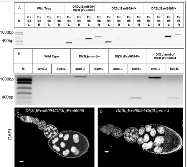
Furthermore, ovaries from Df(3L)Exel6094 over Df(3L)armi-J or Df(3L)Exel6094 over Df(3L)Exel6095 females showed no signs of oogenesis defects (Figure 8). Second, it
was previously found that addition of the CG14971 transgene into Df(3L)armi-J females
either in the presence or absence of the CycJ and armi transgenes had no effect on any of the phenotypes that I observed (Table 1) (Atikukke 2009). Finally, the
Figure 8: A deficiency removing eIF5B (Df(3L)Exel6094) is fully complemented by
both Df(3L)armi-J and a deficiency also removing these three genes
(Df(3L)Exel6095). Transheterozygous females Df(3L)Exel6094 over Df(3L)armi-J or
Df(3L)Exel6095 are viable and have normal oogenesis. (A) and (B) Diagnostic PCR
showing that Df(3L)Exel6094/Df(3L)Exel6095 (A) and Df(3L)Exel6094/Df(3L)armi-J (B)
contain both deficiencies and confirming the endpoints of these deficiencies. (C) and (D)
DAPI stained ovarioles from Df(3L)Exel6094/Df(3L)Exel6095 (C) and
Df(3L)Exel6094/Df(3L)armi-J (D) females. Oogenesis occurs without obvious defects in these transheterozygotes suggesting that eIF5B does not contribute to the three-gene deletion. Size bar = 20 µm. PCR was conducted on template DNA prepared from whole adult flies of the indicated genotypes. Gen=Genotype, Pri=primer pair, M=DNA marker ladder (see methods). Ex94* are primer pairs specific for the *L left end (6094Gpfor/TnLeft) and *R right end (6094Gprev/TnRight) of Exel6094; Ex95* are primer pairs specific for the *L left end (6095Gpfor/TnLeft) and *R right end (6095Gprev/TnRight) of Exel6095. “armi-J” is primer pairs (RB/PA_Df(3L)armiJ_R)
specific for Df(3L)armi-J. All primer pairs consisted of one transposon specific and one
genome primer, sequences listed in Appendix B. Figure has been published in (Atikukke, Albosta et al. 2014) and is represented here with minor revisions.


![{μ 2 [1 (N,N Dimethylamino)ethyl]ferrocene 1,1′ diylbis(diphenylphosphine) κ2P:P′}bis[chlorogold(I)]](data:image/gif;base64,R0lGODlhAQABAIAAAP///wAAACH5BAEAAAAALAAAAAABAAEAAAICRAEAOw==)