REAL-WORLD SOCIAL COGNITION:
Context Effects in Face and Threat Processing
Thesis by Laura A. Harrison
In Partial Fulfillment of the Requirements for the degree of
Doctor of Philosophy
CALIFORNIA INSTITUTE OF TECHNOLOGY Pasadena, California
2015
ACKNOWLEDGEMENTS
This thesis heralds the importance of context, proclaiming that social cognition does not occur in a vacuum. This thesis was not written in a vacuum. Fittingly, I owe many people a debt of gratitude for contributing to the social-developmental milieu that enabled its production.
Academically, I have a long list of people to thank and only have space to highlight a few. Since my kindergarten teacher, Mrs. Kaplow, encouraged my youthful passion for marine biology with “Sea Shell” the sea otter puppet, several other teachers and professors have supported and encouraged me as a potential scientist. A few stand out and deserve special thanks. Mr. Davis from my high school for making physics and calculus fun and for giving me confidence in my scientific abilities. Dr. Mike Barish from City of Hope and Drs. Janette Atkinson and Oliver Braddick from University College London and Oxford University gave me my first research experiences, convincing me to become a scientist. My undergraduate thesis advisor, Dr. Michael Spezio, deserves special thanks for guiding my decision to pursue a Ph.D. and inspiring the kernel of this thesis with my undergraduate thesis work (cf. Spezio, et al. (2012)).
For actually making me a scientist, I am sincerely grateful to Dr. Ralph Adophs. Ralph has been the advisor that most graduate students dream of —insightful,
leaving graduate school excited to embark on my postdoc, optimistic about my career, and still passionate about neuroscience.
Alongside Ralph, the other members of my thesis committee — Drs. John O’Doherty, Pietro Perona, Shinsuke Shimojo, and Doris Tsao — deserve my sincere gratitude. Throughout the past several years, through my thesis committee meetings, they have helped shape the direction of this thesis, improving each of the included projects. In addition to technical advice, I thank them for important advice on both nurturing several projects at once and conducting my research not only diligently but also passionately.
Being a part of Caltech has been an incredible experience for which I am grateful. I have many people to thank for contributing to that experience, including members of the Adolphs lab, past and present, HSS and CNS staff, and my fellow students.
I would like to thank all the undergraduate students I have mentored over the years, and name those whose work contributed to this thesis: Curie Ahn and Poonim Daya.
A scientific career is vocational and leaks into all aspects of one’s life. For his support throughout my Ph.D., as well as dedicating himself to support me throughout my life, including in my chosen vocation, I in turn dedicate this thesis to my dear husband, Ben Harrison. I sincerely thank him and the rest of my family, including my parents, Ray and Cindy Loesch, my siblings Jennifer and Stephen Loesch, and my grandparents Bill and Jo Anne Schutz, for their support and encouragement throughout the years.
Last, but not least, I would like to express gratitude for the funding that supports this research and the participants who both inspire the research and make it possible.
ABSTRACT
As borne out by everyday social experience, social cognition is highly dependent on context, modulated by a host of factors that arise from the social environment in which we live. While streamlined laboratory research provides excellent experimental control, it can be limited to telling us about the capabilities of the brain under artificial conditions, rather than elucidating the processes that come into play in the real world. Consideration of the impact of ecologically valid contextual cues on social cognition will improve the
generalizability of social neuroscience findings also to pathology, e.g., to psychiatric illnesses. To help bridge between laboratory research and social cognition as we experience it in the real world, this thesis investigates three themes: (1) increasing the naturalness of stimuli with richer contextual cues, (2) the potentially special contextual case of social cognition when two people interact directly, and (3) a third theme of experimental believability, which runs in parallel to the first two themes. Focusing on the first two themes, in work with two patient populations, we explore neural contributions to two topics in social cognition. First, we document a basic approach bias in rare patients with bilateral lesions of the amygdala. This finding is then related to the contextual factor of ambiguity, and further investigated together with other contextual cues in a sample of healthy
individuals tested over the internet, finally yielding a hierarchical decision tree for social threat evaluation. Second, we demonstrate that neural processing of eye gaze in brain structures related to face, gaze, and social processing is differently modulated by the direct presence of another live person. This question is investigated using fMRI in people with autism and controls. Across a range of topics, we demonstrate that two themes of
TABLE OF CONTENTS
Acknowledgements ... iii
Abstract ... v
Table of Contents ... vi
List of Illustrations and/or Tables ... vii
Chapter I: Introduction ... 1
1.1 Real Life or Fantasy? A Framework for Evaluating Naturalistic Methods in Social Neuroscience ... 2
1.1.1 Ecological Validity Defined ... 4
1.1.2 Framework for Ecologically Valid Social Neuroscience ... 6
1.1.3 Moving Forward ... 16
Chapter II: The Amygdala and Social Threat ... 17
2.1 The Amygdala and Social Perception ... 19
2.1.1 Introduction……….19
2.1.2 The Amygdala and Fear………..22
2.1.3 The Amygdala in Social Judgment……….26
2.1.4 Social Perception Meta-Analysis………27
2.1.5 Moving Forward: Anatomical Considerations………32
2.2 People with Amygdala Lesions Show a Bias to Approach Faces Even When They are Occluded ... 37
2.2.1 Introduction……….37
2.2.2 Materials and Methods………....41
2.2.3 Results……….………49
2.2.4 Discussion………...…61
2.3 Ecological Structuring of Human Defensive Responses: Evidence from Judgments of Physical and Psychological Threat Scenarios ... 66
2.3.1 Introduction……….67
2.3.2 Materials and Methods………....75
2.3.3 Results……….………87
2.3.4 Discussion………...…97
Chapter III: Face-to-Face Social Cognition ... 110
3.1 Using Face-to-Face Functional Magnetic Resonance Imaging to Investigate the Social Brain in Autism ... 111
3.1.1 Introduction……….. 112
3.1.2 Materials and Methods………..118
3.1.3 Results……….………..127
3.1.4 Discussion………...147
Conclusions and Future Directions ... 153
LIST OF ILLUSTRATIONS AND/OR TABLES
Figure 1.1.1. The Fusiform Face Area is Preferentially Sensitive to the Context of a Face………8
Figure 1.1.2. Example of Contextualized Social Emotional Inference ... 9
Figure 1.1.3. Sample Stimuli from Study on Judgments of Politicians’ Occluded Faces ... 10
Figure 1.1.4. Face-to-Face Curve ... 12
Box 2.2.1. Social Perception ... 21
Figure 2.1.1. Amygdala Nuclei ... 22
Figure 2.1.2. The Brain and Face Processing of Patient SM ... 25
Figure 2.1.3. Social Perception Reverse Inference Map from fMRI Studies ... 30
Table 2.1.1. Association Score Between Each of 11 Social Perception Regions (Figure 2.1.3) and 8 Social Perception Keywords Obtained from fMRI Studies. ... 31
Figure 2.1.4. Amygdala Connectivity ... 34
Figure 2.2.1. Anatomical MRI Scans of the Patients’ Amygdala Lesions ... 42
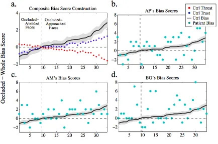
Figure 2.2.2. Stimuli and Construction of Bias Scores ... 44
Figure 2.2.3. German and American Face Ratings ... 47
Table 2.2.1. Results Summary ... 49
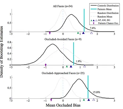
Figure 2.2.4. Composite Bias Score Defined and Compared to Patients’ Scores ... 51
Figure 2.2.5. Mean Patient Occluded Bias Scores Compared to Bootstrapped Mean Control Bias Scores ... 53
Figure 2.2.6. Parametric Bias Visualization ... 56
Figure 2.2.7. Bootstrap Comparison of Patients and Controls Approach-Related “Positivity” Biases ... 57
Table 2.2.2. Bootstrapped Control Samples Exceeding Mean and Individual Patient Values. ... 59
Figure 2.2.8.Amygdala Lesion Patients’ Deviations from Normal Judgments of Approachability (top) and Trustworthiness (bottom) of 100 faces (circles; y-axis) ... 60
Table 2.3.1. Dependent Measures in Experiments ... 76
Table 2.3.2. Threat Scenarios Presented to Subjects ... 79
Figure 2.3.1. Approach/Avoid instructions ... 83
Table 2.3.3. Gender Differences ... 85
Table 2.3.4. Scenario Factor Ratings ... 89
Figure 2.3.2. Factor-Response Option Correlations ... 91
Figure 2.3.3. Decision Tree for Defensive Behaviors to Threatening Scenarios ... 93
Figure 2.3.4. Clustering of Scenario Categories Based on Factor Ratings ... 94
Table 2.3.6. Comparison of Correlations Coefficients between Defensive Behaviors and Scenario Characteristics Obtained in 4 Studies. ... 98
Figure 3.1.1. Face-to-Face Eyetracking in ASD ... 115
Table 3.1.1. Characterization of Participants ... 119
Figure 3.1.2. Live and Video Experimental Conditions ... 120
Figure 3.1.3. Gaze Trial Conditions ... 122
Figure 3.1.4. Session Design ... 123
Figure 3.1.5. Eye Tracking ... 126
Figure 3.1.6. Group Differences in Motion ... 128
Figure 3.1.7. Group Eye Tracking Heatmaps ... 129
Figure 3.1.8. Main Effect of Video>Live Condition ... 131
Figure 3.1.9. Main Effect of Group after Gaze Shift. ... 132
Figure 3.1.10. Average Response to Closed, Averted, and Direct Gaze ... 133
Figure 3.1.11. Main Effect of Specific Direct Gaze ... 134
Figure 3.1.12. Thalamic Activation to Specific Direct Gaze ... 135
Figure 3.1.13. Qualitative Comparison of (1) NT vs. ASD Response to Direct Gaze in Live Condition and (2) Main Effect of Direct Gaze ... 136
Table 3.1.2. Regions of Interest (ROIs) Tested ... 137
Figure 3.1.14. ROI Analysis in pMNS Regions ... 138
Figure 3.1.15. Mirror Region Activation to Joint Gaze Separates NT and ASD Participants ... 139
Figure 3.1.16. ROI Analysis in TOM Regions, Main Effect of Condition ... 140
Figure 3.1.17. ROI Analysis in FFA. ... 141
Figure 3.1.18. ROI Analysis in Amygdala ... 143
C h a p t e r 1
INTRODUCTION
“I had,” said he, “come to an entirely erroneous conclusion, which shows my dear Watson, how dangerous it always is to reason from insufficient data.”
Sherlock Holmes character (Doyle, 1892/2002)
Sherlock Holmes’ observation that it is dangerous to draw conclusions from insufficient data is an extreme statement of an important reminder for cognitive and social neuroscientists. It is unclear when data is sufficient, but it is equally clear that removal of crucial variables from a model will still elicit a model, albeit one that is profoundly different from one that would have included the ignored variables. Currently, the
predominant scientific approach is that of systematic, as opposed to representative, design; variables are carefully isolated, and their effect on behavior, cognition, and neural activity tested. While this approach undeniably generates valuable findings, a lingering concern is that it reveals the brain’s capacity for processing those stimuli when tested in isolation in the laboratory, rather than its real-world processing tendencies when those same variables are contextualized alongside a myriad array of co-occurring variables.
C h a p t e r 1 . 1
REAL LIFE OR FANTASY? A FRAMEWORK FOR EVALUATING NATURALISTIC METHODS IN SOCIAL NEUROSCIENCE
Imagine you’re an undergraduate student at your university. Walking through campus,
you see a sign:
Participate in a psychology experiment!
Earn $20/hr!
$20 can buy you 200 packets of ramen noodles. You approach the sign and tear off
a tab with the experimenter’s contact information.
You decide to participate in the experiment. Later that night, you sign a few forms
and sit down in front of a computer in a university laboratory. A psychology grad student
turns to you:
“In this experiment, you’ll look at pictures of people and judge them. We’re
interested in which faces you find threatening. After seeing a face, please rate it
on a scale of 1 to 7: use 1 if the face isn’t at all threatening and 7 if it’s very
threatening. 4 is neutral. Enter your response as quickly as possible. Do you have
any questions?”
You shake your head ‘no’.
“Okay, press the space-bar to begin.”
A hairless, computer-generated face appears. It stares blankly at you. Is it
threatening? Yeah, he’s a bit dodgy. You don’t feel too strongly about it though. Maybe
Tentatively, you press 5 on the keyboard.
An hour and countless faces later, with $20 in your pocket, you leave the laboratory
and head for the parking lot.
On the way home, you stop at Walmart to exchange the cash for ramen. While
you’re getting back in your car, a man approaches you. Unlike in the lab, you don’t
hesitate. He’s definitely dodgy. Decisively, you jump in your car, jab the lock button, and
drive off.
❧❧❧
As neuroscientists, we have our experimental participants complete experiments
like this one every day. What is the difference between the two described experiences of
fear? There’s the obvious difference: one was real and the other was a laboratory
experiment. Other differences are subtler. The real man had hair. He moved. He was in a
parking lot. Which of these differences matter more to our brains?
As scientists, we propose that the cognitive principles and models we uncover
relate to in situ cognition. However, it is becoming clear that this belief is often
unrealistic. A renewed focus on ecological validity aims to remedy this disconnect
between the laboratory and real life, between the capacity of the brain to behave in a
certain way under specific laboratory conditions and a tendency to react to those same
factor manipulations in a completely different way in the unconstrained outside world.
topic of ecological validity, broadly and widely construed1. Despite heavy theoretical
interest, empirical attempts remain relatively scattershot. To be sure, more than lip
service is paid to the concept, but the overall attempt is minimally organized. In this
chapter, a framework for studying ecological validity is proposed and used as motivation
for the set of studies that follow in this thesis.
Ecological Validity Defined
The first step in providing a framework for this area of research is to define
ecological validity and highlight motivations for its pursuit. While currently fashionable,
the idea of ecological validity is not new:
Increasing numbers of cognitive psychologist and neuropsychologists are moving
beyond the laboratory and attempting to understand human cognitive abilities as
they are manifest in natural contexts (p. xi) (Poon, 1989).
That statement was made in the 1980s when psychologists researching memory
debated the merits of studying “everyday” cognition – analogous to the “real world”
social cognition investigated here. That debate generated two important observations to
keep in mind throughout this thesis. The first is that there are “theoretical and
methodological trade-offs and dilemmas” (Poon, 1989) involved in this kind of research;
as such, we should consider the “how, when, and why” for studying real-world as
opposed to traditional laboratory cognition (Rubin, 1989). The second observation is that
laboratory and real-world research lie on a continuum (Poon, 1989). At one far end of the
spectrum lies experimental manipulation of isolated, low-level variables (e.g., edge
1Including but not limited to the following concepts: cognitive ethology, real-world, real-life or everyday life,
detection); ethology and pure observation lie at the other. Excitingly, mixing this
continuum, computational ethology (Anderson & Perona, 2014) is an emerging field,
employing automated behavioral classification and detailed environmental manipulation,
that makes ethology more experimental. Combined with tools to manipulate neural
systems in organisms, this field has significant potential for detailing neural circuits
underlying ecologically valid behavior.
Within this thesis, ecological validity is conceived as capturing the tendency rather
than capability of the brain, and of utilizing stimuli2 that elicit patterns of response that
capture the most variance in actual everyday behavior. Note that this definition does not
automatically make unnatural stimuli ecologically invalid and naturalistic stimuli
ecologically valid. This distinction draws on the Bruswikian concept of representative
design, developed in the 1950s, which is utilized within the strategy of probabilistic
functionalism. Probabilistic functionalism assumes that “behavior takes place in a
semichaotic medium that contains cues of limited trustworthiness, expressed
vicariously,” (Petrinovich, 1989) requiring a research strategy that samples a broad array
of cues. Petrinovich posits that Brunswik’s approach of representative design can be
“conceived as an exercise is sampling theory” (1989), which pits representative design
against systematic design; both make different assumptions about experimental sampling
but share a common goal of making generalizable theoretical conclusions about the mind
(and, in our case, the brain). Under systematic design, experimenters “systematically
include and exclude factors and manipulate variables systematically as the investigator
2 While this definition focuses on ecological validity of the experimental manipulation, it is of course also important to
deems useful and appropriate.” Alternatively, representative design assumes it is
necessary to “understand the situation in which stimuli are encountered, and then
representatively sample stimuli from that population of situations.” To do this,
representative design requires sampling “distal” stimuli, i.e., situational or contextual
cues that influence the more proximal cause of behavior. Petrinovich makes a strong
claim that the former is nomothetic, seeking general laws, while the latter is idiographic,
observing behavior of isolated cases, but that divide is too severe, especially in modern
treatment: for example, even strict proponents of systematic design consider, and model,
individual differences, something Petrinovich (1989) did not consider compatible with
the traditional systematic approach; similarly, proponents of representative design today
still seek relatively general laws.
To reiterate: the concept of ecological validity used in this thesis is similar to
Bruswikian representative design, which refers to an experiment’s “quality of
naturalness, or lifelikeness” – some of these cues may be strictly “ecologically valid” in
Brunswik’s conceptualization, in which ecological validity relates to the “potential utility
of various cues for organisms in their ecology” (Petrinovich, 1989). Simply put, we are
interested in contextual cues, often naturalistic or lifelike, that have the most influence on
patterns of behavior and cognition as they occur in the real world. While the terms we use
are admittedly “fuzzy”, that imprecision is intentional in order to preserve a broad
concept that encompasses three main classes of ecological validity, discussed below.
Framework for Ecologically Valid Social Neuroscience
Subsequent to defining the concept of ecological validity, a second goal is to
lines of work conducted in ecological validity in social neuroscience, and a third
important area to consider. The first centers around the theme of context, the second
around interaction, and the third around believability.
Adding Contextual Cues. The first and most prominent theme of ecological
validity is to add cues to make a laboratory stimulus perceptually realistic. Often, this
involves making the stimulus as naturalistic as possible (e.g., returning to our original
example, using photographs, or, even better, dynamic videos, instead of
computer-generated facial images). While stimulus manipulation can make stimuli as close to a
real-world representation as possible, another way to manipulate context to make a cue
“realistic” is to instead simply influence high-level representations and beliefs to create
an artificial percept of “realness.” For example, the neural response to manipulation of
interpersonal space (know as “proxemics”) was successfully investigated by convincing
participants that someone was approaching them in the scanning room, not with any
change in perceptual cues, but through verbal instruction, telling participants that
someone was approaching them (Kennedy, Glaescher, Tyszka, & Adolphs, 2009).
Notably, this second type of contextual manipulation (i.e., non-perceptual) can only be
manipulated in humans, whose beliefs can be verbally informed.
Contextual cues influence all levels of cognition. For example, starting with
perception, the fusiform face area, a region that exhibits specialization for faces relative
to other objects, is actually most responsive to the context of a face, rather than an actual
Figure 1.1.1. The Fusiform Face Area is Preferentially Sensitive to the Context of a Face.
Reproduced from Cox et al., (2004). Panel a: the fusiform face area of the brain (top) is
localized in contrasts of responses to faces and objects. Panel b: changes in activity in this
functionally localized region is greatest for the context of a face (highlighted in red).
At the level of social cognition, an example of the power of context influencing
processing tendency rather than capacity is demonstrated by comparing judgments to the
two panels shown in Figure 1.1.2. While individuals can quickly and reliably make
spontaneous trait inferences from photographs of facial features (Willis & Todorov,
2006) as well as judge emotions, (e.g., the woman in panel a is upset), with the addition
of contextual cues from that woman’s environment (Figure 1.1.2b) that social inference
Figure 1.1.2. Example of Contextualized Social Emotional Inference. Contrasts of
emotion judgments elicited by photographs of the same woman, cropped in to only reveal
the face (panel a) and showing the environment surrounding her face (panel b),
demonstrate that a deeper level of social emotional inference is permitted by the
availability of contextual cues.
An experimental example of contextual cues influencing social cognition is the
finding that judgments of politicians based on extra-facial visual cues correspond with
real-world election outcomes (Spezio et al., 2012). It had previously been shown that
subjects’ laboratory two-alternative-forced-choice judgments of who was more
competent and trustworthy of two actual political opponents corresponded with election
outcomes — for example, the candidate more often chosen as more competent in the
laboratory was more often the electoral winner (Ballew & Todorov, 2007). While the
literature surrounding spontaneous trait inferences (rapid judgments of people based on
photographs of their face) traditionally assumes those judgments are driven by facial
features (Todorov & Uleman, 2002), and has manipulated computer-generated faces to
demonstrate visual features that contribute to certain trait judgments (Todorov, Baron,
& Oosterhof, 2008), we showed that judgments of extra-facial cues alone most strongly
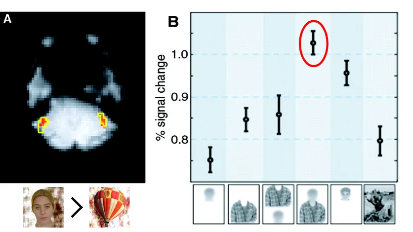
[image:18.612.172.364.167.484.2]correspond with electoral outcomes (Spezio et al., 2012) (Figure 1.1.3).
Figure 1.1.3. Sample Stimuli from Study on Judgments of Politicians’ Occluded Faces.
Reproduced from Spezio, et al., (2012). Subjects made two-alternative forced choice
decisions between candidate pairs from real elections, indicating which face they found
more trustworthy, more competent, and less threatening. Faces were presented in three
conditions: (a) unaltered full facial images, (b) facial-only images that windowed facial
features while excluding external cues, and (c) non-facial images that occluded facial
While it is unclear which of the available visual cues (dress, hairline, posture,
face width, etcetera) influence these judgments, it is clear that some of these contextual,
non-facial features meaningfully influence our appearance-based judgments of others.
So far, we have considered examples of considering contextual cues to improve
ecological validity. A second theme of ecological exists that requires a fundamentally
novel approach to experimentation: interaction.
Social Interaction. In 2007, technology blogger Kathy Sierra described a
surprising phenomena: despite several social media tools for remote communication
being made available at a conference she attended, participants in that conference
preferred travelling and meeting in person over staying home and communicating
through those tools (2007) (Figure 1.1.4). She asked why this was the case. In her
conceptualization, moving up a curve of interactive richness, social communication tools
of increasing representational fidelity (for example, email, to phone, to video-chat),
enabled better communication, yet never approached the case of face-to-face interaction
Figure 1.1.4. Face-to-Face Curve. Reproduced from Sierra (2007).
More scientifically, neuroscientists also propose that distinct neural processing
occurs in interaction (e.g., (Redcay et al., 2010; Risko, Laidlaw, Freeth, Foulsham, &
Kingstone, 2012; Schilbach, Eickhoff, Cieslik, Kuzmanovic, & Vogeley, 2012; L.
Schilbach et al., 2013; Leonhard Schilbach et al., 2013; Tanabe et al., 2012)), in that
social cognition is different when interacting with rather than observing someone.
Interaction can be viewed as a second-person account of social cognition (Schilbach et
al., 2013); in contrast, the two dominant networks described in social neuroscience, the
so-called Mirror Neuron System — a macroscopic brain network that can be recorded
neurons — which offers support for a simulationist account, and the inferential
mentalizing/Theory of Mind (TOM) network, which supports theory-theory accounts,
offer a first and third person account, respectively (Schilbach et al., 2013). It is possible
that evidence in favor of either of these prominent networks is strongly influenced by the
experimental paradigms used: “it has remained unclear whether and how activity in
the[se] networks […] is modulated by the degree to which a person does or does not feel
actively involved in the ongoing interaction and whether the networks might subserve
complimentary or mutually exclusive roles in this case” (Schilbach et al., 2013). To solve
this dilemma, to find the “dark matter” of the neural mechanisms underlying social
interaction, we should study the context of interaction as a potentially necessary form of
ecological validity. A proposed strong contender for explaining second person accounts
of neuroscience is embodied cognition. In contrast to spectatorial accounts, embodied
cognition sees social perceptions as “an active process executed by an organism situated
in the environment, wherein subjects are not isolated from but embedded in and couple
with the perceived world” (Schilbach et al., 2013). Both a hybrid account of the two
spectatorial views (theory-theory TOM/mentalizing and simulation theory mirroring
accounts), as well as accounts emphasizing embodiment are gaining credence, and
second person paradigms (requiring interaction and emotional engagement) may shine
light on them.
Believability. In addition to contextual cues and interaction, a third theme of
ecological validity (which I do not manipulate in this thesis) is believability — making a
participant believe a stimulus or experimental manipulation is ecologically valid and that
studies often consider this element of ecological validity, convincing subjects that they
are actually interacting with other real people and that they are making “real” decisions,
at least one of which will actually be randomly selected and realized.
Ecological Validity in Non-Human Species. Notably, believability is a
high-level manipulation that only works in humans. How can ecological validity be
manipulated in other species? One approach is that ethological approaches can be
scaled-up and computationally assessed (Anderson & Perona, 2014). In our three themes of
ecological validity — (1) contextual cues, (2) interaction, and (3) believability — the
following are possible:
(1) Making the stimulus as rich, dynamic, and contextualized as possible is
feasible in all species. This theme acts on perception.
(2) Making a stimulus interactive, or contingent, on an animal’s actions can be
done in other species. This theme acts at many levels, not just perception. In
addition to other species, this theme would be important to test in development.
(3) Believability can only be manipulated in humans.
An important gap to bridge between social neuroscience in humans and other animals is
that other animals usually do not know that they are in an experiment, and studies with
them are therefore ecologically valid in that sense (Stanley & Adolphs, 2013). On the
other hand, verbal report and explicit instruction are impossible in animals, and
non-ethological testing approaches often require extensive training.
In animal experiments, it is beginning to be recognized that the animal’s rearing
and comfort are crucial aspects of ecological validity in social neuroscience. Tetsuro
research begins to address this issue. In his approach, group-housed, mother-reared
chimpanzees “volunteer” to participate in experiments, coming to testing areas of their
living space as they wish. Further, testing and social life are not separated —
chimpanzees are raised to interact with and trust the experimenter, with their mother
present for interactions with the experimenter. This trusting and fairly natural laboratory
rearing has permitted studying other themes of ecological validity, such as interaction, in
the finding that chimpanzees are capable of altruistic behavior, helping conspecifics
based both on a perceptual understanding of their need and being “asked” for help
(Yamamoto, Humle, & Tanaka, 2012). Observing this altruistic behavior required an
interactive paradigm since the behavior required being asked for help, an interactive
process.
Evolutionary View of Ecological Validity. The following question could be
asked: what does ecological validity actual gain? Are differences just attentional? Aside
from the theoretical reasons put forth thus far, our consideration of other species segues
to answering that question with another question: what did the brain actually evolve to
process? While we do not actually know, there are two very different views. The first is
that it evolved to process real stimuli, in the real world, in an interactive context (a view
espoused by ethologists). A second view, supported by strong proponents of
computational models of the mind, is that the brain evolved to process simpler
computations that can be brought to bear on real-world processes. At minimum, these
real-world processes require incorporating more information or computations to
determine behavior. Regardless of which answer one supports, ecological validity is
manipulations do influence behavior.
Further Aspects of Ecological Validity. While three main themes of ecological
validity have been discussed, there are of course still other aspects of ecological validity
not covered here, including, but not limited to (1) individual variation, (2) the temporal
dynamics of everyday versus laboratory behavior were response times are experimentally
circumscribed, and (3) the need for more varied and comprehensive ways to measure
behavior, including more implicit measures.
Moving Forward
Returning to the concept of representative design, with any factor relevant to
ecological validity tested, it is important to be keenly aware of issues of generalizability.
Additionally, as with all neuroscience, studies sampling many different methods and
providing convergent evidence are preferable. Also, while ethological approaches are
valuable and information, as neuroscientists, we need to not only observe behavior, but
also develop and test models that explain neural processing thereof.
Moving forward, working within the framework of two main themes of ecological
validity, contextual cues, and interaction (with a parallel them of believability), we test
the importance of the those themes in the next two chapters by (1) investigating the
sensitivity of the amygdala to contextual cues, including ambiguity, in threat perception,
and (2) investigating the influence of interaction on the neural processing of gaze in
C h a p t e r 2
THE AMYGDALA AND SOCIAL THREAT
The first chapter of this thesis claimed that ecologically valid social cognition is
influenced by a constellation of situational contextual cues. While offering support for
that claim, this chapter also offers evidence in support of the amygdala serving a general
role; this in turn helps explain the amygdala’s generalized processing of diverse inputs.
This three sections of this chapter all focus on the amygdala and threat; two of the
three sections are published or in press and one is currently under review. The rationale for
focusing on the amygdala is twofold: (1) arguably, it is the most investigated brain
structure in social neuroscience, and will feature later in this thesis as a neuroimaging
region of interest, partially because it is hypothesized to contribute to dysfunctional social
cognition in autism spectrum disorder (cf. Chapter 3.1). Within this chapter, the first
section provides a brief review of what we know about the function of the amygdala; the
second and third sections are empirical papers. A further rationale (2) for studying the
amygdala is that I had access to three extremely rare neurological patients who had focal
bilateral lesions to the amygdala. Because I investigated approach-avoidance behavior to
faces, an important category of social stimuli, prior work that linked face processing to the
amygdala, including in these same patients, increased the relevance of focusing on this
Summarizing our results, this chapter begins exploring the role that the
amygdala plays in social perception, suggesting a general role related to evaluating the
saliency or self-relevance of social and non-social cues for an organism.
In the second section, disruption of this evaluation is shown to promote an
enhanced default approach bias in bilateral amygdala lesion patients – instantiated as a
tendency to default to rating impoverished facial images in which facial features were
occluded as more approachable, i.e., more trustworthy and less threatening than their
unaltered, whole-face counterparts. This second section marks an important reminder
that information content, i.e., ambiguity, rather than specific information, can be an
important contextual dimension along which our brains evaluate stimuli to guide our
social behavior.
This chapter culminates by assessing how ambiguity, alongside nine other
contextual factors, hierarchically guides human defensive responses to both physical
and psychological threat scenarios, mirroring patterns of threat evaluation observed in
other species. In line with the first section of this chapter, this hierarchical guidance of
defensive responses relies upon appraisal of the contextual relevance of potential
C h a p t e r 2 . 1
THE AMYGDALA AND SOCIAL PERCEPTION3
The amygdala is a key structure connecting sensory representations with valuation,
social inference, attentional modulation, and memory encoding. As such, it plays a
prominent role in one particular aspect of social perception: the ability to infer the
meaning of social communicative signals. While this role is best studied in regard to the
recognition of emotions from facial expressions, it extends to more complex social
judgments, other sensory modalities, as well as the incorporation of context. Recent
work attempts to synthesize a more unitary function from all these findings, possibly
related to aspects of evaluating biological significance or saliency. Considerable future
work is required to situate the amygdala’s function within a more extended neural
system, likely featuring close interactions between the amygdala, temporal and prefrontal
cortex, as well as parts of the basal ganglia and brainstem.
Introduction
Bilaterally buried in the medial temporal lobe, the human amygdala is a compact
subcortical structure,unilaterallyoccupying just under 1400 mm3 in postmortem
histology (Schumann & Amaral, 2006) and around 2000 mm3 in live MRI scans
(Schumann et al., 2004). Our understanding of the amygdala’s functional role has
evolved considerably over the past 50 years, most strikingly shifting from a role in social
behavior (in the mid-1900s) to one related to fear (in the late 1900s) to one more
abstractly related to value, saliency, and relevance at the current time.
Historically, and at first glance, the role of the amygdala may appear deceptively
simple — it detects threat through a specific role in associative memory (LeDoux, 1996).
While this function’s simplicity and biological significance constitute an attractive
explanation, decades of investigation support a considerably more complex and diverse
view (Aggleton, 2000; Whalen & Phelps, 2009), to some extent leaving in question what
might be its “basic” function — topics we take up below.
Lesion studies, including the early classic work by Kluver and Bucy (1939),
implicated the amygdala in social processing. Subsequent research focused on detection
of social and non-social threat. In tandem with increased interest in social neuroscience,
social stimuli, especially faces and facial emotions, were studied in detail. Outside of
social neuroscience, threat processing studies evolved into a healthy branch of reward
learning research, supplemented by neuroeconomics. This accumulation of evidence
made it clear that the amygdala does much more than detect threat; in an early synthesis
of these diverse areas of research, we (Adolphs, 2010) summarized the emerging view
that the amygdala processes “a psychological stimulus dimension related to saliency or
relevance.” Clearly, this role would be important to social perception, both at the direct
How tenable is the hypothesis that the amygdala processes salience or relevance?
Attractively, it predicts a rather broad processing role, which is borne out by the
amygdala’s anatomy. The amygdala features dense afferent and efferent connections to
other cortical and subcortical regions, and features interconnections between its
constituent subnuclei (Amaral, Price, Pitkänen, & Carmichael, 1992; Pitkänen, Savander,
& LeDoux, 1997). Interconnections within the amygdala highlight the complexity packed
into this small structure. The human amygdala is a collection of over a dozen nuclei and
sub-nuclei (Figure 2.1.1). Broadly speaking, it consists of two main regions: the
basolateral amygdala and centromedial amygdala. Ultimately, an understanding of the
role of the amygdala in social perception cannot ignore distinctions between its
subnuclei.
Box 2.2.1. Social Perception
Perception is the transduction of sensory stimuli to action potentials, which elicit early neural processing, resulting in an organism detecting, discriminating, and categorizing an attended environmental stimulus. A perceived object is processed to the extent that it can be discriminated from other stimuli, and categorized (further recognition and judgments that rely on memory, and, in the case of humans, naming, typically follow but need not be included under the rubric “perception”).
Social perception is the perception of “social” objects. Notably, “social perception refers to initial
stages in the processing of information that culminates in the accurate analysis of the dispositions and intentions of other individuals” (Allison, Puce, & McCarthy, 2000). Thus, while emotion recognition and face processing are typically considered perceptual, inferring the beliefs of other people (“mentalizing”) is not. The boundary between “initial” and “later” processing is fuzzy and can be debated.
Similarly, the bounds of the amygdala’s role in social perception are not clear. Antecedent to perception, it guides attention. Subsequent to perception, the amygdala helps evaluate social stimuli to guide our behavior. While working within a classic three-stage cognitivist model that separates perception, attention, and behavior can be useful methodologically, attempting to isolate the amygdala’s role in perception ultimately fails to provide a unitary understanding of the amygdala’s function. The amygdala likely contributes to rapid detection, more extended processing, and behavior - all at different points in
time. In fact, it participates all the time, likely through interactions of its various sub-nuclei with different
Figure 2.1.1. Amygdala nuclei. A coronal section of a postmortum human brain (left)
shows the centromedial nucleus outlined in orange and basolateral complex outlined in
red. An MRI scan (right) shows coronal and parasaggital probabilistic locations of
amygdala nuclei in humans. Lefthand panel: BL = basolateral nucleus; BM = basomedial
nucleus; BV = basoventral nucleus; CE = central nucleus; La = lateral nucleus; Me =
medial nucleus; VCo = ventral cortical nucleus; CL = Claustrum; Ent = entorhinal cortex;
F = endorhinal sulcus; Hi = hippocampus; NbM = Nucleus basalis of Meynert; TrO =
Tractus opticus; V = lateral ventricle. Righthand panels: Orange = centromedial
amygdala; red = basolateral amygdala; magenta = superficial amygdala; yellow =
hippocampal-amygdaloid transition area; light blue = subiculum; green = CA regions of
the hippocampus; dark blue = dentate gyrus; purple = entorhinal cortex. Reproduced from
Amunts, et al. (2005). Cytoarchitectonic mapping of the human amygdala, hippocampal
region, and entorhinal cortex: intersubject variability and probability maps. Anatomy and
Embryology, 210(5-6), 343-352, with permission from Spring Science + Business Media.
Here we readdress the question of what the amygdala contributes to social
perception. We do so by (1) reviewing the three main areas of amygdala research - fear,
social judgment, and reward processing; (2) conducting a meta-analysis of human fMRI
The Amygdala and Fear
Behaviorally, some of the most striking consequences of focal amygdala lesions in
nonhuman animals are a lack of fear-like behavior in response to normally threatening
stimuli. This has been shown particularly clearly in the case of rats (Choi & Kim, 2010),
where reversible pharmacological lesions revealed a parametric relationship between
amygdala activity and fearful behavior: muscimol-induced lesions were associated with a
lack of fearful behavior, whereas bicuculline-induced excitation was associated with
exaggerated fearful behavior.
Recently, optogenetics has been used to study fear behavior and conditioning in
rats. Optogenetics uses molecular biology tools to express synthetic ion channels in
particular neurons of an animal’s brain. These ion channels are opened or closed by
light, allowing light pulses to modulate neural activity in real-time in freely moving
animals. Optical stimulation of amygdala neurons can directly serve as a conditioned
stimulus in fear conditioning(Johansen et al., 2010). Meanwhile, optical silencing of
basolateral neurons that project to the centromedial nucleus affects anxiety behavior (Tye
et al., 2011). Optogenetics is a powerful technique, with exciting potential to elucidate
functional interactions of neural populations.
A complementary approach to neuromodulation consists of irreversible chemical
lesions via ibotenic acid, an approach taken in several monkey studies(Emery et al.,
2001; Mason, Capitanio, Machado, Mendoza, & Amaral, 2006; Prather et al., 2001).
Here, too, there is a consensus that amygdala lesions reduce the normally cautionary
break on behavior that is elicited by stimuli that signal potential threat. Interestingly, the
behaviors that are normally inhibited) is seen both for overtly threatening stimuli (e.g.,
snakes) as well as for stimuli that merely signal uncertainty (e.g., novel objects next to
food). The consequences on behavior for complex social stimuli are rather varied, and
depend to some extent on context and individual differences (Mason et al., 2006).
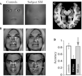
In humans, only a single complete lesion case has been well studied (although there
are a handful of studies on other patients with similar lesions): SM, a patient with
developmental bilateral amygdala lesions, also showed a dramatic lack of fear behaviors
(Feinstein, Adolphs, Damasio, & Tranel, 2011), as well as impairment in directing gaze
and attention to relevant facial features (Figure 2.1.2). Especially valuable in this human
case was the opportunity to (a) test fear elicitation across a wide range of stimuli
(autobiographical recall, actual snakes and spiders, a haunted house, fear experience in
everyday life, horror movies) and (b) investigate other basic emotions as well. A highly
selective absence of fear was found across all the stimuli. Interestingly, while the
amygdala seems necessary to elicit fear from external stimuli, following lesioning of the
amygdala, interoceptive fear can be induced by carbon dioxide inhalation (Feinstein et
Figure 2.1.2. The brain and face processing of patient SM. Bilateral amygdala lesions
impair the use of the eyes and gaze to the eyes during emotion judgment. (A) A patient
with bilateral damage to the amygdala made significantly less use of information from
the eye region of faces when judging emotion. (B) While looking at whole faces, the
patient (right column of images) exhibited abnormal face gaze, making far fewer
fixations to the eyes than did controls (left column of images). This was observed across
emotions (free viewing, emotion judgment, gender discrimination). (C) MRI scan of the
patient’s brain, whose lesion was relatively restricted to the entire amygdala, a very rare
lesion in humans. The two round black regions near the top middle of the image are the
lesioned amygdalae. (D) When the subject was instructed to look at the eyes (“SM eyes”)
in a whole face, she could do this, resulting in a remarkable recovery in ability to
recognize the facial expression of fear. The findings show that an apparent role for the
[image:33.612.112.393.86.349.2]the detection and attentional direction onto features that are socially informative.
Reproduced from Adolphs, (2010). What does the amygdala contribute to social
cognition? In: A. Kingstone & Miler (Eds.), Year in Cognitive Neuroscience 2010 (pp.
42-61). Malden: Wiley, with permission from Wiley.
A few other such amygdala lesion patients have also been studied, and bear out
these main findings (J. Feinstein, personal communication). A point to note is that they
all suffer from a developmental and progressive disease, leaving open questions about
when in development lesions first manifest as well as whether any tissue or passing fibers
might be spared. Some of these patients appear to have damage mostly restricted to the
basolateral nucleus, and also exhibit behavioral variability (Terburg et al., 2012).
Systematic future investigations of these patients will be critical to begin to address the
difficult question of the causal roles of particular amygdala subnuclei in humans.
These findings, and in particular the human case (Feinstein et al., 2011), raise two
broad questions for how to conceive of the amygdala’s role in social perception. First
and foremost: at what point in processing (see Box 2.2.1) does the amygdala come into
play? Does it impair perception as such (detection, discrimination)? Or does it impair
the ability to associate meaning with what is perceived, or to modulate cognition based
on the associated value of what is perceived (e.g., recognition, social judgment)? Across
all of the studies, human and nonhuman, the answer here has been relatively clear:
amygdala lesions impair the associative ability, but not basic perception as such. Yet
even this conclusion is now being modified in light of the amygdala’s role in attentional
processing. The second question is how selective the role is to the domain of fear. Is the
Or, alternatively, is its role in fear reducible to a more computationally abstract or basic
function? We take up these two related questions next.
The Amygdala in Social Judgment
Monkeys with amygdala lesions show complex deficits in their interactions with
other monkeys that are not obviously reducible to an absence of fear (Emery et al., 2001).
In general, the deficits are surprisingly subtle, although they are invariably associated
with negative social consequences (loss of social status, abandonment by the troop and
death in the wild)(Kling & Brothers, 1992). In fact, neonatal amygdala lesions in infant
monkeys result in an exaggerated social fear behavior, something not seen following
adult lesions(Prather et al., 2001).
The deficits in social behavior in humans with amygdala lesions are also very
subtle, although this is perhaps not too surprising given the complex and interactive
nature of the social environment and, specifically in humans, the compensatory aspects
provided by others who know that a patient has an amygdala lesion (Adolphs, 2010).
More experimental control can be obtained in studies that assess social judgments
through ratings given to depictions of scenes, faces, or descriptions of scenarios. In these
studies, while there is also considerable variability, the deficit has been largely consistent
with a lack of fear: patients with bilateral amygdala lesions tend to judge other people as
more trustworthy and more approachable and have difficulty recognizing fear (Adolphs,
Tranel, & Damasio, 1998; Adolphs et al., 1999).
This conclusion in humans is to some extent borne out by functional neuroimaging
studies. For instance, face stimuli designed to parametrically vary in terms of their
in healthy viewers (Winston, Strange, O'Doherty, & Dolan, 2002). Yet there are
notable discrepancies here. In particular, the human amygdala is also potently activated
by appetitive stimuli (e.g., erotic pictures (Hamann, Ely, Hoffman, & Kilts, 2002)); in
fact, it is variably activated by all facial expressions across the board (Fitzgerald,
Angstadt, Jelsone, Nathan, & Phan, 2006), although it may respond in particular to
certain underlying dimensions such as valence (Todorov et al., 2008).
Beyond imaging studies focusing on social stimuli, a unifying attempt comes from
the reward learning literature. There is an extensive literature from lesion and
electrophysiological studies in animals that argues for a role in reward learning and
attention. That literature historically emphasized fear or arousal, in the form of Pavlovian
fear conditioning as well as instrumental avoidance learning (LeDoux, 1996)and
modulation of declarative memory (McGaugh, 2004), but has now been extended to
encompass both appetitive and aversive forms of learning, and both Pavlovian and
instrumental mechanisms(Murray, 2007). Here, attempts to find a simple role have been
frustrated by the sheer range of findings. Results from neuroimaging studies have been
similarly diverse.
Given the very large number of human neuroimaging studies of the amygdala, it is
possible to attempt to extract some possibly basic underlying dimensions from stimuli
and tasks, across several sub-domains of research, which might most consistently activate
the amygdala.
Social Perception Meta-Analysis
To synthesize the neuroimaging research, we turned to Neurosynth
results (Yarkoni, Poldrack, Nichols, Van Essen, & Wager, 2011). At a basic level,
Neurosynth produces forward and reverse inference maps that relate a certain
psychological construct to a brain region. Specifically, forward inference maps represent
the probability of observing activation in a brain region (within 6mm of a coordinate),
given the presence of a particular term, e.g., “social”, in a research article at a certain
frequency (default is once every thousand words); reverse inference maps represent the
probability of a term occurring in an article given activation in a particular brain region.
Probabilities are calculated based on a database of nearly 6000 studies.
Because Neurosynth uses a large database to create these reverse and forward
inference term-to-activation mappings, its developers argue that their tool renders the
reverse inference problem tractable. The reverse inference problem occurs when one
incorrectly assigns a functional role to a region, as is easily done when only looking at
results from a few hand-selected studies. Much more data, which is provided by
Neurosynth, is necessary to make the probabilistic statements necessary to make
functional inferences: a region should be both consistently (forward inference) and
selectively (reverse inference) associated with a functional term.
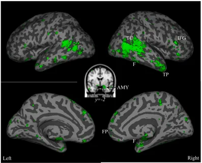
Confirming the premise of this chapter that the amygdala is involved in social
perception, a “social perception” reverse inference map included activation in the
amygdala as well as other regions associated with social perception (Figure 2.1.3).
Interestingly, this map exhibited some right lateralization, both in the amygdala, and
across hemispheres. In the whole brain, 60% of voxels in the reverse inference map
(Figure 2.1.3) are in the right hemisphere.Strong lateralization was observed in the
correction, compared to only 3% of voxels in the left amygdala.
Figure 2.1.3. Social perception reverse inference map from fMRI studies. Combination
of FDR 0.1 (dark green) and FDR 0.05 (bright green) corrected z-score maps of the
probability, given observed activation at a particular voxel, that a study in the Neurosynth
database is related to social perception. These reverse inference maps were created by
searching the database of nearly 6000 papers for those in which the terms “social” and
“perception” both occurred at least once in every 1000 words. 128 papers met this
criteria. AMY = amygdala; TP = temporal pole; FP = frontal pole; F = fusiform gyrus;
IFG = inferior frontal gyrus; TC = temporal cortex. Surface rendering and visualization
for this and following images were completed using the SPM surfrend toolbox
(http://spmsurfrend.sourceforge.net) and Neurolens (neurolens.org).
Left Right
TP IFG
FP
TC
F
F AMY
While the amygdala’s involvement in social perception that emerges from this
data-driven analysis is unsurprising, how does its role differ from that of other “social
perception” areas identified? We tested the specificity of the amygdala’s role in social
perception by calculating association scores between 8 functional keywords and 11
regions identified in the social perception reverse inference map (Table 2.1.1). While
Neurosynth produced a long list of terms associated with activation in each of these
regions, we wanted to summarize those lists to determine whether they were strongly
associated with a small number of social perception concepts. These social perception
“keywords” onto which to map the neurosynth list of terms were obtained by presenting
neuroscientists (n=6) with the top 50 terms associated with each social perception region
by Neurosynth, and asking them to generate words to summarize each list of terms. From
these responses, 8 keywords were distilled and appear as the column headers in Table
2.1.1. A second group of neuroscientists (n=5) rated (0= “No”; 1 = “Maybe”; to 2 =
“Yes”) the relationship between each word associated with a region and these 8
keywords. Neurosynth lists of words were truncated to only consider words receiving a
z-score>3; this resulted in a total of 118 unique terms being rated across all the brain
regions. These ratings were converted into association scores, reported in Table 2.1.1.
Table 2.1.1. Association score between each of 11 social perception regions (Figure
2.1.3) and 8 social perception keywords obtained from fMRI studies. An association
score between each keyword and region was derived by creating a region x term
association score for each subject, averaging across subjects, and thresholding
High/Moderate/Low ratings by a third split of all mean scores. Each subject’s region x
by the number of terms considered. Ratings of 3 (“Yes”) were weighted by 2, ratings of
2 (“Maybe”) were weighted by 0.5, and ratings of 1 (“No”) were weighted by -1. AMY
= amygdala; TP = temporal pole; IFG = inferior frontal gyrus; FP = frontal pole; TC =
temporal cortex; F = fusiform gyrus.
First, this keyword association analysis (Table 2.1.1) confirms that the amygdala is
indeed involved in social cognition and perception, and has an especially strong
association for domain-specific “social perception”, compared to domain general
“perception”. This domain specificity may be skewed by the database of human
neuroimaging articles used by Neurosynth. Beyond confirming the amygdala’s
involvement in social perception, we demonstrate it is moderately to highly associated
with all the other social perception functional concepts, with the sole exception of the
concept language/communication. Appropriately, this last concept was strongly
associated with the temporal cortex. As predicted by the large literature on the amygdala
and faces, emotion, and gaze, the region was highly associated with those three concepts.
However, these associations were not very specific, as several other regions were also
associated with faces, emotion, and gaze. Meanwhile, the amygdala’s moderate
frontal pole were the only regions in the Neurosynth social perception network
associated with valuation. To a degree, these findings agree with the idea that the
amygdala, anatomically intermediary between perceptual and cognitive regions, plays an
intermediary role between perception and cognition by assigning value to (social and
non-social) perceptual objects.
Moving Forward: Anatomical Considerations
As a whole, the above reviews present a puzzle for the amygdala’s role in social
perception. To a degree, the amygdala’s role in this domain can be explained in terms of
its role in basic reward and attentional processes. However, open questions remain, and a
key challenge is resolving several potential explanations. The findings from our
Neurosynth analysis begin to provide a comprehensive answer and highlight a promising
direction.
One solution to the above predicament is to acknowledge that the question “what
does the amygdala contribute to social perception” was rather ill-posed to begin with,
since it leaves out the rest of the social perception network within which the amygdala is
embedded, and whose functions we need to understand if we are to thus situate the
amygdala’s.
One such network approach was taken in a study (Bickart, Hollenbeck, Barrett, &
Dickerson, 2012) that seeded three regions of the amygdala in a resting-state functional
connectivity analyses that elicited three main networks correlated with activity in those
regions: a perception network associated with the ventrolateral amygdala, an affiliation
network with the medial amygdala, and an aversion network with the dorsal amygdala.
literature; the large temporal component of the perception network was evocative of
Freese and Amaral’s (2005) well characterized dense temporal connections (Figure
2.1.4).
Figure 2.1.4. Amygdala connectivity. Amygdala resting-state functional connectivity
perceptual network map (left) from Figure 5 in (Bickart et al., 2012). Map kindly
provided by Brad Dickerson. AMY= amygdala. This connectivity with temporal regions
is reflected in a summary of known anatomical connections between the amygdala and
ventral visual stream areas in the monkey (right). Projections are topographically
organized, with ventral and rostral amygdaloid regions projecting most to rostral visual
areas, and dorsal and caudal amygdaloid regions projecting most to caudal visual areas. A
feedforward/feedback loop exists between area TE and the basal and lateral nuclei of the
amygdala. Right-hand panel reproduced from Freese & Amaral (2005). The organization
of projections from the amygdala to visual cortical areas TE and V1 in the macaque
monkey. Journal of Comparative Neurology, 486(4), 295-317, with permission from
Additionally, the results from Bickart and colleagues (2012; Figure 2.1.4)
partially converged with our meta-analysis results (Figure 2.1.3): their social perception
network, derived from a region similar to the basolateral nucleus, and our social
perception regions partially overlapped, with 27% of the voxels in our Neurosynth social
perception map (combination of FDR 0.1 and FDR 0.05)(Figure 2.1.3) also present in
their map (Figure 4). While the different amygdala nuclei interact, these converging
results suggest that the core social perception function might be assigned mostly to the
basolateral amygdala, which is also the region of the amygdala that is lesioned most
commonly in Urbach-Wiethe disease.
These converging anatomical observations are indicative of one of the most
promising means of advancing our understanding of the amygdala’s role in social
perception: a network approach. The amygdala’s role will not be understood in isolation.
We should strive for a biologically-constrained role that seeks to understand the function
of the amygdala as a group of structures that interact with one another and within
networks. The field of optogenetics has much potential for advancing our network-level
understanding of amygdala function. However, findings must be related across species
(optogenetics research currently being limited mostly to mice) and subfields: here, it is
crucial to conduct meta-analyses within species, and, across species, to carefully consider
how sub-field-defined cognitive functions are related.
Moving forward, especially in the social domain, we must consider environmental
context. Our social environment is rich and complex, and social cognition and perception
are largely inferential. They rely upon correct incorporation of contextual cues. This
amygdala, differs among individuals, and is a product of stimulus history. Approaching
our investigation within this framework is necessary for fleshing out a nuanced
understanding of the amygdala’s network role in modulating perception and assessment
C h a p t e r 2 . 2
PEOPLE WITH AMYGDALA LESIONS SHOW A BIAS TO APPROACH FACES
EVEN WHEN THEY ARE OCCLUDED4
Approach and avoidance constitute a basic dimension of all animal behavior. A
large literature documents approach and avoidance elicited by specific sensory stimuli,
yet comparatively little is known about default approach biases when stimulus
information is reduced. The amygdala is well known to contribute to approach and
avoidance behaviors in response to specific sensory stimuli, and here we test whether the
amygdala’s role might extend to situations where stimulus information is reduced. A
novel task asked three rare patients with bilateral amygdala lesions to make
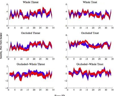
approach-related judgments about photos of faces when intact, and with all internal facial features
occluded. Direct comparisons of these stimuli isolated a stimulus-independent bias. The
patients showed a greater tendency than controls to default to rating occluded faces as
more approachable than whole faces. These findings suggest that the amygdala’s role in
approach behavior extends beyond responses to specific stimuli.
Introduction
From single-celled organisms to humans, all mobile species exhibit
approach-avoidance behavior. In humans, approach-approach-avoidance behavior is regulated by motivation
and influenced by emotion (Elliot, Eder, & Harmon-Jones, 2013); at a more primitive
level, it is related to instinctive defensive behaviors (Blanchard, Griebel, Pobbe, &
Blanchard, 2011; McNaughton & Corr, 2004).
While basic, approach-avoidance behavior shows large individual differences.
Whereas some people would walk into and explore an unfamiliar dark room, others
would pause and gather more information, and some might even flee. What accounts for
this behavioral variability? Prior experience might sway one’s response, but the example
situation offers little information, and may not have been encountered previously.
Nevertheless, a behavioral tendency will be observed. The amygdala is a brain structure
known for its role in memory, learning, and emotion, and implicated in psychiatric
disorders including anxiety. We investigate what role the amygdala might play in
regulating stimulus-independent behavior, termed a “default bias.” A default approach
bias may be normal in certain contexts; here, we ask whether amygdala lesion patients
exhibit an abnormally large approach tendency to low-information stimuli.
An abnormal tendency to approach others and normally threatening stimuli in
amygdala lesioned monkeys (Klüver & Bucy, 1939), rodents (Choi & Kim, 2010) , and
humans (Feinstein et al., 2011; Kennedy et al., 2009) points to the amygdala as being
important in regulating approach-avoidance behavior. However, the basis of this
approach tendency is unclear.
On the one hand, the bias may be specifically tuned for certain stimuli: much of
what we know about the amygdala’s contribution to cognition and behavior has come
from studies investigating faces. Single-unit amygdala response selectivity has been
found for faces in humans (Rutishauser et al., 2011) and monkeys (Gothard, Battaglia,