3693
Introduction
Dorsal spinal cord formation begins with neuronal and glial progenitors exiting the cell cycle in the ventricular zone (VZ), and is followed by sequential migration and further differentiation of migrating neurons and glia. Once neurons have reached their appropriate region of the dorsal spinal cord, they differentiate further into distinct layers or laminae that carry out distinct physiological function (Christensen and Perl, 1970; Rexed, 1952). For example, lamina I primarily contains relay and local interneurons, whereas lamina II (substantia gelatinosa) consists of few projection neurons but contains numerous small-size interneurons, many of which make synaptic contacts with lamina I neurons (Molander and Grant, 1995; Willis, 1995). The primary afferents of dorsal root ganglia (DRG) neurons project to distinct laminae of the spinal cord and establish precise neuronal connections with their targets. Small-diameter DRG neurons that express TrkA, a high-affinity neurotrophin receptor for nerve growth factor (NGF), project to laminae I-II and relay noxious and thermal sensory information, whereas large-diameter DRG neurons that express the neurotrophin receptor TrkC make connections with motoneurons in the ventral horn and relay muscle proprioceptive information (Christensen and Perl, 1970; Huang and Reichardt, 2001; Lawson and Biscoe, 1979; Scott, 1992; Snider, 1994).
During spinal cord development, the specification of dorsal interneurons is mediated by both extrinsic and intrinsic factors (Caspary and Anderson, 2003; Helms and Johnson, 2003; Lee
and Jessell, 1999; Liem et al., 1997). Specifically, the intrinsic factors that control the specification and development of several classes of early-born neurons (dI1-6) have been analyzed in greater detail. In mice lacking transcription factors Lbx1 (Lbx1h – Mouse Genome Informatics) or Rnx (Tlx3 – Mouse Genome Informatics), the specification of dI5 neurons is affected, as indicated by the loss or downregulation of Lmx1b (dI5 marker) (Gross et al., 2002; Muller et al., 2002; Qian et al., 2002). These two genes are also involved in the development of late-born neurons that make up the dorsal horn of the spinal cord (Caspary and Anderson, 2003). In addition, the transcription factors Drg11 (Prxxl1 – Mouse Genome Informatics), Ebf1, Ebf3 and Zic1 have also been shown to be important for the early specification of the dorsal spinal cord neurons (Aruga et al., 1998; Aruga et al., 2002b; Chen et al., 2001; Ebert et al., 2003; Garcia-Dominguez et al., 2003; Garel et al., 1997; Wang et al., 1997). Among them, Zic1 has been shown to be a negative regulator of the differentiation of the dorsal horn neurons in mice (Aruga et al., 2002a; Aruga et al., 1998; Aruga et al., 1996a; Aruga et al., 2002b; Aruga et al., 1994; Aruga et al., 1996b). Despite these studies, our knowledge about the molecular mechanisms that govern the development of early-born neurons and the assembly of the dorsal horn circuits is still rather fragmentary.
Lmx1b is an LIM homeobox-containing gene, and was originally isolated as a mouse ortholog of the chicken Lmx1 (Chen et al., 1998a; Riddle et al., 1995; Vogel et al., 1995). In the chicken, Lmx1 is involved in the specification of the dorsal cell
The differentiation and migration of superficial dorsal horn neurons and subsequent ingrowth of cutaneous afferents are crucial events in the formation of somatosensory circuitry in the dorsal spinal cord. We report that the differentiation and migration of the superficial dorsal horn neurons are regulated by the LIM homeobox gene Lmx1b, and its downstream targets Rnx and Drg11, two transcription factors implicated in the development of dorsal horn circuitry. An analysis of Lmx1b mutants shows that Lmx1b normally acts to maintain the expression of the Ebf genes and to repress the Zic genes. Lmx1b mutants also
exhibit the disruption of the cutaneous afferent ingrowth, suggesting that the dorsal horn cells might provide important cues guiding sensory axons into the dorsal spinal cord. Our results thus indicate that Lmx1b has a pivotal role in genetic cascades that control the assembly of circuitry in the superficial dorsal horn.
Supplemental data available online
Key words: Lmx1b, Dorsal horn, Migration, Differentiation, Cutaneous afferents, Mouse
Summary
Lmx1b controls the differentiation and migration of the superficial
dorsal horn neurons of the spinal cord
Yu-Qiang Ding1,*, Jun Yin1, Artur Kania2, Zhong-Qiu Zhao1, Randy L. Johnson3and Zhou-Feng Chen1,†
1Departments of Anesthesiology, Psychiatry, Molecular Biology and Pharmacology, Washington University School of Medicine Pain Center, St. Louis, MO 63110, USA
2Department of Biochemistry and Molecular Biophysics, Columbia University, New York, NY 10027, USA
3Department of Biochemistry and Molecular Biology, University of Texas M.D. Anderson Cancer Center, Houston, TX 70030, USA *Present address: Laboratory of Neural Development, Institute of Neuroscience, The Chinese Academy of Sciences, 320 Yue Yang Road, Shanghai, 20031, PR China †Author for correspondence (e-mail: chenz@morpheus.wustl.edu)
Accepted 4 May 2004
Development 131, 3693-3703
Published by The Company of Biologists 2004 doi:10.1242/dev.01250
fate in the limb, and the differentiation and morphogenesis of the isthmic organizer (Adams et al., 2000; Matsunaga et al., 2002; Riddle et al., 1995; Vogel et al., 1995). Mice lacking Lmx1b exhibit abnormal limbs and kidneys (Chen et al., 1998a). Mutation of Lmx1b results in nail patella syndrome in humans, an autosomal dominant disease characterized by abnormal skeletal patterning and renal dysplasia (Dreyer et al., 1998). In the central nervous system, Lmx1b is involved in multiple developmental processes, including the formation of eye, dopaminergic neurons, serotonergic neurons and the trajectory of the motor axons in the limb (Cheng et al., 2003; Ding et al., 2003; Kania and Jessell, 2003; Kania et al., 2000; Pressman et al., 2000; Smidt et al., 2000). In the nematode Caenorhabditis elegans, the Lmx1b ortholog, Lim6, has been shown to be important for the differentiation of GABAergic neurons (Hobert et al., 1999).
In the developing dorsal spinal cord, Lmx1b is expressed in dI5 neurons and late-born neurons destined to populate the superficial layer of the dorsal spinal cord (Chen et al., 2001; Gross et al., 2002; Muller et al., 2002). In this study, we performed detailed studies of the dorsal spinal cord development in Lmx1b mutants. Our study reveals that Lmx1b is important for the development of dI5 neurons, late-born dorsal horn neurons and the projection of cutaneous afferents in the dorsal spinal cord. Our study uncovers a central role for Lmx1b in transcriptional cascades that govern the dorsal horn development.
Materials and methods
Genotyping and maintenance of animals
Lmx1b, Rnx and Drg11 mutant mice were generated and genotyped as previously described (Chen et al., 1998a; Roberts et al., 1994). The age of embryos was determined according to the plug date (the plug date is considered to be E0.5). Because the mating time among the mice may differ, the actual developmental stage of the embryos was further ascertained according to the spinal cord morphology by the use of Nissl staining, as well as the criteria described previously (Kaufman, 1998). The Lmx1b+/–, Rnx+/– and Drg11+/– mice were
maintained in a mouse facility according to protocols approved by the Division of Comparative Medicine at Washington University School of Medicine.
BrdU labeling and detection by immunocytochemistry
Pregnant female mice derived from timed matings between Lmx1b or Rnx or Drg11 heterozygous mice were given a single intraperitoneal injection of BrdU (5 mg/ml solution in PBS and 60 µg/g of body weight) at 11.5 days postcoitum (dpc) and 12.5 dpc. After time periods of 2 hours, 1 day, 2 days or 3 days, embryos were removed, genotyped and sectioned as described (Chen et al., 1998a; Chen et al., 2001; Roberts et al., 1994). The sections were processed and stained sequentially with a mouse anti-BrdU antibody (Dako), biotinylated donkey anti-mouse IgG (Jackson Immunoresearch) and ABC Elite reagents (Vector). For double staining with anti-LMX1B antibody (Kania et al., 2000), the slides were first incubated with a guinea pig anti-LMX1B antibody detected enzymatically through production of a brown precipitate. For anti-BrdU antibody detection, a nickel intensification technique was used, producing a black precipitate. To quantify the distribution of BrdU-labeled neurons, we used the Photoshop (Adobe) program after dividing the dorsal horn into two parts: medial one-third and lateral two-thirds (from the midline to the lateral edge of the dorsal horn, Fig. 2). BrdU-labeled neurons in ten sections each from wild-type (n=6) and mutant (n=6) embryos were counted at the thoracic segmental level, and a comparison was performed using Student’s t-test.
Nissl staining, in situ hybridization and immunocytochemistry
Nissl staining, in situ hybridization and immunocytochemistry were performed as described (Chen et al., 1998b; Chen et al., 2001). For double staining of LBX1 with BRN3A, PHOX2A with PAX2, and LMX1B and PHOX2A, Cy3-labeled donkey anti-guinea IgG (Jackson) and FITC-labeled donkey anti-rabbit or anti-mouse IgG (Jackson) were used. Antibodies included: rabbit anti-BRN3A (Fedtsova and Turner, 1995), guinea pig anti-ISL1 (Tanabe et al., 1998), guinea pig anti-LMX1B, guinea pig anti-LBX1 (Muller et al., 2002), mouse anti-MAP2 (Sigma), rabbit anti-PHOX2A antibody (Tiveron et al., 1996), rabbit anti-PAX2 (Zymed Lab), rabbit anti-trkA and rabbit anti-Peripherin 57K (Chemicon). For cell counting, 10-15 consecutive sections each from wild-type (n=6) and homozygous mutant (n=6) embryos at thoracic levels were counted, and statistical analysis was performed by the use of Student’s t-test.
DiI labeling
For study of the projection from the DRG to the spinal cord, a small amount of 1,1′′-dioctadecyl-3,3,3′′,3′′-tetramethylindocarbocyanine perchlorate (DiI; Molecular Probes) crystals was placed in the DRG unilaterally. The samples were kept in the fixative at 37°C for 2-4 days, and then were sectioned transversely with a vibratome at a thickness of 50-100 µm. Labeling was observed with epifluorescent or laser confocal microscopy.
In utero electroporation
For in utero electroporation, previously detailed procedures were followed (Saito and Nakatsuji, 2001). Pregnant mice at 12 dpc were anesthetized with sodium pentobarbital (40 mg/kg), followed by the exposure of the uterus and cutting of the uterine wall on both horns along the antiplacental side. pCAGGS-Lmx1b:EGFP or pCAGGS-EGFP (Niwa et al., 1991) (1-3 µl; 0.5-1.5 µg/µl) was injected into the central canal of the spinal cord using an orally controlled pipette system. After injection, square electrical pulses were delivered by the use of an Electro Square Porator (ECM830) at a rate of one pulse per second (voltage 35V, five pulses, 50 ms) to embryos by holding the utero with forceps-type electrodes. The embryos were repositioned into the abdominal cavity without sewing the uterine wall. The abdomen was filled with warmed saline and the abdominal wall and skin were sutured. Embryos were allowed to survive for 2 days. Genotyping and analysis of gene expression in the spinal cord of electroporated embryos was performed as described above.
Results
Lmx1b is expressed in dI5 and dorsal horn neurons
1D). Thus, Lmx1b expression is largely confined to post-mitotic neurons.
Phox2a marks a subset of dI5 neurons and is partially lost in Lmx1b mutants
Previous studies have shown that Lmx1b expression is either absent or downregulated in dI5 neurons of Lbx1 or Rnx mutant mice (Gross et al., 2002; Muller et al., 2002; Qian et al., 2002). These observations raise the possibility that Lmx1b plays a role in the development of dI5 neurons, and that Lbx1 and/or Rnx may mediate the specification of dI5 neurons via Lmx1b. To characterize the development of dI5 neurons in the absence of Lmx1b, we examined the expression of Lbx1 (dI4-6), Brn3a (Pou4f1 – Mouse Genome Informatics) (dI1-3, dI5) and Pax2 (dI4, dI6), all markers of distinct dorsal neuronal types in Lmx1b mutants (Gross et al., 2002; Muller et al., 2002). Immunocytochemical staining revealed a similar staining pattern of LBX1, BRN3A and PAX2 between Lmx1b mutants and wild-type control at E11.0 (Fig. 1E-H). Our study, however, was complicated by the observations that Lbx1 and Brn3a are also expressed in neurons located adjacent to dI5 neurons. To identify a more dI5-specific marker, we examined the co-expression of LMX1B and PHOX2A at E10.5 and E11.5 spinal cord (Tiveron et al., 1996). We found that PHOX2A expression overlaps with LMX1B in a subset of dI5 neurons, and no neurons expressing PHOX2A alone were found in the same region (Fig. 1I,J). Thus, Phox2a is a dI5 neuron-specific marker (Fig. 1I,J). In Lmx1b mutants, Phox2a expression was either lost or decreased in dI5 neurons in the mutants compared with that in wild-type control (Fig. 1K,L; data not shown). Moreover, the location of PHOX2A+neurons was shifted more ventrally in the spinal cord of Lmx1b
mutants, indicating that dI5 neurons migrated aberrantly in Lmx1b mutants (Fig. 1K,L). Cell counting confirmed that the number of PHOX2A+ neurons was indeed reduced in the mutants (Fig. 1M). Together our data suggests that the development of dI5 neurons is impaired in the absence of Lmx1b.
A migratory defect in the dorsal spinal cord of Lmx1b mutants
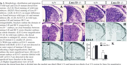
To examine the effect of Lmx1b mutation on the dorsal horns of Lmx1b–/– embryos, we performed Nissl staining. At E12.5, the dorsal horns of Lmx1b–/– embryos and wild-type controls were indistinguishable (Fig. 2A,B). At E15.5, in wild-type embryos, numerous laminae I-II neurons, distinguishable from laminae III-IV neurons by their smaller size, aggregate to form a separate layer, whereas laminae III-IV neurons also become distinct in being more loosely distributed (Fig. 2C). In Lmx1b mutants, these laminar boundaries were not recognizable (Fig. 2D) and the dorsal funiculus was smaller relative to wild-type controls (Fig. 2E,F).
[image:3.612.302.568.71.368.2]We next used BrdU to determine the migratory behavior of the dorsal horn neurons in Lmx1b mutants. To do this, we labeled dorsal horn neurons of wild-type and Lmx1b mutant embryos with BrdU at E11.5 and E12.5, and examined their settling position at E14.5. Interestingly, neurons labeled with BrdU at E11.5 in the wild-type embryo did not migrate to the most superficial region of the dorsal horn (Fig. 2G). By contrast, neurons labeled at E12.5 migrated through earlier-born neurons and occupied the most superficial layer of the dorsal horn, consistent with previous studies (Nornes and Carry, 1978; Nornes and Das, 1974). However, unlike previous studies in which [3H]thymidine autoradiography was used, our Fig. 1. Expression of Lmx1b and specification of early-born
neurons in the spinal cord of Lmx1b–/– mutants. (A-C) Expression
of Lmx1b in the dorsal horn detected by in situ hybridization. (A) Lmx1b is expressed in dI5 interneurons at E10.5 (arrow). (B) At E11.5, in addition to dI5 neurons (small arrow), Lmx1b is expressed in neurons emerging from the dorsal VZ (large arrow). (C) Lmx1b expression is concentrated in laminae I-II neurons at E17.5. (D) Detection of BrdU+(blue)/LMX1B+(brown) in the
spinal cord of wild-type E12.5 embryos. Arrows in D show nascent neurons co-labeled with anti-LMX1B and anti-BrdU antibodies. (E,F) Detection of LBX1 (dI4-6 marker, red) and BRN3A (dI1-3 and dI5 marker, green) in wild-type (E) and Lmx1b mutant (F) embryos at E11. (G,H) Detection of LBX1 (red) and PAX2 (dI4 and dI6 marker, green) in wild-type (G) and Lmx1b mutant (H) spinal cord at E11. (I,J) Detection of LMX1B (red) and PHOX2A (green) in dI5 neurons at E11.5; arrow in I and arrowheads in J (higher magnification of I) indicate double-stained cells. (K,L) Detection of LBX1 (red) and PHOX2A (green) in wild-type (K) and Lmx1b mutant embryos (L) at E11.5. Arrows indicate PHOX2A+cells. (M) Statistical comparison of
the number of PHOX2A+cells in wild-type (white bar) and
BrdU labeling clearly revealed an inside-out migration pattern for laminae I-II neurons. We next examined the migration of BrdU-labeled neurons in Lmx1b mutants. First, we did not find any significant change in the total number of BrdU-labeled neurons between wild-type and Lmx1b mutants (data not shown). However, some neurons labeled with BrdU at E11.5 were present in the most superficial region of the dorsal horn of E14.5 Lmx1b–/– embryos (Fig. 2H), while a significantly higher number of BrdU+neurons were accumulated near the midline region as compared with the control (Fig. 2I-K). In line with this observation, fewer neurons labeled with BrdU at E11.5 were located in the lateral region of the dorsal horn in Lmx1b–/–embryos (Fig. 2J). This altered distribution of dorsal neurons was not due to abnormal neuronal death, as the TUNEL staining pattern was similar in both Lmx1b–/– and wild-type embryos between E11.5 and E15.5 (data not shown). Together, these results indicate that there is a major defect in the migration of neurons in the dorsal horn of Lmx1b–/– embryos.
Lmx1b controls Drg11, Rnx and Ebf1, and Ebf3 expression in the dorsal horn
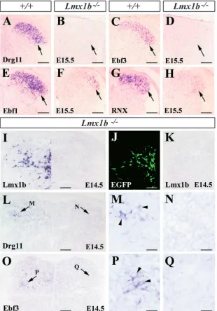
To further characterize the dorsal horn defects in Lmx1b mutants, we examined the expression profile of dorsal horn neuron molecular markers Drg11, Ebf1, Ebf2, Ebf3 and Rnx. In Lmx1b–/– embryos, Drg11 expression was completely abolished from the normal onset of its expression (Fig. 3A,B; see Fig. S1 at http://dev.biologists.org/supplemental). At E11.5, Ebf1, Ebf2, Ebf3 and Rnx appeared to be normally expressed in Lmx1b–/– embryos (see Fig. S1 at http://dev.biologists.org/supplemental). However, by E12.5, Ebf3 and Rnx expression levels were lower in Lmx1b mutants
(see Fig. S1) and by E15.5, Ebf3 expression was completely absent in the Lmx1b mutants (Fig. 3C,D). At this stage, in wild-type embryos, Ebf1 was most strongly expressed in laminae I-II neurons and weakly in laminae I-III-IV neurons. By contrast, Ebf1 expression was markedly reduced in Lmx1b mutant embryos (Fig. 3E,F). Similarly, Rnx was dramatically downregulated in the Lmx1b mutants (Fig. 3G,H).
[image:4.612.47.562.70.336.2]The complete absence of expression of Drg11 and Ebf3 expression in Lmx1b mutants promoted us to examine whether Lmx1b is able to induce the expression of these two genes in Lmx1b mutants. To address this question, Lmx1b-expression vectors were introduced into the dorsal spinal cord of Lmx1b mutant embryos by the use of in utero electroporation (Saba et al., 2003). Although the expression of Drg11 and Ebf3 was absent in the dorsal horn of Lmx1b–/– mutants electroporated with green fluorescent protein expression vectors (Fig. 3J,K; data not shown), Drg11 and Ebf3 expression was detected in the dorsal horn of Lmx1b–/– mutants electroporated with Lmx1b-expression vectors (Fig. 3L,M,O,P). The contralateral unelectroporated side of the dorsal horn showed no Drg11 and Ebf1 expression (Fig. 3L,N,O,Q). We observed that the induced expression was considerably weaker when compared with the endogenous expression, which we attribute to the inefficiency of electroporation. Because of the presence of the residual expression of Rnx and Ebf1 in the dorsal horn of Lmx1b mutants, it was difficult to determine whether Lmx1b is capable of activating these two genes in the mutants. Nevertheless, these data demonstrate that Drg11 and Ebf3 act downstream of Lmx1b in the developing dorsal horn, and Lmx1b is required either for initiating or maintaining expression of these two genes in the dorsal horn.
Fig. 2. Morphology, distribution and migration
of wild-type and Lmx1b mutant dorsal horn neurons. (A,C,E) Nissl staining of wild-type embryos. (B,D,F) Nissl staining of Lmx1b–/–
embryos. (A,B) At E12.5, no major difference was found between wild-type (A) and mutant embryos (B). (C,D) At E15.5, in wild-type, laminae I-II and laminae III-V are
distinguishable, brackets outline the domain of laminae I-II and III-V, respectively. In Lmx1b–/–embryo, no clear boundary between
laminae I-II and III-V exists (bracket outlines the whole domain). (E,F) Lower magnification of C,D. In wild-type embryo, the dorsal funiculus is enlarged (E, arrow), whereas in Lmx1b mutant it is smaller (F, arrow). (G) Wild-type embryo at E14.5: neurons labeled with BrdU at E11.5 are not detected in the outer aspect of laminae I-II (inset
indicating a high magnification). Bracket outlines the most superficial layer. (H) Lmx1b mutant embryo at E14.5: neurons labeled with BrdU at E11.5 are detected in the most superficial layer (bracket in the insert). (I,J) Higher magnification view of G,H
showing the dorsal horn that is divided into the medial one-third (Med 1/3) and lateral two-thirds (Lat 2/3) region by lines for quantitative analysis. (K) Quantitative comparison of BrdU-labeled neurons in the medial one-third region and lateral two-thirds region between wild-type (white bar) and Lmx1b–/–embryos (black bar). Asterisks indicate significant difference using Student’s t-test P<0.0001. Scale bars: 100 µm in
Lmx1b-independent dorsal horn-specific transcription factors
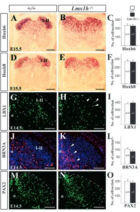
The expression of Lbx1, a lamina IIi-III marker which acts upstream of Lmx1b was examined in wild-type and Lmx1b mutant embryos (Fig. 4G,H). Although a few LBX1+neurons were found in the most superficial layer of the Lmx1b–/– dorsal horn, no significant difference in the number of LBX1+neurons was detected between the Lmx1b mutants and wild-type controls (Fig. 4I). The expression of Brn3a, a laminae III-V marker (Fig. 4J), was examined in Lmx1b mutants (Fig. 4K). Although a seemingly higher number of BRN3A+neurons were located more medially in Lmx1b mutants (Fig. 4K), neuronal counts revealed no difference in the number of BRN3A+ neurons between wild-type and Lmx1b mutants (Fig. 4L).
In the dorsal horn, Lmx1b and Pax2 expression is mutually exclusive (Gross et al., 2002). To determine whether the loss of Lmx1b affects Pax2 expression, PAX2+neurons in the dorsal
horns of the wild-type and Lmx1b–/– embryos were counted. No significant change in the number of PAX2+neurons was detected between wild-type and Lmx1b embryos (Fig. 4M-O).
Hox6 and Hox8 are homeobox genes that are expressed in laminae I-II (Graham et al., 1991). In Lmx1b mutants, Hoxb6 and Hoxb8 expression domains appeared to have expanded (Fig. 4A,B,D,E). Although the expansion of the Hoxb6 and Hoxb8 domains could be attributed to an abnormal location of laminae I-II neurons in the deep dorsal horn, it is also possible that the expression of Hoxb6 and Hoxb8 is upregulated in laminae III-V neurons in the absence of Lmx1b function. If it were the latter case, the total number of Hoxb6 and Hoxb8 neurons would increase in the mutants. However, no significant difference in the number of Hoxb6 and Hoxb8 neurons was evident between wild-type and mutant animals (Fig. 4C,F). Our data thus suggest that abnormal distribution of Hox6/8 neurons is due to migration defects.
Together, these results suggest that expression of Hox6, Hox8, Lbx1, Brn3a and Pax2 is Lmx1b independent; however, the distribution pattern of neurons expressing these markers appears changed in Lmx1b mutants.
Lmx1b, Rnx and Drg11 repress Zic1 and Zic4 genes
[image:5.612.50.360.73.517.2]We next asked whether there is a causal relationship between the expression of Zic genes and Lmx1b as Zic1 appears to be Fig. 3. Expression of laminae I-II markers and rescue
of Drg11 and Ebf3 expression by forced expression of Lmx1b in the dorsal horn of Lmx1b–/–embryos.
(A,B) Drg11 expression in the dorsal horn of wild-type (A, arrow) and Lmx1b–/–(B, arrow) embryos.
(C,D) Ebf3 expression is restricted to laminae I-II neurons in wild-type dorsal horn (arrow in C), but is absent in Lmx1b mutant embryos (arrow in D). (E,F) In wild-type embryos (E) Ebf1 is concentrated in laminae I-II (arrow in E,F), but its expression is markedly reduced in Lmx1b mutant embryos (F, arrow). (G,H) Rnx expression in laminae I-II neurons (arrows) of wild-type (G) and Lmx1b mutant embryos (H). (I,L-Q) Reactivation of Drg11 and Ebf3 expression in Lmx1b–/–spinal cord by exogenously
introduced Lmx1b. (I) Lmx1b expression in Lmx1b–/–
spinal cord after electroporation of Lmx1b-expression vectors. No Lmx1b was found in the contralateral side of the spinal cord. (J,K) Expression of EGFP control plasmids in Lmx1b–/–spinal cord. (K) No Lmx1b was
detected in the electroporated side of Lmx1b–/–spinal
cord after electroporation of EGFP. (L) Induction of Drg11 (arrow) in Lmx1b–/–spinal cord electroporated
with Lmx1b expression vectors. No Drg11 staining was found in the contralateral side of the spinal cord (arrow). (M,N) Higher magnification of the regions indicated by arrows in L. Arrowheads indicate Drg11+cells. (O) Induction of Ebf3 expression (arrow) in the dorsal horn of Lmx1b–/–embryos after
required for maintaining the progenitor state of undifferentiated dorsal neurons by repressing the differentiation of spinal cord progenitors (Aruga et al., 2002b; Ebert et al., 2003).
In wild-type embryos, three Zic genes (Zic1, Zic2 and Zic4) are expressed in the developing dorsal spinal cord (Fig. 5). Their expression is mainly concentrated around the midline region, suggesting that the Zic genes may play a role in modulating the differentiation of the dorsal horn neurons. Strikingly, in Lmx1b–/– mutants, while Zic2 expression remained unaltered (Fig. 5E,F), expression of Zic1 and Zic4 was significantly upregulated (Fig. 5A-D). This was observed not only around the midline region, but also in the superficial layer of the dorsal horn where numerous laminae I-II neurons were present but failed to differentiate further at a later stage (Fig. 5B,D). An upregulation of Zic1 and Zic4 was also detected in Rnx mutants (Fig. 5H,J).
The maturation and differentiation of lamina I-II neurons is impaired in Lmx1b–/–mutants
The aberrant migratory behavior and altered gene expression
profile of the dorsal horn neurons in Lmx1b mutants could also be viewed as the evidence of impaired maturation and differentiation of laminae I-II neurons in the absence of Lmx1b. To further examine the cellular property of laminae I-II neurons, immunocytochemical staining using anti-MAP2 antibody was performed. MAP2 is a microtubule-associated protein expressed in the dendrites, axons and somata of neurons (Lewis et al., 1986). During development, the onset of MAP2 expression concurs with the differentiation, maturation and lamination of laminae I-II neurons. In the dorsal horn of E15.5 wild-type embryos, MAP2 expression was prominent in laminae I-II neurons, especially in the dendrites (Fig. 6A,C). By contrast, MAP2 expression was much weaker in the superficial layer of the Lmx1b mutant (Fig. 6B,D). One possible explanation for the weak expression of MAP2 is that MAP2+ laminae I-II neurons could be aberrantly located in the deeper laminae of Lmx1b mutants. However, no obvious increase of MAP2 staining in these regions was found in Lmx1b mutants (Fig. 6A-D). Therefore, it is most likely that MAP2 expression is largely reduced in laminae I-II neurons of Lmx1b mutants.
Rnx and Drg11 mutants exhibit an aberrant
migration and differentiation of laminae I-II neurons
[image:6.612.43.333.64.503.2]The downregulation of Rnx and the loss of Drg11 expression in Lmx1b mutants suggest that Lmx1b may control some aspects of the migration and differentiation via Rnx and Drg11. A previous study using BrdU labeling detected no migration deficit in the dorsal horn of Rnx mutants (Qian et al., 2002). We revisited this issue by examining the inside-out pattern of the dorsal horn neurons in Rnx and Drg11 mutants and found that neurons labeled with BrdU at E11.5 were detected in the superficial region of the dorsal horn of Rnx and Drg11 mutants (Fig. 7A,B,D,E). Quantitative analysis indicated that in Rnx
Fig. 4. Expression of dorsal horn-specific molecular
markers in wild-type and Lmx1b–/–embryos at E14.5 (G-N)
and E15.5 (A-E). (A,B) Hoxb6 expression in wild-type (A) and Lmx1b mutant embryos (B). (D,E) Spinal cord Hoxb8 expression is mainly evident in laminae I-II of wild type (D); however, in Lmx1b mutant embryo (E) its expression appears expanded ventrally. (C,F) Quantitative analysis of Hoxb6+(C) and Hoxb8+(F) neurons in wild-type (white bar) and Lmx1b–/–embryos (black bar). (G,H) Expression
of Lbx1 in wild-type (G) and Lmx1b mutant embryos (H). Broken white lines outline the dorsal horn and arrowheads indicate mispositioned LBX1+neurons in Lmx1b mutant
embryos. (I) Quantitative analysis of the number of LBX1+
neurons between wild-type and Lmx1b mutant embryos. (J,K) BRN3A+expression in wild-type (J) and Lmx1b–/–
embryos (K) at E14.5. Arrowheads indicate the accumulation of BRN3A+neurons around the midline
(asterisk). (L) Quantitative comparison of the total number of BRN3A+neurons between wild-type and Lmx1b–/–
embryos. (M,N) PAX2 expression in wild-type (M) and Lmx1b mutant embryos (N). (O) Numbers of PAX2+
and Drg11 mutants there were more BrdU+ neurons accumulating in the medial one third of the dorsal horn when compared to controls, whereas fewer BrdU+ neurons were found in the lateral two-thirds of the dorsal horn (Fig. 7C,F). Immunocytochemical staining shows that MAP2 expression was also reduced in the superficial dorsal horn of both mutants (Fig. 7G,H,I,J). Nevertheless, the abnormalities detected in Drg11 mutant are less severe than those in Rnx mutants.
Aberrant projection of TrkA+afferent fibers into the dorsal horn of Lmx1b mutants
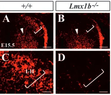
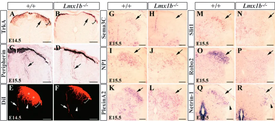
We reasoned that the impaired development of laminae I-II neurons in Lmx1b mutants could influence the projections of the primary cutaneous afferents which innervate the dorsal horn (Sharma and Frank, 1998). To address this possibility, we examined the projections of TrkA+ fibers into laminae I-II (Huang et al., 1999). At E14.5, in wild-type mice, TrkA+ afferents had begun to ramify within laminae I-II of the dorsal horn, and immunostaining showed densely stained laminae I-II, an indication of innervation by primary cutaneous afferents (Ozaki and Snider, 1997) (Fig. 8A). Strikingly, although Lmx1b mutants did not show apparent defects in projections
of muscle afferents, as examined by anti-Peripherin antibody staining (Goldstein et al., 1991) (Fig. 8C,D), no TrkA+ afferents were detected within the gray matter of the dorsal horn of Lmx1b mutants (Fig. 8B).
We next analyzed the sensory afferent projection by DiI labeling of DRG neurons. At E14.5, in wild-type controls, projections to laminae I-IV were intensely labeled with DiI after its application to DRG cell bodies (Fig. 8E). The muscle sensory afferents that project towards the ventral horn were also labeled (Fig. 8E). In Lmx1b mutants, while the presumptive muscle sensory afferent projections appeared normal, there was a dramatic decrease in the intensity of DiI labeling in laminae I-II, although some DiI-labeled DRG afferents appeared to be present in laminae III-IV (Fig. 8F). Together, these results suggest that DRG afferent projection defects in Lmx1b mutants are specific to TrkA+ sensory afferents.
[image:7.612.114.502.72.203.2]Stereotyped expression of attractive or repulsive axonal guidance molecules is crucial to guiding the precise axonal growth of neurons in the developing nervous system (Tessier-Lavigne and Goodman, 1996). To determine whether there was a change of expression of axonal guidance cues/molecules in the dorsal horn of Lmx1b mutants, we examined their expression by in situ hybridization. Sema3a is a secreted cell-surface protein that functions as a chemorepellent in the projection of cutaneous afferents in the spinal cord of the chick and the mouse (Luo et al., 1993; Messersmith et al., 1995). Its expression, however, was normal in Lmx1b mutants (data not shown). We also found that Sema3c (Feiner et al., 2001) was completely lost in the dorsal horn of Lmx1b mutants (Fig. 8G,H). By contrast, expression of the receptors for Sema3a, neuropilin 1 (Nrp1) and plexin A2 (He and Tessier-Lavigne, 1997; Kolodkin et al., 1997; Takahashi et al., 1999), were markedly reduced in the mutants (Fig. 8I-L). We also examined the expression of the members of the Slit family, which have been implicated in axonal guidance and neuronal migration (Brose and Tessier-Lavigne, 2000; Wong et al., 2002). Slit1 and Robo2 were most strongly expressed in laminae I-II (Fig. 8M,O). In the mutants, Slit1 and Robo2 expression was dramatically reduced (Fig. 8N,P). In addition, netrin 1 expression was largely lost in the medial region of the deep dorsal horn of Lmx1b mutants (Serafini et al., 1994) (Fig. 8Q,R). Thus, the altered expression of multiple axonal Fig. 6. MAP2 expression in laminae I-II neurons in the dorsal horn
of wild-type and Lmx1b–/–embryos at E15.5. (A,B) MAP2 staining
[image:7.612.88.265.531.683.2]in laminae I-II of wild-type (bracket in A) and Lmx1b mutant embryos (bracket in B). (C,D) Higher magnification of A,B, respectively. Scale bars: 100 µm in A,B; 20 µm in C,D.
Fig. 5. Comparison of Zic1, Zic2 and Zic4 expression in the dorsal horns of wild-type, Lmx1b–/–and Rnx–/– embryos at E14.5. (A,B,G,H) Zic1
Fig. 8. Selective block of the ingrowth of cutaneous afferents in the dorsal spinal cord of Lmx1b–/–mutant embryos at E14.5.
(A,B) Immunocytochemical detection of TrkA in the dorsal horn of wild-type (A) and Lmx1b–/–mutant (B) embryos. Arrows indicate the
[image:8.612.68.532.414.619.2]superficial laminae region. Brackets outline laminae I-II region. (C,D) Peripherin detection in the dorsal horn of wild-type (C) and Lmx1b mutant embryos (D) appears unchanged. Arrows indicate peripherin-labeled presumptive proprioceptive afferents. (E,F) DiI labeling of primary afferents in the dorsal horn of wild-type (E) and Lmx1b mutant embryos (F). Arrowheads indicate presumptive mechanoreceptor afferents and arrows indicate presumptive muscle proprioceptive afferents. Asterisks and brackets outline laminae I-II region. (G,H) Sema3c expression in wild-type (G) and Lmx1b mutant embryos (H). (I,J) Nrp1 expression in wild-type (I) and Lmx1b mutant embryos (J). (K,L) Plexin A2 expression in wild-type (K) and Lmx1b mutant embryos (L). (M,N) Slit1 expression in wild-type (M) and Lmx1b mutant embryos (N). (O,P) Robo2 expression in wild-type (O) and Lmx1b mutant embryos (P). (Q,R) Netrin 1 expression in wild-type (Q) and Lmx1b mutant embryos (R). Arrows in G-S indicate laminae I-II. Arrowheads in Q and R indicate the lateral region of laminae I-II. Scale bars: 200 µm in A, B; 100 µm in C-R.
Fig. 7. Aberrant migration and defective
differentiation of the dorsal horn neurons in wild-type and Rnx–/– and Drg11–/–
mutant embryos. (A) Neurons labeled with BrdU at E11.5 are not detected in the outer layer of laminae I-II (bracket) in wild-type embryo at E14.5 (inset). (B) Neurons labeled with BrdU at E11.5 are detected in the outer layer of laminae I-II in Rnx–/– embryo (inset, bracket).
(C) Quantitative comparison of the number of BrdU+neurons in the medial
one-third (Med 1/3) and lateral two-thirds (Lat 2/3) regions between wild-type and Rnx–/– embryos (*P<0.001).
(D,E) Comparison of the distribution pattern of neurons labeled with BrdU at E11.5 in the dorsal horn of E14.5 wild-type (D) and Drg11 mutants (E). (F) Quantitative analysis of the numbers of BrdU-labeled neurons in the medial and lateral regions of wild-type and Drg11 mutant embryos (*P<0.001). (G,H) MAP2 expression in the dorsal horn of wild-type (G, bracket) and Rnx mutant embryos (H, bracket). MAP2 expression of the dorsal horn in wild-type (I, bracket) and Drg11–/–mutant embryos
(J, bracket). Scale bars: in B and C, 100
guidance molecules could account for the failure of cutaneous sensory axon ingrowth in Lmx1b mutants.
Discussion
Lmx1b is required for the specification of the dI5 neuronal fate
Recent studies have begun to identify important players such as Math1, Lbx1 and Rnx in the development of early-born postmitotic dorsal neurons (Bermingham et al., 2001; Gross et al., 2002; Muller et al., 2002). In this study, we find that dI5 interneurons can be further divided into two subgroups: one that expresses Phox2a and one that does not. Phox2a is a homeobox gene and has been shown to be an important determinant of several neuronal phenotype in the nervous system (Brunet and Pattyn, 2002). The seemingly normal expression of Lbx1 and Brn3a expression in dI5 neurons of Lmx1b mutants supports the idea that these two genes may act upstream of Lmx1b, whereas the partial loss of Phox2a indicates that Phox2a lies either downstream of or in parallel to Lmx1b. It will be interesting to examine whether Phox2a plays a role in the development of dI5 neurons, and whether Lbx1 and Rnx may specify the fate of dI5 neurons through an Lmx1b-Phox2a pathway.
Lmx1b controls the ingrowth of cutaneous afferents into the dorsal horn
In Lmx1b mutants, the selective blocking of cutaneous afferent ingrowth raises the possibility that Lmx1b may coordinate the projection of TrkA+afferents into the dorsal horn by regulating local axonal guidance cue(s). Altered ingrowth of TrkA+ afferents is also found in Lbx1 mutants and Rnx/Tlx1 double mutants (Gross et al., 2002; Muller et al., 2002; Qian et al., 2002). Nevertheless, unlike Lbx1 and Rnx, Lmx1b is not expressed in DRG neurons, therefore, the TrkA guidance defect most probably resides in the dorsal horn.
The specific block of the entry of TrkA+afferents suggests that certain attractants may be missing in the mutants. Alternatively, the expression level of some repulsive molecules may be increased to repel the cutaneous afferents. Although these possibilities exist, no evidence for increased expression of any repellants examined in Lmx1b mutants was found. Given that multiple axonal guidance cues are expressed in the dorsal horn and their expression is lost in Lmx1b mutants, it is possible that multiple axonal guidance molecules could work synergistically to coordinate the projections of cutaneous afferents. Future analysis of mice lacking multiple axonal guidance cues may be required to reveal the identity of the cues responsible for the ingrowth of TrkA+afferents in the dorsal spinal cord.
Lmx1b guides the differentiation and migration of the dorsal horn neurons
Our study suggests that Zic1, Zic4, Ebf1 and Ebf3 function downstream of Lmx1b and Rnx. Forced expression of Zic1 in chicks represses the differentiation of Math1-expressing neurons in the dorsal neural tube (Ebert et al., 2003). Transgenic mice overexpressing Zic1 exhibit inhibited neuronal differentiation with extension of the progenitor state in the dorsal spinal cord (Aruga et al., 2002b). Loss of Zic1 in mice also leads to premature expression of the βIII tubulin in
the dorsal spinal cord (Aruga et al., 2002b). In the context of these findings, the Zic gene expression upregulation is interesting, and consistent with the migration and differentiation defects in Lmx1b mutants. Zic1 could be an important component of the dorsal spinal cord neuron differentiation pathway.
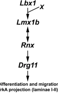
The role of Zic4, Ebf1 and Ebf3 in the development of the dorsal horn is unknown. Whether dysregulation of these genes plays a causal role in aberrant development of the dorsal horn also remains to be determined. Together with previous studies, we hypothesize that the Lbx1/Lmx1b/Rnx/Drg11 pathway represents a major pathway to control the differentiation and migration of laminae I-II neurons and subsequent projection of cutaneous afferents (Fig. 9). The effects of Lmx1b and Rnx may be mediated in part through the Drg11, Ebf and Zic genes and in part via other unidentified transcription factors to control the assembly of the dorsal horn circuits.
The present study reveals the complicated relationship between Lmx1b and other transcription factors in the development of the dorsal spinal cord. For example, Lmx1b and Rnx may depend on each other for their normal expression. Although Lmx1b mutants recapitulate many aspects of the phenotype of Rnx mutants, there are noted differences in the level of regulation of downstream genes such as Zic1 and Drg11. Thus, in addition to the common sets of downstream targets, Lmx1b and Rnx may control additional distinct set of target genes.
[image:9.612.392.495.73.236.2]The finding that an inside-out migration pattern and a normal differentiation program are disrupted in the dorsal horn of Lmx1b mutants raises the question of whether aberrant migratory behavior could be attributed to aberrant differentiation of the dorsal horn neurons or vice versa. Fig. 9. Schematic diagram summarizing the transcriptional cascade
Although we are not able to determine when the migration deficit first occurs in Lmx1b mutants, studies of the possible downstream targets of Lmx1b shed light onto this issue. For example, an alteration of axonal guidance cues such as netrin 1 in Lmx1b mutants might have contributed to aberrant neuronal migration (Brose and Tessier-Lavigne, 2000). Moreover, some of the transcription factors downstream of Lmx1b have also been shown to be required for neuronal migration. In mice, Ebf1 is important for the migration of facial branchiomotor neurons (Garel et al., 2000). In chicks, Ebf1 and Ebf3 appear to control migration and differentiation of the dorsal neurons independently (Garcia-Dominguez et al., 2003; Garel et al., 2000). Thus, Lmx1b may have a unique role in neuronal migration in the developing spinal cord. However, this does not exclude the possibility that aberrant migration reflects some aspects of impaired differentiation of the dorsal horn neurons in the absence of Lmx1b. In fact, in addition to the dorsal horn cells, Lmx1b has also been implicated in neuronal differentiation of dopaminergic neurons and serotonergic neurons in the developing brain (Cheng et al., 2003; Ding et al., 2003; Pressman et al., 2000). Thus, it is likely that Lmx1b could play an important role in both cellular events.
We thank D. J. Anderson, J. Brunet, E. Turner, R. Krumlauf, M. R. Capecchi, T. Jessell, J. Aruga, L. Reichardt, T. Muller, C. Birchmeier, J. Miyazaki, Y. Rao, J. Wu, M. Tessier-Lavigne, H. Fujisawa, A. Kolodkin and Q. Ma for reagents. We are particularly grateful for Dr T. Saito’s advice on in utero electroporation technique. We also thank T. Jessell, J. Sanes, Y. Rao, A. L. Pearlman, I. McIntosh and M. Jacquin for comments on various versions of the manuscript; and H. Xu, S. Li, C. Xiang and Z. Zhang for technical help. This work was supported, in part, by a grant from the McDonnell Center for Cellular and Molecular Neurobiology at Washington University, and a NIH grant NS43968-01 (Z.F.C.). All animal experiments were reviewed and approved by the Animal Studies Committee (ASC) at Washington University.
References
Adams, K. A., Maida, J. M., Golden, J. A. and Riddle, R. D. (2000). The
transcription factor Lmx1b maintains Wnt1 expression within the isthmic organizer. Development 127, 1857-1867.
Aruga, J., Yokota, N., Hashimoto, M., Furuichi, T., Fukuda, M. and Mikoshiba, K. (1994). A novel zinc finger protein, zic, is involved in
neurogenesis, especially in the cell lineage of cerebellar granule cells. J.
Neurochem. 63, 1880-1890.
Aruga, J., Nagai, T., Tokuyama, T., Hayashizaki, Y., Okazaki, Y., Chapman, V. M. and Mikoshiba, K. (1996a). The mouse zic gene family.
Homologues of the Drosophila pair-rule gene odd-paired. J. Biol. Chem.
271, 1043-1047.
Aruga, J., Yozu, A., Hayashizaki, Y., Okazaki, Y., Chapman, V. M. and Mikoshiba, K. (1996b). Identification and characterization of Zic4, a new
member of the mouse Zic gene family. Gene 172, 291-294.
Aruga, J., Minowa, O., Yaginuma, H., Kuno, J., Nagai, T., Noda, T. and Mikoshiba, K. (1998). Mouse Zic1 is involved in cerebellar development.
J. Neurosci. 18, 284-293.
Aruga, J., Inoue, T., Hoshino, J. and Mikoshiba, K. (2002a). Zic2 controls
cerebellar development in cooperation with Zic1. J. Neurosci. 22, 218-225.
Aruga, J., Tohmonda, T., Homma, S. and Mikoshiba, K. (2002b). Zic1
promotes the expansion of dorsal neural progenitors in spinal cord by inhibiting neuronal differentiation. Dev. Biol. 244, 329-341.
Bermingham, N. A., Hassan, B. A., Wang, V. Y., Fernandez, M., Banfi, S., Bellen, H. J., Fritzsch, B. and Zoghbi, H. Y. (2001). Proprioceptor
pathway development is dependent on Math1. Neuron 30, 411-422.
Brose, K. and Tessier-Lavigne, M. (2000). Slit proteins: key regulators of
axon guidance, axonal branching, and cell migration. Curr. Opin. Neurobiol.
10, 95-102.
Brunet, J. F. and Pattyn, A. (2002). Phox2 genes - from patterning to
connectivity. Curr. Opin. Genet. Dev. 12, 435-440.
Caspary, T. and Anderson, K. V. (2003). Patterning cell types in the
dorsal spinal cord: what the mouse mutants say. Nat. Rev. Neurosci. 4, 289-297.
Chen, H., Lun, Y., Ovchinnikov, D., Kokubo, H., Oberg, K. C., Pepicelli, C. V., Gan, L., Lee, B. and Johnson, R. L. (1998a). Limb and kidney
defects in Lmx1b mutant mice suggest an involvement of LMX1B in human nail patella syndrome. Nat. Genet. 19, 51-55.
Chen, Z. F., Paquette, A. J. and Anderson, D. J. (1998b). NRSF/REST is
required in vivo for repression of multiple neuronal target genes during embryogenesis. Nat. Genet. 20, 136-142.
Chen, Z. F., Rebelo, S., White, F., Malmberg, A. B., Baba, H., Lima, D., Woolf, C. J., Basbaum, A. I. and Anderson, D. J. (2001). The paired
homeodomain protein DRG11 is required for the projection of cutaneous sensory afferent fibers to the dorsal spinal cord. Neuron 31, 59-73.
Cheng, L., Chen, C. L., Luo, P., Tan, M., Qiu, M., Johnson, R. and Ma, Q. (2003). Lmx1b, Pet-1, and Nkx2.2 coordinately specify serotonergic
neurotransmitter phenotype. J. Neurosci. 23, 9961-9967.
Christensen, B. N. and Perl, E. R. (1970). Spinal neurons specifically excited
by noxious or thermal stimuli: marginal zone of the dorsal horn. J.
Neurophysiol. 33, 293-307.
Ding, Y. Q., Marklund, U., Yuan, W., Yin, J., Wegman, L., Ericson, J., Deneris, E., Johnson, R. L. and Chen, Z. F. (2003). Lmx1b is
essential for the development of serotonergic neurons. Nat. Neurosci. 6, 933-938.
Dreyer, S. D., Zhou, G., Baldini, A., Winterpacht, A., Zabel, B., Cole, W., Johnson, R. L. and Lee, B. (1998). Mutations in LMX1B cause abnormal
skeletal patterning and renal dysplasia in nail patella syndrome. Nat. Genet.
19, 47-50.
Ebert, P. J., Timmer, J. R., Nakada, Y., Helms, A. W., Parab, P. B., Liu, Y., Hunsaker, T. L. and Johnson, J. E. (2003). Zic1 represses Math1
expression via interactions with the Math1 enhancer and modulation of Math1 autoregulation. Development 130, 1949-1959.
Fedtsova, N. G. and Turner, E. E. (1995). Brn-3.0 expression identifies early
post-mitotic CNS neurons and sensory neural precursors. Mech. Dev. 53, 291-304.
Feiner, L., Webber, A. L., Brown, C. B., Lu, M. M., Jia, L., Feinstein, P., Mombaerts, P., Epstein, J. A. and Raper, J. A. (2001). Targeted disruption
of semaphorin 3C leads to persistent truncus arteriosus and aortic arch interruption. Development 128, 3061-3070.
Garcia-Dominguez, M., Poquet, C., Garel, S. and Charnay, P. (2003). Ebf
gene function is required for coupling neuronal differentiation and cell cycle exit. Development 130, 6013-6025.
Garel, S., Marin, F., Mattei, M. G., Vesque, C., Vincent, A. and Charnay, P. (1997). Family of Ebf/Olf-1-related genes potentially involved in
neuronal differentiation and regional specification in the central nervous system. Dev. Dyn. 210, 191-205.
Garel, S., Garcia-Dominguez, M. and Charnay, P. (2000). Control of the
migratory pathway of facial branchiomotor neurones. Development 127, 5297-5307.
Goldstein, M. E., House, S. B. and Gainer, H. (1991). NF-L and peripherin
immunoreactivities define distinct classes of rat sensory ganglion cells. J.
Neurosci. Res. 30, 92-104.
Graham, A., Maden, M. and Krumlauf, R. (1991). The murine Hox-2 genes
display dynamic dorsoventral patterns of expression during central nervous system development. Development 112, 255-264.
Gross, M. K., Dottori, M. and Goulding, M. (2002). Lbx1 specifies
somatosensory association interneurons in the dorsal spinal cord. Neuron
34, 535-549.
He, Z. and Tessier-Lavigne, M. (1997). Neuropilin is a receptor for the axonal
chemorepellent Semaphorin III. Cell 90, 739-751.
Helms, A. W. and Johnson, J. E. (2003). Specification of dorsal spinal cord
interneurons. Curr. Opin. Neurobiol. 13, 42-49.
Hobert, O., Tessmar, K. and Ruvkun, G. (1999). The Caenorhabditis elegans
lim-6 LIM homeobox gene regulates neurite outgrowth and function of particular GABAergic neurons. Development 126, 1547-1562.
Huang, E. J. and Reichardt, L. F. (2001). Neurotrophins: roles in neuronal
development and function. Annu. Rev. Neurosci. 24, 677-736.
Huang, E. J., Wilkinson, G. A., Farinas, I., Backus, C., Zang, K., Wong, S. L. and Reichardt, L. F. (1999). Expression of Trk receptors in the
developing mouse trigeminal ganglion: in vivo evidence for NT-3 activation of TrkA and TrkB in addition to TrkC. Development 126, 2191-2203.
limb imposed by LIM homeodomain protein regulation of Ephrin-A:EphA interactions. Neuron 38, 581-596.
Kania, A., Johnson, R. L. and Jessell, T. M. (2000). Coordinate roles for
LIM homeobox genes in directing the dorsoventral trajectory of motor axons in the vertebrate limb. Cell 102, 161-173.
Kaufman, M. H. (1998). The Atlas of Mouse Development. San Diego:
Academic Press.
Kolodkin, A. L., Levengood, D. V., Rowe, E. G., Tai, Y. T., Giger, R. J. and Ginty, D. D. (1997). Neuropilin is a semaphorin III receptor. Cell 90,
753-762.
Lawson, S. N. and Biscoe, T. J. (1979). Development of mouse dorsal root
ganglia: an autoradiographic and quantitative study. J. Neurocytol. 8, 265-274.
Lee, K. J. and Jessell, T. M. (1999). The specification of dorsal cell fates in
the vertebrate central nervous system. Annu. Rev. Neurosci. 22, 261-294.
Lewis, S. A., Villasante, A., Sherline, P. and Cowan, N. J. (1986).
Brain-specific expression of MAP2 detected using a cloned cDNA probe. J. Cell
Biol. 102, 2098-2105.
Liem, K. F., Jr, Tremml, G. and Jessell, T. M. (1997). A role for the roof
plate and its resident TGFbeta-related proteins in neuronal patterning in the dorsal spinal cord. Cell 91, 127-138.
Luo, Y., Raible, D. and Raper, J. A. (1993). Collapsin: a protein in brain that
induces the collapse and paralysis of neuronal growth cones. Cell 75, 217-227.
Matsunaga, E., Katahira, T. and Nakamura, H. (2002). Role of Lmx1b and
Wnt1 in mesencephalon and metencephalon development. Development
129, 5269-5277.
Messersmith, E. K., Leonardo, E. D., Shatz, C. J., Tessier-Lavigne, M., Goodman, C. S. and Kolodkin, A. L. (1995). Semaphorin III can function
as a selective chemorepellent to pattern sensory projections in the spinal cord. Neuron 14, 949-959.
Molander, C. and Grant, G. (1995). Spinal cord cytoarchitecture. In The Rat
Nervous System (ed. G. Paxinos). Sydney: Academic Press.
Muller, T., Brohmann, H., Pierani, A., Heppenstall, P. A., Lewin, G. R., Jessell, T. M. and Birchmeier, C. (2002). The homeodomain factor lbx1
distinguishes two major programs of neuronal differentiation in the dorsal spinal cord. Neuron 34, 551-562.
Niwa, H., Yamamura, K. and Miyazaki, J. (1991). Efficient selection for
high-expression transfectants with a novel eukaryotic vector. Gene 108, 193-199.
Nornes, H. O. and Das, G. D. (1974). Temporal pattern of neurogenesis in
spinal cord of rat. I. An autoradiographic study - time and sites of origin and migration and settling patterns of neuroblasts. Brain Res. 73, 121-138.
Nornes, H. O. and Carry, M. (1978). Neurogenesis in spinal cord of mouse:
an autoradiographic analysis. Brain Res. 159, 1-6.
Ozaki, S. and Snider, W. D. (1997). Initial trajectories of sensory axons
toward laminar targets in the developing mouse spinal cord. J. Comp.
Neurol. 380, 215-229.
Pressman, C. L., Chen, H. and Johnson, R. L. (2000). LMX1B, a LIM
homeodomain class transcription factor, is necessary for normal development of multiple tissues in the anterior segment of the murine eye.
Genesis 26, 15-25.
Qian, Y., Shirasawa, S., Chen, C. L., Cheng, L. and Ma, Q. (2002). Proper
development of relay somatic sensory neurons and D2/D4 interneurons requires homeobox genes Rnx/Tlx-3 and Tlx-1. Genes Dev. 16, 1220-1233.
Rexed, B. (1952). The cytoarchitectonic organization of the spinal cord in the
cat. J. Comp. Neurol. 96, 415-496.
Riddle, R. D., Ensini, M., Nelson, C., Tsuchida, T., Jessell, T. M. and Tabin, C. (1995). Induction of the LIM homeobox gene Lmx1 by WNT7a
establishes dorsoventral pattern in the vertebrate limb. Cell 83, 631-640.
Roberts, C. W., Shutter, J. R. and Korsmeyer, S. J. (1994). Hox11 controls
the genesis of the spleen. Nature 368, 747-749.
Saba, R., Nakatsuji, N. and Saito, T. (2003). Mammalian BarHI confers
commissural neuron identity on dorsal cells in the spinal cord. J. Neurosci.
23, 1987-1991.
Saito, T. and Nakatsuji, N. (2001). Efficient gene transfer into the embryonic
mouse brain using in vivo electroporation. Dev. Biol. 240, 237-246.
Scott, S. A. (1992). Sensory neurons: diversity, development, and plasticity.
In The Development of Peripheral Sensory Innervation Patterns (ed. S. A. Scott), pp. 242-263. New York: Oxford Univeristy Press.
Serafini, T., Kennedy, T. E., Galko, M. J., Mirzayan, C., Jessell, T. M. and Tessier-Lavigne, M. (1994). The netrins define a family of axon
outgrowth-promoting proteins homologous to C. elegans UNC-6. Cell 78, 409-424.
Sharma, K. and Frank, E. (1998). Sensory axons are guided by local cues
in the developing dorsal spinal cord. Development 125, 635-643.
Smidt, M. P., Asbreuk, C. H., Cox, J. J., Chen, H., Johnson, R. L. and Burbach, J. P. (2000). A second independent pathway for development of
mesencephalic dopaminergic neurons requires Lmx1b. Nat. Neurosci. 3, 337-341.
Snider, W. D. (1994). Functions of the neurotrophins during nervous system
development: what the knockouts are teaching us. Cell 77, 627-638.
Takahashi, T., Fournier, A., Nakamura, F., Wang, L. H., Murakami, Y., Kalb, R. G., Fujisawa, H. and Strittmatter, S. M. (1999).
Plexin-neuropilin-1 complexes form functional semaphorin-3A receptors. Cell 99, 59-69.
Tanabe, Y., William, C. and Jessell, T. M. (1998). Specification of motor
neuron identity by the MNR2 homeodomain protein. Cell 95, 67-80.
Tessier-Lavigne, M. and Goodman, C. S. (1996). The molecular biology of
axon guidance. Science 274, 1123-1133.
Tiveron, M. C., Hirsch, M. R. and Brunet, J. F. (1996). The expression
pattern of the transcription factor Phox2 delineates synaptic pathways of the autonomic nervous system. J. Neurosci. 16, 7649-7660.
Vogel, A., Rodriguez, C., Warnken, W. and Izpisua Belmonte, J. C. (1995).
Dorsal cell fate specified by chick Lmx1 during vertebrate limb development. Nature 378, 716-720.
Wang, S. S., Tsai, R. Y. L. and Reed, R. R. (1997). The characterization of
the Olf-1/EBF-like HLH transcription factor family: implications in olfactory gene regulation and neuronal development. J. Neurosci. 17, 4149-4158.
Willis, W. D., Westlund, K. N. and Carlton, S. M. (1995). Pain. In The Rat
Nervous System (ed. G. Paxinos). pp. 725-750. Sydney: Academic Press.
Wong, K., Park, H. T., Wu, J. Y. and Rao, Y. (2002). Slit proteins: molecular
guidance cues for cells ranging from neurons to leukocytes. Curr. Opin.