RESEARCH ARTICLE
Control of E-cadherin apical localisation and morphogenesis by a
SOAP-1/AP-1/clathrin pathway in
C. elegans
epidermal cells
Ghislain Gillard1,2,‡, Massiullah Shafaq-Zadah1,2,*,‡, Ophélie Nicolle1,2, Raghida Damaj1,2, Jacques Pécréaux1,2 and Grégoire Michaux1,2,§
ABSTRACT
E-cadherin (E-cad) is the main component of epithelial junctions in multicellular organisms, where it is essential for cell-cell adhesion. The localisation of E-cad is often strongly polarised in the apico-basal axis. However, the mechanisms required for its polarised distribution are still largely unknown. We performed a systematic RNAi screenin vivoto identify genes required for the strict E-cad apical localisation in C. elegans epithelial epidermal cells. We found that the loss of clathrin, its adaptor AP-1 and the AP-1 interactor SOAP-1 induced a basolateral localisation of E-cad without affecting the apico-basal diffusion barrier. We further found that SOAP-1 controls AP-1 localisation, and that AP-1 is required for clathrin recruitment. Finally, we also show that AP-1 controls E-cad apical delivery and actin organisation during embryonic elongation, the final morphogenetic step of embryogenesis. We therefore propose that a molecular pathway, containing SOAP-1, AP-1 and clathrin, controls the apical delivery of E-cad and morphogenesis.
KEY WORDS: Epithelial polarity, E-cadherin, Membrane traffic, AP-1, Caenorhabditis elegans
INTRODUCTION
Adherens junctions and their essential component E-cadherin (E-cad) play a key role in maintaining the integrity of epithelia and preventing epithelium-mesenchyme transitions. Many studies have highlighted the role of membrane traffic in junction maintenance, and intracellular routes have been definedin vivoin Drosophila. In particular, it was shown that dynamin, Rab5, Rab11 or the apical PAR module control the clustering, endocytosis and recycling of E-cad (Blankenship et al., 2007; Georgiou et al., 2008; Harris and Tepass, 2008; Langevin et al., 2005; Leibfried et al., 2008; Levayer and Lecuit, 2013; Levayer et al., 2011; Shaye et al., 2008). In zebrafish embryos, the loss of the adaptor complex AP-1 induces a decrease in E-cad staining at the plasma membrane (Montpetit et al., 2008). In mammalian cells, many studies have also examined the factors implicated in E-cad endocytosis, recycling or degradation (Fujita et al., 2002; Ling et al., 2007; Palacios et al., 2001; Wang et al., 2007). However, whereas E-cad has a polarised distribution along the apico-basal axis in most cell types and tissues (Lynch and Hardin, 2009; St Johnston and Ahringer, 2010),
surprisingly, the sorting mechanisms required for this polarised localisation are largely unknown, except for a potential role of the exocyst subunit Exo70 and of clathrin in mammalian cells (Deborde et al., 2008; Xiong et al., 2012). As a result, the phenotypic consequences of the loss of E-cad polarised distribution are not known.
To identify factors required for E-cad apical localisationin vivoin C. elegansepidermal cells we performed a systematic RNAi screen. We found that clathrin, its adaptor AP-1 and the AP-1 physical interactor p200/HEATR5B are all required to prevent E-cad basolateral localisation. The analysis of the AP-1 loss of function phenotype further shows that the associated embryonic lethality is caused by junction mislocalisation and disorganisation of the actin cytoskeleton during morphogenesis.
RESULTS
Identification of genes required for E-cad apical localisation To identify genes required for E-cad polarised localisation we chose to examine the junctions of the C. elegans epidermis (supplementary material Fig. S1A-C), where E-cad is apical. We observed E-cad in vivo with a functional E-cad::gfp (Achilleos et al., 2010) co-expressed with adlg-1::rfpconstruct (Diogon et al., 2007) to localise the boundary between the apical and lateral domains. In these cells, E-cad is mostly apical, as we could only detect a very weak lateral localisation: quantification showed that the lateral/cytoplasmic ratio of E-cad in control larvae was <1.1 at 48 h after hatching (n=186; Fig. 1C) and down to 1 at 72 h (n=175; supplementary material Table S2). We performed RNAi on 503 conserved genes which were selected based on their predicted function in membrane traffic or cytoskeleton organisation (Shaye and Greenwald, 2011), or on their identification in genome-wide C. elegans membrane traffic screens (Balklava et al., 2007; Winter et al., 2012) (supplementary material Table S1). E-cad localisation was observed after 48 h and 72 h of RNAi. About 10% of these genes affected E-cad expression levels or induced an intracellular accumulation (supplementary material Table S1). We also found that the depletion of ten genes induced a strong and consistent lateral mislocalisation of E-cad; four of these genes encode the clathrin heavy chain (chc-1), theσandγsubunits of the clathrin adaptor AP-1 (aps-1andapg-1) and the AP-1-interacting protein HEATR5B/p200/ Laa1p [c13f10.4, hereafter calledsoap-1for Sorting of apical proteins (Lui et al., 2003)] (Fig. 1Ab-Ah). Each AP-1 µ subunit alone was sufficient to target E-cad to the apical membrane but simultaneous depletion of both µ subunits recapitulated the complete loss of AP-1 (Fig. 1Ad-Af; 3D reconstructions shown in supplementary material Movies 1A,B). After quantification we found an average lateral/ cytoplasmic ratio≥1.7 of E-cad at the basolateral membrane at 48 h and 72 h for all positive genes (Fig. 1C and supplementary material Table S2); the apical/cytoplasmic ratio was not strongly affected (Fig. 1B) except for clathrin, which leads to rapid decrease in E-cad
Received 28 September 2014; Accepted 2 March 2015
1
CNRS, UMR6290, Institut de Génétique et Développement de Rennes, Rennes F-35043, France.2Universitéde Rennes 1, UEB, SFR Biosit, Facultéde Médecine, Rennes F-35043, France.
*Present address: Unitéde Recherche INSERM U1143-CNRS UMR3666, Institut Curie, 26 rue d’Ulm, 75248 Paris Cedex 05, France.
‡
These authors contributed equally to this work
§
Author for correspondence (gmichaux@univ-rennes1.fr)
DEVEL
O
signal. These four factors are therefore essential to prevent E-cad lateral accumulation.
This screen also included other AP-1 interactors (Lui et al., 2003) and genes known to affect E-cad trafficking in other organisms, such as dynamin,rab-5,rab-11, the exocyst complex and the type Iγ phosphatidylinositol phosphate kinase (ppk-1 in C. elegans) (Ivanov and Naydenov, 2013); we also targeted genes implicated in apical sorting in C. elegans, such as glycosphingolipid (GSL) biosynthetic enzymes (Zhang et al., 2011). We found a weaker
[image:2.612.100.515.55.485.2]E-cad signal following the depletion of some GSL biosynthetic enzymes, probably owing to the penetrant larval lethality, but the localisation of E-cad was not affected (supplementary material Table S1 and Fig. S2); the depletion ofppk-1did not alter E-cad localisation despite a highly penetrant larval lethality (supplementary material Fig. S2). Depletion of dynamin, rab-5, rab-11 or the exocyst complex induced a systematic intracellular accumulation close to the apical membrane (Fig. 1Ai-k′; supplementary material Fig. S2), indicating an essential role for Fig. 1. Identification of genes required for E-cad apical sorting.(A) Larvae expressing E-cad::GFP and DLG-1::RFP were observed by confocal microscopy
72 h (or 48 h forchc-1(RNAi)due to the strength of the phenotype) after RNAi induction. Left panels (Apical) show an apical view; middle panels (Lateral)
show sections taken 3 µm below the apical section shown on the left in the same larva; right panels (Z) show theZ-reconstruction obtained in the same larva. In
control larvae, E-cad is only visible apically as two parallel lines separating the seam cells and the dorso-ventral epidermis. Inaps-1(RNAi),apg-1(RNAi),apm-1
(RNAi); unc-101(sy108),soap-1(RNAi)andchc-1(RNAi)larvae, a clear lateral signal (arrowheads) can be observed in the middle panels; see also 3D
reconstructions in supplementary material Movies 1A,B. Note that thechc-1(RNAi)larvae arrest earlier (the picture in Ah shows a 48 h-old larva) as shown by the
presence of seam/seam junctions (arrow in Apical column of Ah; see supplementary material Fig. S1B,C). Indyn-1(RNAi),rab-5(RNAi)andrab-11(RNAi)larvae,
E-cad accumulates inside seam cells at an apical focal place; note that in these larvae, E-cad tends to be lost from its normal junction localisation. In about 50% of
rab-11(RNAi)larvae, E-cad is visible at the lateral membrane (arrowheads) as shown in Ak′. (B,C) Quantification of the apical/cytoplasmic (B) or the lateral/cytoplasmic (C) ratio of E-cad signal at 48 h (quantifications were also performed at 72 h for lateral localisation and gave similar results; see supplementary
material Table S2);ncorresponds to the number of larvae; a-k refers to the RNAi conditions shown in the subpanels of A.†, not quantifiable. To calculate the ratio,
pixel intensity was calculated along two identical lines at the membrane and in the neighbouring cytoplasm as shown foraps-1(see also Material and Methods).
Error bars indicate s.e.m. The lateral localisation observed inrab-5(RNAi)andrab-11(RNAi)larvae is statistically significant (***P<0.001). Ap, apical; Lat, lateral.
Scale bar: 10 µm.
DEVEL
O
these genes in E-cad secretion and/or recycling to the apical membrane. In∼50% ofrab-5- orrab-11-depleted larvae, we could detect a weak basolateral E-cad signal (Fig. 1Ak′,C), suggesting that these factors have also a limited but statistically significant role in E-cad polarised sorting. However, there was no E-cad lateral accumulation following dynamin depletion despite a strong cellular and developmental phenotype. Quantification of the apical signal showed that RAB-5 and RAB-11 are required for the maintenance of the E-cad apical pool, contrary to AP-1 and SOAP-1, which do not affect the apical localisation of E-cad (Fig. 1B). Altogether, this suggests that SOAP-1, AP-1 and clathrin, as well as, to a minor degree, RAB-5 and RAB-11, are required for the apical sorting of E-cad, whereas dynamin, RAB-5, RAB-11 and the exocyst complex are required for E-cad transport.
E-cad localisation in the intestinal epithelium
The first observations that AP-1 and clathrin were implicated in apical sorting were made in theC. elegansintestine (Shafaq-Zadah et al., 2012; Zhang et al., 2012). To test the role of these factors on E-cad localisation in another epithelial tissue we carefully examined the apico-basal distribution of E-cad in the intestine. Using the DLG-1 signal to identify the subapical domain, we found that E-cad is only visible at the lateral membrane in control larvae (n=28; Fig. 2A,B). We next depletedaps-1by RNAi in larvae and never observed E-cad at the apical membrane (n=33; Fig. 2C); similar results were observed followingrab-5and rab-11depletion (data not shown). We concluded that the localisation of E-cad and the function of AP-1 are strikingly different between the intestine and
the epidermis, and we decided to further investigate the apical targeting of E-cad in the epidermis.
Identification of a SOAP-1/AP-1/clathrin interaction in the epidermis
[image:3.612.50.390.383.734.2]We next examined the interactions between SOAP-1, AP-1 and clathrin. AP-1 is a clathrin adaptor capable of recruiting clathrin to membranes (Robinson, 2004). We therefore observed the localisation of clathrin using an integrated chc-1::gfp construct expressed under the control of its own promoter. We found that the loss of AP-1 induces a strong decrease in the number and intensity of clathrin puncta in the epidermis (Fig. 3A-C), consistent with an AP-1-dependent recruitment of clathrin on membranes. We next explored a potential interaction between SOAP-1 and AP-1: it has been shown that the depletion of SOAP-1 in yeast or inDrosophilaleads to a loss of AP-1 punctate accumulation, suggesting that SOAP-1 could contribute to AP-1 recruitment to membranes (Fernández and Payne, 2006; Le Bras et al., 2012). We identified a similar effect on several AP-1 reporters (aps-1::gfp,apm-1::gfpand unc-101::gfp all expressed under the control of their own promoters) followingsoap-1depletion (Fig. 3D-K). Quantifications were performed using a plugin written to also quantify cytoplasmic accumulation (see Materials and Methods); unexpectedly, we found that CHC-1::GFP, followingaps-1depletion, and APS-1::GFP and APM-1::GFP, followingsoap-1depletion, did not accumulate in the cytoplasm (supplementary material Fig. S3), suggesting a global decrease in the level of expression of clathrin and AP-1. We propose that AP-1 controls clathrin membrane recruitment and could itself be recruited by SOAP-1.
Fig. 2. AP-1 independent E-cad lateral sorting in the intestine of larvae.Single confocal sections in the intestine of larvae expressing E-cad::GFP and DLG-1::RFP. P0 larvae were
observed 48 h after the induction ofaps-1(RNAi)
at the L1 stage. (A) E-cad and DLG-1 localisation at the apical membrane focal plane (arrows): E-cad is only visible on the lateral membrane
(arrowheads). The second row (A′) of pictures
shows enlargements of the respective white boxes above. The graph shows a line scan
performed along the blue box in A′: the DLG-1
signal peaks before the E-cad signal. Note the weak DLG-1 signal (*) at the level of the subapical membrane in this focal plane. (B) E-cad and DLG-1 localisation at the lateral membrane focal plane
(arrowheads). The second row (B′) of pictures
shows enlargements of the respective white boxes above. The graph shows a line scan
performed along the blue box shown in B′: the
DLG-1 signal peaks before the E-cad signal. (C) E-cad and DLG-1 localisation at the apical
membrane focal plane (arrows) in control (n=28)
andaps-1(RNAi)(n=33) larvae. E-cad remains lateral (arrowheads) and is not visible at the apical membrane (arrows). The graphs show line scans
performed along the blue box shown in C and C′:
the DLG-1 signal peaks before the E-cad signal in
control andaps-1(RNAi)larvae. Mostaps-1
(RNAi)larvae displayed a significant E-cad lateral signal in epidermal cells, demonstrating that the
APS-1 depletion was efficient. Scale bar: 10μm.
DEVEL
O
AP-1 interaction with RAB-11+endosomes
We found that the depletion of dynamin, RAB-5 or RAB-11 induced an intracellular accumulation of E-cad, indicating that these factors are required for E-cad transport; we also observed a weak but significant E-cad lateral localisation following RAB-5 or RAB-11 depletion. We therefore sought to identify where AP-1 could control E-cad apical sorting. To this aim we examined the integrity of essential membrane traffic organelles following AP-1 depletion. Electron microscopy (EM) analysis revealed that ER, Golgi cisternae, multivesicular bodies (MVBs) and lysosomes were not affected (Fig. 4A,B). By contrast, intestinal cells in the same animals revealed a strong accumulation of MVBs and lysosomes (supplementary material Table S3). To examine the two most important classes of endosomes, which are not easily identified by EM, we investigated the localisation of the early endosome marker RAB-5 and of the recycling endosome marker RAB-11. We observed a significant decrease in GFP::RAB-11 signal ( puncta number and intensity), whereas GFP::RAB-5 was not significantly affected (Fig. 4C-H; supplementary material Fig. S3). We concluded that AP-1 is required for the maintenance of RAB-11+
endosomes. Because epistatic relationships between AP-1 and RAB-11 could not be established (double RNAi with AP-1 does not work), we next tested whether RAB-5, RAB-11 or the exocyst complex were required for the maintenance of the APS-1::mCherry signal. We found that it was not affected by the depletion ofrab-5 (Fig. 4I-K) or the exocyst subunitsec-15(data not shown), whereas it requiredrab-11expression (Fig. 4L-N). To further investigate the link between AP-1, RAB-5 and RAB-11 we performed colocalisation studies; we found that AP-1 and RAB-11, but not RAB-5, were closely apposed and often colocalised, as validated by quantification (Fig. 4O-Q). We therefore propose that the SOAP-1/ AP-1/clathrin module is sorting E-cad to the apical membrane at the level of RAB-11+endosomes.
AP-1 depletion does not affect the apico-basal diffusion barrier in the epidermis
In epithelial cells, the separation between the apical and basolateral domains is made by an apico-basal diffusion barrier. An E-cad lateral mislocalisation could therefore be explained by a defect in the integrity of this barrier. The barrier function is achieved by the tight
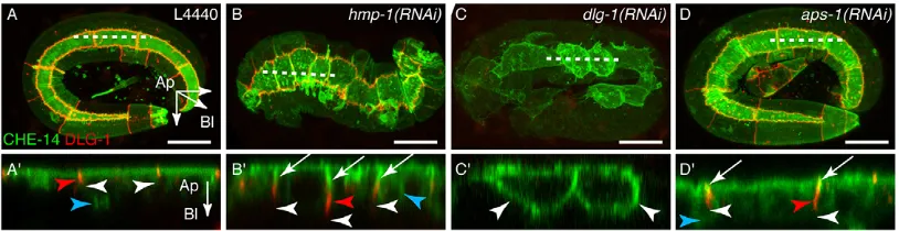
junctions in mammalian cells but it has not yet been defined in DrosophilaorC. elegans. We first tested the potential role of the two known C. elegans junctional complexes (supplementary material Fig. S1A) by observing the localisation of CHE-14, a transmembrane protein which displays a diffuse localisation throughout the epidermal apical membrane (Michaux et al., 2000). These experiments were performed in embryos to observe the known developmental phenotype associated with the loss of junctional complexes (Chisholm and Hardin, 2005). We found that CHE-14 remains apical whenα-catenin (HMP-1 inC. elegans) is targeted (Fig. 5B,B′;n=11/11), whereas it becomes homogenously distributed at the plasma membrane upondlg-1depletion (Fig. 5C,C′; n=9/10). Similarly, E-cad was also observed at the lateral membrane in dlg-1(RNAi) embryos (supplementary material Fig. S4A,B). These results strongly suggest that the diffusion barrier is formed by the DLG-1/AJM-1 complex (DAC) and not by the E-cad complex. Uponaps-1depletion, CHE-14 remained apical to DLG-1 (Fig. 5D,D′; n=14/14) and the DLG-1 belt-like appearance was not affected, demonstrating that the loss of AP-1 does not affect the function of the apico-basal diffusion barrier. Interestingly, we observed that CHE-14 was visible at the lateral membrane above the DLG-1 signal (Fig. 5D′), as in hmp-1(RNAi) embryos (Fig. 5B′). We concluded that the E-cad lateral mislocalisation is not due to a simple diffusion from an apical position along the plasma membrane but to an intracellular missorting defect.
[image:4.612.52.399.58.277.2]AP-1 has a specific function in E-cad apical localisation We next tested whether the sorting function of AP-1 was restricted to E-cad, and we therefore examined the localisation of a variety of polarised markers in AP-1-depleted animals (Fig. 6). First, we observed that a cuticle was visible in threefold stage control and aps-1(RNAi)embryos, a hallmark of apical secretion by the epidermis (Fig. 4A,B). Then, we examined the localisation of the apical PAR proteins, which we found to be mislocalised to the lateral membrane in the intestine (Shafaq-Zadah et al., 2012). We did not observe any mislocalisation in the epidermis, but it should be noted that the expression of these markers becomes too weak to be examined later during morphogenesis at the time of the developmental arrest (Fig. 6A-F). We further confirmed normal apical secretion by observing that the apical transmembrane proteins LRP-1 (Yochem
Fig. 3. A SOAP-1/AP-1/clathrin pathway controls E-cad apical sorting.Maximal projections throughout the epidermis of larvae expressing CHC-1:GFP (clathrin, A-C),
APS-1::GFP (σsubunit, D-F), APM-1::GFP
(μ1-II subunit, G-I) and UNC-101::GFP (μ1-I
subunit, J,K) together with DLG-1::RFP (D-K). Clathrin was examined in P0 larvae 40 h after
the induction ofaps-1(RNAi)at the L1 stage;
AP-1 subunits were examined in F1 larvae from
soap-1(RNAi)-treated young adults. There is a strong and consistent loss of clathrin (A-C) or AP-1 (D-K) punctate staining. (C,F,I) The quantifications of puncta numbers and intensity were performed using a new Fiji macro (see Material and Methods); all parameters and
exactP-values are shown in supplementary
material Fig. S3.n≥15 for each condition.
Owing to its signal mostly restricted to seam cells we were unable to quantify UNC-101.
Scale bar: 10μm.
DEVEL
O
et al., 1999) and VHA-5 (Liégeois et al., 2006) were not affected by AP-1 depletion in larvae (Fig. 6G-J). We next carefully examined the localisation of DLG-1 and AJM-1, which are the main components of the DAC junction, and we found that the belt-like shape of these markers was not altered at the timing of elongation arrest (Fig. 6K-L‴), although AJM-1 and DLG-1 tend to form more
[image:5.612.127.486.55.507.2]intracellular puncta in aps-1(RNAi) embryos. Z-projections also suggest that the DAC junction might be elongated along the apico-basal axis (Fig. 6K‴-L‴). We then observed the localisation of the basolateral polarity determinant LET-413, theC. elegansScribble orthologue (Legouis et al., 2000); this protein is lateral in control embryos and its localisation was unaltered inaps-1(RNAi)embryos Fig. 4. AP-1 interaction with RAB-11+endosomes.(A,B) Control (A,n=4) andaps-1(RNAi)(B,n=6) embryos were observed at the threefold stage by EM. The apical pole is at the top (Ap) and the basal membrane at the bottom (Ba); lateral membrane (La) is visible on each side with electron-dense junctions (Jt). Mitochondria (Mt), Golgi (G), multivesicular bodies (MVB) and lysosomes (Lys) are visible and have a normal ultrastructure. The cuticle (Cu) is also visible.
(C-H) P0 larvae expressing GFP::RAB-5 (C-E) or GFP::RAB-11 (F-H) were examined 72 h after the induction ofaps-1(RNAi). RAB-5 was not affected, whereas
the RAB-11 staining was strongly reduced. Quantifications were performed as in Fig. 3; puncta number (left) and intensity (right) are shown; other parameters and
exactP-values are shown in supplementary material Fig. S3.n=15 for each condition. ns, not significant. (I-N) P0 larvae expressing GFP::RAB-11 and APS-1::
mCherry were examined 48 h after the induction ofrab-5(RNAi)(I-K) orrab-11(RNAi)(L-N). APS-1 was not affected by the depletion of RAB-5 (I-K) but was
strongly reduced following the depletion of RAB-11 (L-N). Quantifications were performed as in Fig. 3; puncta number (left) and intensity (right) are shown; other
parameters and exactP-values are shown in supplementary material Fig. S3.n=15 for each condition. ns, not significant. (O-P‴) Maximal projections throughout
the epidermis of larvae expressing GFP::RAB-5 (O-O‴) or GFP::RAB-11 (P-P‴) together with APS-1::mCherry. The bottom pictures show enlargements
and separate channels of the respective boxes above. No colocalisation was observed with RAB-5, whereas RAB-11 and APS-1 were consistently either
overlapping or closely apposed. (Q) Quantification of the Pearson’s (linear relationship between the two signals) and Manders’( proportion of a signal in one
channel coincident with a signal in the other channel) coefficients for RAB-5 and RAB-11 colocalisation (n=23 for each condition). tM1: proportion of RAB-11
signal coincident with a signal in the APS-1 channel over its total intensity; tM2: proportion of APS-1 signal coincident with a signal in the RAB-11 channel over its
total intensity. Error bars represent s.e.m. Scale bars: 1μm in A for A,B; 10μm in C for C-P.
DEVEL
O
(Fig. 6M-N′). We also probed basal secretion by investigating the localisation of UNC-52, which is secreted as an extracellular matrix component (Mullen et al., 1999), without identifying any defect in aps-1(RNAi)embryos (Fig. 6O,P). We concluded that, contrary to what we found in the intestine, the global cell polarity is maintained and that several polarised markers are not dependent on AP-1: the AP-1 essential apical sorting function seems to be more restricted than in the intestine, and we identified E-cad as a cargo strictly requiring AP-1 for its apical sorting.
AP-1 depletion induces a mislocalisation of the DAC junction and an expansion of the apical pole
As reported previously (Shafaq-Zadah et al., 2012; Shim et al., 2000), RNAi against theσ,γor both µ subunits of AP-1 induces a highly reproducible embryonic arrest during elongation at the threefold stage. We used EM to further characterise the phenotype of threefold stage-arrested embryos. The most striking phenotype was that the electron-dense structures corresponding to the DAC junction (supplementary material Fig. S1A′) (Köppen et al., 2001) were systematically positioned more laterally and were more elongated (n=16 in 6 embryos) than in control embryos (n=12 in 4 embryos) (Fig. 7A-C). The lateral position was not observed in the intestine but the elongation phenotype was similar (Shafaq-Zadah et al., 2012). The expansion of the apical domain and the junction elongation correspond to what we observed with CHE-14 and the AJM-1/DLG-1 localisation. We also observed a rarer phenotype in which the apical pole was sometimes bulging apically from the rest of the body, suggesting that some seam cells undergo at least partial apical extrusion (supplementary material Fig. S5A-B′; apical bulging was observed for 4/36 seam cells in six aps-1(RNAi) embryos and 0/24 seam cells in four control embryos). Finally, we also systematically noticed cellular debris between the eggshell and the embryo itself, as well as inside the intestine lumen (supplementary material Fig. S5B-B‴), which were not observed in control embryos (supplementary material Fig. S5A). We hypothesised that these debris could be formed by parts of seam cells detaching from the body and swallowed by the embryo. We found that the external and intestinal lumen debris were indeed positive for CHE-14::GFP, which is strongly expressed in seam cells but not in the intestine (supplementary material Fig. S5C-C″). These data show that AP-1 is required to maintain the DAC junctions at their normal position and/or to control the size of the apical pole while the diffusion barrier function remains intact.
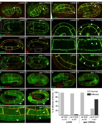
The loss of AP-1 induces the lateral localisation of the E-cad complex and actin disorganisation during morphogenesis Elongation is a morphogenetic process driven first by polarity establishment and junction formation, then by junction reorganisation in seam cells upon actin contraction and, finally, by muscle activity; failure in any of these processes prevents elongation beyond the twofold stage (Chisholm and Hardin, 2005). From the onset of elongation to the twofold stage, these factors were not affected by the loss of AP-1, as assessed by the observation of markers and normal embryo movements inside the eggshell (supplementary material Fig. S4C-H and data not shown). In control embryos, careful observation from the comma stage to the threefold stage showed that E-cad is initially visible at the lateral membrane; however, this signal was almost lost from the twofold stage and very rarely detected in threefold-stage embryos (Fig. 8A,C,E; n=30). In aps-1(RNAi) embryos, the E-cad lateral signal became stronger from the twofold stage (Fig. 8B,D,F; n=29). Three-dimensional (3D) reconstructions showed that E-cad was mislocalised to the lateral membrane rather than intracellularly (Fig. 8E′,F′and supplementary material Movies 2A-C). Other members of the E-cad complex, such as the transmembrane claudin VAB-9 (Simske et al., 2003) and the p120-catenin (JAC-1) (Pettitt et al., 2003), which are strictly apical in threefold-stage control embryos (Fig. 8G,I), were observed at the lateral membrane in aps-1(RNAi) embryos (Fig. 8H-J′ and supplementary material Movies 3A-4B). This suggests that the lateral E-cad pool can recruit other members of its complex.
[image:6.612.103.510.59.164.2]The lateral mislocalisation of E-cad could by itself induce elongation arrest. However, actin is required for embryonic elongation, and E-cad interacts with actin via catenins. We therefore hypothesised that actin itself could also be affected, and examined its localisation with an actin-binding domain fused to GFP (Gally et al., 2009). In control embryos, actin becomes well organised as parallel bundles perpendicular to the antero-posterior axis in the dorso-ventral epidermis from the twofold stage (Fig. 8K,M,O). In aps-1(RNAi) embryos, the initial actin localisation was not perturbed (Fig. 8L,N). However, in threefold-stage embryos, the very precise and regular organisation of actin was lost (Fig. 8O-Q), showing that the AP-1 depletion had a strong impact not only on E-cad-polarised localisation but also on the actin network, which is essential until the end of elongation (Priess and Hirsh, 1986). The late elongation arrest at threefold stage could therefore be explained by the relatively late impact of AP-1 depletion on E-cad-polarised localisation and/or actin disorganisation.
Fig. 5. AP-1 depletion does not affect the apico-basal diffusion barrier.(A-D′) P0 adults expressing CHE-14::GFP and DLG-1::RFP were treated by RNAi for
48 h and F1 embryos examined followinghmp-1,dlg-1oraps-1depletion. CHE-14 is an apical protein which can also be detected in the cytoplasm (blue
arrowheads). Each RNAi condition induces a developmental arrest at a different morphogenetic step:hmp-1(RNAi)embryos arrest after the onset of elongation,
whereasdlg-1(RNAi)embryos arrest at twofold andaps-1(RNAi)arrest at threefold stages. They all express CHE-14 at a similar level. Each picture shows a
maximal projection throughout the epidermis.Z-reconstructions at the level of the dashed lines are shown in the bottom panels (A′-D′): CHE-14 is not seen below
(white arrowheads) the DLG-1 staining except indlg-1(RNAi)embryos, in which DLG-1 is depleted. Note that the DAC junctions (red arrowheads) seem elongated
and slightly detached from the apical pole and that CHE-14 is visible above the DLG-1 signal (white arrows) inhmp-1(B′) andaps-1(D′)-depleted embryos. For all
conditions,n≥10. Scale bars: 10μm.
DEVEL
O
DISCUSSION
We have shown that AP-1 controls E-cad apical localisation both in embryos and larvae and is required for the normal localisation of the E-cadherin-catenin complex and actin organisation in C. elegans epidermal cells. This cellular function of AP-1 is strictly required to prevent late but severe morphogenetic defects leading to lethality during embryonic elongation. Our results also demonstrate that this AP-1 function requires SOAP-1 and clathrin: SOAP-1 probably controls AP-1 membrane recruitment in a manner similar to the recruitment of clathrin by AP-1 itself. This SOAP-1 function is conserved from yeast toDrosophilaand could act in parallel to the more traditionally recognised role of the Arf1 pathway, which was shown to recruit AP-1 in mammalian cells (Robinson, 2004). In the epidermis, the depletion of Arf1 isoforms (arf-1.1orarf-1.2; the latter induces a systematic larval lethality) did not affect E-cad localisation. We therefore propose that a SOAP-1/AP-1/clathrin module controls E-cad apical sorting in the epidermis. In the intestine, AP-1 is also
required for apical sorting but triggers a very different phenotype in which the lateral membrane is converted into an apical membrane (Shafaq-Zadah et al., 2012; Zhang et al., 2012). Interestingly, we observed that E-cad is lateral in the intestine of larvae and not missorted upon AP-1 depletion, whereas the loss ofsoap-1had no effect on the localisation of PAR-6 (Shafaq-Zadah et al., 2012) or CDC-42 (G.G. and G.M., unpublished results) even in larvae displaying a strong developmental arrest phenotype. However, we cannot exclude that other cargos, perhaps less dynamically trafficked and/or less essential than E-cad, might also rely on AP-1 for their polarised localisation in the epidermis. Altogether, these data demonstrate that AP-1 can control essential cargos differently in different tissues, and further work will be required to identify the specificity of these AP-1 functions.
[image:7.612.132.483.57.381.2]In our screen, we identified 50 genes affecting normal E-cad expression or localisation. Among these 50 positive genes, 17 encode subunits or accessory proteins of the COPI, COPII and Fig. 6. AP-1 has a specific function in E-cad apical localisation.In all experiments shown in this figure, we observed a systematic lethality (A-F,K-P: embryonic lethality; G-J: larval lethality) associated with the depletion of AP-1. (A-F) P0 adults were treated by RNAi for 48 h and the F1 embryos examined following
aps-1depletion. Embryos were fixed and stained for PAR-3 (A,B), PAR-6 (C,D) and PKC-3 (E,F). Each picture shows a maximal projection throughout the intestine region and arrowheads point to the epidermal staining. None of these markers was affected at this stage of development. (G,H) Larvae were treated with RNAi for 72 h, then fixed and stained for the apical marker LRP-1. Each picture shows a transverse section in the apico-basal axis. The punctate apical localisation of LRP-1 was not affected. (I,J) Larvae expressing VHA-5::GFP were treated by RNAi for 72 h. Each picture shows a transverse section in the
apico-basal axis. The punctate apical localisation of VHA-5 was not affected. (K-L‴) P0 adults expressing AJM-1::GFP and DLG-1::RFP were treated by RNAi for
48 h and the F1 embryos examined followingaps-1depletion. (K,L) Maximal projections throughout the epidermis. The subapical belt formed by AJM-1 and
DLG-1 is not affected; the intracellular puncta co-stained by these two markers are frequent at that stage of development in control embryos but tend to accumulate
upon AP-1 depletion. (K‴-L‴)Z-reconstructions obtained by reslicing and projections of 4-5 planes, showing a slight thickening of the AJM-1/DLG-1 signal.
(M-N′) P0 adults expressing LET-413::GFP were treated by RNAi for 48 h and the F1 embryos examined followingaps-1depletion. Each picture shows a maximal
projection throughout the epidermis. The LET-413 localisation is not affected at the subapical (arrows) or lateral (arrowheads) levels. M′and N′are enlarged
pictures of the dashed boxes in M and N, respectively. (O,P) P0 adults were treated by RNAi for 48 h and the F1 embryos were fixed and stained for UNC-52
followingaps-1depletion. The basal secretion of this extracellular marker (arrowheads) was not affected. For all conditions,n≥15. Ap, apical; Bl, basolateral.
Scale bars: 10μm.
DEVEL
O
V-ATPase complexes, which control secretion and acidification of organelles. GSL biosynthetic enzymes (fasn-1,acs-1,pod-2,sptl-1 and let-767) were tested separately and displayed the same phenotype; GSLs are an essential component of lipid rafts and control apical sorting in theC. elegansintestine (Zhang et al., 2011). Targeting these genes by RNAi induces a fast and fully penetrant lethality but we did not identify any E-cad localisation defects, in contrast to what was found in the intestine, where GSLs are essential for apical sorting (Zhang et al., 2011). These observations suggest that GSLs do not control E-cad apical sorting in the epidermis.
Where could the SOAP-1/AP-1/clathrin module be implicated in E-cad trafficking? AP-1 could act to prevent E-cad lateral accumulation, for instance by controlling a transcytotic route; this hypothesis could explain easily the lateral localisation of E-cad at the beginning of embryonic elongation. However, we do not favour this possibility because the loss of dynamin does not induce an E-cad lateral accumulation. In addition, E-cad intracellular localisation upon dynamin, RAB-5, RAB-11 or exocyst complex depletion is mostly restricted to the apical region (Fig. 1; supplementary material Fig. S2). We found a very tight link between AP-1 and RAB-11 and a partially overlapping phenotype: an E-cad lateral localisation was observed in about 50% ofrab-11(RNAi)larvae. In epithelial cells, Rab11 plays a central role in the secretory and the recycling pathways (Apodaca et al., 2012), and we therefore propose that AP-1 could control E-cad apical sorting in these two pathways. In the absence of AP-1, E-cad could be targeted randomly to the apical and lateral membranes by a default mechanism not dependent on RAB-11. RAB-11 depletion would lead to a stronger phenotype due to the many functions associated with RAB-11; rab-11(RNAi) induced in P0 hermaphrodites triggers various defects from the one-cell embryo (Bembenek et al., 2010; Sato et al., 2008; Zhang et al., 2008), whereas AP-1-depleted embryos arrest much later at the threefold stage.
While examining AP-1-depleted embryos we found that the DAC domain was elongated. Several groups have reported such a phenotype, in particular upon depletion of the MAGUK protein MAGI-1 (Lynch et al., 2012; Stetak and Hajnal, 2011). This protein probably contributes to junction organisation but is not an essential factor: it was identified as an enhancer of the weak loss-of-function hmp-1(fe4)allele (Lynch et al., 2012), and a likely null allele is viable (Stetak and Hajnal, 2011). Other enhancers ofhmp-1(fe4)
identified include AP-2 subunits (Lynch and Hardin, 2009). Although we did not identify a role for AP-2 in our screen, this observation strongly suggests an AP-2 contribution to junction maintenance. Altogether, these works highlight the need for various screening strategies to identify non-essential factors.
The loss of AP-1 induces a threefold-stage arrest during development. The AP-1 functions identified in neurons (Dwyer et al., 2001) or in the intestine (Shafaq-Zadah et al., 2012; Zhang et al., 2012) cannot account for this lethality. At the developmental level, embryonic elongation requires seam cell elongation driven by actin tension in epidermal cells and functional muscles attached to the epidermis from twofold stage (Chisholm and Hardin, 2005). Because AP-1-depleted embryos display normal movements we believe that the observed embryonic arrest is most likely due to defects linked to epidermal cells.
In AP-1-depleted embryos, the lateral accumulation of E-cad (from twofold stage) was observed first, shortly before the timing of the morphogenetic arrest (threefold stage) when actin disorganisation was observed. This strongly suggests that E-cad mis-targeting and/or actin disorganisation are responsible for the developmental arrest. A potential direct link between E-cad mislocalisation and actin organisation is suggested by the observation that the Hmp-1 and Aps-1 cellular phenotypes are partially similar: both induce a mislocalisation of the DAC junction in the apico-basal axis. This could be due to an arrest of elongation not directly translated into an arrest of membrane addition at the apical pole. If this was the case, the apical membrane could still increase its surface, leading to a more lateral localisation of the DAC junction, and it would be unlikely to affect its diffusion barrier function. However, an important difference is that in AP-1-depleted embryos we did not detect a detachment of actin bundles from the junctions as reported when the E-cad complex is lost (Costa et al., 1998). Instead, we observed a persistence of the E-cad complex at the apical membrane and an irregular organisation and abnormal thickness of actin bundles. Because the E-cad complex can actively recruit and control actin at the level of junctions, E-cad mislocalisation at the lateral membrane could indirectly destabilise actin organisation and affect forces driving elongation.
[image:8.612.126.492.59.207.2]Alternatively, AP-1 could also directly contribute to actin organisation: a proteomic analysis showed that several actin Fig. 7. AP-1 depletion induces a mislocalisation of the DAC junction and an expansion of the apical pole.(A-B′) Embryos were fixed by high-pressure freezing 9-10 h post-fertilisation. Ultrastructures of epidermal seam cells (in green) are shown in threefold-stage embryos. The apical pole is at the top (in yellow) and the basolateral membrane at the bottom (black arrowheads). The electron-dense structure marking the junction (in red) is in subapical position in control
embryos (L4440) and displaced along the lateral membrane inaps-1(RNAi)embryos, confirming observations of apical extension suggested by the CHE-14
localisation (Fig. 5D′). Junctions are also elongated. The dashed boxes in A,B are shown in higher magnification in A′and B′, respectively. (C) Quantification of
defects affecting the electron-dense structure in threefold-stage embryos [control:n=4;aps-1(RNAi):n=6]. Junctions (red in A-B′) are elongated and detached
from the apical pole (yellow) inaps-1(RNAi)embryos. Error bars show s.e.m. Scale bar: 1μm.
DEVEL
O
nucleators were found on AP-1-enriched liposomes (Baust et al., 2006). We therefore cannot exclude that the loss of AP-1 could also have a direct impact on actin organisation through these actin cofactors, independently of E-cad. Although the Gex (gut on the exterior) phenotype associated with the loss of function of these factors (Patel et al., 2008; Soto et al., 2002) occurs earlier than the threefold arrest induced by AP-1 depletion, we cannot rule out a later role of these factors hidden by the earlier phenotype, and therefore a more direct role of AP-1 on actin contractile forces. We therefore propose that AP-1 controls E-cad apical localisation and that the embryonic elongation arrest associated with the loss of AP-1 can be due to E-cad missorting and, directly or indirectly, defective actin contractile forces.
MATERIALS AND METHODS Genetics
C. elegans strains were maintained and crossed as described (Brenner, 1974). The strains used in this study are shown in supplementary material Table S4.
Plasmid construction
Theaps-1::gfpconstruct under the control of its own promoter was generated as described (Shafaq-Zadah et al., 2012). The epidermalaps-1::mCherry
construct was generated using the Multisite Gateway system (Invitrogen). The epidermalaps-1::mCherryconstruct was generated by creating three donor vectors:dpy-7promoter,aps-1genomic sequence andmCherry::unc-543′ UTR (by PCR fusion). The epidermalgfp::rab-11construct was generated by creating three donor vectors:dpy-7promoter fused to GFP (by PCR fusion),
rab-11genomic sequence andunc-543′UTR.
RNAi
[image:9.612.52.399.55.475.2]Embryonic and larval RNAi were performed by feeding as described using the Ahringer-Source BioScience library (Fire et al., 1998; Kamath and Ahringer, 2003; Shafaq-Zadah et al., 2012); L4440 corresponds to the standard control RNAi feeding strain. To bypass the early embryonic requirement of most candidate genes we performed the screen by adding bleach-treated adults to RNAi plates, and we observed E-cad localisation 48 h and 72 h later in the same generation (P0); the embryonic development is therefore normal and the RNAi knockdown only starts after hatching. For other experiments, RNAi was induced in young adults and the phenotypes were observed in the next generation (F1). The penetrance of all phenotypes described following RNAi was >80%. RNAi efficiency was checked by observing the induction of a developmental arrest whenever such a phenotype was expected – e.g. the depletion of GSL enzymes orppk-1induced a larval lethality but no E-cad mislocalisation. aps-1(RNAi) and apg-1(RNAi) induce identical phenotypes.
Fig. 8. AP-1 depletion induces the lateral localisation of the E-cad complex and actin disorganisation during morphogenesis.
(A-F′) P0 adults expressing E-cad::GFP
together with DLG-1::RFP were treated by RNAi for 48 h and F1 embryos examined
following control RNAi (n=30) oraps-1
depletion (n=29). E′,F′show higher
magnifications of the respective boxes in E,F. Each image is a maximal projection throughout the epidermis. E-cad is visible at the lateral membrane (arrowheads) in 1.5-fold (A,B) and twofold-(C,D) stage control and
aps-1(RNAi)embryos; later, it is restricted to the apical membrane in control embryos
(E,E′) but remains easily visible at the lateral
membrane in threefold-stageaps-1(RNAi)
(F,F′). See associated supplementary material
Movies 2A,B for 3D reconstructions of E′,F′
showing a lateral rather than intracellular
localisation. (G-J′) P0 adults expressing
VAB-9::GFP (G-H′) or JAC-1::GFP (I-J′) together
with DLG-1::RFP were treated by RNAi for 48 h
and the F1 embryos examined followingaps-1
orapg-1depletion. Both VAB-9 (G-H′) and
JAC-1 (I-J′) were observed at the lateral
membrane inaps-1(RNAi)threefold-stage
embryos. See associated supplementary material Movies 3A-4B for 3D reconstructions.
For all conditions,n≥10. (K-P′) P0 adults
expressing ABD::GFP (‘Actin’in K-P′) together
with DLG-1::RFP were treated by RNAi for 48 h
and the F1 embryos examined followingaps-1
depletion. There is no difference in actin organisation at 1.5- and twofold stages (K-N), but at the threefold stage actin becomes
disorganised inaps-1(RNAi)embryos, as
shown by the loss of the parallel organisation
of actin bundles (arrowheads). O′,P′show
higher magnifications of the respective boxes in O,P. (Q) Phenotype quantification for actin organisation based on direct observations of
embryos. Scale bar: 10μm.
DEVEL
O
Immunostaining
Fixation of embryos or larvae was performed as described using the freeze-crack methanol protocol (Leung et al., 1999). We used anti-AJM-1 MH27 (1/50) (Köppen et al., 2001), anti-UNC-52 MH2 (1/50) (Francis and Waterston, 1991) and anti-PAR-3 P4A1 (1/50) (Nance et al., 2003) monoclonal antibodies from DSHB (University of Iowa, USA). The anti-PAR-6 (1/50) (Labbe et al., 2006), anti-PKC-3 (1/1000) (Sugiyama et al., 2008) and anti-LRP-1 (1/200) (Yochem et al., 1999) rabbit antibodies were generous gifts from Monica Gotta, Shigeo Ohno and Simon Tuck, respectively. Alexa Fluor 488- or 532-conjugated antibodies (Invitrogen) were used as secondary antibodies.
Electron microscopy
Electron microscopy experiments were performed as described (Shafaq-Zadah et al., 2012). Junction length and detachment from the apical pole (Fig. 7A-C) were quantified as follows: each embryo was sectioned every 5-7 µm to ensure that different cells were observed in different 5-7 µm sections. Only one segment of high quality per section was used to measure electron-dense structures ( junctions). Measurements were performed on 12 junctions from four different control embryos and on 16 junctions from six differentaps-1(RNAi)embryos. An identical approach was used to quantify the number of MVBs and lysosomes in epidermal seam cells and in the intestine: we counted all single-membrane organelles containing internal vesicles or membranes, excluding mitochondria, which have a double membrane. Immuno-electron microscopy was performed as described (Liégeois et al., 2006). The anti-AJM-1 antibody MH27 (1/10, DSHB, University of Iowa) was detected using 5-nm gold particles coupled to a goat anti-mouse antibody (GMHL5, Oxford Instruments).
Confocal microscopy and signal quantifications
Confocal observations were performed using a Leica SPE (for screening) or SP5 confocal microscope equipped with a 63× objective (LAS AF software). The SP5 confocal microscope is equipped with a sensitive hybrid detector. All images were examined using ImageJ 1.43 and assembled using Adobe Photoshop and Illustrator CS3.
The percentage of embryos or larvae displaying a phenotype was obtained either by direct observation or after quantification. Quantifications (Fig. 1) were performed using ImageJ 1.43 along straight lines (length 10 µm, width 0.3 µm) over the apical, lateral and cytoplasmic parts of one cell for each larva. Quantifications were normalised to the cytoplasmic background; a ratio of 1 therefore indicates no specific membrane staining. Quantifications of colocalisation (Fig. 4Q) were performed using the ImageJ JaCoP plugin (Bolte and Cordelieres, 2006) to determine the Pearson’s correlation coefficient (PCC, which measures the linear relationship between the two signals) as well as the Manders’coefficients tM1 ( proportion of RAB-11 signal coincident with a signal in the APS-1 channel over its total intensity) and tM2 ( proportion of APS-1 signal coincident with a signal in the RAB-11 channel over its total intensity). Coefficients were calculated in several regions of interest (ROI) in one plane of each larva examined (n=23 larvae for each condition). Manders’ coefficients were calculated for pixels above a manually determined threshold for each channel. The coefficients obtained in our experiments enabled us to argue that there is a partial colocalisation between APS-1 and RAB-11.
To quantify the total amount of APS-1, APM-1, RAB-5 and RAB-11 in puncta and cytoplasm (Figs 3 and 4) we used a home-written plugin and Fiji software (Schindelin et al., 2012). We computed the sum of intensity over each plane of az-stack containing the entire epidermal signal either masking out the cytoplasm or the puncta to quantify the total amount of labeled protein in puncta or in the cytoplasm, respectively. A mask for each plane was computed: to keep only objects of relevant size, we bandpass-filtered planes (0.16-3μm−1), removed background (rolling-ball diameter: 1.6μm) (Gonzalez and Woods, 2002) and thresholded each plane separately with the Kapur–Sahoo–Wong method modified to use Renyi entropy (Kapur et al., 1985). Because vesicles can spread over severalz-planes, prior to counting particles, we computed a maximum projection and thresholded it as described above, applied the watershed algorithm (Vincent and Soille, 1991) and counted particles with a radius >0.1μm.
Statistical analysis
Parametrict-tests were used when samples had a Gaussian distribution and similar variances. Other cases were treated using Wilcoxon tests. Significance is indicated as follow: *P<0.05, **P<0.01, ***P<0.001.
Acknowledgements
We are very grateful to Roland Le Borgne and Anne Pacquelet for critical reading of the manuscript, to Yann Le Cunff for his help in statistical analysis and to Nicolas Loyer for helpful discussions. Some strains were provided by the CGC, which is funded by NIH Office of Research Infrastructure Programs (P40 OD010440; University of Minnesota, USA), and the NBP (Tokyo University, Japan). We also thank Monica Gotta, Barth Grant, Jeff Hardin, Michel Labouesse, Renaud Legouis, Jeremy Nance, Shigeo Ohno and Simon Tuck for strains and antibodies. We thank the photonic and electron microscopy facilities of the Microscopy Rennes Imaging Center and the IGBMC Imaging Center.
Competing interests
The authors declare no competing or financial interests.
Author contributions
G.G., M.S.-Z. and G.M. designed the experiments. G.G., M.S.-Z., O.N. and R.D. performed experiments and data analysis. J.P. wrote the quantification plug-in. G.G. and G.M. wrote the manuscript.
Funding
This work was mostly supported by the Ligue contre le Cancer [22/29/35/72], and also by an Institut national de la santéet de la recherche médicale (INSERM) Avenir grant [R06488NS], the French Centre national de la recherche scientifique (CNRS) and the Universitéde Rennes 1. The group of J.P. is supported by a Centre national de la recherche scientifique (CNRS) ATIP/Ligue contre le Cancer grant.
Supplementary material
Supplementary material available online at
http://dev.biologists.org/lookup/suppl/doi:10.1242/dev.118216/-/DC1
References
Achilleos, A., Wehman, A. M. and Nance, J.(2010). PAR-3 mediates the initial clustering and apical localization of junction and polarity proteins during C. elegans intestinal epithelial cell polarization.Development137, 1833-1842.
Apodaca, G., Gallo, L. I. and Bryant, D. M.(2012). Role of membrane traffic in the generation of epithelial cell asymmetry.Nat. Cell Biol.14, 1235-1243.
Balklava, Z., Pant, S., Fares, H. and Grant, B. D.(2007). Genome-wide analysis identifies a general requirement for polarity proteins in endocytic traffic.Nat. Cell Biol.9, 1066-1073.
Baust, T., Czupalla, C., Krause, E., Bourel-Bonnet, L. and Hoflack, B.(2006). Proteomic analysis of adaptor protein 1A coats selectively assembled on liposomes.Proc. Natl. Acad. Sci. USA103, 3159-3164.
Bembenek, J. N., White, J. G. and Zheng, Y.(2010). A role for separase in the regulation of RAB-11-positive vesicles at the cleavage furrow and midbody.Curr. Biol.20, 259-264.
Blankenship, J. T., Fuller, M. T. and Zallen, J. A.(2007). The Drosophila homolog of the Exo84 exocyst subunit promotes apical epithelial identity.J. Cell Sci.120, 3099-3110.
Bolte, S. and Cordelières, F. P.(2006). A guided tour into subcellular colocalization analysis in light microscopy.J. Microsc.224, 213-232.
Brenner, S.(1974). The genetics of Caenorhabditis elegans.Genetics77, 71-94.
Chisholm, A. D. and Hardin, J.(2005). Epidermal morphogenesis. InWormBook, pp. 1-22.
Costa, M., Raich, W., Agbunag, C., Leung, B., Hardin, J. and Priess, J. R.(1998). A putative catenin-cadherin system mediates morphogenesis of the Caenorhabditis elegans embryo.J. Cell Biol.141, 297-308.
Deborde, S., Perret, E., Gravotta, D., Deora, A., Salvarezza, S., Schreiner, R. and Rodriguez-Boulan, E.(2008). Clathrin is a key regulator of basolateral polarity.
Nature452, 719-723.
Diogon, M., Wissler, F., Quintin, S., Nagamatsu, Y., Sookhareea, S., Landmann, F., Hutter, H., Vitale, N. and Labouesse, M.(2007). The RhoGAP RGA-2 and LET-502/ROCK achieve a balance of actomyosin-dependent forces in C. elegans epidermis to control morphogenesis.Development134, 2469-2479.
Dwyer, N. D., Adler, C. E., Crump, J. G., L’Etoile, N. D. and Bargmann, C. I.
(2001). Polarized dendritic transport and the AP-1 mu1 clathrin adaptor UNC-101 localize odorant receptors to olfactory cilia.Neuron31, 277-287.
Fernández, G. E. and Payne, G. S.(2006). Laa1p, a conserved AP-1 accessory protein important for AP-1 localization in yeast.Mol. Biol. Cell17, 3304-3317.
Fire, A., Xu, S., Montgomery, M. K., Kostas, S. A., Driver, S. E. and Mello, C. C.
(1998). Potent and specific genetic interference by double-stranded RNA in Caenorhabditis elegans.Nature391, 806-811.
DEVEL
O
Francis, R. and Waterston, R. H.(1991). Muscle cell attachment in Caenorhabditis elegans.J. Cell Biol.114, 465-479.
Fujita, Y., Krause, G., Scheffner, M., Zechner, D., Leddy, H. E. M., Behrens, J., Sommer, T. and Birchmeier, W.(2002). Hakai, a c-Cbl-like protein, ubiquitinates and induces endocytosis of the E-cadherin complex.Nat. Cell Biol.4, 222-231.
Gally, C., Wissler, F., Zahreddine, H., Quintin, S., Landmann, F. and Labouesse, M.
(2009). Myosin II regulation during C. elegans embryonic elongation: LET-502/ ROCK, MRCK-1 and PAK-1, three kinases with different roles.Development136, 3109-3119.
Georgiou, M., Marinari, E., Burden, J. and Baum, B.(2008). Cdc42, Par6, and aPKC regulate Arp2/3-mediated endocytosis to control local adherens junction stability.Curr. Biol.18, 1631-1638.
Gonzalez, R. C. and Woods, R. E.(2002).Digital Image Processing. Upper Saddle River, NJ: Prentice Hall.
Harris, K. P. and Tepass, U.(2008). Cdc42 and Par proteins stabilize dynamic adherens junctions in the Drosophila neuroectoderm through regulation of apical endocytosis.J. Cell Biol.183, 1129-1143.
Ivanov, A. I. and Naydenov, N. G.(2013). Dynamics and regulation of epithelial adherens junctions: recent discoveries and controversies.Int. Rev. Cell Mol. Biol.
303, 27-99.
Kamath, R. S. and Ahringer, J. (2003). Genome-wide RNAi screening in Caenorhabditis elegans.Methods30, 313-321.
Kapur, J. N., Sahoo, P. K. and Wong, A. K. C.(1985). A new method for gray-level picture thresholding using the entropy of the histogram.Comput. Vis. Graph. Image Process.29, 273-285.
Köppen, M., Simske, J. S., Sims, P. A., Firestein, B. L., Hall, D. H., Radice, A. D., Rongo, C. and Hardin, J. D.(2001). Cooperative regulation of AJM-1 controls junctional integrity in Caenorhabditis elegans epithelia.Nat. Cell Biol.3, 983-991.
Labbe, J.-C., Pacquelet, A., Marty, T. and Gotta, M.(2006). A genomewide screen for suppressors of par-2 uncovers potential regulators of PAR protein-dependent cell polarity in Caenorhabditis elegans.Genetics174, 285-295.
Langevin, J., Morgan, M. J., Rossé, C., Racine, V., Sibarita, J.-B., Aresta, S., Murthy, M., Schwarz, T., Camonis, J. and Bellaïche, Y.(2005). Drosophila exocyst components Sec5, Sec6, and Sec15 regulate DE-Cadherin trafficking from recycling endosomes to the plasma membrane.Dev. Cell9, 365-376.
Le Bras, S., Rondanino, C., Kriegel-Taki, G., Dussert, A. and Le Borgne, R.
(2012). Genetic identification of intracellular trafficking regulators involved in Notch-dependent binary cell fate acquisition following asymmetric cell division.
J. Cell Sci.125, 4886-4901.
Legouis, R., Gansmuller, A., Sookhareea, S., Bosher, J. M., Baillie, D. L. and Labouesse, M.(2000). LET-413 is a basolateral protein required for the assembly of adherens junctions in Caenorhabditis elegans.Nat. Cell Biol.2, 415-422.
Leibfried, A., Fricke, R., Morgan, M. J., Bogdan, S. and Bellaiche, Y.(2008). Drosophila Cip4 and WASp define a branch of the Cdc42-Par6-aPKC pathway regulating E-cadherin endocytosis.Curr. Biol.18, 1639-1648.
Leung, B., Hermann, G. J. and Priess, J. R.(1999). Organogenesis of the Caenorhabditis elegans intestine.Dev. Biol.216, 114-134.
Levayer, R. and Lecuit, T. (2013). Oscillation and polarity of E-cadherin asymmetries control actomyosin flow patterns during morphogenesis.Dev. Cell
26, 162-175.
Levayer, R., Pelissier-Monier, A. and Lecuit, T.(2011). Spatial regulation of Dia and Myosin-II by RhoGEF2 controls initiation of E-cadherin endocytosis during epithelial morphogenesis.Nat. Cell Biol.13, 529-540.
Liégeois, S., Benedetto, A., Garnier, J.-M., Schwab, Y. and Labouesse, M.
(2006). The V0-ATPase mediates apical secretion of exosomes containing Hedgehog-related proteins in Caenorhabditis elegans.J. Cell Biol.173, 949-961.
Ling, K., Bairstow, S. F., Carbonara, C., Turbin, D. A., Huntsman, D. G. and Anderson, R. A.(2007). Type I gamma phosphatidylinositol phosphate kinase modulates adherens junction and E-cadherin trafficking via a direct interaction with mu 1B adaptin.J. Cell Biol.176, 343-353.
Lui, W. W. Y., Collins, B. M., Hirst, J., Motley, A., Millar, C., Schu, P., Owen, D. J. and Robinson, M. S.(2003). Binding partners for the COOH-terminal appendage domains of the GGAs and gamma-adaptin.Mol. Biol. Cell14, 2385-2398.
Lynch, A. M. and Hardin, J.(2009). The assembly and maintenance of epithelial junctions in C. elegans.Front. Biosci.14, 1414-1432.
Lynch, A. M., Grana, T., Cox-Paulson, E., Couthier, A., Cameron, M., Chin-Sang, I., Pettitt, J. and Hardin, J.(2012). A genome-wide functional screen shows MAGI-1 is an LMAGI-1CAM-dependent stabilizer of apical junctions in C. elegans.Curr. Biol.
22, 1891-1899.
Michaux, G., Gansmuller, A., Hindelang, C. and Labouesse, M.(2000). CHE-14, a protein with a sterol-sensing domain, is required for apical sorting in C. elegans ectodermal epithelial cells.Curr. Biol.10, 1098-1107.
Montpetit, A., Côté, S., Brustein, E., Drouin, C. A., Lapointe, L., Boudreau, M., Meloche, C., Drouin, R., Hudson, T. J., Drapeau, P. et al.(2008). Disruption of AP1S1, causing a novel neurocutaneous syndrome, perturbs development of the skin and spinal cord.PLoS Genet.4, e1000296.
Mullen, G. P., Rogalski, T. M., Bush, J. A., Gorji, P. R. and Moerman, D. G.(1999). Complex patterns of alternative splicing mediate the spatial and temporal distribution of perlecan/UNC-52 in Caenorhabditis elegans.Mol. Biol. Cell10, 3205-3221.
Nance, J., Munro, E. M. and Priess, J. R.(2003). C. elegans PAR-3 and PAR-6 are required for apicobasal asymmetries associated with cell adhesion and gastrulation.Development130, 5339-5350.
Palacios, F., Price, L., Schweitzer, J., Collard, J. G. and D’Souza-Schorey, C.
(2001). An essential role for ARF6-regulated membrane traffic in adherens junction turnover and epithelial cell migration.EMBO J.20, 4973-4986.
Patel, F. B., Bernadskaya, Y. Y., Chen, E., Jobanputra, A., Pooladi, Z., Freeman, K. L., Gally, C., Mohler, W. A. and Soto, M. C.(2008). The WAVE/SCAR complex promotes polarized cell movements and actin enrichment in epithelia during C. elegans embryogenesis.Dev. Biol.324, 297-309.
Pettitt, J., Cox, E. A., Broadbent, I. D., Flett, A. and Hardin, J.(2003). The Caenorhabditis elegans p120 catenin homologue, JAC-1, modulates cadherin-catenin function during epidermal morphogenesis.J. Cell Biol.162, 15-22.
Priess, J. R. and Hirsh, D. I.(1986). Caenorhabditis elegans morphogenesis: the role of the cytoskeleton in elongation of the embryo.Dev. Biol.117, 156-173.
Robinson, M. S.(2004). Adaptable adaptors for coated vesicles.Trends Cell Biol.
14, 167-174.
Sato, M., Grant, B. D., Harada, A. and Sato, K.(2008). Rab11 is required for synchronous secretion of chondroitin proteoglycans after fertilization in Caenorhabditis elegans.J. Cell Sci.121, 3177-3186.
Schindelin, J., Arganda-Carreras, I., Frise, E., Kaynig, V., Longair, M., Pietzsch, T., Preibisch, S., Rueden, C., Saalfeld, S., Schmid, B. et al.(2012). Fiji: an open-source platform for biological-image analysis.Nat. Methods9, 676-682.
Shafaq-Zadah, M., Brocard, L., Solari, F. and Michaux, G. (2012). AP-1 is required for the maintenance of apico-basal polarity in the C. elegans intestine.
Development139, 2061-2070.
Shaye, D. D. and Greenwald, I.(2011). OrthoList: a compendium of C. elegans genes with human orthologs.PLoS ONE6, e20085.
Shaye, D. D., Casanova, J. and Llimargas, M.(2008). Modulation of intracellular trafficking regulates cell intercalation in the Drosophila trachea.Nat. Cell Biol.10, 964-970.
Shim, J., Sternberg, P. W. and Lee, J.(2000). Distinct and redundant functions of mu1 medium chains of the AP-1 clathrin-associated protein complex in the nematode Caenorhabditis elegans.Mol. Biol. Cell11, 2743-2756.
Simske, J. S., Köppen, M., Sims, P., Hodgkin, J., Yonkof, A. and Hardin, J.
(2003). The cell junction protein VAB-9 regulates adhesion and epidermal morphology in C. elegans.Nat. Cell Biol.5, 619-625.
Soto, M. C., Qadota, H., Kasuya, K., Inoue, M., Tsuboi, D., Mello, C. C. and Kaibuchi, K.(2002). The GEX-2 and GEX-3 proteins are required for tissue morphogenesis and cell migrations in C. elegans.Genes Dev.16, 620-632.
St Johnston, D. and Ahringer, J.(2010). Cell polarity in eggs and epithelia: parallels and diversity.Cell141, 757-774.
Stetak, A. and Hajnal, A.(2011). The C. elegans MAGI-1 protein is a novel component of cell junctions that is required for junctional compartmentalization.
Dev. Biol.350, 24-31.
Sugiyama, Y., Nishimura, A. and Ohno, S.(2008). Symmetrically dividing cell specific division axes alteration observed in proteasome depleted C. elegans embryo.Mech. Dev.125, 743-755.
Vincent, L. and Soille, P. (1991). Watersheds in digital spaces: an efficient algorithm based on immersion simulations.IEEE Trans. Pattern Anal. Mach. Intell.
13, 583-598.
Wang, Q., Chen, X.-W. and Margolis, B.(2007). PALS1 regulates E-cadherin trafficking in mammalian epithelial cells.Mol. Biol. Cell18, 874-885.
Winter, J. F., Höpfner, S., Korn, K., Farnung, B. O., Bradshaw, C. R., Marsico, G., Volkmer, M., Habermann, B. and Zerial, M.(2012). Caenorhabditis elegans screen reveals role of PAR-5 in RAB-11-recycling endosome positioning and apicobasal cell polarity.Nat. Cell Biol.14, 666-676.
Xiong, X., Xu, Q., Huang, Y., Singh, R. D., Anderson, R., Leof, E., Hu, J. and Ling, K.(2012). An association between type Igamma PI4P 5-kinase and Exo70 directs E-cadherin clustering and epithelial polarization.Mol. Biol. Cell23, 87-98.
Yochem, J., Tuck, S., Greenwald, I. and Han, M.(1999). A gp330/megalin-related protein is required in the major epidermis of Caenorhabditis elegans for completion of molting.Development126, 597-606.
Zhang, H., Squirrell, J. M. and White, J. G.(2008). RAB-11 permissively regulates spindle alignment by modulating metaphase microtubule dynamics in Caenorhabditis elegans early embryos.Mol. Biol. Cell19, 2553-2565.
Zhang, H., Abraham, N., Khan, L. A., Hall, D. H., Fleming, J. T. and Göbel, V.
(2011). Apicobasal domain identities of expanding tubular membranes depend on glycosphingolipid biosynthesis.Nat. Cell Biol.13, 1189-1201.
Zhang, H., Kim, A., Abraham, N., Khan, L. A., Hall, D. H., Fleming, J. T. and Gobel, V.(2012). Clathrin and AP-1 regulate apical polarity and lumen formation during C. elegans tubulogenesis.Development139, 2071-2083.