ORIGINAL RESEARCH ARTICLE
CHANGES IN THE RESPIRATORY SYSTEM OF RATS EXPOSED TO GASES FROM THE
COMBUSTION OF CASHEW NUTS SHELL
*
1Daniel Silveira Serra,
2Laura Lorena Fernandes Rosendo,
3Fladimir de Lima Gondim,
4
Rinaldo SantosAraújo,
5Mona Lisa Moura de Oliveira and
6Francisco Sales Ávila Cavalcante
1,3
Institute of Biomedical Sciences, State University of Ceará, Ceará, Brazil,
Av. Dr. Silas Munguba, 1700, zip: 60714-903, Fortaleza-Ceará, Brazil
2, 5, 6Science and Technology Center, State University of Ceará, Ceará, Brazil
4
Department of Chemistry and Environment, Federal Institute of Ceará, Ceará, Brazil
ARTICLE INFO ABSTRACT
The term biomass describes all organic matter that, when burned, decomposed or recycled, can directly and/or indirectly generate some form of mechanical, thermal or electrical energy. Brazil is one of the largest producers of agricultural products in the world, making it also one of the largest producers of residual biomass. Due to its importance in the cashew crop, there is a concern to find alternatives for an environmentally safe disposal for the residual biomass of the cashew nut processing, cashew nut shell (CNS). In view of the above, this work had the objective of evaluating the effects caused by acute exposure to CNS combustion emissions. For this, a CNS combustion reactor was produced to expose animals to exhaust gases. BALB / c mice, body mass of 25 ± 5 g, randomly divided into two groups, Control, exposed for 5 hours to ambient air, and CNS, exposed for 5 hours to CNS combustion exhaust gases were used. Our results show statistically significant changes in all variables analyzed ( , , , , , e PV loop area)of the CNS group in relation to the control group. The use of CNS as a biofuel can be feasible, but our results reinforce the urgent need to seek control methods for the exhaustion of these gases in the atmosphere. Further investigation is necessary in order to know safe parameters for individuals who are continuously exposed to CNS combustion exhaust gases.
Copyright © 2018,Daniel Silveira Serra et al. This is an open access article distributed under the Creative Commons Attribution License, which permits unrestricted use, distribution, and reproduction in any medium, provided the original work is properly cited.
INTRODUCTION
The term biomass describes all organic matter that, when burned, decomposed or recycled, can directly and/or indirectly generate some form of mechanical, thermal or electrical energy. Thus, agricultural residues, animal waste, human waste and organic municipal waste can be used in processes to obtain energy from alternative sources (ANEEL, 2008). In Brazil, some biomass residues contribute to the growth of alternative energy production in the industrial sector, such as coffee grounds (Silva et al.,1998), rice hull (Maffioletti and
*Corresponding author: Daniel Silveira Serra,
Institute of Biomedical Sciences, State University of Ceará, Ceará, Brazil. Av. Dr. Silas Munguba, 1700, zip: 60714-903, Fortaleza-Ceará, Brazil.
Mota, 2013), sugarcane bagasse (Yiannis, 2011), eucalyptus foliage (Possel and Bell, 2012), and the cashew nuts shell (CNS) (Ndalila and Kaunde, 2014), the latter being our object of study. The cashew tree (Anacardium occidentale L.) is a tropical plant, native to Brazil and dispersed throughout most of its territory. Cashew is among the most cultivated fruit trees, standing out in the socioeconomic context of some underdeveloped tropical regions, due to the high nutritional and commercial value of its products, and the labor intensive character, whose production and industrialization guarantee a significant flow of income, making this culture an important
source of job opportunities for the population (Ramos et al.,
2011). According to the International Nut &Dried Fruit (2016), Brazil is the fifth largest producer of cashew nuts in the world.
ISSN: 2230-9926
International Journal of Development Research
Vol. 08, Issue, 03, pp.19410-19415, March,2018
Article History:
Received 20th December, 2017
Received in revised form 06th January, 2018
Accepted 23rd February, 2018
Published online 30th March, 2018
Key Words:
Biomass; Cashew nuts shell, Combustion,
Respiratory system.
Citation: Daniel Silveira Serra, Laura Lorena Fernandes Rosendo, Fladimir de Lima Gondim, Rinaldo SantosAraújo, Mona Lisa Moura de
Oliveira and Francisco Sales Ávila Cavalcante, 2018. “Changes in the respiratory system of rats exposed to gases from the combustion of cashew nuts
shell”, International Journal of Development Research, 8, (03), 19410-19415.
The Brazilian cashew production reached 104,475 tons in 2015, corresponding to a harvested area of 585,966 hectares, the northeast region being responsible for 99.4% of the production (IBGE, 2015). In 2014, Brazil exported approximately 17,023 tons of cashew nuts (MDIFT, 2015). Obtaining the CNS begins through the withdrawal process almond cashew stalk (decorating), held in tanks with cardol, composed of 10% of the liquid extracted from cashew nut itself (CNL). This is heated in boilers at a temperature of ±800 °C. The by-product of this stage is the almond, of great commercial value, and the CNS, drenched in cardol, which hold great potential fuel (Lima, 2008). The high calorific power of CNS (22,48 MJ/kg),is superior to other biomasses such as: rice straw (16,35 MJ/kg) and sugarcane bagasse (18,61 MJ/kg), making the CNS an attractive alternative compared to the use of other fuel sources used by industries, mini-factories and in handicraft productions (Figueiredo et al., 2007), providing an alternative for its use and avoiding its disposal in the environment. However, the combustion of CNS promotes the release of some gases, such as: carbon dioxide
(CO2), carbon monoxide (CO), methane (CH4),ozone (O3),
nitrogen oxides (NOx), nitrogen dioxide (NO2), sulfur dioxide
(SO2), polycyclic aromatic hydrocarbons (PAHs), Volatile
organic compounds (VOCs), Besides other products considered pollutants (Lewne et al.,2004; WHO, 2005). In view of the above, there is an urgent need to evaluate the effects caused by acute exposure to CNS combustion emissions, providing a better knowledge about its probable harmful effects on health, producing parameters of control and safety in its use as biofuel. For this, we analyzed the deleterious effects in the respiratory system of rats exposed to CNS combustion exhaust gases for 5 hours, through the analysis of variables related to the mechanics of the respiratory system of these animals.
MATERIALS AND METHODS
CNS combustion reactor
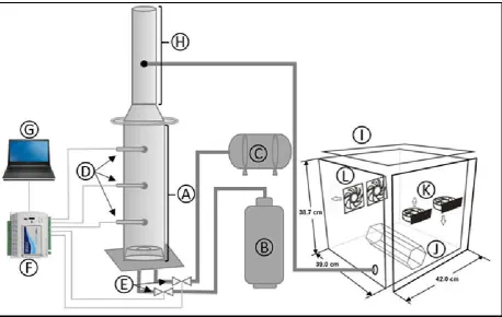
Cashew nut shell was supplied by cascaju agroindustrial S/A, located 65 km from Fortaleza in the municipality of Cascavel in Ceará, Brazil. The combustion and gas exposure system of the CNS (Figure 1). For the animal exposure process, the initial combustion ignition of CNS was performed by supplying liquefied petroleum gas (LPG- Figure 1B) and ambient air from an air compressor (Figure 1C). The
combustion process of CNS was accompanied by
thermocouples (Figure 1D) and flow transducers (Figure 1E) connected to a data acquisition system (FieldLogger-Figure 1F) for the analysis and control of temperature and LPG and air flows (unpublished data), directing information to a notebook (Figure 1G). During the combustion, 500 g of CNS was used every 1 hour of combustion, using a total mass of 2.5 kg during the 5 hours of combustion. The concentration of CO in the exposure chamber for the CNS group was monitored and adjusted every 10 minutes throughout the period, keeping the CO concentration at 10 ppm. The CNS used was not crushed in order to more closely simulate the characteristics of the biomass used in industrial furnaces and boilers.
Animais
[image:2.595.71.530.450.740.2]This study followed all the rules in force for the maintenance of animal welfare. All the protocols used were previously approved by the ethics committee for the use of animals of the State University of Ceará (protocol Nº3113976). We were used sixteen BALB/c mices, female, 8 weeks of age and body weight of 25±5 g were housed in polypropylene cages with temperature of 20 °C to 24 °C with 12 hours light cycle and 12 hours dark, receiving food and water ad libitum.
Figure 1. CNS combustion reactor for animal exposure (CNS group). A- Biomass combustion reactor; B-LGP; C- Air compressor; D- Thermocouples; E- Flow transducers; F- Data acquisition system (Fieldlogger); G- Notebook; H-Chimney; I- Exposure
The animals were randomly divided into two groups: one group exposed for 5 hours to ambient air (Control group, n=8), and another exposed for 5 hours to the CNS exhaust gas (CNS group, n=8). Exposure to exhaust gases from the CNS combustion was performed for 5 hours, an adaptation of the
protocol proposed by Tesfaigzi et al., (2002) used to
investigate the sub chronic effects of exhaust gas exposure from wood smoke in rats.
Experimental protocol
24 hours after the end of the exposure period, the animals were anesthetized with sodium pentobarbital (50 mg/kg, i.p.,
Hypnol® 3%, Syntect, Brazil) and tracheotomized. The
animals were intubated with a 18-gauge cannula (Eastern Medikit, Delhi, India) that was then connected to a
computer-controlled ventilator for small animals (Scirec©-flexVent®,
Montreal, QC, Canada). The animals were ventilated at baseline settings: respiratory frequency of 120 breaths/min,
tidal volume of 10 mL/kg, limiting pressure of 30 cmH2O, and
positive end-expiratory pressure (PEEP) of 3 cmH2O. Animals
were then paralyzed with pancuronium bromide (0.5 mL/kg, i.p., Cristália, Lindoia, MG, Brazil). Initially we standardized the mechanical history of the respiratory system with two deep
inflations (DI, 6-s long, peak pressure: 30 cmH2O). Followed
by 5 minutes of ventilation at baseline. Soon after, the
impedance of the respiratory system (Zrs) was measured with
the forced oscillation technique (Hantos et al., 1992), 12
sequential 30 s sampling intervals, for a total of 6 minutes (Bates, 2009).
The experimental was fitted to the constant phase model as
previously described (Hirai et al., 1999):
= + (2 ) +
( ) Eq. (1)
= tan Eq. (2)
where is the Newtonian resistance, which represents the
central airways resistance, = √−1, f is the frequency (Hz),
represents airway inertance, and and are respectively the dissipative and elastic properties of lung tissue (Hantos et al., 1992). Thereafter, starting at the functional residual capacity (FRC) defined by the PEEP, the flexi Vent delivered 7
inspiratory pressure steps for a total pressure of 30 cmH2O,
followed by 7 expiratory steps, pausing at each step for 1 s. At each step plateau pressure ( ) was recorded and related to the total volume ( ) delivered to produce a quasi-static PV
(pressure-volume) curve. Static compliance ( )was
calculated as the slope of the curve (Salazar and Knowles, 1964). Two quasi-static PV curves were obtained to measure
, an estimate of inspiratory capacity ( ), and PV loop area.
Another forced oscillation technique ensued to determine respiratory system mechanics.
Statistical analysis
Results are presented as mean ± SD, where n represents the
number of samples. Data normal distribution and
homogeneities of variances were tested with Kolmogorov-Smirnov (with Lilliefors’s correction) and Levene median tests, respectively. If both conditions were satisfied, Student’s t-test was used.
If any condition was refused, Mann-Whitney non-parametric test was used instead. A difference was considered significant if p < 0.05.
RESULTS
During the exposure, the animals in the Control group were accommodated independently (Figure 1J) and exposed for 5 hours to the gases of the laboratory ambient conditions. These
values were:0 ppm for CO,20.8 %forO2, 0 ppm for NO, 0 ppm
for SO2 and average temperature of 25 ºC. The animals in the
[image:3.595.354.514.227.568.2]CNS group were accommodated independently (Figure 1J) and exposed for 5 hours to the exhaust gases from the CNS combustion.
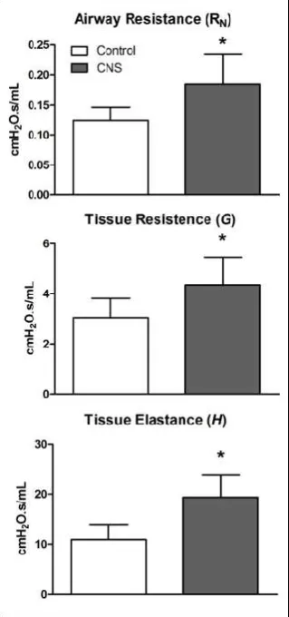
Figure 2. Values for airway resistance( ), tissue resistance ( ) and tissue elastance ( ), of the animals exposed to ambient air,
Control group (white column), and to the combustion exhaust gases of CNS, CNS group (gray column). Values are represented
by mean±standard deviation of the mean. * Represents statistically significant values in comparison
to the control group (p <0,05).
With average values of 8.9 ppm for CO, 20.2 % for O2,
3.1 ppm for NO, 5.8 ppm for SO2and average temperature
of26,3 ºC.In an attempt to avoid the intoxication of the animals by CO, the atmosphere of the CNS group was controlled so that this gas was kept below 10 ppm. Our results regarding the
analysis of the impedance of the respiratory system ( ),
calculated from the forced oscillation technique, are presented in Figure 2. The values of the variables related to airway
resistance ( ), tissue resistance ( ) and tissue elastance ( )
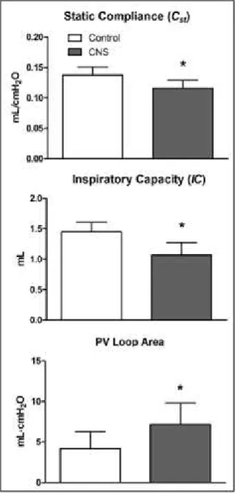
The values of the variables related to the static complacency
( ), estimation of inspiratory capacity ( ) and PV loop area
of the CNS group in relation to the control group, presented statistically significant differences. The absolute values, referring to the variables calculated from the analysis of the
impedance of the respiratory system ( ), and the PV curve of
the groups exposed to the anbiemte air (control group) and to the combustion exhaust gases of the CNS (CNS group), are shown in Table 1.
Figure 3.Values referring to the variables collected from the PV curve. The static complacency ( ), estimation of inspiratory capacity ( ) and PV loop area, of the animals exposed to ambient air, Control group (white column), and the combustion exhaust gases of CNS, CNS group (gray column). Values are represented by mean±standard deviation of the mean. * Represents statistically significant values in comparison to the control group (p <0,05).
DISCUSSION
Although the literature presents works of exposition of animals to the most diverse pollutants, gases and particles generated by the exhaustion and combustion of some biomasses, we do not find works whose objective is to evaluate the probable harmful effects to the respiratory system, due to the direct exposure of animals Combustion exhaust gases from the CNS. However, there is evidence of its harmful effects on health, with regard to the combustion of CNS, the number of respiratory events, increased inhaled therapy, mutagenicity, genotoxicity and
cytotoxicity (Cabral, 2010; Galvão et al., 2014). The
combustion of any biomass emits large amounts of carbon in
the forms CO2, CO, CH4, particulate matter (PM) and their
presence in the air, can transform them into pollutants, depending on the type of biomass burned (chemical composition) and the conditions of burning (Cieslinski, 2014).Exposure to these gases can result in acute and chronic repercussions in the respiratory system, these can occur in both previously healthy individuals and in patients with pre-existing
disease (Galan et al., 2003). Additionally, this exposure may
potentiate immune system response to allergens by increasing epithelial permeability, recruitment and activation of inflammatory cells, and oxidative stress in the airways (Nagato, 2007).
The risk of lung cancer caused by the presence of PM and APHs (acid polycyclic hydrocarbons) present in the biomass combustion gases is higher than the corresponding risk from exposure to exhaust gases from motor vehicles (Sarigiannis et al.,2015).The atmospheric pollution caused by biomass burning in the Brazilian Amazon region shows a positive association between the exposure of its pollutants and the occurrence of respiratory diseases, especially in the most vulnerable age groups (the elderly and children under 5 years
of age) (Ignotti et al., 2010). The MP present in the CNS
combustion exhaust gases can undergo alveolar deposition and endocytosis, translocate the pulmonary epithelioendothelial barrier, expressing a high ratio between surface area and mass, reaching deeper into the respiratory tract, which can reach and damage the pulmonary parenchyma Such as being absorbed into the bloodstream, which amplifie s its biological toxicity (Shimada et al., 2006; Furuyama et al., 2009). Our results, in relation to the parameters obtained through the constant phase model, demonstrate a statistically significant increase in the
values of the Newtonian resistance ( ), tissue resistance ( )
and tissue elastance ( ) (Figure 2) in the CNS group compared
to the Control group. The significant increase in ,may be
[image:4.595.136.462.89.224.2]provided by the inhalation of the PM present in the exhaust fumes from the CNS combustion.
Table 1. Mechanical of respiratory system data of animals exposed to air and CNS. Values are mean ± SD of animals exposed to ambient air (n = 8, Control group) and animals exposed to exhaust gases from the combustion of CNS (n = 8, CNS group). *p<0.05,
statistically significant difference.
Measure Group Value pvalue (t student)
Newtonian Resistence ( ) (cmH2O·s/mL)
Control CNS
0.123±0.022
0.185±0.049 p= 0.0168*
Tissue Resistence ( ) (cmH2O/mL)
Control CNS
3.05±0.76
4.35±1.09 p= 0.0299*
Tissue Elastance ( ) (cmH2O/mL)
Control CNS
10.93±3.05
19.39±4.51 p= 0.0020*
Inspiratory Capacity ( ) (mL)
Static Compliance ( ) (mL/cmH2O)
Loop Area (mLmH2O)
Control CNS Control
CNS Control
CNS
1.45±0.15 1.06±0.20 0.137±0.013 0.115±0.012 4.17±2.07 7.16±2.64
p= 0.0027*
p= 0.0102*
[image:4.595.77.247.345.701.2]These particles, when inhaled induce the expression of
pro-inflammatory mediators and Ca2+ dependent intracellular
signaling pathways (Ermak and Davies, 2002). The biological
functions of the Ca2+ ion in the lungs play a role in the
regulation of various functions, such as mucus secretion, surfactant secretion and ciliary agitation frequency (Conway et al., 2003). Changes in tissue resistance variables ( )and tissue elastance ( ), may be related to the intrinsic properties of the tissue, causing alterations in the rheology of the pulmonary tissue, due to alterations of the extra cellular matrix, tissue remodeling and of other constituents (Bates, 2009). Alterations
in and can also occur due to the increase in the due to a
narrowing of the airways, causing a distortion of the pulmonary parenchyma, resulting in an increase in the intrinsic stiffness of the tissue, causing elastance increase and the development of ventilatory heterogeneities throughout the lung, which causes an increase in tissue stiffness (Bateset al., 1994). Another possibility for changes in the and variables is the presence of mucus in small caliber airways, which could lead to occlusions of these pathways, as the same effect of producing areas with atelectasis.
Regarding the parameters obtained through the realization of the volume pressure curve, we observed significant alterations
in the parameters of static complacency ( ), estimation of
inspiratory capacity ( ) and PV loop area (Figura 3) of the
animals CNS group in relation to the Control group. The
decrease in the parameter may be a reflection of the already
discussed increase in tissue elastance ( ).In experimental studies, inhalation of particles from combustion processes led to decreased lung compliance and an inflammatory response characterized by influx of polymorphonuclear cells and release
of cytokines (Laks et al., 2008; Mazzoli-Rocha et al., 2008).
The decrease in the inspiratory capacity ( ), indicating
stiffening of the lung tissue, observed by the increase in ,
assuming that the animals of the CNS group presented a greater effort in the inspiration. On the other hand, the increase of PV loop area can be explained by possible alterations in the distribution of surfactant on the alveolar surface, associated with the presence of alveolar edema in the lung of the CNS group. The PV loop area is determined by four processes: recruitment/de-recruitment, surface tension, stress relaxation, and gas absorption during PV assays.
Conclusion
Despite the environmental appeal for the energy utilization of a previously discarded biomass in the environment, our results demonstrate that an acute combustion exhaust exposure of the CNS was sufficient to cause changes in the respiratory system of mices. Ours results reinforcing the urgent need to seek control methods for the exhaustion of these gases in the atmosphere. Further investigation is necessary in order to know safe parameters for individuals who are continually exposed to CNS combustion exhaust gases.
REFERENCES
ANEEL (Agência nacional de energia elétrica, [National electrical energy agency]), 2008. Energia elétrica do Brasil [Electric power of Brazil]. 3:65-67. Available in: http://www.aneel.gov.br/arquivos/PDF/atlas3ed.pdf. Access in: 06/23/2016.
Bates, J. 2009. Lung mechanics an inverse modeling approach. Cambridge University press. UK.
Bates, J.H., Lauzon, A.M., Dechman, G.S., Maksym, G.N.
and Schuessler, T.F. 1994.Temporal dynamics of
pulmonary response to intravenous histamine in dogs:
effects of dose and lung volume. Journal of Applied
Physiology. 76(2):616-626.
Cabral, T. M. 2010. Avaliação dos constituintes e do potencial
mutagênico do material particulado oriundo do
beneficiamento artesanal da castanha do caju [Evaluation of the constituents and the mutagenic potential of the particulate matter coming from the artisanal processing of cashew nuts]. Doctoral thesis. Faculdade de Medicina, Universidade do Estado de São Paulo, São Paulo. 126 pp. Cieslinski, J.E.F. 2014. Estudo da emissão e do controle dos
gases e particulados provenientes da queima de biomassa [Study of the emission and control of gases and particulates from biomass burning]. Doctoral thesis. Faculdade de Engenharia do Campus de Guaratinguetá – São Paulo. 23-40 pp.
Conway, J.D., Bartolotta, T., Abdullah, L.H. and Davis, C.W. 2003. Regulation of mucin secretion from human bronchial epithelial cells grown in murine hosted xenografts. American Journal of Physiology-Lung Cellular and Molecular Physiology, 284(6):945-954.
Ermark, G. and Davies, K.J.A. 2002. Calcium and oxidative
stress: from cell signaling to cell death. Molecular
immunology, 38(10):713-721.
Figueiredo, F.A.B., Figueiredo, R.A., Sanchez, C.G., Sanchez, E.M.S., Arauzo, J., Sanchez, J.L. and Gonzalo, A. 2007. Pyrolysis and gasification of cashew nut (Anacardium
Occidentale L.) shell: Liquid, solid and gas products. In:
19th International Congress of Mechanical Engineering,
Available in: http://www.abcm.org.br/pt/wp-content/anais/ cobem/2007/ pdf/COBEM2007-2333.pdf
Furuyama, A., Kanno, S., Kobayashi, T. and Hirano, S. 2009. Extrapulmonary translocation of intratracheally instilled fine and ultrafine particles via direct and alveolar
macrophage-associated routes. Archives of toxicology,
83(5):429-437.
Galan, I., Tobias, A., Banegas, J.R. and Aranguez, E.R. 2003. Short-term effects of air pollution on daily asthma
emergency room admissions. European Respiratory
Journal, 22(5):802-808.
Galvão, M.F., Cabral, T.M., André, P.A., Andrade,
M.F., Miranda, R.M., Saldiva, P.H., Vasconcellos, P.C. and Medeiros, S.R. 2014. Cashew nut roasting: Chemical characterization of particulate matter and genotocixity analysis. Environmental Research, 131(1):145-152. Hantos, Z., Daroczy, B., Suki, B., Nagy, S. and Fredberg, J.J.
1992. Input impedance and peripheral inhomogeneity of dog lungs. Journal of Applied Physiology, 72(1):168–178. Hirai, T., Mckeown, K.A., Gomes, R.F., and Bates, J.H., 1999.
Effects of lung volume on lung and chest wall mechanics in rats. Journal of Applied Physiology, 86(1), 16–21. IBGE (Instituto brasileiro de geografia e estatística [Brazilian
Institute of Geography and Statistics]). 2015. Systematic Survey of Agricultural Production. Access in: 07/13/2016.
Available in: ftp://ftp.ibge.gov.br/Producao_Agricola/
Levantamento_Sistematico_da_Producao_Agricola_[mens al]/Fasciculo/2015/lspa_201506.pdf.
Ignotti, E., Valente, J.G., Longo, K.M., Freitas, S.R., Hacon, S.S. and Netto, P.A. 2010. Impact on human health of particulate matter emitted from burnings in the Brazilian Amazon region. Revista de saúde pública, 44(1):121-130. International Nut & Dried Fruit. 2016. Global statistical review
http://www.nutfruit.org/wp-continguts/uploads/2016/05/ Global-Statist ical-Review-2015-2016.pdf.
Laks, D., Oliveira, R.C., André, P.A., Macchione, M., Lemos, M., Faffe, D., Saldiva, P.H. and Zin, W.A. 2008. Composition of diesel particles influences acute pulmonary toxicity: an experimental study in mice. Inhalation Toxicology, 20(11):1037-1042.
Lewne, M., Cyrys J., Meliefste, K., Hoek, G., Brauer, M., Fischer, P., Gehring, U., Heinrich, J., Brunekreef, B. and Bellander, T. 2004. Spatial variation in nitrogen dioxide in
three European areas. Science of The Total Environment,
332(1-3):217-230.
Lima, S. A. 2008. Análise da viabilidade do uso de cinzas agroindustriais em matrizes cimentícias: estudo de caso da cinza da casca da castanha de caju [Analysis of the feasibility of the use of agroindustrial ashes in cement matrices: a case study of the cashew nut bark ash]. Doctoral thesis. Escola de Engenharia de São Carlos, Universidade de São Paulo. São Carlos, 139 pp
Maffioletti, J. and Mota, M.J. 2013. Electricity generation using rice husks. Brazilian Journal of Energy, 19(1):49-59. Mazzoli-Rocha, F., Magalhãesm C,B., Malmm O., Saldiva,
P.H., Zin, W.A. and Faffe, D.S. 2008. Comparative respiratory toxicity of particles produced by traffi c and sugar cane burning. Environmental research, 108(1):35-41. MDIFT (Ministry of development, industry and foreign trade). 2015. Access in: 03/23/2016. Available in: http://aliceweb. mdic.gov.br/
Nagato, L.K.S. 2007. Acompanhamento temporal da função e histologia pulmonares em camundongos expostos a fuligem da queima de óleo [Temporal monitoring of pulmonary function and histology in mice exposed to soot from oil burning]. Doctoral thesis, Universidade Federal do Rio de Janeiro, Rio de Janeiro, 5-36 pp.
Ndalila, P.D. and Kaunde, O.O. 2014. Combustion and
Kinetics Analysis of Cashew Nut Shells. 6th Int'l
Conference on Mechanical, Production & Automobile Engineering (ICMPAE'2014), Nov. 27-28, Cape Town (South Africa)
Possel, M. and Bell, T.L. 2012. The influence of fuel moisture
content on the combustion of Eucalyptus foliage.
International Journal of Wildland Fire, 22(3):343-352 Ramos, J.E.T., Duarte, T.C., Rodrigues, A.K.O., Silva Jr, I.J.,
Cavalcante, C.L. and Azededo D.C.S. 2011. On the production of glucose and fructose syrups from cashew
apple juice derivatives. Journal of Food
Engineering,102(4):355-360.
Salazar, E. and Knowles, J.H. 1964. An analysis of
pressure-volume characteristics of the lungs. Journal of Applied
Physiology, 19(1):97-104.
Sarigiannis, D.A., Kermenidou, M., Nikolaki, S., Zikopoulos, D. and Karakitsios, S.P. 2015. Mortality and morbidity attributed to aerosol and gaseous emissions from biomass
use for space heating. Aerosol and Air Quality Researsh,
15(7)2496-2507.
Shimada, A., Kawamura, N., Okajima, M., Kaewamatawong, T., Inoue, H. and Morita, T. 2006. Translocation pathway of the intratracheally instilled ultrafine particles from the
lung into the blood circulation in the mouse. Toxicologic
Pathology, 34(7):949-957.
Silva, M.A., Nebra, S.A., Machado, S.M.J. and Sanchez, C.G. 1998. The use of biomass residues in the brazilian soluble coffee industry. Biomass and bioenergy, 14(5-6):457-467. Tesfaigzi, Y., Singht, S.P., Foster, J.E., Kubatko, J., Barr, E.B.
Fine, P.M., Mcdonald, J.D., Hahn, F.F. and Mauderly, J.L. 2002. Health Effects of Subchronic Exposure to Low
Levels of Wood Smoke in Rats. Toxicological Sciences,
65(1):115-125.
WHO (World health organization). 2005. Who air quality guidelines global update 2005: report on a working group meeting, Bonn, Germany.Access in: 04/13/2016. Available
in: http://www.euro.who.int/__data/assets/pdf_file/0008/
147851/E87950.pdf.
Yiannis, A.L., Kulbhushan, J., Reza, K. and Adel, F.S. 2011. Combustion behavior in air of single particles from three different coal ranks and from sugarcane bagasse. Combustion and flame, 158(3):452-465.