ISSN Online: 2163-0585 ISSN Print: 2163-0569
Continuous Vagus Nerve Monitoring during
Carotid Endarterectomy
Tamaki Tomonori
1*, Kubota Minoru
2, Node Yoji
1, Morita Akio
31Departmrnt of Neurological Surgery, Nippon Medical School, Tamanagayama Hospital, Tokyo, Japan 2Department of Clinical Laboratory, Nippon Medical School, Tamanagayama Hospital, Tokyo, Japan 3Departmrnt of Neurological Surgery, Nippon Medical School, Tokyo, Japan
Abstract
Backgrounds: Injury to the vagus nerve or one of its branches during carotid endarterectomy can result in vocal fold paralysis but the exact mechanism of injury responsible for vocal fold paralysis after carotid endarterectomy is unclear. Aims: This study was performed to identify potential predictors of vagus nerve injury and obtain feedback by application of intraoperative continuous vagus nerve monitoring. Materials and Methods: Seventy-four patients undergoing carotid endarterectomy were enrolled. A new vagus nerve electrode was designed for less invasive continuous vagus nerve stimulation and monitoring of the vocal fold electromyogram without disturbing the surgical procedure. The device was rectangular (13 mm × 9 mm), with two small round electrodes set on a flexible silicon plate and tube. The electrode was fully implantable during carotid endarterectomy and was positioned at the most distal site of the vagus nerve by suturing to the connective tissue without nerve dissection. All patients underwent laryngoscopy to assess postoperative vocal fold and pharyngeal wall palsy at one week after carotid endarterectomy. Results: Sudden loss of the vocal fold electromyogram was noted in two patients (during plaque removal and during arterial wall suture in one each). In these two patients, incomplete vocal fold and pharyngeal palsy was confirmed by laryngoscopy. The cause of vagus nerve injury may have been traction at the time of distal internal carotid artery manipulation. The vocal fold electromyogram remained normal during the operation in the other 72 patients. However laryngoscopy revealed postoperative vocal fold and pharyngeal palsy in six patients. These findings suggested that delayed vagus nerve injury can occur after carotid endarterectomy. Conclusion: The continuous vagus nerve monitoring may be worthwhile for elucidating the mechanism of vagus nerve injury related to carotid endarterectomy.
Keywords
Carotid Endarterectomy, Vagus Nerve, Vocal Fold, Neuromonitoring, Complication How to cite this paper: Tomonori, T.,
Minoru, K., Yoji, N. and Akio, M. (2017) Continuous Vagus Nerve Monitoring dur- ing Carotid Endarterectomy. Open Journal of Modern Neurosurgery, 7, 1-9.
http://dx.doi.org/10.4236/ojmn.2017.71001
Received: August 7, 2016 Accepted: December 9, 2016 Published: December 12, 2016
Copyright © 2017 by authors and Scientific Research Publishing Inc. This work is licensed under the Creative Commons Attribution International License (CC BY 4.0).
1. Introduction
Injury to the vagus nerve (VN) is a well-recognized problem in patients under- going carotid endarterectomy (CEA) [1][2]. VN damage can lead to undesirable consequences, including vocal disturbance, aspiration, and dysphagia. Although VN palsy is a serious complication of CEA, the causes of VN injury are not well understood [3][4]. In patients undergoing neck surgery, intraoperative neuro-monitoring has been advocated as a method of localizing and identifying the re-current laryngeal nerve (RLN), as well as being used to predict postoperative vocal fold function [5][6]. We previously reported a method of intermittent VN monitoring during CEA using a hand-held monopolar electrode for VN stimu-lation [7]. However, the hand-held stimulator had certain limitations. Besides difficulty in precisely identifying the VN, assessment of functional integrity was limited to the periods of intermittent stimulation. To overcome these limita-tions, a method that allowed continuous monitoring would be desirable. Ac-cordingly, we performed the present investigation to assess the effectiveness of a new flexible plate electrode for continuous VN monitoring (VNM) by retrospec-tive evaluation in 74 patients undergoing CEA.
2. Materials and Methods
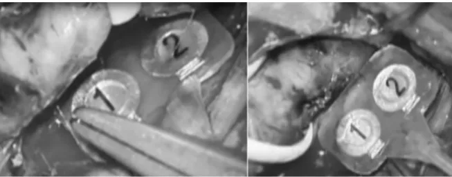
patients, CEA was performed by the same standard surgical technique, which involved dissection of the carotid artery while causing the minimum possible damage to any cranial nerves in the operating field. Special attention was paid to careful dissection of the VN from the carotid artery, especially at the sites of proximal and distal clamping. We did not use self-retaining wound retractors. An internal shunt and a Hemashield patch graft were employed in all patients, and the procedure was always performed under an operating microscope. Our intention was to develop a less invasive flexible plate VN stimulation electrode that would not disturb the CEA procedure. The electrode was designed so that dissection of the VN was not required for attachment and so that it remained in contact with the nerve for continuous VNM. Accordingly, two small round stimulation electrodes (cerebral surface electrodes for motor evoked potential monitoring during craniotomy) were placed on a flexible silicon plate and tube (Figure 1). The electrode was fully implantable during CEA and was connected to a Nihon Kohden MEB-2312. Prior to positioning this continuous VNM electrode, we used a conventional handheld bipolar stimulation electrode (stimulus intensity of 0.5 - 2.0 mA, pulse width of 100 μs, and frequency of 3Hz) to identify the VN and we placed the continuous VNM electrode at the most caudal point of the nerve. To maintain contact with the VN, we sutured the silicon plate of the electrode to connective tissue (Figure 2). The stimulus conditions for continuous VNM were as follows: sense: 500 μV, filter: 2 KHz ~50 Hz, stimulus intensity: <2 mA, single train, duration: 0.2 msec, stimulation rate: 1 Hz, number of stimuli: 10, monitoring time (/div): 20 msec, analysis time (/div): 3 msec, and tend interval: 1 ~ 3 minutes. The initial stimulus intensity was 0.5 mA using a negative square wave impulse and it was gradually increased in 0.5-mA intervals to the upper limit of 2 mA until the first stable EMG signal from the vocal fold was identified to evaluate the influence of signal amplitude.
Figure 1. This photograph shows the new stimulation electrode for continuous vagus
Figure 2. This photograph shows using the new stimulation electrode during carotid endarterectomy.
The Nihon Kohden MEB-2312 was used for both stimulation and measurement, with the latency, amplitude, and duration being assessed as quantitative parameters. Latency was defined as the time (in milliseconds) between the stimulus artifact and the onset of EMG activity and amplitude was defined as the magnitude of the EMG wave (in microvolts). Data are expressed as the mean ± standard deviation. EMG changes were classified as signal loss (a signal was initially obtained from the VN, but could not be elicited subsequently at 2.5 mA and an event threshold of 100 mV) or impairment (latency delayed by less than 20% and amplitude decreased by less than 50%). For evaluation of cardiac and pulmonary side effects, the heart rate, blood pressure, and SaO2 were measured before, during, and after continuous VNM. The electrode was removed from the VN just before wound closure. In all patients, vocal fold and pharyngeal movements were observed by laryngoscopy at one week after CEA. When vocal fold dysfunction was identified, follow-up was done at 2-month intervals. We used the Student’s t-test for continuous variables and P-values < 0.05 were con-sidered statistically significant.
3. Results
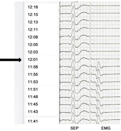
one case each (Figure 3). In these two patients, we could not detect the EMG by stimulating the VN at any site after signal loss. Laryngoscopy demonstrated in-complete vocal fold and pharyngeal wall palsy, with recovery after 6 months in one patient and 12 months in the other. In the remaining 72 CEA procedures, there was no signal loss or impairment of the vocal fold EMG during continuous VNM. Although six patients showed incomplete vocal fold and pharyngeal palsy on laryngoscopy, they recovered after 2 to 12 months (Figure 4). There were no complications of continuous VNM.
Figure 3. The sensory evoked potential (left side) and ipsilateral vocal fold electromyogram
(right side) were revealed. The black arrow shows vocal fold electromyogram disappeared suddenly during plaque removing. SEP: Sensory evoked potential, EMG: Vocal fold electromyogram.
Figure 4. This figure shows the sumary of vocal fold electromyogram and laryngoscope
[image:5.595.268.479.531.702.2]4. Discussion
study. The overall VN identification rate was 81%. Giovagnorio investigated variations of the VN using ultrasonography [10]. They reported that the VN ran anterior to the common carotid artery in 4.3% of 144 subjects and medial to it in 1.2%. Ha also investigated the variations of VN anatomy, reporting that the nerve ran posterior to the common carotid artery in 13.8% and medial to the vessel in 0.3% [11]. During CEA, the surgeon cannot find a variant VN located medial or posterior to the carotid artery. Visual identification of the recurrent laryngeal nerve (RLN) during thyroid surgery is associated with lower rates of permanent RLN palsy and is considered the gold standard by many authors [12] [13] [14]. Jatzko et al. reviewed 10 reports covering 12,211 thyroid operations and found a lower rate of RLN palsy in the patients with nerve identification than in those without nerve identification (2.7% vs 7.9% for temporary palsy and 1.2% vs 5.2% for permanent palsy) [14]. Especially in patients with visual integ-rity, the mechanisms of nerve injury are still not well understood [14]. We also must consider the possible side effects of continuous VNM. The same mecha-nism of VN stimulation was used during continuous VNM as that employed to treat drug-resistant epilepsy and depression [15][16][17]. Chronic VN stimula-tion over several months may lead to side effects, with various types of laryngo-pharyngeal dysfunction, including hoarseness, cough, pharyngitis, throat dis-comfort, laryngeal muscle spasm, and dyspnea, being detected after long-term stimulation for weeks or months [15][16][17]. However, no complications were seen in our patients after short-term stimulation. In this study, the mean con-tinuous VNM time was 68 minutes, which is a very short duration of VN stimu-lation, but we cannot exclude the possibility that VN palsy may be induced by continuous VNM. Regarding complete EMG signal loss, the effect of VN fatigue must be considered [18][19]. However, the lower frequency limit for fatigue of vegetative nerves is more than 10 Hz, while we used 3 Hz for stimulation. In conclusion, continuous VNM was safe and it seems worthwhile to continue in-vestigations in order to elucidate the mechanism of VN injury during CEA.
5. Conclusion
This is the first report of continuous VNM during CEA using new small plated form stimulation electrode. We performed postoperative laryngoscope examina-tion for all cases. From continuous VNM and post CEA laryngoscope findings, we speculated the cause of VN injury was stretch effect during the caudal side ICA manipulation. Although vocal fold EMG was normal at the end point of CEA, there were six cases revealed postoperative VN palsy. These findings cre-ated a hypothesis the delayed VN injury mechanism after CEA may exist. The VN monitoring may worth trying for elucidate the mechanism of VN injury of CEA.
References
Stent-ing with Endarterectomy in Symptomatic Carotid Artery Stenosis. Cerebrovascular Diseases, 18, 69-74. https://doi.org/10.1159/000078753
[2] Silver, F.L., Mackey, A., Clark, W,M,. Brooks, W., Timaran, C.H., Chiu, D., Goldstein, L.B., Meschia, J.F., Ferguson, R.D., Moore, W.S., Howard, G. and Brott, T.G. (2011) Safety of Stenting and Endarterectomy by Symptomatic Status in the Carotid Revascularization Endarterectomy Versus Stenting Trial (CREST). Stroke, 42, 675-680. https://doi.org/10.1161/STROKEAHA.110.610212
[3] Curran, A.J., Smyth, D., Sheehan, S.J., Joyce, W., Hayes, D.B. and Walsh, M.A. (1997) Recurrent Laryngeal Nerve Dysfunction Following Carotid Endarterectomy.
J R Coll Surg Edinb, 42, 168-170.
[4] Beasley, W.D. and Gibbons, C.P. (2008) Cranial Nerve Injuries and the Retrojugular Approach in Carotid Endarterectomy. Ann R Coll Surg Engl, 90, 685-688.
https://doi.org/10.1308/003588408X318138
[5] Chiang, F.Y., Lu, I.C., Kuo, W.R., Lee, K.W., Chang, N.C. and Wu, C.W. (2008) The Mechanism of Recurrent Laryngeal Nerve Injury during Thyroid Surgery—The Application of Intraoperative Neuromonitoring. Surgery, 143, 743-749.
https://doi.org/10.1016/j.surg.2008.02.006
[6] Roberts, J.W. (2008) Elucidating Mechanisms of Recurrent Laryngeal Nerve Injury during Thyroidectomy and Parathyroidectomy. Journal of the American College of Surgeons, 206, 123-130. https://doi.org/10.1016/j.jamcollsurg.2007.07.017
[7] Tomonori, T., Minoru, K., Norihiro, S., Katsuya, U., Takayuki, K. and Yoji, N. (2012) Vagus Nerve Neuromonitoring during Carotid Endarterectomy. Perspectives in Vascular Surgery and Endovascular Therapy, 24, 137-140.
https://doi.org/10.1177/1531003512472239
[8] Tamaki, T., Node, Y., Saitou, N., Saigusa, H., Yamazaki, M. and Morita, A. (2013) Vernet’s Syndrome after Carotid Endarterectomy. Perspectives in Vascular Surgery and Endovascular Therapy, 25, 65-68. https://doi.org/10.1177/1531003514525476
[9] Tamaki, T., Node, Y., Saitou, N. and Saigusa, H. (2013) Observation of Vocal Fold and Pharyngeal Paralysis after Carotid Endarterectomy Using a Magnifying Laryn-goscope. World Journal of Surgery, 37, 911-914.
https://doi.org/10.1007/s00268-013-1920-1
[10] Giovagnorio, F. and Martinoli, C. (2001) Sonography of the Cervical Vagus Nerve: Normal Appearance and Abnormal Findings. American Journal of Roentgenology, 76, 745-749. https://doi.org/10.2214/ajr.176.3.1760745
[11] Ha, E.J., Baek, J.H., Lee, J.H., Kim, J.K. and Shong, Y.K. (2011) Clinical Significance of Vagus Nerve Variation in Radiofrequency Ablation of Thyroid Nodules. Euro-pean Radiology, 21, 2151-2157. https://doi.org/10.1007/s00330-011-2167-6
[12] Dionigi, G., Chiang, F.Y., Rausei, S., Wu, C.W., Boni, L., Lee, K.W., Rovera, F., Cantone, G. and Bacuzzi, A. (2010) Surgical Anatomy and Neurophysiology of the Vagus Nerve (VN) for Standardised Intraoperative Neuromonitoring (IONM) of the Inferior Laryngeal Nerve (ILN) during Thyroidectomy. Langenbeck’s Archives of Surgery, 395, 893-899. https://doi.org/10.1007/s00423-010-0693-3
[13] Lorenz, K., Sekulla, C., Schelle. J., Schmeiss. B., Brauckhoff, M. and Dralle, H. (2010) What Are Normal Quantitative Parameters of Intraoperative Neuromoni- toring (IONM) in Thyroid Surgery? Langenbeck’s Archives of Surgery, 395, 901- 909. https://doi.org/10.1007/s00423-010-0691-5
https://doi.org/10.1186/1477-7827-12-28
[15] Agnew, W.F. and McCreery, D.B. (1990) Considerations for Safety with Chronically Implanted Nerve Electrodes. Epilepsia, 31, 27-32.
https://doi.org/10.1111/j.1528-1157.1990.tb05845.x
[16] Bernards, C.M. (2004) An Unusual Cause of Airway Obstruction during General Anesthesia with a Laryngeal Mask Airway. Anesthesiology, 100, 1017-1018.
https://doi.org/10.1097/00000542-200404000-00037
[17] Groves, D.A. and Brown, V.J. (2005) Vagal Nerve Stimulation: A Review of Its Ap-plications and Potential Mechanisms That Mediate Its Clinical Effects. Neuro- science & Biobehavioral Reviews, 29, 493-500.
https://doi.org/10.1016/j.neubiorev.2005.01.004
[18] Dralle, H., Sekulla, C., Haerting, J., Timmermann, W., Neumann, H.J., Kruse, E., Grond, S., Mühlig, H.P., Richter, C., Voss, J., Thomusch, O., Lippert, H., Gastinger, I., Brauckhoff, M. and Gimm, O. (2004) Risk Factors of Paralysis and Functional Outcome after Recurrent Laryngeal Nerve Monitoring in Thyroid Surgery. Surgery, 136, 1310-1322. https://doi.org/10.1016/j.surg.2004.07.018
[19] Ulmer, C., Friedrich, C., Kohler, A., Rieber, F., Basar, T., Deuschle, M., Thon, K.P. and Lamadé, W. (2011) Impact of Continuous Intraoperative Neuromonitoring on Autonomic Nervous System during Thyroid Surgery. Head Neck, 33, 976-984.
https://doi.org/10.1002/hed.21564
Submit or recommend next manuscript to SCIRP and we will provide best service for you:
Accepting pre-submission inquiries through Email, Facebook, LinkedIn, Twitter, etc. A wide selection of journals (inclusive of 9 subjects, more than 200 journals)
Providing 24-hour high-quality service User-friendly online submission system Fair and swift peer-review system
Efficient typesetting and proofreading procedure
Display of the result of downloads and visits, as well as the number of cited articles Maximum dissemination of your research work