ScholarWorks @ Georgia State University
ScholarWorks @ Georgia State University
Chemistry Dissertations Department of Chemistry
Summer 8-7-2012
Synthesis and Energetics of Gold Nanoclusters Tailored by
Synthesis and Energetics of Gold Nanoclusters Tailored by
Interfacial Bonding Structure
Interfacial Bonding Structure
Tang Zhenghua
Georgia State University
Follow this and additional works at: https://scholarworks.gsu.edu/chemistry_diss
Recommended Citation Recommended Citation
Zhenghua, Tang, "Synthesis and Energetics of Gold Nanoclusters Tailored by Interfacial Bonding Structure." Dissertation, Georgia State University, 2012.
https://scholarworks.gsu.edu/chemistry_diss/67
This Dissertation is brought to you for free and open access by the Department of Chemistry at ScholarWorks @ Georgia State University. It has been accepted for inclusion in Chemistry Dissertations by an authorized
administrator of ScholarWorks @ Georgia State University. For more information, please contact
TURE
by
ZHENGHUA TANG
Under the Direction of Gangli Wang
ABSTRACT
In addition to the well known quantum confinement effects resulted from size and shape, interfacial
bond structure is another factor, affecting the properties of the nanomaterial that is rarely studied.
In-spired by the “Au-S-Au” staple motif discovered from the crystal structure of monothiol protected
Au102 nanocluster (Science, 2007, 318, 430), dithiol molecules (e. g. 1, 2-dithiol, 1, 4-dithiol, etc.) with
molecular structural constraint have been employed to create dithiolate protected clusters or mixed
monothiolate and dithiolate protected clusters. The structure and properties of the Au clusters are
ex-pected to change due to two effects: The entropy gain of dithiol over monothiol protection and the
con-straint to the formation of the thiol bridging motif. DMPS (1, 2-dithiol molecule) stabilized clusters with
characteristic absorption bands have been obtained, and characterized by multiple techniques.
Mono-layer reaction on gold core surface between the monothiol tiopronin and dithiol DMPS has been
4-the optical and electrochemical energetics have been successfully correlated based on core and
core-ligand energy states. Furthermore, the impact of 1, 4-dithiolate-Au bonding on the near infrared
lumi-nescence has been studied.
INDEX WORDS: Au MPCs, Staple motif, DMPS, Au DTCs, Au4, Tiopronin, Monolayer reaction, Durene-DT, Au MTCs, Au130, Optical energetic, Electrochemistry, Near infrared luminescence, 1,
TURE
by
ZHENGHUA TANG
A Dissertation Submitted in Partial Fulfillment of the Requirements for the Degree of
Doctor of Philosophy
in the College of Arts and Sciences
Georgia State University
Copyright by Zhenghua Tang
TURE
by
ZHENGHUA TANG
Committee Chair: Gangli Wang
Committee: Markus W. Germann
Shahab A. Shamsi
Electronic Version Approved:
Office of Graduate Studies
College of Arts and Sciences
Georgia State University
DEDICATION
I want to dedicate this dissertation to my parents (Hongfa Tang and Genxiu Duan). Without you,
I could not go so far. You always give the most valuable spiritual support as well as material support in
all my way of studying and growing up as a man. Your love and care is the origin of my motivation to do
research, to conquer difficulties I meet. I also want to dedicate this dissertation to my brother Dehua
ACKNOWLEDGEMENTS
All the work has been done under the direction and support of Dr. Gangli Wang. I owe my
deep-est gratitude to him, and I am so thankful he is my Ph. D advisor. His inspiring suggdeep-estions on experiment
design, rigorous thoughts to research results and self-motivated attitude to research itself will benefit
my whole life, not only as a researcher, but also as a person.
The committee members of my dissertation Professor Markus W. Germann and Professor
Sha-hab A. Shamsi are gratefully acknowledged.
Special thanks to Professors Jenny J. Yang and Binghe Wang for all the help.
I thank all the staff in Department of Chemistry at Georgia State University, especially mass
spectrometry manager Dr. Siming Wang and her assistant Dr. Yanyi Chen.
I would like to thank all the current and past members (Dr. Bin Xu, Dr. Tarushee Ahuja, Dr.
Bao-hua Wu, Donald A. Robinson, Nadia Bokossa, Juan Liu, Warren Brown, Dengchao Wang, Yan Li, Maksim
Kvetny, Cecil Conroy) in Dr. Gangli Wang's group. Thank you for your help during those years of study,
and I enjoyed working together with you.
My US brother Yuehua (Michael) Tang helped me with a lot of things, and I thank him for his
great support!
I acknowledge the fellowship (2011-2012) from Center of Diagnostics and Therapeutics (CDT) at
Department of Chemistry, Georgia State University.
The financial support for my research, including Gangli Wang startup at GSU, Research Initiation
Grant at Georgia State University and National Science Foundation funding (CHE 1059022), are gratefully
TABLE OF CONTENTS
ACKNOWLEDGEMENTS... v
LIST OF SCHEMES ... xi
LIST OF TABLES ... xii
LIST OF FIGURES ... xiii
LIST OF ABBREVIATIONS ... xviii
1 INTRODUCTION ... 1
1.1 Au NPs: Synthesis, Characterizations and Fundamental Properties ... 1
1.2 Au NPs Used in Catalytic Regime ... 20
1.3 Au NPs Used in Biomedical Applications ... 24
1.3.1 Au NPs as Drug-Delivery Systems (DDS) ... 25
1.3.2 Au NPs as Imaging Contrast Agents ... 27
1.3.3 Au NPs as Biosensors, Biolabeling Agents ... 28
1.3.4 Au NPs for Surface-Enhanced Raman Scattering (SERS) ... 31
1.3.5 Au NPs for Cancer Treatments and Photothermal Therapy ... 33
1.3.6 Nanotoxicity of Au NPs in Biomedical Applications ... 34
1.4 Au NPs Used in Other Areas ... 36
2 SYNTHESIS AND STRUCTURAL DETERMINATION OF MULTIDENTATE 2, 3-DITHIOL STABILIZED GOLD CLUSTERS ... 36
2.2 Results and Discussion ... 39
2.2.1 UV-Visible Absorbance of DMPS clusters ... 39
2.2.2 Hydrodynamic Size Distribution of DMPS DTCs by NMR Studies ... 41
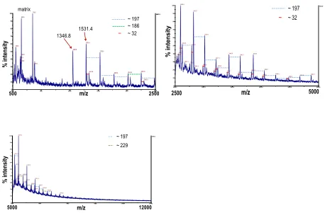
2.2.3 Composition of 3X DMPS DTCs by MALDI-MS Studies ... 45
2.2.4 Size on a Solid Support Imaged by AFM ... 47
2.3 Experimental Section ... 56
2.3.1 Chemicals ... 56
2.3.2 Measurements ... 57
2.3.3 Synthesis of DMPS Clusters ... 58
2.4 Summary ... 59
3 MONOLAYER REACTIONS OF PROTECTED GOLD NANOCLUSTERS WITH MONOTHIOL TIOPRONIN AND 2, 3-DITHIOL DIMERCAPTOPROPONIC SULFONATE ... 60
3.1 Background and Research Strategy ... 60
3.2 Results and Discussion ... 63
3.2.1 Optical Properties Monitored by Absorbance and Luminescence of Tiopronin MPCs with DMPS Molecules ... 63
3.2.2 Characterization of Monolayer Reaction Products of Tiopronin MPC with DMPS Ligands………65
3.2.3 NMR Studies of Reaction Process of Tiopronin MPCs with DMPS ... 67
3.2.4 Kinetics of Monolayer Reaction of Tiopronin MPCs with DMPS ... 70
3.2.6 UV-Visible Absorbance and Luminescence Features of DMPS DTCs with Tiopronin
Molecules……….…..75
3.2.7 Characterization of DMPS DTCs with Tiopronin Molecules ... 77
3.3 Experimental Section ... 83
3.3.1 Chemicals ... 83
3.3.2 Measurements ... 83
3.3.3 Synthesis of Tiopronin Au MPCs and DMPS Au DTCs ... 83
3.3.4 Monolayer Reactions of Au Clusters –Monothiol Tiopronin versus Dithiol DMPS ………84
3.4 Summary ... 84
4 MIXED DITHIOLATE DURENE-DT AND MONOTHIOLATE PHENYLETHANETHIOLATE PROTECTED AU130 NANOPARTICLES WITH DISCRETE CORE AND CORE-LIGAND ENERGY STATES ... 85
4.1 Background and Research Strategy ... 85
4.2 Results and Discussion Results and Discussion ... 87
4.2.1 UV-Visible Absorbance of Mixed Thiolate Au Clusters ... 87
4.2.2 Molecular Ion Identification by MALDI-MS ... 89
4.2.3 Ligand Composition in the Monolayer Determined by 1H NMR ... 94
4.2.4 Diffusion Coefficient and Monodispersity Probed by Diffusion–Ordered NMR Spectroscopy (DOSY) ... 98
4.2.6 Molecular Composition of the Au MTCs ... 100
4.2.7 Near Infrared (IR) Luminescence of Au130 MTCs ... 101
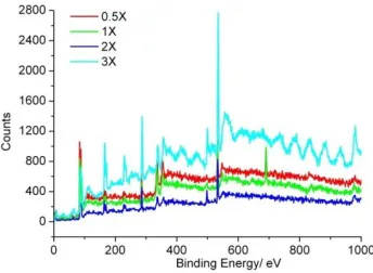
4.2.8 Binding Energy by X-ray Photoelectron Spectroscopy (XPS) Studies ... 104
4.2.9 Electrochemical Properties of Au130 MTCs ... 106
4.2.10 The Correlation of Electrochemical and Optical Energetics: Core Charging and Core-Ligand Charge Delocalization ... 111
4.3 Experimental Section ... 113
4.3.1 Chemicals ... 113
4.3.2 Measurements ... 113
4.3.3 Synthesis and Purification ... 114
4.3.4 Iodine Death Reaction ... 114
4.4 Summary ... 115
5 NEAR INFRARED LUMINESCENCE OF GOLD NANOCLUSTERS AFFECTED BY THE BONDING OF 1, 4- DITHIOLATE DURENE AND MONOTHIOLATE PHENYLETHANETHIOLATE ... 115
5.1 Background and Research Strategy ... 115
5.2 Results and Discussions ... 118
5.2.1 Optical Properties of the Reaction between Monothiolate Au MPCs with Durene-DT 1, 4-Dithiols ... 118
5.2.2 Dithiol-Monothiol Exchange Monitored by Proton NMR ... 121
5.2.4 Optical Properties of Durene-DT Au DTCs with PhC2S Monothiols ... 124
5.2.5 Mass Spectrometry Characterization of the Reaction Products ... 126
5.3 Materials and Method ... 128
5.3.1 Chemicals ... 128
5.3.2 Methods ... 128
5.3.3 Synthesis and Purifications ... 128
5.4 Summary ... 129
6 CONCLUSIONS AND MAJOR DISCOVERY ... 129
7 REFERENCES ... 130
LIST OF SCHEMES
Scheme 1.1 The synthetic scheme of nonpolar gold nanoclusters with Brust-Schiffrin method and place
exchange (PE) reaction ... 4
Scheme 3.1 Proposed reaction mechanism of tiopronin MPCs with DMPS ... 75
LIST OF TABLES
Table 2.1 Hydrodynamic sizes of monothiol and dithiol clusters. ... 44
Table 3.1 Mass spectrometric peak assignments of Figure 3.2, Figure 3.15 and Figure 3.16. ... 81
Table 4.1 The comparison of experimental data and the representative compositions proposed of the Au
MTCs. ... 101
LIST OF FIGURES
Figure 1.1 Cartoon of thiolate protected gold nanoclusters ... 2
Figure 1.2 The TEM image of tiopronin MPCs (3X) ... 6
Figure 1.3 The absorbance spectra of Au25 nanoparticles ... 10
Figure 1.4 Emission spectrum of Au25(SC2H4Ph)18 nanoparticle ... 11
Figure 1.5 Differential pulse voltammogram (DPV) of Au25(SC2H4Ph)18 ... 12
Figure 1.6 Absorbance spectrum of Au144(SCH2CH2Ph)60 nanoparticle ... 16
Figure 1.7 Quantized double-layer charging behaviors of Au144 nanoparticle protected by hexanethi-olate ... 17
Figure 1.8 Absorbance and emission spectra of 3X tiopronin MPCs ... 19
Figure 1.9 Absorbance comparison of tiopronin MPCs, 1X, 3X and 100X ... 20
Figure 2.1 Structural illustration of two monothiol molecules versus one dithiol molecule on Au core sur-face. ... 37
Figure 2.2 UV-Visible absorbance spectra of DMPS Au DTCs. ... 40
Figure 2.3 Absorbance spectra of pre-DTC intermediates ... 41
Figure 2.4 A: NMR spectra of DMPS ligand and Au DTCs; B: Comparison of diffusion coefficient calculated from diffusion NMR studies... 42
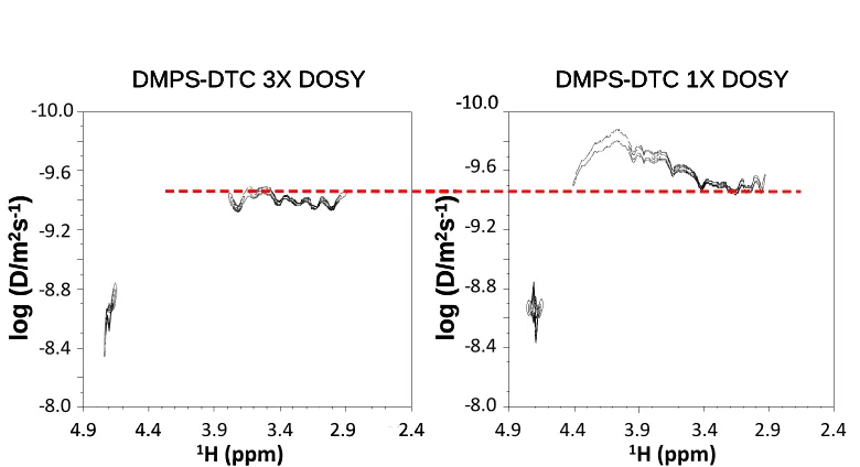
Figure 2.5 NMR DOSY spectra of 3X and 1X DMPS Au DTCs ... 44
Figure 2.6 MALDI MS results of the 3X DMPS Au DTCs. ... 45
Figure 2.7 3X DTC MALDI MS spectrum of another synthetic batch. ... 46
Figure 2.8 3X Pre-DTC MALDI MS spectra ... 47
Figure 2.9 Representative AFM images of 1X and 3X DTCs ... 48
Figure 2.1013C- 1H multiplicity edited F2 coupled HSQC spectrum of 3X DMPS Au DTCs. ... 49
Figure 2.12 XPS spectra of DMPS DTCs ... 52
Figure 2.13 XPS Survey Scan of DMPS DTCs ... 53
Figure 2.14 The proposed Au4L4 and Au4L3 cluster structure ... 53
Figure 2.15 The relaxed 3D structure of Au4L4 by molecular mechanics... 54
Figure 2.16 The relaxed 3D structure of Au4L3 by molecular mechanics... 54
Figure 2.17 Infrared spectra of DMPS DTCs ... 55
Figure 2.18 Thermogravimetric analysis of DMPS DTCs ... 56
Figure 3.1 Absorbance and luminescence spectra of monolayer reaction of tiopronin MPCs with DMPS molecules ... 64
Figure 3.2 MALDI MS spectrum of DMPS DTCs formed by monolayer reaction of tiopronin MPCs with DMPS. ... 66
Figure 3.3 Comparison of normalized absorbance spectra of the synthesized DMPS Au4DTCs and the products of tiopronin Au MPC-DMPS monolayer reaction. ... 67
Figure 3.41H NMR spectra of monolayer reaction of tiopronin MPCs with DMPS ... 68
Figure 3.5 Monolayer reaction kinetics of tiopronin Au MPCs with DMPS studied by 1H NMR. ... 69
Figure 3.6 Absorbance and luminescence spectra of tiopronin MPCs reaction with DMPS. ... 69
Figure 3.7 Absorbance and luminescence spectra of tiopronin MPCs reaction with DMPS. ... 71
Figure 3.8 Kinetics of monolayer reactions at different reactant Au MPC concentration and ligand mole ratio R (DMPS:tiopronin) ... 72
Figure 3.9 A: Representative first order fitting of luminescence changes. B: Linear correlation of fitted first order rate constants of luminescence changes with DMPS concentration………74
Figure 3.11 Absorbance and luminescence spectra of 2-5 nm DMPS Au DTC reaction with tiopronin
mo-lecules. ... 77
Figure 3.121H NMR spectra of monolayer reaction of 1.5 nm DMPS DTCs with tiopronin ... 78
Figure 3.131H NMR spectra of monolayer reaction of 2-5 nm DMPS DTCs with tiopronin ... 79
Figure 3.14 MALDI MS characterization of monolayer reaction product of 1.5 nm DMPS Au DTCs with
tiopronin ... 80
Figure 3.15 Expanded view of m/z patterns of Au4L3 and Au6S5 anions that display isotope peaks ... 81 Figure 3.16 MALDI MS results of monolayer reaction product of 2-5 nm DMPS Au DTCs with tiopronin
... …81
Figure 4.1 A. The absorbance change during the reduction. B. Absorbance spectrum of the purified
product in methylene chloride………...87
Figure 4.2 MALDI mass spectra of Au MTCs at different laser intensities... 89
Figure 4.3 Left: MALDI mass spectrum of external standard Bolvine Serum Albumin (BSA) in sinapinic
acid matrix. Right: MALDI mass spectrum of MTC-1-2-1-X. ... 90
Figure 4.4 The MALDI mass spectra of the Au MTCs with reflectron negative mode (Left), and
representative fragments observed in linear positive mode (Right) ... 91
Figure 4.5 The representative chromatogram of Au MTCs separated by HPLC ... 92
Figure 4.6 Normalized absorbance spectra of original injected Au MTC sample, 4.6 min eluate and 7.0
min eluate. ... 93
Figure 4.7 Rest potential comparison of original MTCs and HPLC collected eluates ... 93
Figure 4.8 NMR spectra of the purified MTCs (Top) and the decomposed products (Bottom) ... 94
Figure 4.9 Enlarged view of 1H NMR spectrum of the purified MTCs (Left) and decomposed sample (Right)………...94
Figure 4.111H -1H TOCSY spectrum of decomposed MTCs ... 97
Figure 4.12 Full peak assignment of protons in the decomposed sample in 1H NMR spectrum ... 97
Figure 4.13 DOSY spectrum of Au MTCs in CD2Cl2 ... 99
Figure 4.14 Thermogravimetric analysis (TGA) of the MTCs ... 100
Figure 4.15 Luminescence of dilute Au130 MTCs... 101
Figure 4.16 Original emission spectra of the Au130 MTCs at 450-850 nm and 850-1500 nm without cor-rection ... 102
Figure 4.17 Excitation spectra of MTCs, emission collected at 700 nm and 900 nm ... 102
Figure 4.18 Au XPS spectrum of the Au130 MTCs ... 104
Figure 4.19 The survey scan of the Au130 MTCs XPS spectrum ... 104
Figure 4.20 S(2p) focusing scan XPS spectrum of the Au130 MTCs. ... 105
Figure 4.21 A: Square wave voltammogram (SWV) of Au MTCs. B: Zoom in of oxidation scan in SWV with one corresponding sampled current shown above ... 106
Figure 4.22 Cyclic voltammograms of the Au130 MTCs ... 107
Figure 4.23 SWV oxidation scan of the Au130 MTCs from Figure 4.21 with 1st derivative plot shown in bottom panel ... 108
Figure 4.24 Differential pulse voltammogram of the Au130 MTCs ... 109
Figure 4.25 Cyclic voltammogram of Durene-DT ligand at room temperature ... 109
Figure 4.26 Energy diagram that correlates optical and electrochemical features ... 111
Figure 5.1 The spectrum changes of absorbance and luminescence in the reaction between Au25(SC2Ph)18 with durene-DT. ... 119
Figure 5.2 The proton NMR spectra of the reaction between Au25(SC2Ph)18 with durene-DT. ... 121
Figure 5.4 The absorbance (Left) and luminescence (Right) change of Durene DTCs reacted with
phenyle-thanethiol. ... 124
Figure 5.5 Mass spectra of Au25(PhC2S)18 (Top), the final products of Au25 MPCs with durene-DT (Middle)
LIST OF ABBREVIATIONS
Au NPs Gold Nanoparticles
Au MPCs Gold Monolayer-Protected Clusters
Au DTCs Gold Dithiol-Protected Clusters
Au MTCs Gold Mixed Thiolate Clusters
UV-Vis Ultraviolet-Visible
XAFS X-ray Absorption Fine structure Spectroscopy
SAXC Small-Angle X-ray Scattering
XANES X-ray Absorption Near-Edge Spectroscopy
XRD X-Ray Diffraction
AFM Atomic Force Microscopy
TEM Transmission Electron Microscopy
STM Scan Tunneling Microscopy
NMR Nuclear Magnetic Resonance
COSY Correlated Spectroscopy
DOSY Diffusion Ordered Spectroscopy
TOCSY Total Correlation Spectroscopy
HSQC Heteronuclear Single-Quantum Coherence
HMBC Heteronuclear Mutiple-Bond Correlation Spectroscopy
MS Mass Spectrometry
ESI Electrospray Ionization
MALDI-TOF Matrix-Assisted Laser Desorption Ionization-Time of Flight
EA Elemental Analysis
XPS X-ray Photoelectron Spectroscopy
ESCA Electron Spectroscopy for Chemical Analysis
BE Binding Energy
DMPS Dimercaptopropanesulfonic Acid
MWCO Molecular Weight Cutoff
CHCA α-Cyano-4-hydroxycinnamic acid
FT-IR Fourier-Transform Infrared Spectroscopy
PE Place Exchange
Durene-DT Durene-α1, α2-dithiol
DCTB trans-2-[3-(4-tert-butylphenyl)-2-methyl-propenylidene]-malononitrile
HPLC High Performance Liquid Chromatography
TBAP Tetrabutylammonium Perchlorate
CV Cyclic Voltammetry
DPV Differential Pulse Voltammetry
SWV Square Wave Voltammetry
PhC2S Phenylethanethiol(ate)
OCPT Open Circuit Potential Test
TOAB Tetraoctylammonium Bromide
HOMO Highest Occupied Molecular Orbital
1 INTRODUCTION
1.1 Au NPs: Synthesis, Characterizations and Fundamental Properties
Nanotechnology, an emerging technology, has penetrated into every aspect of daily life all over
the world. It focuses on the study and dealing with ultra-small matters or devices, specifically, in the
range of 1 to 100 nm even hundreds of nm. As we know, 1 nm = 10-9 m, and it is about 105 times the
width of a single human hair. In another word, in the regime of nanotechnology, the matter or device at
ultrasmall range named “nanoscale” is investigated. Nanotechnology, ranging from the conventional
physical devices to new approaches based on molecular assembly, stretches across physics, chemistry,
material science, electronics, biology, medicine, environmental science, energy technology and so on.
On the other hand, many problems, issues and concerns have been raised, such as the toxicity of
nano-materials, the environmental and ecological impacts of nanomaterials and the potential conflicts of
tra-ditional ethics with applications of nanotechnology.
Nanoparticles, one of the most important materials in nanotechnology, are defined as bridges
be-tween bulk materials and single molecular or atomic structures. The well studied nanoparticles include
quantum dots, metal nanoparticles, bio-constructed nanoparticles (DNA, protein based nanocomposite),
colloid or gel, polymer nanoparticles and so on.
Gold nanoparticles (Au NPs), including gold nanocrystals or gold nanoclusters, are a classic type of
metal nanoparticle of great importance. Generally speaking, nanocrystals are particles with dimensions
over 100 nm, which can be considered “larger nanoparticles” or “microparticles”, while nanoclusters
refer to much smaller particles with diameter less than 2 nm, so-called “molecular level”. My research
mainly focuses on small gold nanoparticles, specifically, the synthesis, characterizations and property
Gold is a subject of the most ancient theme of investigations in science, technology, medicine,
en-gineering, decorating arts and so on.1 In ancient China or Egypt, gold colloids had been used as
thera-peutic agents. These colloids, in fact, we now believe, were gold gel solutions. They were microparticles
instead of nanoparticles. As the emergence of nanotechnology, gold nanoparticles gained a great deal of
research interests, probably due to their extra-ordinary stability, tunable surface functionalities and
size-dependent properties.
Ultra-small gold nanoparticles, also called gold monolayer-protected clusters (MPCs), are
com-posed of gold core and self-assembled monolayers (SAMs), which are usually made of halide, phosphate
and thiolate molecules. The gold core consists of ten to a few hundreds of gold atoms, and the gold
atoms are packed with Au-Au bonding. The monolayers, can be not only organothiolate molecules, but
also bio-molecules, for example, DNA, protein etc. Figure 1.1 presents the cartoon of thiolate protected
gold nanoclusters. The core and the surface are connected by the Au-S bond, or another way to say, the
monolayer protects the gold core through Au-S interaction on the interface. The gold core enables the
particle unique size-dependent capabilities; meanwhile the protection from the monolayer provides the
particles robust stabilities. Moreover, the monolayer can be easily modified, which imparts the particle
surface functionalities, such as chemical reactions. These gold nanoclusters, usually in a dimension less
than 2 nm, are an intense research subject recently1-5, thanks to their fundamental properties such as
optical behaviors, surface reactivity and electrochemistry.
[image:24.612.75.181.556.661.2]
There are several ways to obtain Au NPs, mostly starting from the reduction of Au (III) derivatives.
For the Au NPs normally employed in biological systems with ~20 nm diameter or even larger, the most
widely used method has been developed by Turkevitch in 1951.6 The stabilizer in his approach is
triso-dium citrate, and it is well recognized in colloid science that citrate molecules weakly coordinate with
the gold core through multiple sites. Currently, this method is still being widely employed to prepare the
precursor of more stable AuNP-biomolecule nanocomposite for further biomedical applications.
By using X-ray absorption fine structure spectroscopy (XAFS), the initial kinetic nucleation of such
gold nanocrystals under the grain size of 1 nm has been reported,7 which can be described as the
forma-tion of Cl3-Au-AuCl3- dimer and the subsequent higher complexes “AunCln+x” in the early stage. The bulk
gold nanocrystal formation mechanism has been deduced by four steps recently,8 including nucleation
(~2 nm), growth by aggregation (~3 nm), slow further growth (~5.5 nm) and fast final growth (~7.7 nm)
through small-angle X-ray scattering (SAXS) and X-ray absorption near-edge spectroscopy (XANES)
inves-tigations.
In terms of preparing ultra-small gold nanoclusters, the Brust-Schiffrin method, first reported in
19949 and 199510, has a considerable impact on the overall field in less than 20 years. The synthetic
scheme of nonpolar gold nanoclusters is shown in Scheme 1.1. Basically, chloroauric acid (Au(III)) was
first transferred from aqueous solution to organic phase by tetraoctylammonium bromide (TOAB), and
reduced by organic thiol molecules to form gold-thiolate (Au(I)) polymers. The organic soluble Au-S
lymer can be further reduced by aqueous sodium borohydride solution. The produced materials are
po-lydisperse gold nanoclusters with wide size distribution, ranging from ~ 5 nm to less than 1 nm. It is
worth noting that, the metal can be gold, palladium, copper or other metals or alloy. Among all the
met-al nanoclusters, gold is the most widely prepared one by this method, probably due to their
extraordi-nary stability and versatile applications. The as-prepared clusters are thermally stable and air stable, and
de-composition of the structures. The facile method allows the reproducible synthesis of stable clusters
with controllable quantities for the first time. Gold nanoclusters obtained by this method can be
han-dled or modified as chemical reagents or molecular compounds, and one of the typical modifications is
place exchange (PE) reaction, which is also shown in scheme 1.1. The ligand on the monolayer can be
partially or totally exchanged by other thiol-containing molecules, depending on the reactivity of the
incoming ligand, the chemical equivalents and other experimental conditions. This reaction can be
per-formed to obtain new nanoparticles with modified surface, while the original ligand and the incoming
ligand are not necessarily the same type, e. g. the original ligand is citrate and the incoming ligand is
or-ganic molecule or biomolecule with –SH group included.
Scheme 1.1 The synthetic scheme of nonpolar gold nanoclusters with Brust-Schiffrin method and place
exchange (PE) reaction.
There is ongoing debate on the mechanism of the nanocluster formation by this biphasic synthetic
method. It has been reported recently by Lennox11 and coworkers that the metal precursor before
NaBH4 reduction is not the generally believed a [Au(I)-SR] polymer but rather the [TOA][MX2] complex.
better control in forming different metal (Au, Ag, and Cu) NPs with different organo-chalcogen ligands as
the stabilizers.12 It is worth noting that, size evolution of thiol protected gold nanoclusters can be
as-sisted by proton acids and halogen anions.13 By employing mercaptobenzoic acid (MBA) as ligand,
theo-retical work of the AuNP growth mechanistic scheme including formation of a stable Au4(MBA)4 complex
and Aun NP growth and stabilization upon the interaction with the Au4(MBA)4 complexes has been
rec-orded.14
The easy handling properties of the gold nanoclusters facilitate their structural and compositional
analysis, which can be further divided into the core and the monolayer analysis.1,2
The formula of molecular gold nanoclusters can be described as Aun(SR)m, and the compositions of
these clusters can be revealed by mass spectrometry. For clusters with multiple positive or negative
charges, electrospray ionization (ESI)15-20 is a good choice and has been extensively employed. For singly
charged clusters or neutral clusters associated with singly charged counter ion, laser desorption
ioniza-tion mass spectrometry (LDI-MS) especially matrix assisted laser desorpioniza-tion ionizaioniza-tion mass
spectrome-try (MALDI-MS)21-32 has proven to be a powerful tool to identify the molecular ion. Other mass
spectro-metric techniques, such as fast atom bombardment (FAB)33 and ion-mobility mass spectrometry
(IM-MS)34,35 have been exploited to demystify the molecular structures of those clusters. Fragmentation in-formation regarding the composition of exchanged nanoparticle can be obtained by tandem mass
spec-trometry (MS-MS),36 while several successful cases can be found to detect the molecular ions with the
combination of liquid chromatography or gas chromatography and mass spectrometry, which will be
discussed later in separation section.
For the gold core, the most straightforward method to “see” it is high resolution transmission
electron microscopy (HR-TEM). The gold core can be photographed by TEM technique. TEM has been
widely used to characterize the gold metal clusters37-44 since 1990s. The shape of the nanoparticle can
easi-ly identified. Besides imaging the real particle, another advantage of TEM is that by counting the
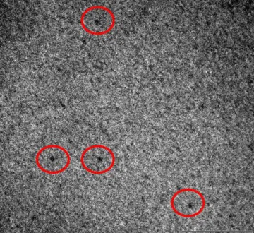
par-ticles in a microscopic window, the particle size histogram can be obtained. The TEM image of tiopronin
MPCs (3X, while X= Ligand-to-gold ratio during synthesis) is shown in Figure 1.2.
However, the gold core dimensions can also be characterized by other techniques, such as atomic
force microscopy (AFM),45-52 scanning tunnel microscopy (SEM),53-59 small-angle X-ray scattering
[image:28.612.74.254.234.399.2](SAXS),8,60-63 X-ray diffraction (XRD)64-68 and so on.
Figure 1.2 The TEM image of tiopronin MPCs (3X). The black dots circled by red line in the graph are the
gold cores of the nanoparticles (unpublished results). The scale bar is 50 nm and the diameter of the
gold core is ~2 nm.
For the monolayer, the ligand structure can be analyzed by nuclear magnetic resonance (NMR),
and in most cases, polydisperse materials were examined, thus the data represents an average.
Com-pared with sharp peaks of the ligand, theNMR peaks are significantly broadened, known as
line-broadening effect.69 Ongoing efforts in understanding the source of the line-broadening effects have
been carrying on continuously.70-74 The extent of such effects is strongly dependent on the nanoparticle
size, shape, ligand type, monodispersity as well as the proximity to the gold core.25,75-80 Moreover, NMR
with either place exchange reaction, chemical reactions, or dynamic study.81-83 It is interesting to note
that intramolecular ligand dynamics in organophosphate stabilized gold nanoparticles investigated by 2H
NMR has been reported recently.84
Two dimensional (2D) NMR techniques have also found versatile applications in structural analysis
of the monolayer attached on the gold core. Nuclear overhauser enhancement spectroscopy (NOESY)
and heteronuclear multiple quantum coherence (HMQC) spectra have been generated to assign all the
NMR sensitive nuclei in the tiopronin ligand while diffusion-ordered spectroscopy (DOSY) provided
in-formation about the particle size distribution of water soluble Au MPCs in Murray’s work.77 In a recent
report, DOSY has been described as a reliable alternative to TEM for determining the size of gold
nano-particles in organic solutions.85 The identification of the uniquitin-gold nanoparticle interaction sites has
been reported by [15N-1H]-heteronuclear single quantum coherence (HSQC) NMR technique in
protein-nanoparticle interaction.86
The broad peaks in NMR resulted from line-broadening effects often lead to low resolution
spec-tra, hence not much information can be obtained from the interpratation. To overcome this limitation,
high resolution magic angle spinning (HRMAS) 1H NMR technique has been introduced to characterize
the organic molecules attached to the gold core recently.87
The mass fraction of the monolayer in the total nanoparticle can be analyzed by
thermogravime-tric analysis (TGA). In a TGA experiment, the monolayer of the gold clusters decomposes from the gold
core as volatile disulfide, leaving elemental gold core. Hence, the organic/metal ratio can be calculated
from TGA experiment, and this ratio has been widely applied to determine the average composition of
the polydisperse gold clusters.88-91 For monodisperse gold clusters with uniform size, the combination of
elemental analysis (EA)92,93 with this technique makes it more powerful for determining the molecular
The nature of the gold core of these clusters can be examined by X-ray photoelectron
spectrosco-py (XPS). XPS is also known as electron spectroscospectrosco-py for chemical analysis (ESCA), and is a quantitative
technique that can measure the elemental composition, empirical formula, chemical state and
electron-ic state of the elements present in the material.94,95 The basic principle of this technique is that, the
ana-lyte is irradiated by an X-ray beam while the kinetic energy and the number of the electrons that escape
from the top 1-10 nm of the surface are measured simultaneously. High vacuum experimental
condi-tions are required, and this surface chemical analysis technique can be used to analyze all kinds of
mate-rials, such as inorganic compounds, alloys, polymers, semiconductors, ceramics, catalyst, glasses, and so
on.
For gold nanoclusters, XPS can be performed to probe the charge state and the binding energy of
the elements, in most cases gold, e. g. Au(4f5/2) and Au(4f7/2)25,70,96-98, sometimes, sulfurs, e.g. S(2p)99,100,
or both.101,102 It is interesting to point out that the survey scan in XPS experiment can always be treated
as an elemental analysis experiment, which can be the proof of the existence of one element.
For metal nanocrystals, because all the molecular orbitals are filled with free electrons, the
absor-bance shows featureless exponential decay. Metal nanoclusters, specifically gold nanoclusters, often
consist of tens to hundreds of atoms, and their size typically ranges from sub-nanometer to
approx-imately 2 nm in core diameter. In such extremely small size regime, strong quantum confinement of
electrons results in the occurrence of electron energy quantization, which strongly alter the
fundamen-tal characteristics of the gold clusters, such as UV-Visible absorbance, luminescence and
electrochemi-stry. For example, instead of the appearance of plasmon excitation in nanocrystals, molecular gold
na-noclusters normally show pronounced absorption bands, reflecting the discrete electron energy
states.1,3,5
One of the best examples to demonstrate nanoparticle’s unique fundamental properties including
li-gand here can be glutathione, phenylethanethiolate, hexanethiolate, or dodecanethiolate. Au25L18 has
been a good target of studying nanoparticles because of their molecule-like properties, such as facile
synthetic accessibility, easy isolation with great monodispersity and extraordinary stabilities.
Pioneering work to obtain this nanoparticle was done by Whetten and coworkers,15,22 but
unfor-tunately, it was labeled as Au28(SG)16 (SG= Glutathione) because of the analytical instrument limitations.
Murray’s group synthesized monodisperse Au25(SC2H4Ph)18 nanoparticle and studied the optical
beha-viors and electrochemistry on it thoroughly,104,105 but they also mislabeled it as Au38(SC2H4Ph)24. By using
ESI-MS technique, Tsukuda and coworkers97 corrected the identity of glutathione protected gold
nanoc-lusters as Au25(SG)18. The large scale synthesis of Au25(SG)18 through ligand exchange reaction of
phos-phine-stabilized Au11 clusters has also been documented by Tsukuda’s group.106 High resolution ESI-MS
identification of Au25(SC2H4Ph)18- anion has been done by Murray’s group,17,18 and thus the molecular
composition finally has been settled down as Au25L18.
The crystal structure of Au25(SC2H4Ph)18- anion with TOA+ cation as counter-ion was reported by
both Murray’s group107 and Jin’s group108 in 2008. X-ray crystal structure shows Au25 nanoparticle
in-cludes icosahedral Au13 core, which is surrounded by six Au2(SR)3 semi-rings. Note that, Au25
nanopar-ticle can possess three different charge states,-1, 0 and +1, which has been revealed by Tsukuda et al. in
ESI-MS investigations109. The following work on the crystal structure of Au25(SC2H4Ph)180 neutral species
showed that the neutral Au25 has almost identical structure with the anionic one except some small
dif-ferences,110 specifically, the slight structural distortion in the anionic Au25 were not observed in neutral
one. Theoretical investigations111-114 also play an important role in advancing the understanding of the
electronic structure and the origin of the stability of this nanoparticle.
As shown in Figure 1.3, Au25 nanoparticles exhibit strong absorbance peaks at ~400 nm (3.11 eV),
~ 450 nm (2.76 eV) and ~670 nm (1.86 eV), with optical band gap of ~1.33 eV.105 Notably, no matter
phenylethanethi-olate, as long as the nanoparticle adopts Au25 structure, the absorbance bears the similar features.29
However, the absorbance of Au25 nanoparticle shows a slight difference at different charge states. For
Au25(SC2H4Ph)18, it has been reported that the anionic species exhibit a distinct, broad shoulder at ~800
nm while the neutral one does not,110 similar case of Au25(SG)1829 and other Au25 nanoparticles stabilized
by organothiolate ligands. From here, we can conclude that, both Au25 nanoparticles shown in Figure 1.3
are anionic species, as ~800 nm shoulder can be clearly observed in both spectra. The ~670 nm
absorp-tion can be correlated with the electronic and geometric structure of the icosahedral Au13 core through
density theory functional calculations.108
Figure 1.3 Absorbance spectra of Au25 nanoparticles, unpublished results. Left: Au25(SC2H4Ph)18, Right:
Au25(SC12H25)18.
The near infrared photoluminescence (PL) of Au25(SC2H4Ph)18 has also been reported105. However,
this intensity is fairly weak, probably due to the polydispersity of the sample tested. Figure 1.4 presents
the emission spectrum of Au25(SC2H4Ph)18. Broad band from 600 nm to 850 nm even longer wavelength
centered at ~720 nm can be observed from the emission spectrum. By using electron withdrawing
li-gands, the emission intensity can be improved significantly.115 The origin of this PL is believed to arise
from the surface states of the nanoparticle. The surface states can localize the free electrons hence
serum albumin (BSA) protected Au25 nanoclusters.117 Recent report found that charge state of the gold nanocluster and surface ligand can play an important role in enhancing the near-IR PL of Au25
nanoc-lusters.118 Interestingly, the surface ligand can influence the luminescence either by charge transfer from
the ligand to the metal core through Au-S bond or by direct donation of delocalized electrons of
elec-tronic-rich atoms or groups of the ligand to the metal core.118
Figure 1.4 Emission spectrum of Au25(SC2H4Ph)18 nanoparticle, excited at 450 nm, unpublished results.
Functionalized gold nanoclusters exhibit unusual electrochemical reactivities, quantized
double-layer charging voltammetry and molecule-like voltammetry.4 The quantized double-layer charging
beha-viors can be observed in clusters with relative larger cores, e. g. Au144 nanoparticle, and this will be
dis-cussed later in Au144 part. The electrochemical properties of Au25(SC2H4Ph)18 and ligand exchange
prod-uct have been probed by Murray and coworkers.105Figure 1.5 shows the voltammetric measurement of
Au25(SC2H4Ph)18 nanoparticles. Basically, for Au25(SC2H4Ph)18 nanoparticle, voltammetry in CH2Cl2 reveals
a 1.62 eV energy gap between the first one-electron oxidation and the reduction gap, which
corres-ponds with the sum of the corrected charging energy ca. 0.29 eV and the LUMO-HOMO energy gap 1.33
model regarding the correlation of the optical energy gap and electrochemical gap has also been
pro-posed in this report.
Figure 1.5 Differential pulse voltammogram (DPV) ofAu25(SC2H4Ph)18 in CH2Cl2 at room temparature.119
Adapted with permission from J. Am. Chem. Soc. 2004, 126, 6193-6199. Copyright 2004 American
Chem-ical Society.
Another good example of molecular gold nanoclusters with interesting absorbance, luminescence
and electrochemical properties is Au38 nanoparticle. The Au38 nanoparticle passivated by glutathione
pertains to 8k Da of molecular mass was first reported by Schaaff et al.120 However, at that time, the
ex-act composition was not confirmed by MS, since LDI-MS often results in complicated fragments instead
of molecular ions. Later on Au-SG cluster compounds from Au11 to Au39, including Au38, have been
isolated by polyacrylamide gel electrophoresis (PAGE) by Tsukuda’s group.97 Au38(SC6H13)22 has been
suc-cessfully synthesized through an excess thiol etching method by Quinn’s group,121 and this 8k gold
clus-ter of Au38 protected either by hexanethiolate or dodecanethiolate have also been obtained by Chaki et
al. with the structural implication by MS.122 The size focusing synthesis of Au38(SC2H4Ph)24 has been
de-veloped by Jin’s group.123 In their synthetic strategies, two steps are included: the formation of
temperature. Similar work on the facile and large-scale synthesis of Au38(SC12H25)24 has been
docu-mented by them.124
Au38 nanoparticles, stabilized by glutathione,97 phenylethanethiolate,123 hexanethiolate121 or
do-decanethiolate, exhibit discrete electronic energy states, mainly at ~1050 nm (1.18 eV), ~750 nm (1.66
eV), ~620 nm (2.00 eV), ~560 nm (2.21 eV) and ~520 nm (2.39 eV) with ~0.92 eV band gap. Among them,
the most distinct peak is the one at 2.00 eV, which can be considered the distinct one of Au38
nanopar-ticle. Temperature-dependent emission spectra of Au38(SC6H13)24 has been investigated. At room
tem-perature only one emission band at near-IR region was observed, while at low temtem-perature fine
struc-tural spectra can reveal at least four bands.125
Notably, the Au38 nanoparticle also exhibit different charge states, and electrochemistry provides
elegant means to probe the electronic structure and chemical stabilities.121 Au38(SC6H13)22 with charge
states of -1, 0, +1, +2 have been characterized by cyclic voltammetry (CV) and differential pulse
voltam-metry (DPV) by Toikkanen et al.121 The HOMO-LUMO band gap was around ~1.2 eV, which agrees well
with Au38(SC2H4Ph)24123 reported by Jin’s group.
A structural model of Au38(SR)24 has been established by Jiang et al,126 and density-functional
theory calculations have been performed by Aikens and coworkers127 on the charity and electronic
struc-ture of Au38(SR)24 nanoparticle. Zeng’s group proposed a face-fused bi-icosahedral Au23 core in the
structure of Au38(SR)24 nanoparticle.128 The theoretical work advanced the structure and property
under-standing of Au38 nanoparticle.
Besides Au25 and Au38 nanoparticles, other molecular nanoclusters have also been widely
inves-tigated. “Magic number” [Au13(dppe)5Cl2]Cl3 clusters have been obtained through a convergence of
nuc-learity strategy promoted by hydrochloric acid, and very strong near-IR emission was exhibited.129 The
synthesis of atomic precise Au19(SC2H4Ph)13 nanoparticle has been performed by Wu et al. recently
pro-nounced absorption can be observed in this cluster. Molecular pure Au20(SC2H4Ph)16 has been obtained
by Zhu et al., and this cluster exhibit pronounced absorption peak at ~485 nm (2.56 eV) and a broad
band at ~420 nm (2.95 eV) with a larger LUMO-HOMO band gap of ~2.15 eV.93 As the crystal structure of
the Au20 nanoparticle was not resolved, theoretical predictions done by Zeng’s group showed that
Au20(SC2H4Ph)16 nanoparticle may adopt a structure of a prolate Au8 core with new Au3(SR)4 motif.131
Recently, the synthesis of Au36 nanoclusters completely protected by the aromatic thiol, Au36(SPh)23,
has been documented by Dass’s group. This nanomolecule shows a peak at ~566 nm (2.20 eV) and two
shoulders at ~430 nm (2.89 eV) and ~370 nm (3.36 eV).132
Au40(SC2H4Ph)24, another molecular cluster, which possess ~8 kDa mass reported by Jin’s group,30
has been found to co-exist with Au38(SC2H4Ph)24. The Au40 nanoparticle was isolated from Au38
nano-particle through size exclusion chromatography (SEC) and its composition was confirmed by ESI-MS and
MALDI-MS. No pronounced absorption peak was observed and the optical band gap is ~1.0 eV, which is
comparable with Au38 (0.9 eV). Recent work regarding semi-preparative scale separation of
Au38(SC2H4Ph)24, Au40(SC2H4Ph)24 and larger clusters by SEC has been recorded by Knoppe et al.133
Au55 clusters protected by alkanethiolate with mass around 11 kDa can be obtained by
chroma-tographic isolation in Tsukuda’s report.134 Au55(PPh3)12Cl6 nanoparticle, with a two core-shell
close-packed cuboctahedron structure of Au13 surrounded by 42 gold atoms, was first reported in the early
1980s.135,136 However, this phosphine-stabilized 1.4 nm gold nanoparticle has limited stability, because
the phosphine ligand can dissociate easily. It is worth noting that the reaction of Au55(PPh3)12Cl6 with
hexane thiol or other thiols can lead to the formation of Au75 nanoparticle, typically Au75(SR)40.137
Au68(SC2H4Ph)34 nanoparticle has been identified by mass spectrometry during the size-focusing
transition process of Au25(SC2H4Ph)18 nanoparticle formation by Dass’s group, however, they were not
Au102 nanoparticle protected by para-mercaptobenzoic acid (p-MBA), Au102(p-MBA)44, is a water
soluble polar cluster with molecular purity. Interestingly, the first report on this cluster is its crystal
structure.138 Note that, this is the first crystal structure of gold nanoparticle. X-ray diffraction is a
power-ful analytical approach that provides unambiguous information concerning the nanoparticle structure.
The single crystal was obtained from a mixture by Kornberg’s group in 2007. The 1.1 Å resolution crystal
structure is very informative. The central 79 gold atoms in the Au102 core are packed with a markus
de-cahedron and surrounded by the additional layers of gold and sulfur atoms of unexpected geometry
“staple motif”. Three gold atoms and two sulfur atoms form “staple motif” on the interfacial gold core
and monolayer surface, and the Au102 core is an Au79 core protected staple motives. It is worth noting
that although the ligand was non-chiral, the nanoparticle exhibited chirality, as two enantiomers
alter-nating in the crystal lattice described in this paper.
The landmark achievement regarding the crystal structure of Au102(p-MBA)44 and the revelation of
the “staple motif” has greatly influenced the nanogold research community. First of all, the traditional
simple Au-S bonding has been challenged by the “staple motif”, and this “staple motif” may be the key
of the extraordinary stability of these gold nanoclusters. Our research ideas and strategies are based on
this “staple motif”, which will be discussed later. Numerous reports, especially some theoretical
calcula-tion work, have been published to unravel the relacalcula-tionship of “staple motif” and the robust stability.
Second, the success of the first crystal structure of gold nanoparticle strongly promoted and stimulated
the great efforts of obtaining monodisperse material. A lot of research attention has been devoted to
gain monodisperse nanoclusters, accompanying attempts at crystallization.
The synthesis and characterizations of Au102(p-MBA)44 nanoparticle on a preparative scale in a high
yield has been reported recently by the same group,139 and this nanoparticle exhibits a featureless
The crystal structure of Au102(p-MBA)44 immediately triggered a lot of research interest in
theoreti-cal theoreti-calculations. Ab initio study of Au102(p-MBA)44 on this cluster to model the electronic structure has
been performed by Gao et al,140 and Li et al. presented the first principles, density functional theory
cal-culations of the structural and electronic properties of this cluster in 2008.141 Based on this crystal
struc-ture, chemical analysis of the super-atom model of thiol-stabilized gold nanoparticle has been
docu-mented to gain the insight of the thermodynamic stability.142 The electronic and vibrational signatures of
Au102(p-MBA)44 nanoparticle has been performed recently by Hakkinen and coworkers.143
The next molecular gold nanocluster will be discussed later is Au144 nanoparticle. Au144(SR)60
na-noparticle was previously assigned or reported as Au(144-150)(SR)(50-60), which corresponds with 29 kDa
mass. It was first revealed by MS by Schaaff et al.144 and Chen et al.145 Murray’s group and Quinn’s group
described the quantized double-layer charging behaviors, which will be discussed separately in the next
paragraph. Chaki et al. obtained dodecanethiolate protected Au144 nanoparticle and assigned the
mole-cular composition as Au144(SC12H25)59 by LDI-MS.122 Qian et al. reported a facile, two step synthetic
me-thod to prepare monodisperse Au144(SC2H4Ph)60 nanoparticle,92 which shows prominent absorption
bands at ~510 nm (2.44 eV) and ~700 nm (1.78 eV). By following the method they reported,
monodis-perse Au144(SCH2CH2Ph)60 has been synthesized successfully, and the absorbance of Au144 is presented
Figure 1.6. Step-like absorbance band at ~510 nm and ~695 nm can be observed, which is at least
Figure 1.6 Absorbance spectrum of Au144(SCH2CH2Ph)60 nanoparticle, unpublished results.
Gold nanoclusters with core diameter less than 2 nm exhibit very small effective capacitance (C),
which leads to single electron changes in the core occur at large voltage intervals (V=e/C), hence
quan-tized double-layer charging happens.146,147 The electronic charging behaviors of the high monodisperse
Au144 nanoparticle protected by hexanethiolate, governed by electrical doubler layer properties, have
been reported by Murray and coworkers,148 as shown in Figure 1.7. The extraordinary electrochemical
resolution of 15 oxidation states of this nanoparticle has been recorded by Quinn’s group.149
Figure 1.7 Quantized double-layer charging behaviors of Au144 nanoparticle protected by
hexanethi-olate.148 Adapted with permission from J. Am. Chem. Soc. 2002, 124, 13322-13328. Copyright 2002
For nonpolar gold clusters, the Brust-Schiffrin biphasic synthetic method offers a straightforward
way to prepare nonpolar ligand stabilized Au NPs. For the polar gold clusters, Brust-like syntheses use
water soluble functional thiols such as tiopronin,88 glutathione (SG),22 poly(ethylene glycol) (PEG)150,151
and para-mercaptobenzoic acid (p-MBA)138 etc. The best examples of molecular water soluble gold
na-noclusters are the well studied Au25(SG)1829,97,152 and Au102(p-MBA)44,138,139,143 which have been discussed
above. Ackerson et al. summarized and screened 36 water-soluble organothiolates for their ability to
form water-soluble MPCs in 2005.153 Aggregation-resistant water soluble gold nanoparticles stabilized by
zwitterionic ligands have been produced by Schlenoff’s group.154 The preparation and the properties of
water-soluble gold nanoparticles protected by perfluorinated amphiphilic thiolates have been described
by Pasquato and coworkers.155
One interesting water-soluble Au nanoparticle is tiopronin protected gold MPCs that are
unfortu-nately polydisperse. The average composition of this gold clusters has been determined as
Au201(Tiopronin)85 with core diameter of ~1.8 nm.88 The size of the nanoparticle can be tuned by varying
the ligand-to-gold ratio during the synthesis as demonstrated by the size-dependent absorbance. Figure
1.8 shows the absorbance and emission spectra of 3X tiopronin MPCs, while X represents the
ligand-to-gold ratio. The absorbance shows an exponential decay with two small bumps at ~260 nm and ~350 nm,
while broad emission band rangies from 650 nm to 850 nm and even higher wavelength. The quantum
Figure 1.8 Absorbance and emission spectra of 3X tiopronin MPCs. Left: Absorbance spectra, Right:
Emission spectra.
Interestingly, the nanoparticle size can be controlled by altering the ligand-to-gold ratio during the
synthesis, and the optical behaviors such as absorbance are size-dependent due to the size confinement
effects. As we know, with higher ligand-to-gold ratio employed in the synthesis, smaller sized particles
are obtained. The nanoparticle formation is a core agglomeration process while more excess thiol can
passivate the gold surface hence terminating the process at the initial stage to form smaller particles.
Figure 1.9 presents absorbance spectra of tiopronin MPCs with different ligand-to-gold ratios. The 1X
sample display a band at around 522 nm, which is called surface plasmon band (SPB) and will be
dis-cussed in the next paragraph. The UV absorbance of the 3X and 100X sample decays in approximately an
exponential fashion into visible, with no detectable SPB. Note that a much sharper decay occurs in the
100X sample than the 3X sample, indicating smaller core size in average. In fact, the core diameter of 1X
Figure 1.9 Absorbance comparison of tiopronin MPCs, 1X, 3X and 100X (X= ligand-to-gold ratio).
Surface plasmon band (SPB), reflected physically as a red deep color of gold nanoparticle solution,
normally appears at about 520 nm. The mechanism of the band is believed to be due to the electron
collective oscillation at the nanoparticle surface, which correlates the electromagnetic field of the
in-coming light.1 The characterizations of the SPB are as follows: 1. it normally shows at around 520 nm,
but the position is size dependent for nanoparticle larger than 2 nm, 2. the intensity decreases as the
core size decreases for Au NPs with 1-5 nm core diameters because of the onset of the quantum
con-finements. The position and width of SPB are also influenced by the particle shape, solvent used,
dielec-tric constant of the solvent, temperature and so on. The SPB is absent for bulky gold as well as Au NPs
with core diameter less than 2 nm. Recent experimental results demonstrated that the critical size for
the observation of quantum confinement of gold clusters is ~2.2 nm.156
1.2 Au NPs Used in Catalytic Regime
Bulk gold was thought to be catalytically inert for a long time.157,158 However, the first
break-through of nanogold catalysis was made in 1987 by using gold nanoparticles mixed with supported
cataly-sis has become a hot topic in chemistry and significant progress of heterogeneous gold nanocatalyst has
been achieved. In 1998, the investigations made by Valden and coworkers regarding the onset on the
catalytic activity of gold cluster on titania revealed that supported gold clusters might be valuable
cata-lyst for CO oxidation or other reactions.160 In the last decade, especially the last 5 years, the main focus
of nanocatalysis has been on the study of the reactivity of small gold nanoparticles, hence called
“Gold-Rush” Era.161,162 The identification of active gold nanoclusters on iron supports on CO oxidation has been
performed by Hutchings’s group, and the high catalytic activity was correlated to the small clusters with
diameter of ~0.5 nm with ~10 gold atoms contained.163 The following study on iron-supported gold
clus-ter catalysis demonstrated that, it was not mandatory for gold clusclus-ters with diameclus-ter of ~0.5 nm to gain
high activity.164 Very low temperature CO oxidation by heterogeneous colloidally deposited gold clusters
on MgO or Mg(OH)2 has been achieved as demonstrated by Jia et al,165 and charge-mediated adsorption
behaviors of CO on MgO-supported gold clusters have been found to play an important role in the
cata-lytic process.166
Besides CO oxidation, gold clusters supported by transition metal oxides have also been found to
be exceptionally active in various organic reactions, including hydrogenation, selective oxidation,
nuc-leophilic additions, alkylation, carbon-carbon coupling reactions and so on.167-169 Propene epoxidation
with dioxygen catalyzed by Al2O3 or TiO2 supported gold clusters has been documented.170 TiO2
sup-ported gold clusters with 1.3 nm core diameter have been found to be markedly effective catalyst as
hydrogen dissociation.171 The demonstration of a new and green catalyst of gold nanoclusters supported
by inorganic materials hydrotalcite for de-oxygenation of a diverse range of epoxides has been
ex-ploited,172 meanwhile room-temperature de-oxygenation of epoxides with CO catalyzed by hydrotalcite
supported gold nanoparticles in water was recorded.173 Recent findings indicated that hydrotalcite
sup-ported gold clusters can selectively catalyze the de-oxygenation of the epoxides in the presence of
nano-particle and basic sites on a support.174 The heterogeneous catalyst of TiO2 or Al2O3 supported gold
clus-ters has been reported dominating the process of sonogashira coupling reaction.175 Gold nanoparticle
supported on TiO2 catalyzing the unprecedented oxidative cycloaddition of 1,1,3,3-tetramethyldiloxane
to alkynes at R. T. and at very mild conditions has been published recently.176
In addition to heterogeneous catalyst, significant progress has also been made on the catalytic
effi-ciency, selectivity, recovery and recyclability of homogeneous catalyst. To gain the mechanistic insight of
the catalytic process, especially to unravel the relationship between the structure and property of the
catalyst and the catalytic activity, homogeneous catalytic materials are prerequisite. Upon this goal, a
great deal of research attention has been directed to obtain monodisperse gold nanoclusters as
cata-lyst.177 Gold nanoclusters stabilized by a hydrophilic polymer, poly(N-vinyl-2-pyrrolidone) (PVP:
(C6H9ON)n), with core diameter of 1.5 nm and 2.2 nm produced by conventional batch method have
been employed as catalyst of aerobic oxidation of benzylic alcohol in water at ambient temperature by
Tsukuda’s group.178 Size effects have been observed, indicating oxygen adsorption plays a key factor in
catalytic process. Later on, the microfluidic synthesis and catalytic applications of PVP stabilized ~1 nm
gold clusters has been reported.179 The follow-up investigations on the catalyst made of gold
nanoclus-ters stabilized by PVP, revealed that electronic structure of the gold clusnanoclus-ters is directly related to the
catalytic performance, as the enhanced catalytic activity corresponds with increasing negative charge on
the core.180 This aerobic oxidation reaction of benzylic alcohol catalyzed by reusable and durable gold
nanoclusters with less than 4 nm dimension stabilized by well defined vinyl ether star polymer has been
recorded.181 Other effects on catalysis such as Ag doping on catalytic activity of this PVP-stabilized gold
clusters has also been investigated.182 Polymeric thiolate-ligand poly-(2-aminothiophenol) (PATP)
stabi-lized gold nanoparticles have demonstrated unexpectedly high catalytic activity for Suzuki-Miyaura cross
been described.183 Other gold nanoparticle nanocatalyst employing polymer as the stabilizer has also
documented.184
A recent research focus of the gold nanocatalysis community is the synthesis of monodisperse
gold nanoclusters with precise number of atoms and their exploration on catalytic behaviors. Selective
oxidation of styrene to benzaldehyde, styrene epoxide and acetophenone by Au55(PPh3)12Cl6
nanopar-ticle on inert support has been described.185 In this report, a size threshold effect in catalytic activity has
been found, that is, particles with diameters of ~2 nm and above were completely inactive.
Au25(SC2H4Ph)18 nanoparticle demonstrated the remarkably strong capability of the chemoselective
hy-drogenation of α, β-unsaturated ketones and aldehydes to unsaturated alcohols with near 100%
selec-tivity.186 The speculative mechanism is that electron-rich Au13 core can facilitate the absorption and
acti-vation of the C=O bond. Efficient and selective epoxidation of styrene with tert-butyl hydroperoxide
(TBHP) catalyzed by Au25(SG)18 on hydroxyapatite (HAP) has been exploited with 100% conversion and
92% selectivity.187 The exploration of thiolate-protected Aun(SR)m nanoparticles as catalysts for selective
oxidation and hydrogenation processes of styrene have been performed and the molecular clusters
in-cluded Au25(SC2H4Ph)18, Au25(SC6H13)18, Au38(SC2H4Ph)24, Au38(SC12H25)24, Au144(SC2H4Ph)60 and
Au144(SC12H25)60.188 Similarly, Aun clusters (n=10, 18, 25, 39) with atomically controlled size on
hydroxya-patite (HAP) as catalysts for selective oxidation of cyclohexane to cyclohexanol and cyclohexanone has
been investigated recently by Tsukuda and coworkers, and they found Au39 cluster demonstrated the
best catalytic activity.189
Interestingly, the aggregation of gold nanoparticle also plays an important role in catalytic activity.
In a recent published report of hydrosilylation reaction, when exposed to UV irradiation, the particles
aggregate and catalysis is effectively switched off, and when exposed to visible light, the particles
1.3 Au NPs Used in Biomedical Applications
The use of nanomaterials merges with biology and medicine, hence a new cutting-edge science
and technology burgeons as nanomedicine. In the past twenty years, the biomedical applications of
na-noparticles have been a hot subject, reflected by the explosive growth of the related publications. The
unique chemical and physical properties of the nanoparticles provide them with a fantastic platform to
be engineered in the biological systems. The nanoparticles widely employed in biomedicine regime
in-clude: quantum dot, e. g. CdS, silica nanoparticles, e. g. SiO2, metal oxide, e. g. Fe3O4, magnetic
nanopar-ticles, e. g. FePt, novel metal nanoparnanopar-ticles, e. g. gold or silver nanoparnanopar-ticles, polymeric nanoparnanopar-ticles, e.
g. nanogel and so on.191
Among all sorts of nanoparticles, gold nanoparticles command a great deal of attention in
bio-medical applications. Several review papers are summarized as references.192-197 Au NPs have been
con-sidered a “gold standard” to evaluate the biological response by National Institute of Standard and
Technology (NIST). First, gold nanoparticles, either nanocrystal or colloid, can be obtained by easy
syn-thetic approaches, and their synsyn-thetic methods are reliable, reproducible, and often accompanied with
high yields. The resulting nanoparticles are robust with high stabilities, easily isolated or separated, and
can be handled like small organic molecules. The sizes and shapes of such nanoparticles can also be
al-tered by controlling the synthetic thermodynamics or kinetics. Second, gold nanoparticles hold a lot of
attributes that are suitable for biomedical applications. These attributes include unique size comparable
to bio-molecules, e. g. DNA, protein, bio-polymers; rich optical properties such as absorbance,
fluores-cence, special electronic features, e. g. surface plasmon excitations; and high surface area to volume
ratio for biomolecule loading or targeting. Third, Au NPs hold tunable enriched surface functionalities.
The monolayer can be readily modified with ligands containing functional groups, such as thiols,
phos-phines, and amines. Additional moieties can be anchored to the ligands through these functional groups,
fur-ther impart the particles with even greater functionalities. The as-formed nanoconjugates or
nanocom-posites have engineered the nanoparticles with a wide range of investigations, which express great
po-tential in biology and medicine. Fourth and the most important point for biomedical applications, Au
NPs are biocompatible with low toxicities. In most reported experimental results, gold nanoparticles
ex-hibit relativity low or even no acute cytotoxicities,198 which will be discussed further separately. The
tox-icities of nanoparticles for biomedical applications have been attracting great attention in both basic
research, pharmaceutical industry and the society.
The potential applications of engineered gold nanoparticles include: drug or gene delivery,
imag-ing contrast agents, biosensors or biolabelimag-ing, surface enhanced raman scatterimag-ing (SERS), cancer
treat-ments and photothermal therapy, and so on.
1.3.1 Au NPs as Drug-Delivery Systems (DDS)
Drug-delivery systems (DDS) are drug carriers, which can carry or transport drugs to designated
destinations. DDS can significantly enhance the efficacy of the pharmaceutical payloads, not only by
im-proving the solubility, stability and the pharmacokinetics of drugs, but also by the interaction with
spe-cific tissues and cell types through the functionalized ligands.199 To deliver the drug precisely and safely
to its targeted sites, DDS must be designed to be capable of providing prolonged blood circulation,
en-dosome and lysosome processes, and controllable drug release at designated sites.200
Gold nanoparticles can be used as DDS, mainly due to the ligand on the monolayer can hold
cova-lent attachment to the drug to form bio-conjugates. Au NPs are great candidates for DDS because of the
many desirable attributes. Au NPs are non-toxic, and can be easily and reproducibly obtained with a
wide size range of 1.0 nm to 10 nm and even larger. Moreover, Au NPs are stable and have large surface