Gene Expression in Rat Hearts Following Oral Administration
of a Single Hepatotoxic Dose of Acetaminophen
Seon Mi Jin,
1Hong Ryang Kil,
2Kwangsik Park,
3and Chung Il Noh
4 1Division of Cardiology, Department of Pediatrics, College of Medicine, Eulji University, Daejeon;2Department of Pediatrics, College of Medicine, Chungnam National University, Daejeon;
3College of Pharmacy, Dongduk Women’s University, Seoul;
4Department of Pediatrics, College of Medicine, Seoul National University, Seoul, Korea.
Received: January 5, 2011 Revised: February 19, 2011 Accepted: March 16, 2011
Corresponding author: Dr. Seon Mi Jin, Department of Pediatrics, College of Medicine, Eulji University, 14 Hangeulbiseok-gil, Nowon-gu, Seoul 139-711, Korea. Tel: 82-2-970-8220, Fax: 82-2-976-5441 E-mail: sunmijin@medimail.co.kr
∙ The authors have no financial conflicts of interest.
© Copyright:
Yonsei University College of Medicine 2012 This is an Open Access article distributed under the terms of the Creative Commons Attribution Non-Commercial License (http://creativecommons.org/ licenses/by-nc/3.0) which permits unrestricted non-commercial use, distribution, and reproduction in any medium, provided the original work is properly cited.
Purpose: Toxicity caused by acetaminophen and its toxic mechanisms in the liver have been widely studied, including effects involving metabolism and oxidative stress. However, its adverse effects on heart have not been sufficiently investigated. This study evaluated the cardiac influence and molecular events occurring within the myocardium in rats treated with a dose of acetaminophen large enough to induce conventional liver damage. Materials and Methods: Male rats were orally adminis-tered a single dose of acetaminophen at 1,000 mg/kg-body weight, and subsequently examined for conventional toxicological parameters and for gene expression altera-tions to both the heart and liver 24 hours after administration. Results: Following treatment, serum biochemical parameters including aspartate aminotransferase and alanine aminotransferase were elevated. Histopathological alterations of necrosis were observed in the liver, but not in the heart. However, alterations in gene expres-sion were observed in both the liver and heart 24 hours after dosing. Transcriptional profiling revealed that acetaminophen changed the expression of genes implicated in oxidative stress, inflammatory processes, and apoptosis in the heart as well as in the liver. The numbers of up-regulated and down-regulated genes in the heart were 271 and 81, respectively, based on a two-fold criterion. Conclusion: The induced expres-sion of genes implicated in oxidative stress and inflammatory processes in the myo-cardium reflects molecular levels of injury caused by acetaminophen (APAP), which could not be identified by conventional histopathology.
Key Words: Acetaminophen, toxicity, gene expression, heart, liver
INTRODUCTION
Acetaminophen (APAP, Tylenol®, also known as paracetamol,
determine whether alterations that occur in heart tissue dif-fer from those known to occur in the liver following adminis-tration of a toxic dose of acetaminophen. Deregulation of nor-mal gene expression by APAP may be indicative of potential adverse effects in the absence of the occurrence of overt tox-icity. For this study, microarray technology was used for heart gene profiling as it provides a rapid and efficient method for large-scale profiling of gene expression changes.
MATERIALS AND METHODS
Chemicals and animals
APAP, Tween 80, and carboxymethyl cellulose (CMC) were purchased from Sigma-Aldrich (St. Louis, MO, USA). APAP was dissolved in a 0.5% (w/v) CMC solution contain-ing 0.1% Tween 80. Male Sprague-Dawley rats, 6 weeks of age, were obtained from Orient Co. Ltd (Seongnam, Ko-rea). The animals were acclimatized to laboratory condi-tions for one week, and maintained on a 12 h light/dark cy-cle in the animal room at 23±1°C and humidity 50±5%. They were provided with food and drinking water ad libi-tum. APAP was orally administered to the rats as a single dose of 1,000 mg/kg. The dose level was set at the maximum tolerated dose level, based on clear signs of toxicity with little or no lethality.11 The respective vehicle control was
ad-ministered to the non-treated control group through the same oral route.
Serum biochemistry and hematology
Sera were prepared from whole blood obtained from the treated group and the non-treated control group at 24 hours after APAP treatment. Clinical chemistry analysis was per-formed using an automated clinical chemistry analyzer (Hit-achi7180, Hitachi, Japan). Analysis included the total pro-tein, albumin, blood urea nitrogen (BUN), creatinine, total cholesterol, and the activities of marker enzymes such as as-partate aminotransferase (AST), alanine aminotransferase (ALT), γ-glutamyl transpeptidase (γ-GTP), and alkaline phosphatase (ALP). Hematology analysis was performed on whole blood collected using ethylenediaminetetraacetic acid (EDTA) as an anticoagulant. Erythrocyte, platelet, and leu-kocyte counts, hematocrit and hemoglobin concentrations, mean cell volumes (MCV), mean cell hemoglobin (MCH), and mean cell hemoglobin concentrations (MCHC) were determined using an automated hematology analyzer (He-maVet850, Drew-Scientific Group, San Diego, CA, USA). are abundant enzymes in liver cells. The active NAPQI
me-tabolite can be detoxified by conjugation with glutathione (GSH), a non-protein thiol with both oxidant-scavenging and redox-regulating capacities. Following overdose with APAP, NAPQI may be generated at much higher levels than can be detoxified by conjugation with available GSH. As cellular pools of GSH become depleted, NAPQI freely binds to functional proteins within the cells, leading to hepatotox-icity. Metabolism of APAP is also accompanied by the for-mation of reactive oxygen species (ROS), which initiate lipid peroxidation. This peroxidation is a critical factor that causes destruction and damage to cell membranes and finally leads to necrosis or apoptosis of the liver cells.2
The number of intoxications by APAP is worrisome.3 In
2006 alone, the American Association of Poison Control Centers implicated acetaminophen in nearly 140,000 poi-soning cases, in which more than 100 patients died.4 Its
in-cidence has been steadily increasing over the past decade, and acetaminophen is now the most common cause of acute liver failure.
Toxicity studies of APAP are now expanding to the mo-lecular level with the use of microarray techniques to depict gene expression profiles in the liver. Toxicogenomics is a new discipline of molecular toxicology that incorporates the emerging technologies of genomics and bioinformatics for the quantitation and characterization of changes in gene expression in response to exposure to toxicants. Gene ex-pression profiling in several laboratories has also allowed for prediction of subtle injuries to the liver induced by APAP. Functional classification of the differentially expressed genes identified those associated with stress responses, the cell cycle, growth inhibition, cell death, structural components, cell signaling, and inflammation.5,6
However, few studies have been conducted on cardiac toxicity induced by APAP. Myocardial damage with suben-docardial hemorrhage and focal necrosis was reported fol-lowing an APAP overdose.7,8 Details on the cardiotoxicity
caused by APAP have never been further investigated, and little is known regarding the precise molecular events oc-curring within the myocardium following APAP insult. Al-though the cytochrome p450 enzymes are most abundant in the liver, some studies on metabolic enzymes in the heart have recently indicated the presence of cytchrome p450s,9,10
so the possibility of metabolic activation of APAP in the heart cannot be excluded.
fymetrix data were extracted, normalized, and summarized using the robust multi-average method implemented in the Affymetrix Expression Console. Highly expressed genes that showed a 2-fold change in expression were selected. The results were classified using hierarchical and K-mean clustering algorithms implemented in TMEV software 4.0. Differentially expressed genes were then classified based on their known biological functions using the Database for Annotation, Visualization, and Integrated Discovery soft-ware. Functional classification was based on both Gene on-tology and KEGG pathway analysis.
RESULTS
Serum biochemistry, hematology, and histopathology
Clinical observations and neurobehavioral assessment showed no treatment-related changes for 24 hours after a high dose (1,000 mg/kg-bw) of APAP. Body weight and consumption of water and food did not change significantly after treatment with APAP. The toxic effects of APAP were evaluated by measuring serum levels of tissue-specific en-zymes and by histopathological examination. In APAP-treated rats, significant increases in ALT and AST activity were noted 24 hours after treatment of APAP. Changes in enzyme activity in serum and blood cells after administra-tion of APAP are shown in Table 1 and 2. Serum levels of ALT were 300.80±322.29 IU/L (mean±SD) in the treated group (n=5) and 41.00±7.07 IU/L in the controls (n=4). AST levels were 702.80±491.11 IU/L in the treated group (n=5) and 104.25±11.08 IU/L in the controls (n=4). BUN, an indi-cator of kidney damage, was also increased to 14.00±2.44 mg/dL in the treated group; the control group level was 9.75±0.50 mg/dL. Other parameters of serum biochemistry, including total protein, albumin, total cholesterol, creatinine, γ-GTP, and ALP were not changed by APAP administration (Table 1). Conventional analysis revealed no changes in he-matological parameters including white blood cells, red blood cells, hemoglobin, hematocrit, MCV, MCH, MCHC, platelets, segmented neutrophils, lymphocytes, monocytes, eosinophils, and basophils (Table 2).
Histopathological changes observed in livers from APAP-treated rats consisted of minimal to moderate degrees of cen-trilobular hepatocellular necrosis and inflammatory cell infil-tration, which supported the results of serum transaminases. The arrows in Fig. 1 indicate the abnormal regions of necro-sis in liver. However, no histopathological alterations were
Histopathology
Livers and hearts were removed and prefixed in 10% (v/v) neutral buffered formaldehyde for histopathological exami-nation. The fixed heart and liver tissues were trimmed, dehy-drated, embedded in paraffin, sectioned, mounted on glass slides, stained with hematoxylin and eosin, and examined by light microscopy. A histopathologist evaluated all heart and liver sections in a blinded fashion and recorded observations.
RNA isolation and gene expression profiling
Gene expression analyses were performed using Affyme-trix GeneChip® Rat Gene 1.0 ST oligonucleotide arrays. The
sample preparation was performed according to the instruc-tions and recommendainstruc-tions provided by the manufacturer. The treated rats that showed definite histopathological ab-normalities in the liver but not in the heart by conventional toxicology were selected for comparison with non-treated rats. Small pieces of tissue from three rats were pooled and RNA was extracted. In brief, the total RNA was isolated us-ing RNeasy Mini Kit columns as described by the manu-facturer (Qiagen, Hilden, Germany). RNA quality was as-sessed with an Agilent 2100 bioanalyzer using the RNA 6000 Nano Chip (Agilent Technologies, Amstelveen, The Netherlands), and RNA quantity was determined using an ND-1000 Spectrophotometer (Thermo Fisher Scientific, Wilmington, DE, USA). The 260/280 nm ratio of total RNA used in the study was 1.8-2.0. For each RNA sample, 300 ng of total RNA was converted to double-stranded cDNA, as recommended by the manufacturer. Using a random hexam-er incorporating a T7 promothexam-er, amplified RNA (cRNA) was generated from the double-stranded cDNA template though an in-vitro transcription reaction and purified with the Affymetrix sample cleanup module. The corresponding cDNA was regenerated through a random-primed reverse transcription using a deoxyribonucleotide triphosphate (dNTP) mix containing deoxyuridine triphosphate (dUTP). The cDNA was then fragmented by uracil DNA glycosylase (UDG) and apurinic/apyrimidinic (AP) endonuclease 1 (APE 1) restriction endonucleases and end-labeled by ter-minal transferase reaction incorporating a biotinylated dide-oxynucleotide. Fragmented end-labeled cDNA was hybrid-ized to the GeneChip® rat Gene 1.0 ST arrays for 16 hours
Af-Table 1. Changes in Blood Biochemistry after Treatment with Acetaminophen
Control group Treated group
Number of animals 4 5
Total protein (g/dL) 5.55±0.23 5.72±0.19
Albumin (g/dL) 2.52±0.12 2.58±0.08
BUN (mg/dL) 9.75±0.50 14.00±2.44*
Creatinine (mg/dL) 0.20±0.00 0.26±0.05
AST (IU/L) 104.25±11.08 702.80±491.11*
ALT (IU/L) 41.00±7.07 300.80±322.29*
γ-GTP (U/L) 1.30±2.46 0.66±0.78
ALP (IU/L) 1,027.50±172.22 1,033.20±262.44
Total cholesterol (mg/dL) 92.00±13.03 82.20±11.96
BUN, blood urea nitrogen; AST, aspartate aminotransferase; ALT, alanine aminotransferase; γ-GTP, γ-glutamyl transpeptidase; ALP, alkaline phosphatase.
[image:4.595.98.515.79.216.2]There were four animals in the control group and five in the treated group. Data are expressed as mean±SD. *Indicates p<0.05, significantly different by Mann-Whitney's U-test.
Table 2. Changes in Blood Cell Count after Treatment with Acetaminophen
Control group Treated group
WBC (103/mm3) 6.64±2.19 5.65±2.57
RBC (103/mm3) 5.84±0.21 6.32±0.73
Hemoglobin (g/dL) 13.03±0.15 13.66±0.61
Hematocrit (%) 32.15±1.07 35.56±4.33
MCV (fL) 55.13±1.85 56.22±1.31
MCH (pg) 22.33±0.60 21.78±1.95
MCHC (g/dL) 40.55±0.90 38.74±3.45
Platelet (103/mm3) 969.00±140.71 949.60±407.71
Segmented neutrophil (%) 28.20±8.12 39.16±7.17
Lymphocyte (%) 66.23±7.68 56.82±7.32
Monocyte (%) 4.83±2.15 3.34±0.63
Eosinophil (%) 0.53±0.22 0.54±0.32
Basophil (%) 0.23±0.10 0.14±0.05
WBC, white blood cells; RBC, red blood cells; MCV, mean cell volumes; MCH, mean cell hemoglobin; MCHC, mean cell hemoglobin concentrations.
There were four animals in the control group and five in the treated group. Data are expressed as mean±SD.
Fig. 1. Histopathology of the liver and heart 24 hours after treatment. Tissue sections were stained with hematoxylin and eosin. Arrows in-dicate the regions of necrosis in liver. (A) Control liver. (B) Acetaminophen-treated liver. (C) Control heart. (D) Acetaminophen-treated heart (original magnification ×200). Histological observation was done in the control (n=4) and treated groups (n=5) and the representa-tive data are presented. CV, central vein.
A
C
B
[image:4.595.99.511.283.453.2] [image:4.595.100.511.492.689.2]of some genes in the heart were significantly higher than those in the liver, while other genes showed the opposite.
A partial list of the genes induced in the heart and their expression ratios in the heart and liver is shown in Table 4. In the liver, the expression of genes related to inflammation, oxidative stress, and apoptosis were induced, as reported previously.12,13 Many of the genes induced in the liver were
also induced in significant levels in the heart. For example, CXCL10, a chemokine ligand 10 gene, was up-regulated 19.2-fold in the heart and 21.6-fold in the liver. Similarly, expression of interferon (IFN) regulatory factor 7 (IRF-7) was increased 10.1-fold in the heart and 13.8-fold in the liver. Heme oxygenase 1 (Hmox1), a well-known molecular bio-marker of oxidative stress, was increased 6.1-fold in the heart and 4.8-fold in the liver. The expression of caspase (Casp) enzymes, which are involved in cellular apoptosis, was sig-nificantly increased both in the heart and the liver. As men-tioned, the results of conventional toxicity endpoints, includ-ing serum biochemistry and histopathology, showed some observed in the hearts of the APAP-treated rats in the study.
Gene expression profiling
Using microarrays, the expression levels of more than 20,000 genes were analyzed in heart and liver tissues. Twenty-four hours after treatment with a single dose of 1,000 mg/kg of APAP by oral gavage, 271 genes were up-regulated and 81 genes were down-regulated in the heart, while 551 genes were up-regulated and 404 genes were down-regulated in the liver, based on a two-fold criterion.
[image:5.595.83.495.357.438.2]Of the 271 up-regulated genes in the heart, 150 gene coun-terparts were also up-regulated in the liver. However, 121 genes were uniquely up-regulated in the heart, while 401 genes were up-regulated only in the liver. Among the 81 genes regulated in the heart, 13 genes were also down-regulated in the liver, while the remaining 68 were uniquely regulated in the heart. Among the 404 genes down-regulated in the liver, 391 were uniquely down-down-regulated in the liver and not in the heart (Table 3). The induction levels
Table 3. Differentially Expressed Genes in the Heart and Liver of Acetaminophen-Treated Rats
Up regulation Down regulation Total
Liver 551 404 955
Heart 271 81 352
Liver and heart 150 13 163
Only in heart 121 68 189
Only in liver 401 391 792
Table 4. Genes Induced in the Heart and Their Expression Ratios after Acetaminophen Treatment
Gene
symbol Gene description Gene accession ratio in heartExpression ratio in liverExpression
CXCL10 Chemokine (C-X-Cmotif) ligand10 NM_139089 19.18 21.59
CXCL9 Chemokine (C-X-Cmotif) ligand9 NM_145672 12.93 10.14
IRF7 Interferon regulatory factor7 NM_001033691 10.11 13.83
Hmox1 Heme oxygenase (decycling) 1 NM_012580 6.06 4.75
VCAM1 Vascular cell adhesion molecule1 NM_012889 3.78 1.03
IRF1 Interferon regulatory factor1 NM_012591 3.14 4.17
Gsta2 Glutathione-S-transferase, alpha type 2 NM_017013 3.00 1.71
IRF9 Interferon regulatory factor9 NM_001012041 2.98 2.06
Xdh Xanthine dehydrogenase NM_017154 2.80 1.35
Casp1 Caspase1 NM_012762 2.78 1.73
Casp12 Caspase12 NM_130422 2.25 19.03
FGF7 Fibroblast growth factor7 NM_022182 2.24 0.85
Lox1 Oxidized low density lipoprotein (lectin-like) receptor1 NM_133306 2.18 10.09
ICAM1 Intercellular adhesion molecule1 NM_012967 2.16 1.36
Casp4 Caspase4, apoptosis-related cysteine peptidase NM_053736 2.13 4.44
Mgst1 Microsomal glutathioneS-transferase1 NM_134349 2.10 1.17
Edn1 Endothelin1 NM_012548 2.10 2.23
[image:5.595.86.496.464.731.2]with neoplasms who undergo cancer therapy are now at a substantial risk for deterioration of their cardiovascular health. However, the cardiotoxic mechanisms of anticancer drugs have not yet been well studied. Oxidative stress seems to be the common pathway leading to mitochondrial dam-age, which then progresses to DNA damdam-age, ATP blockade, cellular apoptosis, and finally cardiac dysfunction.14
Many other drugs in addition to anticancer drugs, such as APAP, are known to induce oxidative stress and to cause side effects during their metabolism in the liver.1,2 However, no
similar studies have yet been conducted on the cardiotoxicity of APAP. Although APAP is considered safe at therapeutic doses, it may generate oxidative stress in the heart as well as the liver, thereby causing adverse cardiac side effects. In the absence of significant toxicity by conventional toxicity end-points, changes in gene expression are critical for monitoring biological effects in the heart, and these changes may predict possible cardiotoxicity in response to APAP administration.
In Fig. 1, serious liver damage was observed, based on histopathological criteria, when rats were orally treated with APAP 1,000 mg/kg-bw, but no histopathological alterations were observed in the heart. However, significant alterations in gene expression were observed in the heart, as shown in Table 4 and 5. Among the induced genes, inflammation-re-lated genes were conspicuous.
For example, chemokines are a family of chemotactic cy-tokines that regulate migration and tissue localization of cells that initiate inflammatory responses. The cytokines participate in inflammatory leukocyte recruitment, in lym-phocyte recirculation, and even in cancer metastasis. CXC, one subfamily of the chemokines, has two conserved cyste-ines separated by one amino acid in the N-terminal residue. abnormalities only in the liver, but not in the heart (Table 1,
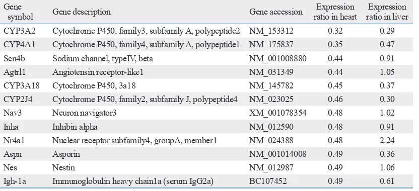
2 and Fig. 1). However, the expression of many genes related to oxidative stress, inflammation, and apoptosis were altered in the heart as well as in the liver. This suggested the possi-bility of intoxication in the heart based on molecular level standards. The partial list of genes repressed in the heart and their expression ratios in the heart and liver are shown in Table 5. In the heart, many cytochrome p450 enzymes were repressed. The CYP3A group of cytochrome p450 proteins, a well-known group of enzymes that metabolize drugs, including APAP, was significantly repressed in both the heart and liver. Other groups of cytochrome p450s were also repressed by APAP. Among the repressed genes in the heart, the identity and function of many of the revealed genes are still unknown (data not shown).
DISCUSSION
[image:6.595.100.516.544.734.2]Drug-induced cardiotoxicity is emerging as an important is-sue following anticancer treatments. The symptoms encom-pass a number of heterogeneous side effects including ar-rhythmias, changes in blood pressure, myocardial ischemia, thrombosis, and impairment in myocardial contraction and/ or relaxation. In the past, the risk of heart complications was less evident because life span after chemotherapy was too short for the cardiovascular effects to be of major concern. With the progress in cancer therapy, however, cardiotoxicity has now become a pivotal issue. A number of targeted drugs in addition to conventional anthracyclines now represent the current mainstay of treatment for several forms of cancer, but their use is hampered by cardiac side effects. Many patients
Table 5. Genes Repressed in the Heart and Their Expression Ratios after Acetaminophen Treatment
Gene
symbol Gene description Gene accession ratio in heartExpression ratio in liverExpression CYP3A2 Cytochrome P450, family3, subfamily A, polypeptide2 NM_153312 0.32 0.29 CYP4A1 Cytochrome P450, family4, subfamily A, polypeptide1 NM_175837 0.35 0.47
Scn4b Sodium channel, typeIV, beta NM_001008880 0.44 0.91
Agtrl1 Angiotensin receptor-like1 NM_031349 0.44 1.05
CYP3A18 Cytochrome P450, 3a18 NM_145782 0.45 0.37
CYP2J4 Cytochrome P450, family2, subfamily J, polypeptide4 NM_023025 0.46 0.30
Nav3 Neuron navigator3 XM_001078354 0.48 1.02
Inha Inhibin alpha NM_012590 0.48 0.91
Nr4a1 Nuclear receptor subfamily4, groupA, member1 NM_024388 0.48 2.24
Aspn Asporin NM_001014008 0.49 0.36
Nes Nestin NM_012987 0.49 1.06
in the pathogenesis of cardiovascular disease.21 ICAM-1
pro-motes adhesion between leucocytes and endothelial cells, al-lowing for tissue-specific localization of leucocytes for im-mune and inflammatory responses. Cell-cell recognition, leukocyte rolling, and trafficking processes are dependent on cell surface ICAM-1 expression.22 ICAM-1 mRNA
expres-sion levels are increased in response to some stimuli, and several inflammatory cytokines and hydrogen peroxide can stimulate ICAM-1 expression.23 ICAM-1 is also increased in
the rat myocardium in various models of cardiac ischemia reperfusion injury. The oxidative stress and cytokine release that occur in the infarcted heart are considered to activate NF-κB, while NF-κB activation triggers gene expression of adhesion molecules including VCAM-1 and ICAM-1.24 In
the present study, ICAM-1 was induced by APAP in the heart (2.2-fold), but its expression was not altered significantly in the liver. The significance of the induction of inflammation-related genes as a consequence of APAP treatment is very difficult to interpret because the functions of CXCLs and IRFs are varied and intricate.
The induced expression of VCAM-1 and ICAM-1 in heart tissue reflects molecular levels of inflammation and oxidative damage caused by APAP, which were not identi-fied by conventional histopathological measurements. Some cellular stresses, including oxidative stress, were generated in the heart by APAP administration, and consequently, the inflammation-related genes were induced as a means of maintaining homeostasis. Confirmation of this was evident in the significant induction of the Hmox1 gene (6.1-fold), which is a well-known biomarker of oxidative stress in cells. Previ-ous in vivo studies showed that oxidative stress in the liver, represented by an increased Hmox1, was induced by a high dose of APAP.12,13 The up-regulation of Hmox1 by
stress-causing agents could mediate cytoprotection against subse-quent noxious stimuli, which may be an important physio-logical process.12 Thus, physiological induction of Hmox1
may represent a protective response to oxidative stress in-duced by high doses of APAP. The xanthine dehydrogenase gene, which is induced in cells during oxidative stress, was also induced by APAP (2.8-fold).
Based on the induction of these stress genes, oxidative stress was evidently generated in the hearts of rats treated with APAP. In general, oxidative stress may trigger inflam-mation and ultimately lead to cellular toxicity through the apoptotic process. The imbalance between ROS production and antioxidant mechanisms leads to myocyte dysfunction, injury, and necrosis. ROS can cause lipid peroxidation, re-In the CXC subfamily, more than 17 ligands have been
iden-tified.15 The expression of the CXCL10 [chemokine (CXC
motif) ligand 10] gene was dramatically higher in both the hearts and livers of the APAP-treated group (19.2-fold in heart, 21.6-fold in liver) than in the control group. CXCL9 was also significantly induced in the heart (12.9-fold).
IRFs, implicated as regulators of the IFN gene, represent a family with a total of nine members with multiple func-tions.16,17 IFNs are a family of multi-functional cytokines
that mediate cellular resistance against viral infection and also have diverse functions in immune responses to patho-gens, immunomodulation, and hematopoietic development. Expression of IFNs is regulated by several IRFs. Following treatment with APAP, IRF-7 was induced 10.1-fold in the heart. IRF-1 and IRF-9 were also induced in the heart (3.1- and 3.0-fold, respectively). IRF-1 has been assigned a dual role in apoptosis, as it serves both pro- and anti- apoptotic functions. In general, IRF-1 activity does not lead to cell death. However, under certain physiological and pathologi-cal conditions, it promotes apoptosis. The induction of Casp1 by IRF-1 has been shown to be an essential component of apoptosis induction.16 Transcription of other members of the
family of caspases is also induced by IRF-1. Furthermore, IRF-1 can act as a mediator of cytokine-induced apoptosis, and IRF-1 induction appears to be an important signaling event in the initiation of apoptosis by IFN-γ.16,17
Up-regula-tion of IRF-1 suggests the possibility that apoptotic processes might be induced in the rat heart after APAP intoxication.
Vascular cell adhesion molecule 1 (VCAM-1) is one of the endothelial cell adhesion molecules that mediates leu-kocyte binding and regulates leuleu-kocyte migration from the blood into tissues.18 VCAM-1 activates reduced
nicotin-amide adenine dinucleotide phosphate (NADPH) oxidase in endothelial cells for generation of ROS, while these ROS activate matrix metalloproteinases (MMPs). Hydrogen per-oxide from the VCAM-1 signaling pathway also modulates actin restructuring, causing cytoskeletal changes in endotheli-al cells. Degradation of the extracellular matrix by MMPs and changes to endothelial cell actin structure are required for VCAM-1 dependent lymphocyte migration. Previous studies have reported that VCAM-1 expression was induced in endothelial cells under inflammatory conditions.18,19 In
addition, VCAM-1 was increased in the liver by APAP, re-flecting the vascular damage it produced.20 In the present
calcium may shorten phase 2 of the cardiac action potential, providing a negative inotropic effect and decreasing myo-cardial oxygen and energy consumption. Up-regulation of FGF7 may be a protective response to oxidative stress in-duced by APAP in the heart, and cGMP and nitric oxide would be involved in this cytoprotective response. In our study, the Nrf2 gene, which is one of the important transcrip-tion factors for oxidative stress, was not altered by APAP. Al-though Nrf2 has been known to be one of the important transcription factors for oxidative stress or detoxification, it is known that Nrf2 induction is not always observed con-comitantly with the induction of Nrf2-mediated oxidative stress related genes. Furthermore, decreased expression of Nrf2 gene was reported in the presence of APAP.31
In contrast to the large numbers of the genes induced by APAP in the heart, the number of repressed heart genes was relatively small and most of these genes have yet to be identi-fied. Among those with established identities were many cy-tochrome p450 genes, including CYP3A, which is known to be actively involved in the metabolism of APAP. The ex-pression ratio was 0.32 in this study. Other groups of cyto-chrome p450s, such as CYP4A and CYP2J, were also re-pressed, with ratios of 0.35 and 0.46, respectively. During metabolism of APAP, toxic metabolites including NAPQI were generated; these metabolites may attack the mRNA responsible for translation of metabolic enzymes. Further-more, toxic metabolites generated by the liver may cause adverse effects when they reach the heart. There are also other potential roles of liver injury and systemic inflamma-tion that can have undesirable effects on heart funcinflamma-tion. This was not clarified in this study.
In summary, liver injury was induced in rats sacrificed 24 hours after oral administration of 1,000 mg/kg APAP. In contrast, heart intoxication was not identified at the same dosage level by conventional histopathology. However, the induction of many genes related to the processes of oxida-tive stress, inflammation, and apoptosis was identified in the heart using microarray analysis. These results suggest that APAP may cause unfavorable pathological changes in the heart, and that further studies are needed to identify the molecular mechanisms of cardiotoxicity induced by this frequently used over-the-counter drug.
REFERENCES
1. Laine JE, Auriola S, Pasanen M, Juvonen RO. Acetaminophen
sulting in disruption of membrane architecture and subse-quent lysosomal enzyme release, while DNA and amino acid oxidation causes genetic mutations and enzyme dys-functions or proteolysis. Oxidative stress also induces heart remodeling.25
Activated caspases cleave a variety of substrates, includ-ing proteins involved in signal transduction (apoptosis reg-ulators, cytokines, serine/threonine kinases, etc.), structural proteins, and proteins involved in the regulation of transcrip-tion, translatranscrip-tion, and RNA editing.26 The caspases are
divid-ed into two subgroups: pro-apoptotic and pro-inflammato-ry, depending on their major functions. Members of the first group, Casp1-3 and 6-10, have a role in the process of apop-tosis and are essential for the activation and implementation of cellular demise.27 Members of the second group, Casp1,
4, 5 and 12, are involved in cytokine maturation and, con-sequently, the inflammatory response. Active Casp1 is es-sential for cleavage of pro-interleukin 1β (pro-IL-1β) and pro-IL-18 into their active forms. IL-1β is related to many immune reactions, including the recruitment of inflamma-tory cells to the site of infection, while IL-18 is important for the production of IFN-γ and enhancement of the cytolytic ac-tivity of natural killer cells.28 However, gathering evidence
indicates that caspases play roles in many other cellular processes that cannot be classified as apoptotic or pro-inflammatory. The activation of pro-inflammatory caspases can clearly induce apoptosis.26 Caspases are known to play
a key role in APAP-induced apoptosis and necrosis of the liver.20 In the present study, among the 11 caspases
investi-gated, Casp1, 4, and 12 were induced in the heart by APAP administration (2.8-, 2.1- and 2.3-fold respectively). Casp4 and 12 were also induced in the liver by APAP (4.4- and 19-fold respectively).
Among 23 fibroblast growth factors (FGFs), only FGF7 was increased (by 2.2 fold) in the heart of the APAP-treated group, compared to the control, but unchanged in the liver. FGF21 and FGF23 were increased in the liver, but not in the heart. The FGFs constitute a family of closely related poly-peptides, initially considered to be mitogens. However, sev-eral previous studies have reported nonmitogenic actions of FGFs. FGFs can participate as endogenous cardioprotective agents and improve cardiac resistance to ischemia-reperfu-sion injury.29 FGFs increase cytosolic cGMP levels in a
myo-cardial ischemia-reperfusion setting.30 In turn, cGMP
16. Kröger A, Köster M, Schroeder K, Hauser H, Mueller PP. Activi-ties of IRF-1. J Interferon Cytokine Res 2002;22:5-14.
17. Taniguchi T, Ogasawara K, Takaoka A, Tanaka N. IRF family of transcription factors as regulators of host defense. Annu Rev Im-munol 2001;19:623-55.
18. Cook-Mills JM. VCAM-1 signals during lymphocyte migration: role of reactive oxygen species. Mol Immunol 2002;39:499-508. 19. Iiyama K, Hajra L, Iiyama M, Li H, DiChiara M, Medoff BD, et
al. Patterns of vascular cell adhesion molecule-1 and intercellular adhesion molecule-1 expression in rabbit and mouse atheroscle-rotic lesions and at sites predisposed to lesion formation. Circ Res 1999;85:199-207.
20. Liu J, Li C, Waalkes MP, Clark J, Myers P, Saavedra JE, et al. The nitric oxide donor, V-PYRRO/NO, protects against acetamino-phen-induced hepatotoxicity in mice. Hepatology 2003;37:324-33. 21. Hope SA, Meredith IT. Cellular adhesion molecules and cardio-vascular disease. Part II. Their association with conventional and emerging risk factors, acute coronary events and cardiovascular risk prediction. Intern Med J 2003;33:450-62.
22. Hubbard AK, Rothlein R. Intercellular adhesion molecule-1 (ICAM-1) expression and cell signaling cascades. Free Radic Biol Med 2000;28:1379-86.
23. Roebuck KA, Finnegan A. Regulation of intercellular adhesion molecule-1 (CD54) gene expression. J Leukoc Biol 1999;66:876-88.
24. Benson V, McMahon AC, Lowe HC. ICAM-1 in acute myocardi-al infarction: a potentimyocardi-al therapeutic target. Curr Mol Med 2007;7: 219-27.
25. Seddon M, Looi YH, Shah AM. Oxidative stress and redox signal-ling in cardiac hypertrophy and heart failure. Heart 2007;93:903-7. 26. Li J, Yuan J. Caspases in apoptosis and beyond. Oncogene
2008;27:6194-206.
27. Franchi L, Eigenbrod T, Muñoz-Planillo R, Nuñez G. The inflam-masome: a caspase-1-activation platform that regulates immune responses and disease pathogenesis. Nat Immunol 2009;10:241-7. 28. Arend WP, Palmer G, Gabay C. IL-1, IL-18, and IL-33 families of
cytokines. Immunol Rev 2008;223:20-38.
29. Cuevas P, Carceller F, Giménez-Gallego G. Fibroblast growth fac-tors in myocardial ischemia / reperfusion injury and ischemic pre-conditioning. J Cell Mol Med 2001;5:132-42.
30. Cuevas P, Carceller F, Martinez-Coso V, Cuevas B, Fernandez-Ayerdi A, Reimers D, et al. Cardioprotection from ischemia by fi-broblast growth factor: role of inducible nitric oxide synthase. Eur J Med Res 1999;4:517-24.
31. Fukushima T, Kikkawa R, Hamada Y, Horii I. Genomic cluster and network analysis for predictive screening for hepatotoxicity. J Toxicol Sci 2006;31:419-32.
bioactivation by human cytochrome P450 enzymes and animal microsomes. Xenobiotica 2009;39:11-21.
2. Moore M, Thor H, Moore G, Nelson S, Moldéus P, Orrenius S. The toxicity of acetaminophen and N-acetyl-p-benzoquinone im-ine in isolated hepatocytes is associated with thiol depletion and increased cytosolic Ca2+. J Biol Chem 1985;260:13035-40. 3. Schilling A, Corey R, Leonard M, Eghtesad B. Acetaminophen:
old drug, new warnings. Cleve Clin J Med 2010;77:19-27. 4. Bronstein AC, Spyker DA, Cantilena LR Jr, Green J, Rumack
BH, Heard SE. 2006 Annual Report of the American Association of Poison Control Centers’ National Poison Data System (NPDS). Clin Toxicol (Phila) 2007;45:815-917.
5. Priyadarsiny P, Khattar SK, Malik R, Udupa V, Seshaiah A, Rah-man S, et al. Differential gene expression analysis of a known hepatotoxin, N-acetyl-p-amino-phenol (APAP) as compared to its non-toxic analog, N-acetyl-m-amino-phenol (AMAP) in mouse liver. J Toxicol Sci 2008;33:163-73.
6. Jeong SY, Lim JS, Park HJ, Cho JW, Rana SV, Yoon S. Effects of acetaminophen on hepatic gene expression in mice. Physiol Chem Phys Med NMR 2006;38:77-83.
7. Pimstone BL, Uys CJ. Liver necrosis and myocardiopathy follow-ing paracetamol overdosage. S Afr Med J 1968;42:259-62. 8. Sanerkin NG. Acute myocardial necrosis in paracetamol
poison-ing. Br Med J 1971;3:478.
9. Zordoky BN, El-Kadi AO. H9c2 cell line is a valuable in vitro model to study the drug metabolizing enzymes in the heart. J Pharmacol Toxicol Methods 2007;56:317-22.
10. Thum T, Borlak J. Gene expression in distinct regions of the heart. Lancet 2000;355:979-83.
11. Fukushima T, Hamada Y, Yamada H, Horii I. Changes of micro-RNA expression in rat liver treated by acetaminophen or carbon tetrachloride--regulating role of micro-RNA for RNA expression. J Toxicol Sci 2007;32:401-9.
12. Chiu H, Brittingham JA, Laskin DL. Differential induction of heme oxygenase-1 in macrophages and hepatocytes during acet-aminophen-induced hepatotoxicity in the rat: effects of hemin and biliverdin. Toxicol Appl Pharmacol 2002;181:106-15.
13. Reilly TP, Bourdi M, Brady JN, Pise-Masison CA, Radonovich MF, George JW, et al. Expression profiling of acetaminophen liver toxicity in mice using microarray technology. Biochem Biophys Res Commun 2001;282:321-8.
14. Albini A, Pennesi G, Donatelli F, Cammarota R, De Flora S, Noonan DM. Cardiotoxicity of anticancer drugs: the need for car-dio-oncology and cardio-oncological prevention. J Natl Cancer Inst 2010;102:14-25.