http://dx.doi.org/10.4236/ojog.2016.67055
How to cite this paper: Leroy, D., Slachmuylder, E., Popijn, M., Cassart, M., Massez, A., D'Haene, N., Désir, J., Vander-maelen, A., Daelemans, C., Ceysens, G. and Donner, C. (2016) Antenatal Diagnosis of Isolated Total Arhinia in the Second Trimester of Pregnancy. Open Journal of Obstetrics and Gynecology, 6, 419-423.
http://dx.doi.org/10.4236/ojog.2016.67055
Antenatal Diagnosis of Isolated
Total Arhinia in the Second
Trimester of Pregnancy
D. Leroy
*, E. Slachmuylder, M. Popijn, M. Cassart, A. Massez, N. D'Haene, J. Désir,
A. Vandermaelen, C. Daelemans, G. Ceysens, C. Donner
Erasme (Fetal Medicine), Brussels, BelgiumReceived 19 April 2016; accepted 13 June 2016; published 16 June 2016
Copyright © 2016 by authors and Scientific Research Publishing Inc.
This work is licensed under the Creative Commons Attribution International License (CC BY).
http://creativecommons.org/licenses/by/4.0/
Abstract
Congenital arhinia is a very rare condition especially when it is isolated. Most of arhinia are iden-tified after birth and only five prenatal cases are described in the literature. Generally, arhinia is associated with other malformations mainly craniofacial anomalies. Genetics aberrations are un-common. Our case was diagnosed in the second trimester of pregnancy and we found no associ-ated anomaly except for a single umbilical artery. Autopsy confirmed the diagnosis and neuropa-thology analysis revealed the absence of olfactory bulbs and tracts.
Keywords
Antenatal, Ultrasound, Arhinia, Diagnosis, Pregnancy
1. Introduction
Complete arhinia is defined as the congenital absence of the nose. In the majority of cases, arhinia is associated with other craniofacial anomalies. Isolated arhinia is a very rare condition only reported antenatally in five cases. The majority of cases are sporadic.
2. Case Report
A 30-years-old primigravida was referred at 23 weeks’ gestation for a suspected facial anomaly. Her medical and familial histories were unremarkable. A first trimester sonography performed at 13 weeks’ gestation showed a
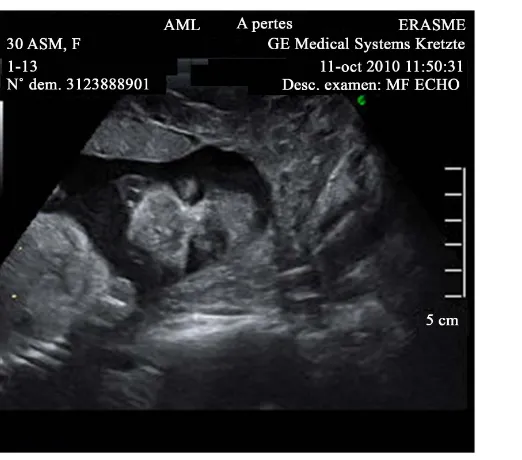
normal nuchal translucency but the nasal bone was not visualized. A second ultrasound performed at 16 weeks was reported normal but the fetal profile could not be obtained. The anatomy scan at 23 weeks’ gestation de-tected the absence of the nose (Figure 1andFigure 2). This anomaly was documented using 2D and 3D imag-ing. The profile was flat and the upper lip appeared prominent. No other malformation was detected and the in-tracranial structures appeared normal. The only associated feature was a single umbilical artery. An amniocente-sis was performed and showed a normal karyotype 46XX and a normal CGH array (Agilent 60 K).
After receiving extensive information about the possible management of that condition from a genetician, a paediatrician and a paediatric surgeon, the couple opted for termination of pregnancy. The delivery was induced one week later.
After termination, the macroscopic examination confirmed the arhinia (Figure 3) and no additional malfor-mations were observed.
The autopsy confirmed arhinia with absence of nostrils, moderate exophtalmy, a small oral orifice and a sin-gle umbilical artery. No other fetal abnormalities were identified and placental examination was unremarkable.
Examination of the brain, although not completely contributive because of partial autolysis showed an ab-sence of olfactive bulbs and tracts.
[image:2.595.214.412.300.451.2]The post mortem CT scan (Figure 4) confirmed the absence of the nasal bone and also showed cribriform plate aplasia and absence of ethmoid sinuses and cells.
Figure 1. 23 wks, sagittal view, absence of nose, prominent upper lip.
[image:2.595.195.456.477.707.2]Figure 3. Confirmation of arhinia after termination.
Figure 4. Postnatal CTscan, transverse view (a) our case: absence of nasal and ethmoid bones (b) normal anatomy for com-parison.
3. Discussion
[image:3.595.87.541.350.614.2]prenatal [1]-[4] cases.
Although the majority of cases are sporadic, two familial cases were described with a possible autosomal re-cessive mode of inheritance in one and autosomal dominant in the second [5] [6]. The chromosomal analysis in patients with arhinia showed normal results, excepting for 3 cases that had an abnormal karyotype: mos46, XX/47, XX, +9 [7]; 46, XY, inv(9) [8]; 46, XX, t(3;12) (q13.2; p11.2) (de novo) [9]. The third patient was also discovered to be the carrier of a 19 Mb deletion spanning from 3q11.2 to 3q13.31 at the 3q breakpoint of the translocation [10]. As the deleted segment at 3q was a strong candidate region for a putative arhinia gene, array CGH was performed in other arhinia patients, as well as mutation analysis of candidate genes [10]. No consis-tent gene mutations have been discovered so far; therefore, genetic testing for a putative arhinia gene is not yet available.
In a review of 27 neonatal cases no sex predominance was found. Associated maternal diabetes was suggested in 3 cases [11].
The antenatal diagnosis of arhinia is more often associated with other anomalies of the skull, the face and the brain (abnormalities of the lips and palate, maxillary hypoplasia, microphtalmia, hypertelorism, coloboma, cho-anal atresia, absence or occlusion of the lacrymal ducts, holoprosencephaly, encephalocele, absence of the ol-factory bulbs and tracts).
Some authors have identified cases of arhinia with hypogonadotropic hypogonadism, but no genetic loci were found to explain this association [12]-[14].
To our knowledge, only 5 cases of complete isolated arhinia diagnosed antenatally have been described in the literature, respectively at 23, 25, 27 and 29 weeks [1]-[4] [15]. In our case the diagnosis was also only made at 23 weeks. In the case of Majewski [1], the patient had an ultrasound at 18 weeks where the fetal position did not allow the adequate visualization of the fetal face and the cardiac outflow tracts. A second ultrasound was offered at 22 weeks and the anomaly was then diagnosed. In the case by Olsen [3], the patient had an ultrasound at 17 weeks reporting diffuse midfacial anomalies with edema. The absence of the external nose was diagnosed at 25 weeks. In the case by Thornburg [4], the diagnosis was made during the first ultrasound performed at 27 weeks. In the case by Cusik [2], a first ultrasound was made at 18 weeks but the fetal face and profile could not be visu-alized due to fetal position. It is the second scan at 29 weeks indicated by gestational diabetes that made the di-agnosis possible.
In our case, the patient had ultrasounds at 13 and 16 weeks but the diagnosis was only made at 23 weeks. At the 13 andt 16 weeks ultrasound the face was not adequately visualized as in two other cases [1] [2].
On the other hand, in several cases of isolated arhinia diagnosed at birth, the authors noted that antenatal ul-trasound were reported as normal even in one case where an amniocentesis was performed for a positive second trimester triple test [11].
The missed diagnosis is probably due to the rarity of the malformation and the fact that the lack of additional anomalies makes it more difficult to detect it in early pregnancy and in situations where the fetal visualization is not optimal.
A systematic approach during first trimester scan, could help for the earlier diagnosis of arhinia. The use of transvaginal ultrasound when images are suboptimal transabdominally should also be promoted. The 3D ultra-sound scanning can also help to demonstrate the absence of nose (Figure 2).
When the diagnosis of arhinia is established, it is important to exclude other facial, skeletal and cerebral ano-malies or extra-cerebral malformations. The amniocentesis or chorionic villus sampling is useful to exclude chromosomal abnormalities.
In several studies, the prevalence of fetal abnormalities associated with single umbilical artery is 33.6% [14]. Interestingly, this particularity was present in our case and in the case by Olsen and al., because of the very small number of cases and the non specific character of a single umbilical artery, we believe this is more of an anec-dotical finding than an associated sign.
4. Conclusion
References
[1] Majewski, S., Donnenfeld, A.E., Kuhlman, K. and Patel, A. (2007) Second-Trimester Prenatal Diagnosis of Total Arhinia. Journal of Ultrasound in Medicine, 26, 391-395.
[2] Cusik, W., Sullivan, C.A., Rojas, B., Poole, A.E. and Poole, D.A. (2000) Prenatal Diagnosis of Total Arhinia. Ultra-sound in Obstetrics & Gynecology, 15, 259-261. http://dx.doi.org/10.1046/j.1469-0705.2000.00081.x
[3] Olsen, Ø.E., Gjelland, K., Reigstad, H. and Rosendahl, K. (2001) Congenital Absence of the Nose: A Case Report and Literature Review. Pediatric Radiology, 31, 225-232. http://dx.doi.org/10.1007/s002470000419
[4] Thornburg, L.L., Christensen, N., Laroia, N. and Pressman, E.K. (2009) Prenatal Diagnosis of Total Arhinia Associ-ated with Normal Chromosomal Analysis: A Case Report. The Journal of Reproductive Medicine, 54, 579-582. [5] Ruprecht, K.W. and Majewski, F. (1978) Familiary Arhinia Combined with Peters’Anomaly and Maxilliar
Deformi-ties: A New Malformation Syndrome (Author’s Transl). Klin Monbl Augenheilkd, 172, 708-715.
[6] Thiele, H., Musil, A., Nagel, F. and Majewski, F. (1996) Familial Arhinia, Choanal Atresia, and Microphthalmia. American Journal of Medical Genetics, 63, 310-313.
http://dx.doi.org/10.1002/(SICI)1096-8628(19960503)63:1<310::AID-AJMG51>3.0.CO;2-N
[7] Kaminker, C.P., Dain, L., Lamas, M.A. and Sanchez, J.M. (1985) Mosaic Trisomy 9 Syndrome with Unusual Pheno-type. American Journal of Medical Genetics, 22, 237-241. http://dx.doi.org/10.1002/ajmg.1320220204
[8] Cohen, D. and Goitein, K.J. (1987) Arhinia Revisited. Rhinology, 25, 237-244.
[9] Hou, J.W. (2004) Congenital Arhinia with de Novo Reciprocal Translocation t(3;12)(q13.2;p11.2). American Journal of Medical GeneticsA, 130A, 200-203. http://dx.doi.org/10.1002/ajmg.a.30268
[10] Sato, D., et al. (2007) Congenital Arhinia: Molecular-Genetic Analysis of Five Patients. American Journal of Medical GeneticsA, 143A, 546-552. http://dx.doi.org/10.1002/ajmg.a.31613
[11] McGlone, L. (2003) Congenital Arhinia. Journal of Paediatrics and Child Health, 39, 474.
http://dx.doi.org/10.1046/j.1440-1754.2003.00193.x
[12] Tryggestad, J.B., Li, S.B. and Chernausek, S.D. (2013) Hypogonadotropic Hypogonadism Presenting with Arhinia: A Case Report. Journal of Medical Case Reports, 7, 52. http://dx.doi.org/10.1186/1752-1947-7-52
[13] Graham Jr., J.M. and Lee, J. (2006) Bosma Arhinia Microphthalmia Syndrome. American Journal of Medical Genetics A, 140, 189-193. http://dx.doi.org/10.1002/ajmg.a.31039
[14] De Figueiredo, D., Dagklis, T., Zidere, V. and Allan, L. (2010) Nicolaides KH. Isolated Single Umbilical Artery. Ul-trasound in Obstetrics & Gynecology, 36, 553-555. http://dx.doi.org/10.1002/uog.7711
[15] Li, X., Zhang, L. and Wang, F. (2015) Prenatal Diagnosis of Total Arhinia by RMN. Japanese Journal of Radiology, 33, 672-674. http://dx.doi.org/10.1007/s11604-015-0473-7

