4-Hydroxy-3-methoxybenzaldehyde–
nicotinamide (1/1)
Fiona N.-F. How,aM. S. Amalina,aHamid Khaledib* and Hapipah Mohd Alib
aDepartment of Biomedical Sciences, Kulliyah of Science, IIUM Kuantan, Jalan
Sultan Ahmad Shah, Bandar Indera Mahkota, 25200 Kuantan, Pahang Darul Makmur, Malaysia, andbDepartment of Chemistry, University of Malaya, 50603
Kuala Lumpur, Malaysia
Correspondence e-mail: [email protected] Received 24 October 2011; accepted 31 October 2011
Key indicators: single-crystal X-ray study;T= 100 K; mean(C–C) = 0.003 A˚;
Rfactor = 0.045;wRfactor = 0.112; data-to-parameter ratio = 11.7.
In the title compound, C6H6N2OC8H8O3, an equimolar
co-crystal of nicotinamide and vanillin, the aromatic ring and the amide fragment of the nicotinamide molecule make a dihedral angle of 32.6 (2). The vanillin molecule is almost planar, with
an r.m.s. deviation for all non-H atoms of 0.0094 A˚ . The vaniline and nicotinamide aromatic rings are nearly coplanar, the dihedral angle between them being 3.20 (9). In the
crystal, the two components are linked through N—H O and O—H N hydrogen bonds into chains along the aaxis. The chains are connectedviaC—H O interactions, forming a three-dimensional polymeric structure.
Related literature
For the crystal structure of nicotinamide, see: Miwa et al.
(1999); Li et al. (2011). For the structure of vanillin, see: Velavanet al.(1995).
Experimental
Crystal data
C6H6N2OC8H8O3
Mr= 274.27
Triclinic,P1
a= 4.8979 (1) A˚
b= 8.5440 (2) A˚
c= 15.4713 (4) A˚
= 98.108 (1) = 92.810 (2)
= 94.784 (2)
V= 637.52 (3) A˚3
Z= 2
MoKradiation
= 0.11 mm1
T= 100 K
0.220.140.04 mm
Data collection
Bruker APEXII CCD diffractometer
Absorption correction: multi-scan (SADABS; Sheldrick, 1996)
Tmin= 0.977,Tmax= 0.996
3432 measured reflections 2243 independent reflections 1862 reflections withI> 2(I)
Rint= 0.019
Refinement
R[F2> 2(F2)] = 0.045
wR(F2) = 0.112
S= 1.05 2243 reflections 191 parameters 4 restraints
H atoms treated by a mixture of independent and constrained refinement
max= 0.66 e A˚3
[image:1.610.313.565.304.383.2]min=0.28 e A˚3
Table 1
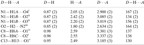
Hydrogen-bond geometry (A˚ ,).
D—H A D—H H A D A D—H A
N1—H1A O4i 0.87 (2) 2.05 (2) 2.900 (2) 167 (2) N1—H1B O2ii
0.87 (2) 2.42 (2) 3.085 (2) 134 (2) N1—H1B O3ii
0.87 (2) 2.20 (2) 3.019 (2) 156 (2) O2—H2 N2iii
0.85 (2) 1.80 (2) 2.634 (2) 164 (2) C8—H8A O1iv
0.98 2.59 3.381 (3) 137 C8—H8C O2i
0.98 2.55 3.337 (2) 138 C13—H13 O1v
0.95 2.49 3.185 (3) 130
Symmetry codes: (i) x1;y;z; (ii) xþ1;yþ1;z; (iii) xþ1;y;z; (iv) xþ1;yþ1;zþ1; (v)xþ1;yþ2;zþ1.
Data collection:APEX2(Bruker, 2007); cell refinement:SAINT
(Bruker, 2007); data reduction:SAINT; program(s) used to solve structure:SHELXS97(Sheldrick, 2008); program(s) used to refine structure: SHELXL97 (Sheldrick, 2008); molecular graphics: X-SEED (Barbour, 2001); software used to prepare material for publication:SHELXL97andpublCIF(Westrip, 2010).
IIUM is acknowledged for funding this study (Endowment fund A No. EDWA10–150–0697).
Supplementary data and figures for this paper are available from the IUCr electronic archives (Reference: PV2471).
References
Barbour, L. J. (2001).J. Supramol. Chem,1, 189–191.
Bruker (2007).APEX2andSAINT. Bruker AXS Inc., Madison, Wisconsin, USA.
Li, J., Bourne, S. A. & Caira, M. R. (2011).Chem. Commun.47, 1530–1532. Miwa, Y., Mizuno, T., Tsuchida, K., Taga, T. & Iwata, Y. (1999).Acta Cryst.
B55, 78–84.
Sheldrick, G. M. (1996).SADABS. University of Go¨ttingen, Germany. Sheldrick, G. M. (2008).Acta Cryst.A64, 112–122.
Velavan, R., Sureshkumar, P., Sivakumar, K. & Natarajan, S. (1995).Acta Cryst.C51, 1131–1133.
Westrip, S. P. (2010).J. Appl. Cryst.43, 920–925.
organic compounds
o3168
Howet al. doi:10.1107/S1600536811045648 Acta Cryst.(2011). E67, o3168Acta Crystallographica Section E
Structure Reports Online
supplementary materials
sup-1
Acta Cryst.
(2011). E
67
, o3168 [
doi:10.1107/S1600536811045648
]
4-Hydroxy-3-methoxybenzaldehyde-nicotinamide (1/1)
F. N.-F. How
,
M. S. Amalina
,
H. Khaledi
and
H. Mohd Ali
Comment
The crystal structures of nicotinamide (Miwa
et al.
,1999; Li
et al.
, 2011) and 4-hydroxy-3-methoxybenzaldehyde, vanillin,
(Velavan
et al.
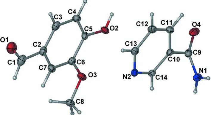
,1995) have been previously reported. The title compound is an equimolar cocrystal of nicotinamide and
vanillin (Fig. 1). The nicotinamide aromatic ring and the plane of the amide fragment, N1—C9—O4, are twisted with respect
to each other, making a dihedral angle of 32.6 (2)°. The vanillin molecule is essentially planar, the highest deviation from the
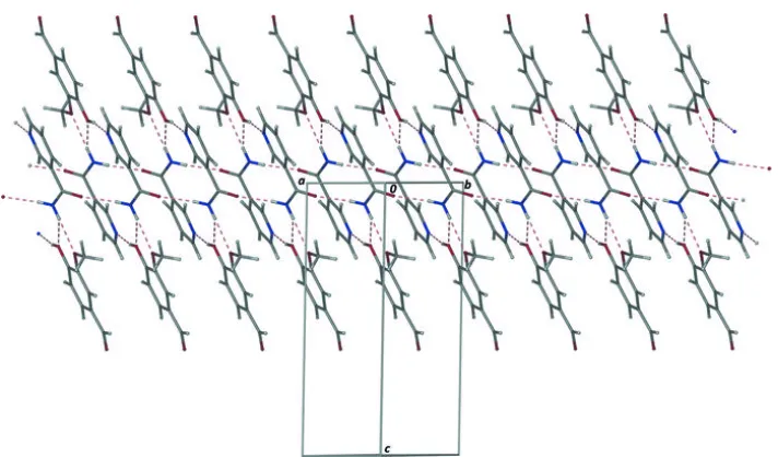
best plane passing through all non-H atoms being 0.0156 (13) Å for O3 atom. In the crystal, the molecules of nicotinamide
and vanillin are linked through N—H···O and O—H···N hydrogen bonds into infinite chains along the
a
axis (Fig. 2). The
chains are connected
via
C—H···O interactions (Table 1 and Fig. 2) to form a three-dimensional polymeric structure.
Experimental
A mixture of vanillin (1.52 g, 0.1 mol) and nicotinamide (1.22 g, 0.1 mol) in ethanol (30 ml) was heated for 1 hr. The solvent
was then evaporated partially and the solution was left at room temperature. The colorless crystals of the title compound
were obtained in a day.
Refinement
The C-bound H atoms were placed at calculated positions and were treated as riding on their parent C atoms with C—H
distances of 0.95 (aryl) and 0.98 (methyl) Å. The N– and O-bound H atoms were located in a difference Fourier map,
and refined with distance restraints of O—H = 0.84 (2) Å and N—H = 0.88 (2) Å. For all H atoms,
U
iso(H) was set to
1.2–1.5
eq(carrier atom). An additional rigid-bond type restraint (
DELU
in
SHELXL97
) was placed on the displacement
parameters of C1 and C2.
Figures
Fig. 1. Molecular structure of the title compound with displacement ellipsoids drawn at 50%
probability level. Hydrogen atoms are drawn as spheres of arbitrary radii.
4-Hydroxy-3-methoxybenzaldehyde–nicotinamide (1/1)
Crystal data
C6H6N2O·C8H8O3 Z = 2
Mr = 274.27 F(000) = 288
Triclinic, P1 Dx = 1.429 Mg m−3
Hall symbol: -P 1 Mo Kα radiation, λ = 0.71073 Å
a = 4.8979 (1) Å Cell parameters from 1226 reflections
b = 8.5440 (2) Å θ = 2.6–29.7°
c = 15.4713 (4) Å µ = 0.11 mm−1
α = 98.108 (1)° T = 100 K
β = 92.810 (2)° Lath, colorless
γ = 94.784 (2)° 0.22 × 0.14 × 0.04 mm
V = 637.52 (3) Å3
Data collection
Bruker APEXII CCD
diffractometer 2243 independent reflections
Radiation source: fine-focus sealed tube 1862 reflections with I > 2σ(I)
graphite Rint = 0.019
φ and ω scans θmax = 25.3°, θmin = 2.4°
Absorption correction: multi-scan
(SADABS; Sheldrick, 1996) h = −5→5
Tmin = 0.977, Tmax = 0.996 k = −10→10
3432 measured reflections l = −18→18
Refinement
Refinement on F2 Primary atom site location: structure-invariant directmethods
Least-squares matrix: full Secondary atom site location: difference Fourier map
R[F2 > 2σ(F2)] = 0.045 Hydrogen site location: inferred from neighbouringsites
wR(F2) = 0.112 H atoms treated by a mixture of independent andconstrained refinement
S = 1.05 w = 1/[σ2(Fo2) + (0.0439P)2 + 0.4685P]
where P = (Fo2 + 2Fc2)/3
2243 reflections (Δ/σ)max < 0.001
191 parameters Δρmax = 0.66 e Å−3
supplementary materials
sup-3
between e.s.d.'s in cell parameters are only used when they are defined by crystal symmetry. An approximate (isotropic) treatment of cell e.s.d.'s is used for estimating e.s.d.'s involving l.s. planes.
Refinement. Refinement of F2 against ALL reflections. The weighted R-factor wR and goodness of fit S are based on F2, convention-al R-factors R are based on F, with F set to zero for negative F2. The threshold expression of F2 > σ(F2) is used only for calculating R
-factors(gt) etc. and is not relevant to the choice of reflections for refinement. R-factors based on F2 are statistically about twice as large
as those based on F, and R- factors based on ALL data will be even larger.
Fractional atomic coordinates and isotropic or equivalent isotropic displacement parameters (Å
2)
x y z Uiso*/Ueq
O1 0.3651 (3) 0.83011 (19) 0.61085 (10) 0.0359 (4)
O2 0.8107 (3) 0.63328 (16) 0.23075 (9) 0.0195 (3)
H2 0.949 (4) 0.698 (2) 0.2250 (15) 0.029*
O3 0.4028 (3) 0.44026 (16) 0.25734 (9) 0.0218 (3)
C1 0.3070 (5) 0.7306 (3) 0.54863 (14) 0.0286 (5)
H1 0.1529 0.6570 0.5522 0.034*
C2 0.4516 (4) 0.7113 (2) 0.46701 (13) 0.0210 (4)
C3 0.6726 (4) 0.8148 (2) 0.45164 (13) 0.0221 (5)
H3 0.7380 0.9016 0.4951 0.026*
C4 0.7972 (4) 0.7912 (2) 0.37307 (13) 0.0204 (4)
H4 0.9482 0.8620 0.3629 0.024*
C5 0.7035 (4) 0.6648 (2) 0.30888 (12) 0.0166 (4)
C6 0.4793 (4) 0.5600 (2) 0.32443 (12) 0.0176 (4)
C7 0.3561 (4) 0.5847 (2) 0.40255 (13) 0.0208 (5)
H7 0.2040 0.5146 0.4128 0.025*
C8 0.1709 (4) 0.3323 (2) 0.26827 (14) 0.0217 (5)
H8A 0.2110 0.2765 0.3179 0.033*
H8B 0.1328 0.2551 0.2150 0.033*
H8C 0.0103 0.3917 0.2793 0.033*
O4 0.8254 (3) 0.71964 (19) −0.04761 (9) 0.0299 (4)
N1 0.3766 (4) 0.6341 (2) −0.07875 (11) 0.0205 (4)
H1A 0.207 (3) 0.645 (3) −0.0672 (14) 0.025*
H1B 0.415 (4) 0.586 (2) −0.1293 (11) 0.025*
N2 0.2385 (3) 0.80224 (19) 0.18395 (10) 0.0170 (4)
C9 0.5840 (4) 0.7129 (2) −0.02755 (13) 0.0197 (4)
C10 0.5123 (4) 0.7959 (2) 0.05932 (12) 0.0169 (4)
C11 0.6653 (4) 0.9350 (2) 0.09779 (13) 0.0201 (4)
H11 0.8121 0.9801 0.0687 0.024*
C12 0.5998 (4) 1.0066 (2) 0.17913 (13) 0.0220 (5)
H12 0.6997 1.1023 0.2066 0.026*
C13 0.3864 (4) 0.9363 (2) 0.21971 (13) 0.0192 (4)
H13 0.3429 0.9856 0.2757 0.023*
C14 0.3015 (4) 0.7346 (2) 0.10499 (12) 0.0162 (4)
Atomic displacement parameters (Å
2)
U11 U22 U33 U12 U13 U23
O1 0.0440 (10) 0.0363 (9) 0.0266 (9) 0.0056 (8) 0.0038 (7) 0.0006 (7)
O2 0.0182 (8) 0.0217 (7) 0.0174 (7) −0.0031 (6) 0.0053 (6) 0.0004 (6)
O3 0.0223 (8) 0.0214 (7) 0.0198 (7) −0.0045 (6) 0.0064 (6) −0.0016 (6)
C1 0.0356 (13) 0.0308 (12) 0.0189 (10) 0.0104 (10) −0.0028 (9) −0.0012 (9)
C2 0.0226 (11) 0.0246 (11) 0.0170 (10) 0.0074 (8) 0.0004 (8) 0.0045 (8)
C3 0.0270 (11) 0.0214 (10) 0.0165 (10) 0.0060 (9) −0.0036 (8) −0.0020 (8)
C4 0.0187 (10) 0.0193 (10) 0.0218 (11) −0.0018 (8) −0.0001 (8) 0.0014 (8)
C5 0.0157 (10) 0.0209 (10) 0.0145 (9) 0.0054 (8) 0.0026 (8) 0.0043 (8)
C6 0.0193 (10) 0.0176 (10) 0.0157 (10) 0.0033 (8) 0.0002 (8) 0.0007 (8)
C7 0.0193 (11) 0.0242 (11) 0.0199 (10) 0.0020 (8) 0.0046 (8) 0.0055 (8)
C8 0.0185 (11) 0.0198 (10) 0.0259 (11) −0.0031 (8) 0.0035 (8) 0.0021 (8)
O4 0.0144 (8) 0.0516 (10) 0.0228 (8) 0.0064 (7) 0.0038 (6) −0.0003 (7)
N1 0.0172 (9) 0.0279 (9) 0.0153 (9) 0.0046 (7) 0.0035 (7) −0.0030 (7)
N2 0.0171 (9) 0.0194 (8) 0.0148 (8) 0.0031 (7) 0.0012 (6) 0.0027 (6)
C9 0.0178 (11) 0.0255 (11) 0.0168 (10) 0.0055 (8) 0.0018 (8) 0.0042 (8)
C10 0.0144 (10) 0.0210 (10) 0.0156 (10) 0.0038 (8) −0.0008 (7) 0.0030 (8)
C11 0.0149 (10) 0.0240 (10) 0.0221 (10) 0.0003 (8) 0.0020 (8) 0.0058 (8)
C12 0.0227 (11) 0.0180 (10) 0.0234 (11) −0.0030 (8) −0.0011 (8) −0.0002 (8)
C13 0.0217 (11) 0.0192 (10) 0.0160 (10) 0.0031 (8) 0.0002 (8) 0.0001 (8)
C14 0.0157 (10) 0.0162 (9) 0.0161 (10) 0.0009 (7) −0.0013 (8) 0.0012 (7)
Geometric parameters (Å, °)
O1—C1 1.197 (3) C8—H8B 0.9800
O2—C5 1.343 (2) C8—H8C 0.9800
O2—H2 0.854 (16) O4—C9 1.236 (2)
O3—C6 1.365 (2) N1—C9 1.330 (3)
O3—C8 1.435 (2) N1—H1A 0.868 (16)
C1—C2 1.474 (3) N1—H1B 0.869 (16)
C1—H1 0.9500 N2—C13 1.336 (2)
C2—C3 1.391 (3) N2—C14 1.338 (2)
C2—C7 1.396 (3) C9—C10 1.500 (3)
C3—C4 1.384 (3) C10—C14 1.388 (3)
C3—H3 0.9500 C10—C11 1.392 (3)
C4—C5 1.391 (3) C11—C12 1.384 (3)
C4—H4 0.9500 C11—H11 0.9500
C5—C6 1.410 (3) C12—C13 1.384 (3)
C6—C7 1.375 (3) C12—H12 0.9500
C7—H7 0.9500 C13—H13 0.9500
supplementary materials
sup-5
C2—C1—H1 116.9 C9—N1—H1B 117.5 (15)
C3—C2—C7 119.56 (18) H1A—N1—H1B 120 (2)
C3—C2—C1 122.83 (19) C13—N2—C14 117.72 (17)
C7—C2—C1 117.59 (19) O4—C9—N1 123.82 (19)
C4—C3—C2 119.93 (19) O4—C9—C10 119.84 (18)
C4—C3—H3 120.0 N1—C9—C10 116.34 (17)
C2—C3—H3 120.0 C14—C10—C11 118.15 (18)
C3—C4—C5 120.68 (18) C14—C10—C9 121.73 (18)
C3—C4—H4 119.7 C11—C10—C9 120.07 (18)
C5—C4—H4 119.7 C12—C11—C10 118.88 (18)
O2—C5—C4 124.72 (18) C12—C11—H11 120.6
O2—C5—C6 115.90 (17) C10—C11—H11 120.6
C4—C5—C6 119.38 (18) C13—C12—C11 118.77 (19)
O3—C6—C7 125.62 (18) C13—C12—H12 120.6
O3—C6—C5 114.82 (17) C11—C12—H12 120.6
C7—C6—C5 119.56 (18) N2—C13—C12 123.12 (18)
C6—C7—C2 120.88 (19) N2—C13—H13 118.4
C6—C7—H7 119.6 C12—C13—H13 118.4
C2—C7—H7 119.6 N2—C14—C10 123.34 (18)
O3—C8—H8A 109.5 N2—C14—H14 118.3
O3—C8—H8B 109.5 C10—C14—H14 118.3
H8A—C8—H8B 109.5
Hydrogen-bond geometry (Å, °)
D—H···A D—H H···A D···A D—H···A
N1—H1A···O4i 0.87 (2) 2.05 (2) 2.900 (2) 167 (2)
N1—H1B···O2ii 0.87 (2) 2.42 (2) 3.085 (2) 134.(2)
N1—H1B···O3ii 0.87 (2) 2.20 (2) 3.019 (2) 156 (2)
O2—H2···N2iii 0.85 (2) 1.80 (2) 2.634 (2) 164 (2)
C8—H8A···O1iv 0.98 2.59 3.381 (3) 137.
C8—H8C···O2i 0.98 2.55 3.337 (2) 138.
C13—H13···O1v 0.95 2.49 3.185 (3) 130.