3
Aspergillus fumigatus mounted by CD4
+
T cells
from cystic fibrosis patients with allergic
bronchopulmonary aspergillosis
James L. Kreindler, … , Anuradha Ray, Jay K. Kolls
J Clin Invest.
2010;
120(9)
:3242-3254.
https://doi.org/10.1172/JCI42388
.
Allergic bronchopulmonary aspergillosis (ABPA) is caused by a dominant Th2 immune
response to antigens derived from the opportunistic mold
Aspergillus
, most commonly
Aspergillus fumigatus
. It occurs in 4%–15% of patients with cystic fibrosis (CF); however,
not all patients with CF infected with
A. fumigatus
develop ABPA. Therefore, we compared
cohorts of
A. fumigatus
–colonized CF patients with and without ABPA to identify factors
mediating tolerance versus sensitization. We found that the costimulatory molecule OX40
ligand (OX40L) was critical in driving Th2 responses to
A. fumigatus
in peripheral CD4
+T
cells isolated from patients with ABPA. In contrast, CD4
+T cells from the non-ABPA cohort
did not mount enhanced Th2 responses in vitro and contained a higher frequency of
TGF-b
–expressing regulatory T cells. Heightened Th2 reactivity in the ABPA cohort correlated
with lower mean serum vitamin D levels. Further, in vitro addition of 1,25 OH-vitamin D
3substantially reduced DC expression of OX40L and increased DC expression of TGF-
b
.
This in vitro treatment also resulted in increased Treg TGF-
b
expression and reduced Th2
responses by CD4
+T cells from patients with ABPA. These data provide rationale for a
therapeutic trial of vitamin D to prevent or treat ABPA in patients with CF.
Research Article
Pulmonology
Find the latest version:
Vitamin D
3
attenuates Th2 responses to
Aspergillus fumigatus
mounted by CD4
+
T cells from cystic fibrosis patients with
allergic bronchopulmonary aspergillosis
James L. Kreindler,1 Chad Steele,2 Nikki Nguyen,3 Yvonne R. Chan,4 Joseph M. Pilewski,4
John F. Alcorn,1 Yatin M. Vyas,1 Shean J. Aujla,1 Peter Finelli,3 Megan Blanchard,1
Steven F. Zeigler,5 Alison Logar,1 Elizabeth Hartigan,1 Marcia Kurs-Lasky,6
Howard Rockette,6 Anuradha Ray,4 and Jay K. Kolls1,3
1Children’s Hospital of Pittsburgh, Pittsburgh, Pennsylvania, USA. 2University of Alabama at Birmingham Division of Pulmonary, Allergy & Critical Care,
Birmingham, Alabama, USA. 3Department of Genetics, Louisiana State University Health Sciences Center (LSUHSC), New Orleans, Louisiana, USA. 4Division of Pulmonary, Allergy and Critical Care Medicine, University of Pittsburgh, Pittsburgh, Pennsylvania, USA. 5Benaroya Research Institute at Virginia Mason,
Seattle, Washington, USA. 6Department of Biostatistics, Graduate School of Public Health, University of Pittsburgh, Pittsburgh, Pennsylvania, USA.
Allergic bronchopulmonary aspergillosis (ABPA) is caused by a dominant Th2 immune response to antigens
derived from the opportunistic mold
Aspergillus
, most commonly
Aspergillus fumigatus
. It occurs in 4%–15% of
patients with cystic fibrosis (CF); however, not all patients with CF infected with
A. fumigatus
develop ABPA.
Therefore, we compared cohorts of
A. fumigatus
–colonized CF patients with and without ABPA to identify
factors mediating tolerance versus sensitization. We found that the costimulatory molecule OX40 ligand
(OX40L) was critical in driving Th2 responses to
A. fumigatus
in peripheral CD4
+T cells isolated from patients
with ABPA. In contrast, CD4
+T cells from the non-ABPA cohort did not mount enhanced Th2 responses
in vitro and contained a higher frequency of TGF-
β
–expressing regulatory T cells. Heightened Th2
reactiv-ity in the ABPA cohort correlated with lower mean serum vitamin D levels. Further, in vitro addition of
1,25 OH-vitamin D
3substantially reduced DC expression of OX40L and increased DC expression of TGF-
β
.
This in vitro treatment also resulted in increased Treg TGF-
β
expression and reduced Th2 responses by CD4
+T cells from patients with ABPA. These data provide rationale for a therapeutic trial of vitamin D to prevent
or treat ABPA in patients with CF.
Introduction
The development of Th2 responses, as in asthma and allergic bron-chopulmonary aspergillosis (ABPA), is driven by both genetic and environmental factors. Mechanistically, inhaled allergens are present-ed by lung DCs to naive T cells, which leads to induction of allergen-specific Th2 cells (1–3). In mouse models of experimental asthma, T cell anergy and allergen tolerance have been shown to be critical to prevent the development of Th2 responses. Recently, our group has shown that CD4+Foxp3+ Tregs that express membrane TGF-β are
critical to the development of allergen tolerance in the lung (1), and inhibition of these cells augments antigen-induced Th2 responses in the lung (4). Conversely, it has been demonstrated that cytokine products of the airway epithelium such as thymic stromal lympho-poietin (TSLP) or IL-25 can augment Th2 differentiation (5–9). Notably, 90% of children who have similar exposures to environmen-tal allergens fail to develop Th2 sensitization or clinical asthma, indi-cating robust mechanisms of immune tolerance in the lung.
One example of failure of immune tolerance in the lungs is the development of ABPA in cystic fibrosis (CF) patients. CF is the most common severely life-shortening genetic disease among peo-ple of mixed European descent and has a smaller but significant
prevalence in Hispanics, African Americans, and Asians, affecting approximately 30,000 people in the United States (10, 11) and another 70,000 people worldwide. CF results from mutations in CFTR, an anion channel found in the apical plasma membrane of epithelial cells throughout the body. Lack of CFTR function in airway epithelia leads to impaired mucociliary clearance, allow-ing for altered microbial colonization of the lungs of CF patients with bacterial species, especially Pseudomonas aeruginosa, and in up to 50% of patients with fungi (12). Among fungal organisms that colonize the respiratory tracts of patients with CF, the ubiquitous environmental mold Aspergillus fumigatus is the most prevalent. In fact, in one study, up to 80% of children with CF demonstrate IgG antibody to Asp f1, an immunodominant Aspergillus peptide anti-gen, by an early age (13). The presence of A. fumigatus in a patient’s sputum and immune recognition may or may not manifest in overt clinical disease. However, when A. fumigatus does cause clini-cal symptoms, they are most often along the spectrum of ABPA, which occurs in 4%–15% of all CF patients (14) and is characterized clinically by wheezing, pulmonary infiltrates, bronchiectasis, and parenchymal fibrosis. Because of the high prevalence of A. fumiga-tus colonization but relatively low prevalence of ABPA, we hypoth-esized that factors other than CFTR dysfunction would contribute to development of ABPA in CF patients.
In patients with ABPA, immunological responses to a variety of A. fumigatus antigens result in a heightened Th2 response and an elevated IgE level (15, 16). However, what controls Th2 versus
Authorship note: James L. Kreindler and Chad Steele contributed equally to this work.
Conflict of interest: The authors have declared that no conflict of interest exists.
Treg lineage choices and, therefore, what controls tolerance versus allergy in patients remains unclear. In particular, the contribution of signals from the lung epithelium versus nonepithelial-derived signals to DCs remains to be defined.
To identify factors mediating Th2 sensitization versus toler-ance, we studied 2 groups of CF patients (ABPA patients ver-sus A. fumigatus–exposed patients without ABPA [non-ABPA patients]) to test the hypothesis that Th2 sensitization may be controlled by epithelial TSLP. Moreover, as TSLP can induce OX40 ligand (OX40L) on DCs and OX40L is a critical factor for Th2 inflammation the lung (17) and can break immune toler-ance (18), we also investigated the role of OX40L in A. fumigatus
Th2 responses in patients with ABPA. Last, we hypothesized that A. fumigatus–colonized patients without ABPA would have higher percentages of Foxp3+CD4+ T cells in response
to A. fumigatus stimulation and that these cells suppress Th2 responses in these individuals.
Here, we show that TSLP induced potent Th2-skewing activity by CD11c+ DCs from ABPA patients, which was dependent on
OX40L. Furthermore, we discovered that ABPA correlated with vitamin D deficiency, and supplementation with vitamin D poten-tiated Treg-mediated regulation of Th2 reactivity. These data sup-port the development of a clinical trial of vitamin D to prevent or treat ABPA in CF and other Th2-related diseases.
Results
Patient demographics. All patients were accrued from the CF Center at the Children’s Hospital of Pittsburgh and the University of Pitts-burgh, at which A. fumigatus exposure is defined by at least one posi-tive sputum culture in the year prior to enrollment. A. fumigatus expo-sure exceeds 50% in our subjects in the CF registry of approximately 450 human subjects, and of these colonized patients, 15.5% meet diagnostic criteria for ABPA (the ABPA cohort) (14). The remaining 84.5% did not have elevated IgE levels or clinical ABPA yet had cul-tured A. fumigatus on at least one occasion (the non-ABPA cohort).
We enrolled a total of 25 CF patients with ABPA and 16 CF patients with A. fumigatus exposure, defined by a positive sputum culture for A. fumigatus within one year of enrollment into the study but no immunological evidence of ABPA as defined by con-sensus criteria (14). The patient characteristics are listed in Table 1. There was no difference in sex, forced expiratory volume in 1 sec-ond (FEV1), or CF genotype between the ABPA+ cohort and the
A. fumigatus–colonized non-ABPA cohort. The patients with ABPA were significantly younger (Table 1). The difference in age may be attributable to the fact that some patients with ABPA were con-sented at the time of hospitalization for a clinically suspected ABPA exacerbation, as younger patients were more likely to be hospitalized than older adult CF patients. As expected serum total and A. fumigatus–specific IgE levels were significantly higher in the ABPA cohort at the time of enrollment. As we did not exclude patients with a previous history of ABPA who were controlled with less than 0.5 mg/kg of prednisone per day, there was a wide range of total IgE levels ranging from 2 to 2,000 IU/ml. Thus, it is impor-tant to note that these values represent the IgE level at the time of enrollment, and we permitted patients on low-dose prednisone to participate in this study. However, there was no difference in ste-roid use or use of ursodiol between the 2 groups (Table 1).
CD14+-derived DCs are poor inducers of A. fumigatus–specific Th2
responses in patients with ABPA, while CD11c+ DCs are potent inducers. To better understand the role of DCs in the development of ABPA, we initially examined monocyte-derived DCs from CD14+ cells
obtained from 9 patients with confirmed ABPA. CD14+ DCs were
purified from peripheral blood and were grown in recombinant human GM-CSF and IL-4 for 6 days. At the end of the culture, DCs were more than 90% positive for CD80, CD86, and class II MHC. DCs were pulsed with zymosan, heat-killed swollen conidia (HKSC) (19), or A. fumigatus extract (ASPEXT) for 1 hour, after which autol-ogous CD4+ T cells were added to the culture for 96 hours. As a
positive control CD4+ T cells were stimulated with CD3/CD28
[image:3.585.56.532.111.323.2]beads. As shown in Supplemental Figure 1 (supplemental material
Table 1
Patient characteristics
ABPA-positive patients ABPA-negative patients P value
FEV1 (l) 2.755 ± 1.511 (0.96–5.71) 2.120 ± 1.187 (0.72–4.89) 0.1463
FEV1 (% predicted) 69.28 ± 5.288 (31–113) 62.60 ± 5.689 (26–98) 0.6053
BMI 21.92 ± 2.993 (17.06–27.68) 21.96 ± 4.571 (17.19–32.24) 0.6283
IgE (IU/ml) 461.3 ± 105.3 (2–2,000) 151.8 ± 53.49 (7–666) 0.0088
A. fumigatus–specific IgE (kUA/I) 14.02 ± 3.323 (0.35–62.00) 5.168 ± 2.35 (0.35–25.70) 0.0024
Age (yr) 26.08 ± 2.203 (16–56) 36.63 ± 3.304 (19–62) 0.004
Sex 16 males, 9 females 9 males, 7 females NS
Genotype 14 homozygous ΔF508; 6 homozygous ΔF508; NS
2 ΔF508/2789+5G-A; 1 ΔF508/R347H;
1 homozygous 711ΔT; 1 ΔF508/2789+5G-A;
1ΔF508/621+1G-T; 3 ΔF508/unknown allele;
2 ΔF508/R533X; 1 G542X/unknown allele;
3 ΔF508/unknown allele; 1 621+1GT/unknown allele;
1 G542X/unknown allele; 2 unreported
1 unreported
Steroid use 4/25 4/16 NS
Ursodiol use 7/25 3/16 NS
available online with this article; doi:10.1172/JCI42388DS1), DCs pulsed with zymosan (a particle derived from S. cerevisiae enriched for β1,3 glucan), HKSC (also shown to express β1,3 glucan), or ASPEXT resulted in minimal production of the Th2 cytokines IL-4, IL-5, and IL-13 (Supplemental Figure 1). In contrast, CD3/CD28 stimulation resulted in significant induction of IL-4, IL-5, and IL-13 in T cells from all subjects. Both zymosan and HKSC resulted in a significant increase in the expression of CD86 and class II MHC, and thus the lack of T cell response was not due to a lack of expres-sion of these molecules (Supplemental Figure 2).
Based on the negative results with CD14+-derived DCs, we
reen-rolled these same subjects to compare CD14+-derived DCs with
CD11c+ DCs, because CD11c+ cells have been shown to elicit better
Th2 immune responses (20). Compared with CD14+ DCs, CD11c+
DCs pulsed with ASPEXT, followed by the addition of autologous CD4+ T cells, elicited significantly greater IL-5 responses in patients
with ABPA (Figure 1A). In fact, 5 out of 8 patients with ABPA had IL-5 levels that exceeded the mean CD3/CD28-stimulated IL-5 response (depicted as a black dashed line), whereas CD14+ DCs from the same
patients did not elicit this response. Similar to IL-5 responses, IL-13 responses were significantly greater with ASPEXT-pulsed CD11c+
DCs compared with CD14+ DCs (Figure 1B). Unpulsed DCs (either
CD14 or CD11c) elicited IL-5 or IL-13 from autologous CD4+ T cells
that were below or near the limit of detection (data not shown). We next asked whether Th2 responses elicited by ASPEXT-pulsed CD11c+ DCs could discriminate between patients with ABPA
ver-sus sex-matched non-ABPA control CF patients. CD11c+ DCs from
ABPA patients pulsed with ASPEXT elicited significantly greater production of IL-4 (Figure 1C) and IL-5 (Figure 1D) from cocul-tured CD4+ T cells than did cells from non-ABPA patients. Similar
to IL-5, ASPEXT elicited substantially greater IL-13 production in CD4+ T cells from ABPA patients compared with those from
patients with A. fumigatus exposure (non-ABPA). In fact, cells from 18 out of 24 subjects with ABPA exceeded 1,000 pg/ml of IL-13 in this coculture assay, whereas no patients in the non-ABPA cohort showed this response. In contrast to Th2 responses, there were no differences between ABPA and non-ABPA patients in IL-17, IL-10, or IFN-γ responses to ASPEXT (Supplemental Figure 3).
As Th2 cytokine responses were greater with CD11c+ cells pulsed
with A. fumigatus compared with CD14+ DCs, we assessed expression
[image:4.585.41.542.82.404.2]of CD80, CD86, and of cytokines that may induce Th2 responses in both subsets of DCs. Treatment of CD14+ or CD11c+ DCs with LPS
Figure 1
CD11c+ DCs elicit stronger Th2 responses in patients with ABPA. CD14+ DCs (n = 9) grown in GM-CSF and IL-4 for 6 days or CD11c+ DCs from
patients with confirmed ABPA were pulsed with media (data not shown) or ASPEXT. CD4+ T cells were added to DCs for 96 hours. Supernatants
were harvested and analyzed using Luminex for (A) IL-5 and (B) IL-13 production. The dashed line represents the mean CD3/CD28-stimulated response. Next CD11c+ DCs (n = 9) from sex-matched patients with documented A. fumigatus colonization without ABPA (non-ABPA) or CD11c+
DCs (n = 19) from patients with confirmed ABPA were pulsed with media (MED) (data not shown) or ASPEXT. CD4+ T cells were added to DCs
resulted in equivalent increases in the percentage of cells positive for CD86 (gated on HLA-DR positive cells). Moreover, there were no dif-ferences in the MFI of CD86, regardless of treatment conditions (Sup-plemental Figure 2). There were no significant differences in IL-12p40 (a component of IL-23) induction between CD14+ or CD11c+ cells
pulsed with zymosan (to stimulate β-glucan signaling) or ASPEXT (data not shown). Zymosan induced IL-12p70 responses were less than 50 pg/ml. In contrast, IL-6 was induced to a much greater extent in CD11c+ cells than in CD14+ DCs by zymosan or ASPEXT
in patients, regardless of if they were diagnosed with ABPA or if they were classified as non-ABPA (Figure 2, A and B). In contrast to IL-6, there was no difference in the induction of TNF-α by zymosan or ASPEXT between CD11c+ and CD14+ DCs, regardless of ABPA status
(Supplemental Figure 4). Taken together, these data demonstrate that CD11c+ cells can elicit more robust Th2 responses than CD14+ DCs;
however, these differences in eliciting T cell responses could not be fully explained by differences in induction of CD80, CD86, or IL-6.
TSLP-DCs from ABPA patients elicit more potent Th2 responses via an OX40L-dependent pathway. Several investigators have shown that the TSLP receptor (TSLPR, encoded by CRLF2) is expressed on CD11c+
DCs but not CD14+-derived DCs (5, 20). We confirmed similar
increases in CRLF2 mRNA by real-time PCR in CD11c+ DCs obtained
from patients with CF in either the ABPA or non-ABPA cohort (Fig-ure 2C). TSLP has been shown to prime Th2 responses through the upregulation of OX40L (5, 20). Based on this, we examined OX40L (TNFSF4) mRNA levels in CD14+-derived DCs versus CD11c+ DCs
from patients with CF with non-ABPA or ABPA (Figure 2D). Simi-lar to TSLPR, CD11c+ DCs had substantially higher expression of
OX40L transcripts compared with CD14+-derived DCs (Figure 2D).
CD11c+ DCs from ABPA patients had higher levels of OX40L
expres-sion compared with those with non-ABPA (Figure 2D).
Based on these data, we examined whether CD11c+ DCs
exposed to TSLP (TSLP-DCs) and OX40L were required for
A. fumigatus–specific Th2 responses in CF patients with ABPA.
TSLP-DCs pulsed with ASPEXT elicited nearly a half log greater IL-5 response (Figure 3A) in patients with ABPA. Moreover, TSLP (TSLP-DCs) resulted in significantly greater IL-13 responses in patients with ABPA and significantly reduced the standard error compared with CD11c+ DCs alone (Figure 3B). TSLP-DCs
did not elicit nonspecific increases in IL-5 or IL-13 responses in non-ABPA patients (Figure 3, A and B). To investigate whether these responses were dependent on OX40L, we blocked OX40L in the pulsed DC cultures prior to the addition of CD4+ T cells.
The addition of anti-OX40L to the DC/T cell cocultures signifi-cantly blocked the IL-13 and IL-5 response to ASPEXT in TSLP-DCs compared with that of an isotype control antibody (Figure 3, C and D). We also observed that the lower amount of Th2 responses to ASPEXT in non-ABPA patients was also dependent on OX40L (Supplemental Figure 5). These data strongly implicate a critical role of TSLP and OX40L in the Th2 response that is criti-cal in the pathogenesis of ABPA.
[image:5.585.42.403.78.342.2]Cytokines produced by lung epithelium such as TSLP and IL-25 have been shown to regulate the ability of DCs to prime Th2 immune responses (5–9). To determine whether the increased prevalence of ABPA in CF patients versus non-CF asthmatics is due to epithelial factors such as TSLP or IL-25, we examined whether A. fumigatus conidia would differentially induce TSLP or IL-25 in CF versus non-CF bronchial epithelial cells in vitro. For these studies, we apically applied resting conidia (RC) or HKSC, which express more β1,3 glucan (19), to the apical sur-face of non-CF human bronchial epithelial (HBE) cells or cells from patients homozygous for the ΔF508 CF mutation (Fig-ure 4). RC, HKSC, and the positive control polyinosinic-poly-cytidylic acid (poly I:C) (21) induced TSLP production by HBE cells derived from non-CF and CF donors (Figure 4A) to similar extents. In contrast to RC and HKSC, only poly I:C resulted in induction of IL-25 production by epithelial cells, and again there was no difference between HBE cells grown from non-CF
Figure 2
CD11c+ DCs produce more IL-6 in
response to zymosan and Aspergillus
extract compared with CD14+ DCs. (A)
CD14+ DCs (n = 5–7) grown in
GM-CSF and IL-4 for 6 days or CD11c+ DCs
(n = 19) from patients with confirmed ABPA pulsed with media (data not shown), zymosan (ZYM), or ASPEXT for 24 hours. Supernatants were harvested and analyzed using Luminex for IL-6. (B) CD14+ DCs (n = 5–7) grown in
GM-CSF and IL-4 for 6 days or CD11c+ DCs
(n = 19) from patients with A. fumigatus
colonization (non-ABPA) were pulsed with media (data not shown), zymosan, or ASPEXT for 24 hours. Supernatants were harvested and analyzed using Luminex for IL-6. Horizontal bars indicate the mean. Relative transcript expression (compared with 18s rRNA) of (C) TSLPR (CRLF2) and (D) OX40L (TNFSF4) in CD11c+ DCs compared with that of
CD14+ DCs in patients with ABPA (n = 7
for CD14+ DCs and n = 8–10 for CD11c+
donors or patients with homozygous ΔF508 mutations (Figure 4B). These data suggest that differential production of epithe-lial cell TSLP or IL-25 or DC maturation do not explain the relative high prevalence of ABPA in CF.
A. fumigatus–colonized CF patients without ABPA have differences in Treg populations compared with CF patients with ABPA.As we observed no differences in DC maturation or epithelial TSLP production, we examined whether there were differences in putative Treg popu-lations in patients with A. fumigatus colonization (non-ABPA) com-pared with ABPA patients. After a 4-day incubation of peripheral blood CD4+ cells with media or ASPEXT, CD4+ cells were stained
for CD4, CD25, intracellular Foxp3, surface TGF-β, and IL-10 and assayed by multicolor flow cytometry. Patients without ABPA had higher percentages of CD4+Foxp3+ cells compared with those
patients with ABPA (Figure 5A). There was also a trend toward higher percentages of CD4+, Foxp3+TGF-β+, and CD4+Foxp3+IL-10+
cells after stimulation with ASPEXT, but due to variability among subjects these differences were not statistically significant (Figure 5A). Because we have recently shown that allergen-induced toler-ance in mice can be mediated by TGF-β+ Tregs (22), we specifically
examined the MFI of surface TGF-β. After 4 days of stimulation with ASPEXT, there was significantly higher expression of Foxp3 and surface TGF-β in CD4+CD25+ cells (Figure 5B). We did not
observe an increase in the amount of intracellular IL-10 or the MFI of IL-10 or TGF-β when simply gated on bulk CD4+ T cells
(Figure 5B). To determine whether these CD4+CD25+ cells were
[image:6.585.43.391.80.382.2]functional suppressor cells in A. fumigatus–colonized patients, we depleted CD4+CD25+ cells from total CD4+ cells and then added
Figure 3
TSLP-DCs elicit stronger Th2 responses in ABPA and require OX40L. CD11c+ DCs with (n = 19)
from patients with confirmed ABPA were treated with media or TSLP (5 ng/ml) and then pulsed with zymosan or ASPEXT. Purified CD4+
T cells were added for 96 hours. Supernatants were harvested and analyzed using Luminex for (A) IL-5 and (B) IL-13 production. Horizontal bars indicate the mean. *P < 0.05, **P < 0.01 by Mann-Whitney. In a sub-group of ABPA patients, TSLP-DCs were pulsed with ASPEXT and then treated with anti-OX40L (10 ng/ml, BD PharMingen) or isotype control prior to the addition of CD4+ T cells for
96 hours. (C) IL-5 and (D) IL-13 were measured at 96 hours in cell super-natants using Luminex. **P < 0.01 by paired t test.
Figure 4
Induction of TSLP by Aspergillus in HBE cells. Homozygous ΔF508 CF or non-CF HBE cells were grown at ALI, followed by the apical applica-tion of 106 RC, 106 HKSC, or polyinosinic-polycytidylic acid (25 μg/ml) for 24 hours. Basolateral supernatants were harvested at 24 hours, and
[image:6.585.91.498.577.698.2]the remaining CD4+ cells to ASPEXT-pulsed CD11c+ DCs in a
sub-set of 4 non-ABPA (A. fumigatus–colonized) patients. Depletion of CD4+CD25+ cells was confirmed by FACS and resulted in
signifi-cantly higher induction of IL-5 and IL-13 in response to ASPEXT (Figure 6), demonstrating that the CD4+CD25+ cell population
contained suppressor activity.
Vitamin D deficiency and ABPA. Vitamin D has been implicated in Treg development (23, 24), and vitamin D treatment along with dexamethasone can induce Tregs as well as IL-10 production (24). As up to 80% of CF patients have exocrine pancreatic insufficiency and therefore may have malabsorption of fat-soluble vitamins, even in the presence of exogenous pancreatic enzymes, we assessed serum levels of vitamin A, D, and E in our cohort (Table 2). Patients with ABPA had significantly lower 25-OH vitamin D (the major circulat-ing form of vitamin D3) levels compared with non-ABPA controls
(Table 2), and the mean level was significantly below the recom-mended level of 30 ng/ml in CF (25). Notably, there was no signifi-cant difference in BMI (Table 1) or vitamin A and E levels between ABPA and non-ABPA patients, suggesting that the relative vitamin D deficiency was not associated with relative malnutrition of this cohort of patients. To exclude potential environmental/geographi-cal differences in our cohort, we used geographienvironmental/geographi-cal information systems (GIS) mapping to determine whether there was
geographi-cal clustering of ABPA patients versus non-ABPA patients; however, this analysis did not identify significant clustering in western or northern Pennsylvania (data not shown). To exclude age as a covari-ant, we analyzed vitamin D levels in only the patients accrued as outpatients, where age was no longer significantly different in the ABPA versus non-ABPA cohort. In this subset of patients as in the overall cohort, 25-OH vitamin D levels remained statistically differ-ent between the 2 cohorts (Supplemdiffer-ental Table 1). Finally, there was no significant difference in terms of month of accrual in the study between the ABPA or non-ABPA cohort that would potentially account for differences in vitamin D status (Supplemental Figure 7). Taken together, these findings strongly suggest that the relative 25-OH vitamin D deficiency observed in ABPA patients was not a surrogate marker of another variable but rather may be a causal factor in the development of ABPA.
Therefore, we next examined whether addition of 1,25 OH-vitamin D3 could reduce Th2 cytokine responses in CD4+ cells
from patients with ABPA. Patients with confirmed ABPA were reenrolled, and we again restimulated CD4+ cells with
ASPEXT-pulsed CD11c+ DCs in the presence and absence of TSLP.
Addi-tion of 0.1 μM 1,25 OH-vitamin D3 significantly attenuated
[image:7.585.49.422.72.469.2]A. fumigatus–induced increases in IL-5 and IL-13 (Figure 7, A and B). Moreover, the addition of TSLP had no effect on the ability of 1,25
Figure 5
CD4+Foxp3+ subsets in ABPA versus
non-ABPA A. fumigatus–colonized patients. CD11c+ DCs were pulsed
with media or ASPEXT, followed by the addition of autologous CD4+
T cells from patients with ABPA (n = 9) or non-ABPA (n = 11). Cells were incubated for 96 hours followed by staining for CD4, intracellular CD25, Foxp3, IL-10 and TGF-β, fol-lowed by analysis on a FACSaria. (A) Data are plotted as the percent-age of Foxp3+, TGF-β+, Foxp3+, or
IL-10+/Foxp3+ cells gated on CD4
for each subject. The dashed line indicates the average CD4+FoxP3
percentage of the ABPA and non-ABPA cohort in unstimulated cul-tures. Horizontal bars indicate the mean. (B) Data are plotted as the MFI of CD25, Foxp3, IL-10, or TGF-β on CD4+CD25+ cells. *P < 0.05 by
OH-vitamin D3 to reduce A. fumigatus–induced IL-5 and IL-13
production (Figure 7, C and D). In contrast to its effects on Th2 cytokine production, vitamin D had no affect on IL-10 production in the culture, whether experiments were performed with CD11c+
DCs (Figure 7E) or TSLP-DCs (Figure 7F), suggesting that IL-10 production alone cannot account for the vitamin D–dependent reductions in IL-5 and IL-13. Moreover, the addition of 1,25 OH-vitamin D3 reduced IFN-γ response to ASPEXT, but this difference
was not significant (Supplemental Figure 6).
Incubation with 1,25 OH-vitamin D3 was associated with an
increase in the number of CD4+CD25+TGF-β+ cells from a mean of
1.24% to over 3% (Figure 8A), although this difference was not statis-tically significant. However, we observed a significant increase in the MFI of TGF-β on CD4+CD25+ cells treated with 1,25 OH-vitamin D
3
(Figure 8B). A representative dot plot is shown in Figure 8C (without 1,25 OH-vitamin D3) and Figure 8D (with 1,25 OH-vitamin D3). The
shift in intensity of TGF-β staining was assessed using the patient as their own control in the presence of vitamin D, and the approximate 3-fold increase in TGF-β staining was consistent and statistically significant compared with the absence of vitamin D. The increase in TGF-β1 and Foxp3 expression by vitamin D in DCs pulsed with
ASPEXT was also confirmed by real-time PCR analysis. There was also a trend toward greater amounts of secreted TGF-β in cultures treated with 1,25 OH-vitamin D3, but this difference was not
sta-tistically significant (Supplemental Figure 6). Treatment with 1,25 OH-vitamin D3 in vitro also significantly reduced OX40L expression
in these cultures (Figure 8E). IL-10 expression was not significantly altered by 1,25 OH-vitamin D3 treatment in vitro.
To determine whether TGF-β was a functional suppressor of Th2 responses, we studied a subgroup of enrolled CF patients with ABPA. CD11c+ DCs were stimulated with or without TSLP and pulsed with
ASPEXT, followed by the addition of autologous CD4+ T cells. DC/T
cell cocultures were then incubated with 1 μM 1,25 OH-vitamin D3
alone or in the presence of anti–IL-10 or sTGF-βRII/Fc to antagonize IL-10 or TGF-β activity, respectively. Incubation of DC/T cell cocul-tures with sTGF-βRII significantly reversed the suppressive effects of 1,25 OH-vitamin D3 on Aspergillus-stimulated IL-5 (Figure 9A) and
IL-13 levels (Figure 9B). In contrast, the addition of anti–IL-10 had no effect on IL-5 or IL-13 responses (Figure 9, C and D).
Vitamin D regulates the expression of TGF-β and OX40L in murine CD11c+ DCs. To examine whether nutritional vitamin D defi-ciency could regulate DC function, we generated vitamin
D–defi-Table 2
Serum levels of vitamin A, D, and E
ABPA-positive patients ABPA-negative patients P value
Vitamin D (ng/ml) 22.04 ± 1.999 36.56 ± 5.021 0.0201
Vitamin D with IL-5 concentration >1,300 pg/ml 18.22 ± 2.160 ND ND
Vitamin D with IL-5 concentration <1,300 pg/ml 22.45 ± 2.175 ND ND
Vitamin A (μg/dl) 50.93 ± 2.955 53.27 ± 4.188 0.989
Vitamin E (μg/ml) 9.754 ± 0.9482 10.13 ± 1.092 0.73
Vitamin A, D, and E levels in ABPA-positive versus ABPA-negative patients are shown as well as Vitamin D levels in the ABPA cohort with high levels of
[image:8.585.74.488.72.308.2]A. fumigatus–induced IL-5 (>1,300 pg/ml) versus those with low IL-5 responses (<1,300 pg/ml). Figure 6
Depletion of CD25+ cells in non-ABPA patients exacerbates Aspergillus-induced Th2 responses. CD11c+ DCs from patients with non-ABPA
(n = 4) were pulsed with ASPEXT followed by addition of autologous bulk CD4+ T cells or CD4+ T cells depleted of CD25+ T cells. Cells were
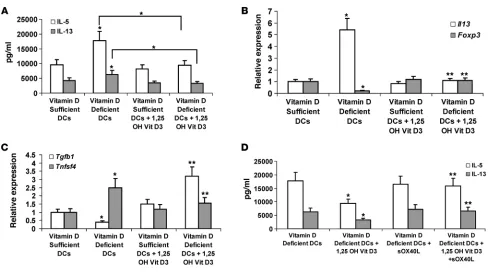
[image:8.585.56.532.643.721.2]cient mice, as previously described (26). After 4 weeks of being fed a vitamin D–deficient diet, mice had a mean serum 25-OH vitamin D3 level of 10.3 ± 1.9 ng/ml versus 31.6 + 2.9 ng/ml for
pair-fed controls. After 4 weeks, we obtained splenic CD11+ DCs
and assayed their capacity to induce Th2 differentiation to OVA peptide using naive CD4+ T cells from DO11.10 mice, as
previ-ously described (27). CD11c+ cells from vitamin D–deficient mice
primed significantly greater IL-5 and IL-13 responses in vitro when cells were restimulated with antigen compared with con-trol vitamin D–sufficient DCs (Figure 10A). Addition of 0.1 μM 1,25 OH-vitamin D3 to the culture had no effect on the Th2
prim-ing of CD11c+ DCs from vitamin D–sufficient mice but
signifi-cantly reduced the enhanced Th2 differentiation observed with CD11c DCs from vitamin D–deficient mice (Figure 10A). As we had observed that OX40L is critical in Th2 priming, we exam-ined the level of transcripts for TGF-β and OX40L in CD11c+ DCs
from vitamin D–deficient versus –sufficient mice. Similar to the human DCs, CD11c+ DCs from vitamin D–deficient mice had
sig-nificantly lower levels of Tgfb mRNA and higher levels of OX40L expression (Figure 10B). Treatment of these DCs with 0.1 μM 1,25 OH-vitamin D3 significantly increased TGF-β expression and
[image:9.585.90.484.72.543.2]reduced OX40L expression (Figure 10B). As TGF-β and OX40L have been implicated in Treg and Th2 differentiation respective-ly, we examined whether vitamin D affected subsequent Foxp3
Figure 7
1,25 OH-vitamin D3 suppresses Aspergillus-induced Th2 responses in CD4+ T cells from patients with ABPA. CD11c+ DCs or TSLP-DCs from
patients with ABPA (n = 6) were pulsed with ASPEXT, followed by addition of autologous bulk CD4+ T cells, followed by addition of 1,25
OH-vitamin D3 (1,25 Vit D3) or vehicle. Cells were incubated for 96 hours, and IL-5 (A and C), IL-13 (B and D), and IL-10 (E and F) were measured
and IL-13 expression in the Th2 differentiation assay. Similar to what we observed at the protein level (Figure 10A), vitamin D–deficient DCs induced significantly higher IL-13 expression in T cells undergoing Th2 differentiation compared with DCs from vitamin D–sufficient mice (Figure 10C). This enhanced IL-13 response in Th2 cells stimulated with DCs from vitamin D–defi-cient mice was associated with significantly reduced expression of Foxp3 mRNA (Figure 10C). No IL-13 or Foxp3 expression was observed in cultures that lacked the addition of naive CD4+ T cells
(data not shown). Furthermore, addition of 1,25 OH-vitamin D3
to Th2 cultures primed with DCs from vitamin D–deficient mice significantly reduced IL-13 induction and increased the induc-tion of Foxp3 expression in the cells (Figure 10C). These data suggest that 1,25 OH-vitamin D3 increases the ratio of TGF-β to
OX40L in the cell favoring induction of Tregs versus Th2 cells. To test this possibility, we examined whether the addition of soluble OX40L (sOX40L) could reverse the effect of exogenous 1,25 OH-vitamin D3 on vitamin D–deficient DCs. As observed on Figure
10A, the addition of 1,25 OH-vitamin D3 reduced the ability of
DCs from vitamin D–deficient mice to induce IL-5 and IL-13 production in cells undergoing Th2 differentiation; however, the addition of sOX40L restored this response nearly to the level of that of untreated DCs (Figure 10D). Addition of sOX40L to
cultures of vitamin D–deficient DCs not treated with exogenous 1,25 OH-vitamin D3 had no affect on Th2 cytokine production,
suggesting that OX40L expression was already maximal in this condition (Figure 10D).
Discussion
[image:10.585.49.523.78.386.2]Tolerance to many inhaled antigens is the normal immunological response in the lung and is mediated by both anergy and Tregs (1–3). When tolerance is not established, sensitization to inhaled antigens can result in asthma, hypersensitivity pneumonitis, and ABPA. ABPA is rare in the non-CF population, though seen in 0.1% to 0.5% of asthmatic patients. In patients with CF, ABPA prevalence ranges from 4% to 15%. Several risk factors have been identified to date. The most studied risk factor is the class II MHC haplotype of the affected patient. Alleles encoding HLA-DR2 and -DR5 confer susceptibility, whereas HLA-DQ2 has shown protec-tion (28, 29). However, experience in other disease models sug-gests there are likely risk factors other than the MHC haplotype that are critical to development of ABPA (30). For example, in another chronic inflammatory disease, Crohn disease, MHC is a relatively minor allele compared with polymorphism in the IL-23 receptor or other loci (31). Thus, other factors beyond MHC are likely critical for the development of ABPA.
Figure 8
1,25 OH-vitamin D3 increases the level of cell-associated TGF-β in patients with ABPA. CD11c+ DCs from patients with ABPA (n = 6) were
pulsed with ASPEXT, followed by the addition of autologous bulk CD4+ T cells, followed by addition of 1,25 OH-vitamin D3 or vehicle. Cells
were incubated for 96 hours, followed by staining for CD4, CD25, and TGF-β, followed by analysis on a FACSaria. (A) Data are plotted as the percentage of CD4+CD25+ and the percentage of CD4+CD25+TGF-β+ cells. (B) Relative MFI of TGF-β on CD4+CD25+ cells, before and after the
addition of 1,25 OH-vitamin D3. (C and D) Representative dot plot of TGF-β+ on CD4+CD25+ cells exposed to A. fumigatus–pulsed DCs, with or
without 1,25 OH-vitamin D3. (E) Relative gene expression of TNFSF4, FOXP3, TGFB1, and IL10 in DC/T cell cocultures at the end of the 96-hour
There are several advantages to studying ABPA in CF as a model to better understand mechanisms of immune tolerance. First, its preva-lence in CF is high enough to be studied. Second, most centers screen for ABPA at least annually, and most CF subjects in North America and Europe are already participating in disease registries. Third, as opposed to allergen-induced asthma, the inciting allergen is known in ABPA, as the vast majority of patients have disease due to A. fumiga-tus sensitization (15). Therefore, we sought to define T cell subsets in these patients to better elucidate mechanisms of immune tolerance versus sensitization in a cohort of ABPA patients versus non-ABPA patients that had documented colonization with A. fumigatus.
In this study, we found that CD11c+ DCs and TSLP-DCs elicited
more robust Th2 cytokine production from CD4+ T cells in an
OX40L-dependent fashion after pulsing with A. fumigatus antigens compared with similarly treated CD14+ DCs. We hypothesized that
CF epithelium may produce more TSLP or IL-25 in response to either Aspergillus or to poly I:C, but this was not the case, as CF HBE
cells produced similar amounts of TSLP and IL-25 compared with non-CF HBE cells. These data therefore do not favor a hypoth-esis that CF-specific epithelial factors are responsible for the high prevalence of ABPA in CF.
As mentioned above, tolerance may be mediated by anergy or by the induction of Tregs. In A. fumigatus–colonized individuals with-out ABPA, we observed greater percentages of CD4+CD25+Foxp3+
cells and particularly a higher surface TGF-β expression in CD4+CD25+ cells. Evidence in a subgroup of ABPA supports the
contention that these cells are suppressor cells, as depletion of the CD25+ subset enhanced Th2 responses to A. fumigatus antigens. Due
to the fact that this was a subgroup analysis, we cannot exclude a role of anergy in some patients that failed to develop ABPA despite
[image:11.585.73.504.78.483.2]A. fumigatus colonization. GIS mapping of patients did not reveal environmental clues to why some patients develop ABPA, and thus we turned our attention to dietary factors. In particular, we inves-tigated vitamin D, because patients with CF are at greater risk of
Figure 9
Blockade of TGF-β but not IL-10 antagonizes the 1,25 OH-vitamin D3 suppression of Aspergillus-induced Th2 responses in CD4+ T cells from patients
with ABPA. CD11c+ DCs or TSLP-DCs from patients with ABPA (n = 5) were pulsed with ASPEXT, followed by the addition of autologous bulk CD4+
T cells, followed by addition of 1,25 OH-vitamin D3, with or without recombinant human TGF-βRII Fc chimera (A and B) or anti–IL-10 (C and D). Cells
vitamin D deficiency and vitamin D has been implicated in Treg development (23, 24, 32). Moreover, vitamin D receptor knockout mice show exacerbated experimental allergic asthma compared with sensitized and challenged control mice (33). We observed that the ABPA cohort had significantly lower levels of serum vitamin D compared with the non-ABPA cohort. Despite this finding, there was no difference in BMI or vitamin A or E levels, suggesting that the vitamin D deficiency is not a marker of a more global nutri-tional defect in the patients studied. Treatment of CD4+ T cells in
vitro with 1,25 OH-vitamin D3 significantly reduced A. fumigatus–
induced increases in IL-5 and IL-13, regardless of whether the T cells were primed with CD11c+ DCs or TSLP-DCs. This suppression of
Th2 responses is mediated by an increase in TGF-β expression in CD4+CD25+ cells. This is supported by the fact that treatment with
1,25 OH-vitamin D3 increases the MFI of TGF-β on CD4+CD25+
cells and the expression of TGF-β1 transcripts as well as the fact that blockade of TGF-β signaling in vitro attenuates the suppres-sive affects of 1,25 OH-vitamin D3 on Th2 cytokine responses to
Aspergillus antigens. For these studies, we used 10% autologous AB serum and the measured final 1,25 vitamin D3 in the media was
30.16 pg/ml, and thus the responses we observed were unlikely affected by active vitamin D in the media. This is appears to be due to the effect of 1,25 OH-vitamin D3 on the DCs and not T cells.
This is supported by the fact that in vitro treatment with 1,25 OH-vitamin D3 suppressed OX40L expression in human DC/T cell
cocultures. Also, CD11c+ DCs from vitamin D–deficient mice had
higher levels of OX40L and primed stronger Th2 responses. Again, in vitro 1,25 OH-vitamin D3 treatment reduced OX40L expression
and reduced Th2 priming. Th2 priming could be restored with the addition of sOX40L. Lastly, 1,25 OH-vitamin D3 only reduced the
IL-5 and IL-13 response to ASPEXT-pulsed DCs and not to CD3/ CD28 stimulation (data not shown), further suggesting that the affect of 1,25 OH-vitamin D3 is through the DC.
Additionally, 1,25 OH-vitamin D3 has been shown to also inhibit
relB (34) and DC maturation (35, 36) in vitro, which may inhibit Th2 T cell development. In this study, we found a specific effect on OX40L expression. In fact, what may be critical is the relative expres-sion of OX40L and TGF-β1, as OX40L has been shown recently to block the induction of Foxp3 in T cells (18). LPS has been shown to antagonize airway tolerance to antigen and is mediated by OX40L induction on both DCs and B cells. The upregulation of OX40L increased both Th2 and Th1 responses to antigen. In addition to altering OX40L expression, vitamin D treatment of DCs has been shown to lead to apoptosis of alloreactive T cells.
There is a great deal of interest in vitamin D and asthma, anoth-er Th2-driven disease (37–39). In a cohort of children with asthma from Costa Rica, Brehm et al. found that vitamin D deficiency was significantly and inversely associated with total IgE and eosinophil counts (38). In a recent study, vitamin D deficiency was also associ-ated with increased use of inhaled corticosteroids (40). Addition-ally, in vitro 1,25 OH-vitamin D3 synergized to increase IL-10 levels
[image:12.585.47.537.78.351.2]in Tregs (24), an affect not seen in our DC/T cell coculture system.
Figure 10
Vitamin D deficiency increases Th2 priming of CD11c+ DCs. (A) Ova-specific IL-5 and IL-13 response in Th2-polarized cells differentiated with splenic
CD11c+ DCs from vitamin D–sufficient or –deficient mice (n = 6–8). (B) Relative gene expression of Tgfb1 and Tnfsf4 in splenic CD11c+ DCs from
vitamin D–deficient or –sufficient mice, before and after treatment with 1,25 OH-vitamin D3. (C) Relative gene expression of Il13 and Foxp3 in DC/T
cell cultures, in which CD11c+ DCs were from vitamin D–deficient or –sufficient mice, before and after treatment with vehicle or 1,25 OH-vitamin D3. (B
and C) *P < 0.05 by Mann-Whitney, compared with the vitamin D–sufficient group; **P < 0.05 by Mann-Whitney compared with the vitamin D–deficient group. (D) Effect of sOX40L on Th2 differentiation in the presence or absence of 1,25 OH-vitamin D3. *P < 0.05 by Mann-Whitney, compared with the
It has recently been shown that 1,25 OH-vitamin D3 can increase
TLR9 expression in inducible Tregs and that activation via TLR9 can reduce their regulatory function (41). These data suggest that vitamin D has profound affects on both DCs and iTregs. It is also possible that this TLR9 pathway may be another pathway in which iTregs lack efficacy in a chronic infectious condition such as CF.
The basis for vitamin D3 deficiency in our cohort remains
unclear at the present but does not appear to be a direct effect of geographical location. There was no difference in steroid or ursodi-ol use (the latter, a crude measure of biliary disease) in our cohort, and all patients were prescribed a combination of vitamin A, D, E, and K. The fact that vitamin A and E levels were not different sug-gests that nonadherence was unlikely a factor. One possible factor is genetic polymorphisms in the vitamin D pathway. In a recent study, SNPs were assessed to determine relative risk in an asthma cohort in the vitamin D receptor (VDR) as well as CYP2R1 (the 25 hydroxylase) and CYP24A1, which mediates the clearance of 1,25 OH-vitamin D3 (37). SNPs in CYP2R1 and CYP24A1 were
associ-ated with asthma, and in a 2-gene model, SNPs in VDR and IL10
were also associated. We are currently performing exon sequenc-ing of these genes in our cohort, as SNPs in this pathway could clearly determine efficacy of vitamin D treatment in this cohort. Taken together, our data strongly implicate vitamin D deficiency as a risk factor for ABPA and lay the ground work for clinical trials of enhanced vitamin D supplementation to prevent or treat ABPA. Moreover, our data suggest that enumeration of OX40L expres-sion on DCs and Treg frequency may be useful biomarkers to fol-low response to vitamin D augmentation.
Methods
HBE cultures. Primary HBE cells were provided by the Tissue Core Labora-tory at the University of Pittsburgh or purchased from Cambrex (Lonza). Primary cells were grown as polarized air-liquid interface (ALI) cultures, as previously described (42). HBE cells were stimulated apically with RC, HKSC (19), or poly I:C for 24 hours, and TSLP was measured by ELISA.
Analytical assays. TSLP was measured by sandwich ELISA as previously described (7). IL-4, IL-5, IL-10, IL-13, and IL-17 were measured using Luminex (Millipore).
Human subjects, cell harvest, and sorting. Accrual of all study subjects occurred after approval of the research protocol by the Institutional Review Board at the University of Pittsburgh. Human CD4+, CD11c+, and
CD14+ cells were obtained from whole blood from CF donors who gave
their written informed consent. The blood was then ficolled in Vacutainer CPT tubes (BD Pharmingen) and then resuspended in cell buffer com-posed of EDTA (Gibco), BSA (Sigma-Aldrich), and 1X PBS (Fisher). All cells were isolated by magnetic bead–activated sorting using microbeads and MidiMacs (Miltenyi). The CD4+ cells were first isolated by positive
separation in MS columns using CD4 Microbeads (Miltenyi). CD11c+
cells were subsequently isolated from the negative fraction of the CD4+
isolation using CD11c-APC microbeads and then anti-APC microbeads for the final magnetic separation. CD14+ DCs were selected by CD14+
microbeads (Miltenyi) and cultured in human recombinant GM-CSF (50 ng/ml; PeproTech) and IL-4 (20 ng/ml; PeproTech) for 6 days.
Robotic plate set up. DCs were then plated in flat bottom 96-well plates at a density of 5 × 10⁵ and 5 × 10⁴ DCs per well in medium containing RPMI,
l-glutamine, penicillin/streptomycin (pen/strep), FBS (Gibco), and Human
AB serum (Atlanta Biologicals). Cells in the plate were then stimulated with the following stimulators: TSLP (5 ng/ml; R&D Systems), Zymosan (50 mg/ml; Molecular Probes Inc.), or ASPEXT (1 μg/ml; Hollister-stier). A fourth condi-tion included both ASPEXT (1 μg/ml) and TSLP (5 ng/ml). One well in the
plate was left unstimulated as a control, and then 5 × 10⁵ CD4+ cells were
added. Control wells received CD4+ T cells that were cultured in media or
stimulated with CD3/CD28 beads (Dynal, Invitrogen). Additionally, recombi-nant IL-2 was added to all wells on the plate (7.5 ng/ml). Medium containing RPMI, 5% l-glutamine, 5% pen/strep, 10% FBS (Gibco), and 5% Human AB serum (Atlanta Biologicals) was then added to each well to bring up to final volume. All cells were then incubated at 37°C and 5% CO2 for 96 hours.
In experiments with 1,25 OH-vitamin D3, The conditions mentioned
above were plated in duplicate, in which one set was additionally incubat-ed with 1α, 25-OH-dihydroxyvitamin D3 (Biomol) at a concentration of
0.1 μM per condition versus an ethanol control. In some experiments, anti-human IL-10 (1 μg/ml final concentration) or recombinant human TGF-B sRII Fc chimera (10 μg/ml final concentration) were added.
Cell collection and FACS analysis. Cells in the plate were collected after 96-hour incubation, after which the release of cytokines was stopped by admin-istering Golgi Plug (BD Pharmingen) for the last 6 hours of stimulation. The cells from each well were then harvested for flow cytometry, and super-natants were assessed for cytokine contents using Luminex (Lincoplex). The cells harvested for flow cytometry were stained with a cocktail containing Pacific Blue–conjugated CD4 and PECy7-conjugated CD25 (BD Pharmin-gen). After initial staining, the cells were then fixed and permeabilized using a FOXP3 Staining Buffer Set (eBIOscience) and subsequently stained with another cocktail containing the following: FITC-conjugated Foxp3 (eBIOsci-ence), PE-conjugated TGF-β (R&D Systems), and additional Pacific Blue– conjugated CD4 (BD Pharmingen). All cells were analyzed on a FACSAria.
Vitamin D–deficient diet. All mouse experiments were approved by the IACUC at LSUHSC. Six-week-old female Balb/c mice (Charles River) were placed on a vitamin D–deficient diet (Bio-Serv AIN-93G) and maintained on the diet for at least 4 weeks. Serum 25-OH vitamin D levels were mea-sured by ELISA (IDS) to ensure vitamin D deficiency. Control mice were kept on regular mouse chow, supplied by the Division of Animal Care at LSUHSC (Harlan Teklad 2019S). Splenic CD11c+ cells were purified from
the spleen using CD11c+-coated magnetic beads (Miltenyi) and used for
Th2 differentiation assays as previously described (27). In certain experi-ments sOX40L (R&D Systems) was added.
Statistics. All data are presented as the mean ± SEM. For paired experi-ments, statistical significance was determined by a 2-tailed, paired t test. For comparisons of data from 2 independent groups, we used the nonpara-metric 2-tailed Mann-Whitney test, since the data often appeared incon-sistent with an assumption of normality. A P value of less than 0.05 was considered significant.
Acknowledgments
The authors would like to acknowledge support from the follow-ing Public Health Service grants: 5R01HL079142 (to J.K. Kolls), P50HL084932 (to J.K. Kolls, J.M. Pilewski, A. Ray, and C. Steele), P30DK072506 (to J.K. Kolls and J.M. Pilewski), and 5R01AI048927 (to A. Ray). The authors would like to thank Sandra Hurban and Adrienne Horn for their help with study accrual.
Received for publication January 20, 2010, and accepted in revised form July 7, 2010.
Address correspondence to: Jay K. Kolls, Department of Genet-ics, Louisiana State University Health Sciences Center, CSRB 657, 533 Bolivar Street, New Orleans, Louisiana 70112, USA. Phone: 504.568.6117; Fax: 504.568.8500; E-mail: jkolls@lsuhsc.edu.
1. Ostroukhova M, Qi Z, Oriss TB, Dixon-McCar-thy B, Ray P, Ray A. Treg-mediated immunosup-pression involves activation of the Notch-HES1 axis by membrane-bound TGF-beta. J Clin Invest. 2006;116(4):996–1004.
2. Hammad H, Lambrecht BN. Dendritic cells and epi-thelial cells: linking innate and adaptive immunity in asthma. Nat Rev Immunol. 2008;8(3):193–204. 3. Montagnoli C, et al. Immunity and tolerance to
Aspergillus involve functionally distinct regula-tory T cells and tryptophan catabolism. J Immunol. 2006;176(3):1712–1723.
4. Ray A, Khare A, Krishnamoorthy N, Qi Z, Ray P. Regulatory T cells in many flavors control asthma.
Mucosal Immunol. 2010;3(3):216–229.
5. Liu YJ, et al. TSLP: an epithelial cell cytokine that regulates T cell differentiation by condition-ing dendritic cell maturation. Annu Rev Immunol. 2007;25:193–219.
6. Wang YH, et al. IL-25 augments type 2 immune responses by enhancing the expansion and func-tions of TSLP-DC-activated Th2 memory cells.
J Exp Med. 2007;204(8):1837–1847.
7. Zhou B, Headley MB, Aye T, Tocker J, Comeau MR, Ziegler SF. Reversal of thymic stromal lymphopoietin-induced airway inflammation through inhibition of Th2 responses. J Immunol. 2008;181(9):6557–6562.
8. Zhou B, et al. Thymic stromal lymphopoietin as a key initiator of allergic airway inflammation in mice. Nat Immunol. 2005;6(10):1047–1053. 9. Goswami S, et al. Divergent functions for airway
epithelial matrix metalloproteinase 7 and reti-noic acid in experimental asthma. Nat Immunol. 2009;10(5):496–503.
10. Donaldson SH, Boucher RC. Update on pathogen-esis of cystic fibrosis lung disease. Curr Opin Pulm Med. 2003;9(6):486–491.
11. Boucher RC. New concepts of the pathogen-esis of cystic fibrosis lung disease. Eur Respir J. 2004;23(1):146–158.
12. Pihet M, et al. Occurrence and relevance of fila-mentous fungi in respiratory secretions of patients with cystic fibrosis--a review. Med Mycol. 2009; 47(4):387–397.
13. el Dahr JM, Fink R, Selden R, Arruda LK, Platts-Mills TA, Heymann PW. Development of immune responses to Aspergillus at an early age in chil-dren with cystic fibrosis. Am J Respir Crit Care Med. 1994;150(6 pt 1):1513–1518.
14. Stevens DA, et al. Allergic bronchopulmonary aspergillosis in cystic fibrosis--state of the art: Cys-tic Fibrosis Foundation Consensus Conference.
Clin Infect Dis. 2003;37(suppl 3):S225–S264.
15. Moss RB. Pathophysiology and immunology of allergic bronchopulmonary aspergillosis. Med Mycol. 2005;43(suppl 1):S203–S206.
16. Agarwal R. Allergic bronchopulmonary aspergil-losis. Chest. 2009;135(3):805–826.
17. Salek-Ardakani S, et al. OX40 (CD134) controls memory T helper 2 cells that drive lung inflamma-tion. J Exp Med. 2003;198(2):315–324.
18. Duan W, So T, Croft M. Antagonism of airway tol-erance by endotoxin/lipopolysaccharide through promoting OX40L and suppressing antigen-specific Foxp3+ T regulatory cells. J Immunol. 2008;181(12):8650–8659.
19. Steele C, et al. The beta-glucan receptor dectin-1 recognizes specific morphologies of Aspergillus fumigatus. PLoS Pathog. 2005;1(4):e42.
20. Ziegler SF, Liu YJ. Thymic stromal lymphopoietin in normal and pathogenic T cell development and function. Nat Immunol. 2006;7(7):709–714. 21. Kato A, Favoreto S Jr, Avila PC, Schleimer RP.
TLR3- and Th2 cytokine-dependent production of thymic stromal lymphopoietin in human airway epithelial cells. J Immunol. 2007;179(2):1080–1087. 22. Ostroukhova M, et al. Tolerance induced by inhaled antigen involves CD4(+) T cells expressing mem-brane-bound TGF-beta and FOXP3. J Clin Invest. 2004;114(1):28–38.
23. Xystrakis E, et al. Reversing the defective induc-tion of IL-10-secreting regulatory T cells in glu-cocorticoid-resistant asthma patients. J Clin Invest. 2006;116(1):146–155.
24. Barrat FJ, et al. In vitro generation of interleukin 10-producing regulatory CD4(+) T cells is induced by immunosuppressive drugs and inhibited by T helper type 1 (Th1)- and Th2-inducing cytokines.
J Exp Med. 2002;195(5):603–616.
25. Green D, et al. Current treatment recommenda-tions for correcting vitamin D deficiency in pedi-atric patients with cystic fibrosis are inadequate.
J Pediatr. 2008;153(4):554–559.
26. Misharin A, et al. Vitamin D deficiency modulates Graves’ hyperthyroidism induced in BALB/c mice by thyrotropin receptor immunization. Endocrinol-ogy. 2009;150(2):1051–1060.
27. McKinley L, et al. TH17 cells mediate steroid-resistant airway inf lammation and airway hyperresponsiveness in mice. J Immunol. 2008; 181(6):4089–4097.
28. Chauhan B, et al. The association of HLA-DR alleles and T cell activation with allergic bron-chopulmonary aspergillosis. J Immunol. 1997; 159(8):4072–4076.
29. Chauhan B, et al. Evidence for the involvement of two different MHC class II regions in
suscep-tibility or protection in allergic bronchopulmo-nary aspergillosis. J Allergy Clin Immunol. 2000; 106(4):723–729.
30. Duerr RH, et al. A genome-wide association study identifies IL23R as an inflammatory bowel disease gene. Science. 2006;314(5804):1461–1463. 31. Duerr RH. Genome-wide association studies
herald a new era of rapid discoveries in inflam-matory bowel disease research. Gastroenterology. 2007;132(5):2045–2049.
32. Devereux G, Macdonald H, Hawrylowicz C. Vita-min D and asthma: time for intervention? Am J Respir Crit Care Med. 2009;179(9):739–740. 33. Wittke A, Weaver V, Mahon BD, August A,
Can-torna MT. Vitamin D receptor-deficient mice fail to develop experimental allergic asthma. J Immunol. 2004;173(5):3432–3436.
34. Dong X, et al. Regulation of relB in dendritic cells by means of modulated association of vita-min D receptor and histone deacetylase 3 with the promoter. Proc Natl Acad Sci U S A. 2005; 102(44):16007–16012.
35. Penna G, Adorini L. 1 Alpha,25-dihydroxyvitamin D3 inhibits differentiation, maturation, activa-tion, and survival of dendritic cells leading to impaired alloreactive T cell activation. J Immunol. 2000;164(5):2405–2411.
36. Griffin MD, Lutz W, Phan VA, Bachman LA, McKean DJ, Kumar R. Dendritic cell modulation by 1alpha,25 dihydroxyvitamin D3 and its analogs: a vitamin D receptor-dependent pathway that promotes a persis-tent state of immaturity in vitro and in vivo. Proc Natl Acad Sci U S A. 2001;98(12):6800–6805.
37. Bosse Y, et al. Asthma and genes encoding com-ponents of the vitamin D pathway. Respir Res. 2009;10:98.
38. Brehm JM, et al. Serum vitamin D levels and mark-ers of severity of childhood asthma in Costa Rica.
Am J Respir Crit Care Med. 2009;179(9):765–771. 39. Litonjua AA, Weiss ST. Is vitamin D deficiency to
blame for the asthma epidemic? J Allergy Clin Immu-nol. 2007;120(5):1031–1035.
40. Searing DA, Zhang Y, Murphy JR, Hauk PJ, Gol-eva E, Leung DY. Decreased serum vitamin D lev-els in children with asthma are associated with increased corticosteroid use. J Allergy Clin Immunol. 2010;125(5):995–1000.
41. Urry Z, et al. Ligation of TLR9 induced on human IL-10-secreting Tregs by 1alpha,25-dihydroxyvita-min D3 abrogates regulatory function. J Clin Invest. 2009;119(2):387–398.