melanoma development
Barbara Bedogni, … , Amato J. Giaccia, Marianne Broome
Powell
J Clin Invest. 2008;
118(11)
:3660-3670.
https://doi.org/10.1172/JCI36157
.
Melanomas are highly aggressive neoplasms resistant to most conventional therapies.
These tumors result from the interaction of altered intracellular tumor suppressors and
oncogenes with the microenvironment in which these changes occur. We previously
demonstrated that physiologic skin hypoxia contributes to melanomagenesis in conjunction
with Akt activation. Here we show that Notch1 signaling is elevated in human melanoma
samples and cell lines and is required for Akt and hypoxia to transform melanocytes in vitro.
Notch1 facilitated melanoma development in a xenograft model by maintaining cell
proliferation and by protecting cells from stress-induced cell death. Hyperactivated PI3K/Akt
signaling led to upregulation of Notch1 through NF-
k
B activity, while the low oxygen content
normally found in skin increased mRNA and protein levels of Notch1 via stabilization of
HIF-1
a
. Taken together, these findings demonstrate that Notch1 is a key effector of both Akt
and hypoxia in melanoma development and identify the Notch signaling pathway as a
potential therapeutic target in melanoma treatment.
Research Article
Oncology
Find the latest version:
Notch1 is an effector of Akt and hypoxia
in melanoma development
Barbara Bedogni,1 James A. Warneke,2 Brian J. Nickoloff,3 Amato J. Giaccia,1 and Marianne Broome Powell1
1Division of Radiation and Cancer Biology, Stanford University, Stanford, California, USA. 2Arizona Cancer Center and Department of Surgery, University of Arizona, Tucson, Arizona, USA. 3Department of Pathology, Loyola University Medical Center, Maywood, Illinois, USA.
Melanomas are highly aggressive neoplasms resistant to most conventional therapies. These tumors result
from the interaction of altered intracellular tumor suppressors and oncogenes with the microenvironment in
which these changes occur. We previously demonstrated that physiologic skin hypoxia contributes to
mela-nomagenesis in conjunction with Akt activation. Here we show that Notch1 signaling is elevated in human
mel-anoma samples and cell lines and is required for Akt and hypoxia to transform melanocytes in vitro. Notch1
facilitated melanoma development in a xenograft model by maintaining cell proliferation and by protecting
cells from stress-induced cell death. Hyperactivated PI3K/Akt signaling led to upregulation of Notch1 through
NF-
κ
B activity, while the low oxygen content normally found in skin increased mRNA and protein levels of
Notch1 via stabilization of HIF-1
α
. Taken together, these findings demonstrate that Notch1 is a key effector
of both Akt and hypoxia in melanoma development and identify the Notch signaling pathway as a potential
therapeutic target in melanoma treatment.
Introduction
Cutaneous melanoma is one of the most aggressive malignancies, with a steadily increasing incidence over the past few decades. Its high mortality rate is mainly the result of resistance to standard therapies and a propensity to metastasis, especially to the central nervous system.
Alterations of several signaling pathways, such as NRas (1), BRaf (2), PTEN/PI3K/Akt (3), and p16/ARF (4), are found in melano-mas and lead to acquisition of growth advantages, resistance to apoptosis, and invasion/metastatic behavior. A considerable effort has been put into the development of therapies aimed at inhibit-ing these protumorigenic functions as well as at identifyinhibit-ing novel critical signaling pathways involved in melanoma.
Notch signaling controls a variety of processes, involving cell fate specification, differentiation, proliferation, and survival (5). In mammals, the Notch family consists of 4 transmembrane recep-tors (Notch1–Notch4) and 5 ligands (Jagged-1, Jagged-2, Delta-1, Delta-3, and Delta-4). Binding of ligand to its receptor induces metalloproteinase-mediated and γ-secretase–mediated cleavage of the Notch receptor. The Notch1 intracellular domain (Notch1-NIC)
is released from the plasma membrane and translocates into the nucleus, where it forms a complex with RBP-Jk/CBF1, Su(H), Lag-2 (RBP-Jk/CSL), and mastermind-like (MAML). This complex medi-ates the transcription of target genes such as that encoding Deltex, genes in the hairy enhancer of split (HES) and HES-related families of basic helix-loop-helix transcription factors, and others (6).
Increased attention to Notch signaling in cancer biology began with the identification of the chromosomal translocation t(7;9) in human T cell acute lymphoblastic leukemia (T-ALL), which results
in constitutive activation of Notch1 signaling (7). Since this find-ing, somatic activating mutations in Notch1 have been found in more than half of human T-ALL samples (8). Recently, inactivating mutations of FBW7, an H3 ligase that targets Notch1 and other proto-oncogenes for degradation, were found in a large fraction of human T-ALL cell lines and primary tumors (9) and in other can-cers (10). In addition, aberrant Notch signaling has previously been observed in small cell lung cancer (11), neuroblastoma (12), cervical cancer (13), prostate cancer (14), and renal cell carcinomas (15).
Notch can function as a tumor promoter or a suppressor depending on the cell type and context. In experimental mouse models of skin tumorigenesis, Notch1 shows antitumor activity. Ablation of Notch1 in keratinocytes leads to spontaneous devel-opment of basal cell carcinoma–like tumors (16), likely as a result of reactivation of the β-catenin signaling pathway. Similarly, abla-tion of presenilin-1, an important component of the γ-secretase cleavage machinery, leads to keratinocyte-derived skin cancer via reactivation of the β-catenin/cyclin D axis (17).
Evidence suggesting a role of Notch1 signaling in melanoma is only now emerging. HES related with yrpw motif 1 (HEY1), a known direct target of Notch, was found to be elevated in a subset of melanoma cell lines in a gene array analysis (18), which implies that Notch activity may be involved in melanomagenesis. Inhibi-tion of γ-secretase activity was shown to induce apoptosis in mela-noma cells while sparing normal melanocytes, suggesting that Notch may be required for melanoma cell survival (19). Overex-pression of Notch1 has been shown to increase the aggressiveness of primary melanoma cell lines by activating the β-catenin and the PI3K/Akt and MEK/Erk pathways (20, 21). The results of these prior studies suggest that Notch1 exerts its tumorigenic activity by acting upstream of these key signaling pathways.
Activation of Notch can also be downstream of these signal-ing cascades. Oncogenic Ras activates Notch1 signalsignal-ing, which is required to maintain the neoplastic phenotype in Ras-transformed human fibroblasts (22). In endothelial cells, the activation of the MAPK pathway leads to an arterial fate specification by
activat-Nonstandard abbreviations used: DAPT, N-[N-(3,5-difluorophenacetyl-L-alanyl)]-S-phenylglycine t-butyl ester; HES, hairy and enhancer of split; HEY1, HES related with yrpw motif 1; IκBαM, degradation-resistant mutant of IκBα; Notch1-NIC,
Notch1 intracellular domain; qRT-PCR, quantitative real-time RT-PCR; T-ALL, T cell acute lymphoblastic leukemia; TM-Notch1, transmembrane Notch1.
ing the Notch1 signaling cascade (23). Constitutively active Akt induces Notch1 activity in some T-ALL cell lines (24), and it has recently been shown to confer resistance to γ-secretase inhibitors in T-ALL cell lines that show loss of PTEN function (25), which suggests that in these cells the 2 pathways can act in parallel in conferring cell growth advantages.
Notch1 can play an active role in mediating effects of the micro-environment on the surrounding cells. For example, Notch1 is required by tissue hypoxia to maintain cells in an undifferenti-ated state (26). This is particularly relevant to melanoma, because we and others previously demonstrated that the skin is normally mildly hypoxic and that melanocytes reside in this environment (27, 28). Taken together, these findings strongly suggest that the Notch1 signaling pathway can be under control of both oncogene activation and environmental conditions, although how these fac-tors regulate Notch1 is still not fully understood.
We have previously shown that skin hypoxia plays an impor-tant role in melanocyte transformation (27). Oncogenes such as Akt transform melanocytes only when these cells are grown in an hypoxic environment recapitulating skin hypoxia, and this effect is mediated by HIF-1α. We hypothesize that Notch1 acts downstream of Akt and hypoxia and that it is one of the key effectors mediating melanocyte transformation and melanoma development.
In this study we found elevated Notch1 signaling in melanomas. We showed that Notch1 expression was controlled by both the PI3K/Akt pathway through NF-κB and by tissue hypoxia through HIF-1α. Finally, we demonstrated that Notch1 was required by Akt to transform melanocytes under hypoxic conditions and to allow tumor growth in vivo by protecting cells from stress-dependent cell death and by maintaining cells in a highly proliferative state. Our findings provide evidence that Notch1 signaling mediates the interaction between Akt and hypoxia in melanoma development and reinforce an earlier suggestion for targeting the Notch signal-ing pathway in melanoma therapy (29).
Results
Notch1 signaling is elevated in melanomas. Array data from the Onco-mine database (30) showed higher levels of HEY1 expression in pri-mary melanoma samples than in normal skin or benign nevi (Figure 1A). HEY1 is a Notch target gene (31) and was previously found to be elevated in a gene array analysis done on melanoma cell lines (18). To further corroborate the expression array data, we performed West-ern blot analysis on a number of human melanoma samples. Of 19 tumors, 15 (79%) exhibited expression of Notch1 protein, with 10 of 19 (53%) showing substantially elevated transmembrane Notch1 (TM-Notch1) and Notch1-NIC levels compared with congenital nevi
(Figure 1B). These data show that Notch1 signaling is elevated in melanomas and may therefore play a role in the disease.
Notch1 expression is controlled by the PI3K/Akt pathway. Major sig-naling pathways altered in melanoma include the Ras pathway and downstream effectors BRaf/MEK/Erks and PTEN/PI3K/Akt (32, 33). It was previously shown that active Notch1 (i.e., Notch1-NIC)
can promote melanoma progression by activating both the PI3K/ Akt and the Raf/MEK pathways (21). Therefore, we sought to determine how Notch1 activity is associated with alteration of these pathways using a number of low-passage human melanoma cell lines. Western blot analysis revealed that the majority of these cell lines have one or both pathways activated compared with nor-mal melanocytes and that 17 of 26 melanomas (65%) showed the presence of Notch1-NIC (Figure 2A). Because in most cases, the
presence of Notch1-NIC protein is associated with loss of PTEN
expression and with phosphorylation of Akt and/or Erk1/2, this observations suggests that both pathways may be involved in Notch1 regulation. Inhibition of PI3K by Ly294002 virtually elimi-nated Notch1-NIC protein, whereas inhibition of MEK by U0126
did not show any effect on Notch1, despite markedly inhibiting Erk1/2 phosphorylation (Figure 2B). In cells treated with the com-bination of Ly294002 and U0126, the decrease in Notch1-NIC was
comparable to that in cells treated with the PI3K inhibitor alone, further suggesting that Notch1-NIC regulation is not dependent
on the Raf/MEK/Erk pathway. Genetic inhibition of PI3K by the dominant-negative Δp85 reduced Notch1-NIC protein levels, also
corroborating a role of PI3K signaling in regulating Notch1 (Fig-ure 2C). Total Akt and total Erk1/2 were not affected by chemi-cal or genetic inhibition. The importance of PI3K/Akt in regulat-ing Notch1 expression and/or activity was substantiated by the analysis of mouse melanocytes engineered to overexpress either oncogenic human Akt or oncogenic human BRaf (27). Only cells expressing active Akt showed increased levels of Notch1-NIC, while
in BRaf-expressing melanocytes, Notch1-NIC levels did not change
compared with control cells (Figure 2D).
These data suggest the existence of an interesting reciprocal regu-lation of Notch1 and PI3K pathways. Not only can Notch1 induce PI3K/Akt signaling (21), but Notch1 is also downstream of Akt. Similar results were previously observed in some T-ALL cell lines and were reported to be dependent on posttranslational regula-tion of Notch1 by Akt (24). In contrast, melanoma cells expressing an active Akt showed a substantial increase in TM-Notch1, Notch1
mRNA, and a significant increase in Notch1 activity, measured as the capacity to induce a HES1-dependent promoter reporter construct (Figure 2, E–G), which suggests that Akt may control Notch1 at the transcriptional level.
Akt regulates Notch1 via NF-κB. To determine how Akt regulates Notch1, we focused on NF-κB signaling. NF-κB is one of the tran-scription factors activated by Akt that plays a role in melanoma (34, 35), and cross-talk between Notch and NF-κB has also been described (36). We examined whether Akt transcriptionally con-trols Notch1 through regulation of NF-κB. Introduction of a deg-radation-resistant mutant of IκBα (IκBαM; ref. 37) in Akt-express-ing melanocytes resulted in decreased nuclear p65 accumulation (Figure 3A) and reduced NF-κB activity, as measured both by the expression level of IκBα and c-myc and by a NF-κB reporter assay (Figure 3, B and C). Reduction of NF-κB activity resulted in inhibi-tion of TM-Notch1, Notch1-NIC protein, and Notch1 mRNA (Figure
3, D and E) as well as decreased Notch1 activity, measured as the expression of its target genes HES1 and HEY1 (Figure 3, F and G). Taken together, these data strongly support the existence of a func-tional relationship between the activation of the PI3K/Akt/NF-κB pathway and Notch1 signaling in melanoma development.
Notch1 colocalizes with hypoxia and is regulated by HIF-1α. In order to analyze the potential role of Notch1 in vivo, we stained Akt-dependent melanoma xenografts with an antibody recognizing Notch1-NIC.
Interestingly, we found that Notch1-NIC was abundant in these
tumors, particularly in the nuclei, which suggests that Notch1 plays an active role in Akt-mediated tumorigenesis (Figure 4A).
and poor prognosis (38, 39). Notch1 was previously reported to be required by hypoxia to maintain stem and precursor cell popula-tions in an undifferentiated state (26); more recently, hypoxia was shown to induce Notch1 expression and activity in lung adeno-carcinoma cell lines (40). In order to test whether Notch1-NIC
pro-tein is associated with hypoxia, we stained Akt-dependent tumors with the hypoxia marker EF5 (Figure 4B and ref. 41). Notch1-NIC
staining was more intense in areas of hypoxia (Figure 4C), which suggests that the microenvironment as well as Akt may influence Notch1 expression and activity in an additive manner.
We next investigated whether hypoxic treatment of Akt-express-ing melanocytes induces Notch1 expression and whether this effect is mediated by HIF-1α. Akt-expressing melanocytes were grown either in normoxia (21% O2) or in hypoxia (2% O2) for
24 h, and mRNA and protein levels of Notch1 were analyzed. We found that hypoxia increased Notch1 mRNA and protein as well as Notch activity, measured as HES1 and HEY1 expression and HES1 promoter activity (Figure 4, D–G, and Supplemental Figure 1, A–C; supplemental material available online with this article; doi:10.1172/JCI36157DS1). This effect was dependent on HIF-1α: in the presence of an shRNA against HIF-1α (referred to herein as shHIF-1α), Notch1 mRNA and protein were no longer induced by hypoxia compared with control GFP shRNA (referred to herein as shGFP; Figure 4, D and E). The efficacy of shHIF-1α
was assessed as inhibition of HIF-1α protein expression (Figure 4E) and as the capacity to inhibit the induction, under hypoxia, of the HIF-1α target gene VEGF (Supplemental Figure 2). The results presented thus far suggest that Notch1, like other protu-morigenic molecules, is under the control of oncogenes and the tissue microenvironment and is therefore a downstream effector of both pathways in melanoma development.
Notch1 is required for Akt-dependent melanocyte transformation in hypoxia and for tumor growth in vivo. To test whether Notch1 is an
effector of Akt and hypoxia in melanoma development, we treated Akt-expressing melanocytes with either N-[N-(3,5-difluorophen-acetyl-L-alanyl)]-S-phenylglycine t-butyl ester (DAPT), a γ-secretase inhibitor that blocks the processing of TM-Notch1 to Notch1-NIC, or a Notch1-specific shRNA (referred to herein as shNotch1).
Cells were grown in soft agar for 3 wk in a hypoxic atmosphere. We cultured cells in hypoxia because, as shown previously (27), Akt-expressing melanocytes require a physiologically relevant oxygen environment for growth in soft agar. We achieved a sig-nificant reduction in colony formation either by blocking Notch activation with DAPT or by inhibiting Notch1 expression, which strongly suggests that Notch1 activity is required by Akt to trans-form melanocytes in hypoxia (Figure 5A and Figure 6A). More-over, when these cells were implanted in SCID mice, tumor growth was significantly delayed in a dose-dependent manner by topical application of DAPT or by the presence of shNotch1 (Figure 5B and Figure 6B). The level of inhibition of Notch1 was assessed by Western blot analysis of Notch1-NIC and by quantitative real-time
RT-PCR (qRT-PCR) of Notch1, HES1, and HEY1 transcripts. Fig-ure 5C shows that DAPT treatment greatly reduced the amount of Notch1-NIC released in the cells. In the presence of shNotch1,
virtually no protein was detectable (Figure 6C). As expected, DAPT did not have a significant effect on Notch1 transcript (Figure 5D) but did reduce the amount of HES1 and HEY1 expressed (Figure 5, E and F). In contrast, shNotch1 significantly decreased Notch1,
HES1, and HEY1 expression (Figure 6, D–F). Tumors excised at the end time point were stained with an anti–Notch1-NIC antibody.
shNotch1-expressing tumors showed reduction of Notch1-NIC
lev-els compared with controls (Figure 7, H and I), demonstrating that shRNA expression was retained throughout the experiment.
[image:4.585.46.360.80.361.2]Tumor growth delay by inhibition of Notch1 is associated with reduced cell growth, reduced cyclin D1 expression, and increased cell death. Notch1 is known to regulate a number of targets having a role in cell
Figure 1
Notch1 is elevated in melanoma. (A) HEY1
expression levels in normal skin, benign nevi, and primary melanoma tumor samples. Boxes denote interquartile range; lines denote median; whiskers denote first and fourth quartiles, exclud-ing outliers (dots). Melanoma vs. nevi, melano-ma vs. skin, and melanomelano-ma vs. combined benign samples, P < 0.001; skin vs. nevi, P < 0.005; Student’s t test for all comparisons. (B) West-ern blot analysis of congenital nevus (Cong nev) and melanoma samples. The double band in the upper panel refers to TM-Notch1 (TM) and Notch1-NIC (NIC). The same samples were also
Figure 2
Notch1-NIC expression is a function of PI3K/Akt pathway activation and is transcriptionally regulated by Akt. (A) We analyzed 26
low-pas-sage melanoma cell lines for activation of the PI3K/Akt and Raf/MEK/Erk pathways with respect to normal human melanocytes in correlation with Notch1-NIC. M, melanocytes; p-, phosphorylated; t-, total. (B) Two of the cell lines analyzed in A (WM266, lane 3; K457, lane 8) were
treated with 50 μM Ly294002, 10 μM U0126, or the combination (Ly/U). Effectiveness of the treatment was assessed as the inhibition of phosphorylation of Akt and Erk1/2. (C) K457 and WM266 expressing dominant-negative PI3K (Δp85) were assessed for Notch1-NIC protein.
(D) Mouse melanocytes overexpressing oncogenic Akt or oncogenic BRaf were assessed for Notch1-NIC expression. The ratio of Notch1-NIC
to α-tubulin was 1.3 for Babe, 4.8 for Akt, and 1.2 for BRaf. (E) Western blot for TM-Notch1 showed higher expression in Akt-expressing cells. (F) qRT-PCR for Notch1. As an internal control, 18S was used for normalization. (G) Notch1 activity, measured as induction of a HES1-dependent reporter construct. Data in F and G are mean ± SD. *P < 0.05 versus Babe control, Student’s t test. In A–E, α-tubulin and
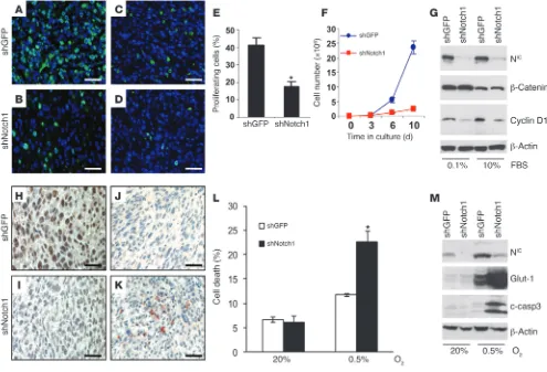
growth, and inhibition of Notch1 was previously shown to induce cell death in melanoma cells (19) and in lung adenocarcinomas (40). In order to assess whether proliferation and/or survival are affected by Notch1 inhibition in vivo, we stained Akt-dependent tumors with Ki67 and cleaved caspase-3 antibodies, respectively. Tumors originating from the shNotch1-expressing cells showed a significant reduction in the fraction of proliferating (i.e., Ki67-positive) cells (Figure 7, A, B, and E) compared with the tumors formed by the melanocytes expressing shGFP. Similarly, in an in vitro growth assay, cells expressing shNotch1 grew significantly slower than did control cells (Figure 7F).
Among the cell cycle regulators reported to be under the influ-ence of Notch1, cyclin D1, c-myc, NF-κB, p21Cip1, and p27Kip1 have
previously been shown to be involved in Notch1-dependent primary rat kidney epithelial (RKE) cell transformation (42), breast tumor development (43), pancreatic cancer invasion (44), and renal cell car-cinoma growth (15). We examined the expression levels of these regu-lators in Akt melanoma cells expressing either shGFP or shNotch1. Notch1 knockdown had a marked effect on cyclin D1 induction: cells expressing shNotch1 were unable to induce cyclin D1, in contrast to control cells, even after serum stimulation (Figure 7G). Tumors developing from shNotch1-expressing cells also showed a substan-tial decrease in cyclin D1 expression (Figure 7, C and D). In contrast, c-myc, NF-κB, p21Cip1, and p27Kip1 were not affected by inhibition of
Notch1 expression in melanomas (Supplemental Figure 4). We also observed positive staining for cleaved caspase-3 in vivo (Figure 7K) associated with tumors formed by the
shNotch1-expressing cells (Figure 7I), which suggests that these tumor cells are more sensitive to apoptosis. Because earlier studies in lung tumor cell lines showed that inhibition of Notch activity by a
γ-secretase inhibitor sensitized cells to hypoxia-induced cell death, we sought to determine whether lack of Notch1 function would increase melanoma cell death in stringent hypoxia (0.5% O2). Cells
expressing shNotch1 showed a significant increase in cell death after being treated with stringent hypoxia for 48 h (Figure 7L and Supplemental Figure 3A). This effect was accompanied by altera-tion of the Bax/BCL2/BCL-xL ratio (Supplemental Figure 3B) and
by caspase-3 cleavage (Figure 7M). Cells expressing shNotch1 showed reduced expression of BCL-xL and a partial reduction of
BCL2 compared with shGFP-expressing controls, whereas Bax
lev-els remained unchanged. Such alterations are likely to cause an imbalance in the Bax/BCL2/BCL-xL levels in favor of the
proapop-totic protein Bax. Induction of the HIF-1α direct target Glut-1 was used as a control to indicate an effective hypoxia response. These findings indicate that the inhibition of cell and tumor growth we observed was dependent on both increased cell death and decreased proliferation.
Discussion
[image:6.585.114.475.77.335.2]In this study, we demonstrated that Notch1 signaling is amplified in human melanomas and that Notch1 contributes to Akt-depen-dent melanocyte transformation in hypoxia and facilitates mela-noma growth. Elevated Notch1 activity was detected in primary melanomas compared with benign nevi and normal skin. Notch1
Figure 3
Notch1 is transcriptionally regulated by Akt through NF-κB activity. (A) Western blot analysis for p65 in nuclei of Akt-expressing melanocytes stably transfected with IκBαM. Tata-binding protein (TBP) was used as loading control for nuclear protein lysates. (B) qRT-PCR analysis of cells in A for IκBα and c-myc. (C) NF-κB reporter assay in melanocytes expressing an empty vector (pBabe), Akt, or Akt/IκBαM. (D) Western blot for TM-Notch1 and Notch1-NIC protein in Akt cells expressing either an empty vector (pBabe) or IκBαM. Tata-binding protein and α-tubulin were
receptor levels substantially increased above nevi levels in 53% of the melanoma samples analyzed by Western blot. Furthermore, we showed that Notch1 was transcriptionally regulated by NF-κB downstream of Akt and by HIF-1α in hypoxia and that Notch1 function was necessary for melanomagenesis. Taken together, these data provide a rationale for therapeutically targeting the Notch signaling pathway in melanoma.
Hyperactivation of the BRaf/MEK and PI3K/Akt pathways has previously been observed in 70% and 40%–60%, respectively, of human melanomas (2, 3). We found Notch1-NIC protein to be
associated with the activation of both pathways, although only the PI3K/Akt cascade was responsible for regulating Notch1 expres-sion. Interestingly, in the panel of cell lines analyzed, few showed only activation of BRaf/MEK in association with Notch1 protein. It is possible that in these cells, signaling pathways other than PI3K/Akt are active and may regulate Notch1 expression. Ras, for example, was previously shown to increase levels and activity of Notch1-NIC and to upregulate Delta-1 and presenilin-1 through
a p38-mediated pathway (22). Notch, in turn, may activate the MEK/Erks cascade, as has been shown previously (21).
Akt regulates a number of downstream effectors involved in melanomagenesis (45). Among them, NF-κB has been proposed as a potential therapeutic target (46). NF-κB was previously shown to increase Notch1 activity indirectly, by increasing the expression of Notch ligand Jagged1 in HeLa, lymphoma, and myeloma cells (47). However, in our melanoma model, Akt regulated Notch1 by increasing Notch1 transcription through the activity of NF-κB. Our findings suggest the existence of cell type–specific cross-talk between Notch1 and NF-κB. However, it is not clear whether
Notch1 is directly or indirectly responsive to NF-κB. In a prelimi-nary analysis of the proximal 2-kb promoter of Notch1, we found several putative NF-κB binding sites, but none that controlled Notch1 expression in a reporter assay (our unpublished observa-tions). Determining how NF-κB controls Notch1 transcription will require further investigation.
Melanocytes reside in a distinct niche in the skin and are under the influence of the surrounding microenvironment, which, after oncogene activation, continues to exert its selective pressure on the genetically altered melanocytes (48). We previously found that normal skin hypoxia acts as a tumor-promoting factor in melanomas (27). Melanocytes that have acquired oncogenic mutations and are genetically unstable showed a fully trans-formed phenotype only when under the influence of the low oxygen concentration found in skin.
Notch1 activity is influenced by tissue hypoxia (26, 40). In our system, hypoxia resulted in increased expression of Notch1 through HIF-1α–dependent induction of Notch1 mRNA. Similarly, in hypoxic lung cancer cells, Notch1 is transcriptionally regulated (40). On the other hand, in renal cell carcinomas, alteration of the Notch pathway is independent of HIF-1α and HIF-2α (15), while in stem and precursor cells, hypoxia regulates Notch1 activity posttranslationally via HIF-1α (26). Notch1 activity has also been reported to be regulated by factor inhibiting HIF-1α (FIH), and Notch1 itself potentiates the cellular hypoxic response by increas-ing the recruitment of HIF-1α to the HRE sequences of canonical HIF-1 target genes (49). Furthermore, it has previously been shown that both Akt and hypoxia can induce an increase in reactive oxy-gen species that can play a role in regulating Akt downstream
tar-Figure 4
Notch1-NIC colocalizes with hypoxia, and its expression is regulated by HIF-1α. (A–C) Akt-dependent tumors were stained for Notch1-NIC (A) and
with the hypoxia marker EF5 (B), showing colocalization (C). Scale bars: 50 μm. (D) qRT-PCR analysis of Akt-expressing melanocytes treated with normoxia (Nx; 21% O2) or hypoxia (Hx; 2% O2) and expressing either shGFP or shHIF-1α. (E) Western blot analysis for HIF-1α and
Notch1-NIC in nuclear lysates. Tata-binding protein was used as loading control. (F and G) HES1 and HEY1 expression, as measured by qRT-PCR. As
[image:7.585.83.505.79.341.2]gets and HIF-1α stability (50–52). This observation suggests that Notch1 regulation could potentially be under the influence of such species as well. The diverse findings of these studies under-line an intricate mechanism of regulation of the Notch complex by oncogenes and the tumor microenvironment through HIF-1α that is possibly dependent on different tissues of origin of the tumors. Inhibition of Notch signaling induces cell death in a number of malignant cell types (40, 53, 54) and influences tumor cell prolif-eration (42, 43, 53). Inhibition of Notch1 in our system resulted in both cell death and cell growth inhibition in vitro and in vivo. Inter-estingly, melanoma cells lacking Notch1 did not apoptose in nor-mal culture conditions, but they were sensitive to stress-induced cell death. Tumors experience ranges of oxygenation that go from mild hypoxia to near anoxia. While mild hypoxia (~1.5% O2)
is normally compatible with cell proliferation and survival of a number of cell types (55–58), more stringent hypoxia (<0.5% O2) is
toxic for normal and tumor cells and promotes tumor progression by selecting cells with mutations that allow them to survive in these extreme conditions (59). Akt-expressing melanocytes lack-ing Notch1 were more sensitive to apoptosis when treated with stringent hypoxia, and this effect was associated with increased caspase-3 cleavage.
Apoptosis is finely regulated by the level of expression of pro-apoptotic and antipro-apoptotic proteins. Antipro-apoptotic BCL2 family
members BCL2 and BCL-xL maintain mitochondrial membrane
integrity, whereas multidomain proapoptotic proteins such as Bax facilitate the release of apoptogenic factors from mitochon-dria, initiating the caspase cascade (60). We observed alterations in the balance of BCL2, BCL-xL, and Bax. While expression of BCL-xL,
and to a lesser degree BCL2, was reduced in cells expressing
shNotch1, Bax expression was unchanged with respect to the pres-ence or abspres-ence of Notch1 or to hypoxia treatment. This is likely to increase the Bax/BCL-xL ratio, which in turn may be responsible
for the observed activation of caspase-3. Notch1 was previously shown to positively regulate BCL-xL expression in T cells and
pan-creatic cancer cells, thus contributing to resistance to apoptosis in these systems (53, 61). These findings suggest that Notch1 aids in tumor development and progression, at least in part, by providing protection against hypoxia induced cell death.
We also observed that Notch1 knockdown significantly decreased cell growth and was associated with inhibition of cyclin D1. These findings imply that inhibition of cell and tumor growth can also be dependent on reduced cyclin D1 signaling. It has previously been proposed that Notch1 promotes melanoma progression in part by inducing β-catenin (20), which can then regulate cyclin D1 expres-sion. In our system, however, β-catenin appeared unaffected by inhi-bition of Notch1, which suggests that, as in other cells, cyclin D1 is under direct control of Notch1 activity (42). This is quite intriguing in melanoma, since deregulation of the cyclin D pathway has been implicated in melanoma development and progression (62, 63).
Our findings highlight what we believe to be a new role for Notch1 in melanomagenesis as a key mediator of the interaction of oncogenic Akt, through NF-κB, and the low-oxygen microenvi-ronment found in skin, through HIF-1α. Furthermore, we showed that Notch1 function contributed to melanoma development in part by allowing melanoma cells to survive environmental stresses and to actively proliferate.
Methods
[image:8.585.49.571.79.350.2]Cells and tumor samples. Mouse Ink4a/ARF knockout melanocytes transfect-ed with either an empty vector (pBabe) or activattransfect-ed Akt were previously described (27). Tumor samples were collected from 1990 to 1995 at the University of Arizona Medical Center, snap frozen, and stored in liquid nitrogen. Samples were completely deidentified; excess tissue was removed at the time of therapeutic resection and routine patient care. A general surgical consent form was signed by the patients that stated that tissues
Figure 5
Chemical inhibition of Notch1 activ-ity reduces melanocyte transforma-tion and delays tumor growth. (A) Akt-expressing melanocytes were seeded in soft agar in the presence of 10 μM DAPT or DMSO vehicle and incubated in hypoxia for 3 wk. Mean ± SD colony numbers were 99 ± 3 for vehicle control and 10 ± 5 for DAPT. (B) Akt-expressing cells were injected s.c. into SCID mice, which were treat-ed topically with vehicle or DAPT (10, 100, or 500 μM) every other day for the duration of the experiment. Shown are mean ± SEM tumor volumes. (C) Western blot for Notch1-NIC in
Akt-expressing melanocytes treated with vehicle or 10 μM DAPT. α-Tubulin was used as loading control. (D–F) qRT-PCR for Notch1, HES1, and
could be used for research. Procurement of tumor samples was in compli-ance with University of Arizona IRB regulation. Human melanoma cell lines were derived from freshly collected primary and metastatic lesions and maintained in DMEM supplemented with 10% FCS, 1% glutamine, and 1% penicillin-streptomycin.
shRNAs. shHIF-1α (GTCTAGAGATGCAGCAAGA) was inserted into RNAi-Ready pSiren-RetroQ vector (BD Biosciences) between BamHI and EcoRI restriction sites (27). shNotch1 was from Open Biosystems (catalog no. RMM3981-9595162).
Western blot analysis. Cells (106 cells/dish, 100-mm dishes) were plated in
RPMI-1640 plus 10% FCS, allowed to adhere, and then placed for 16 h in low-serum media (0.5% FCS). Total protein was extracted with urea lysis buf-fer (9 M urea; 75 mM Tris-HCl, pH 7.5; and 100 mM 2-ME), and 40–50 μg per sample were separated on an 8% SDS-PAGE gel and transferred onto nitrocellulose membranes. Nuclear proteins were extracted as follows: cells were disrupted with a Dounce homogenizer in a hypotonic buffer (20 mM HEPES, pH 7; 10 mM KCl; 1 mM MgCl2; 0.1% Triton X-100; 20% glycerol;
2 mM PMSF; 5 μg/ml aprotinin; and 5 μg/ml leupeptin) and centrifuged at 850 g to obtain a pellet of nuclei. Nuclear lysates were prepared in urea buffer, and 40–50 μg were loaded onto 8% SDS-PAGE gels. Tumor samples were homogenized in RIPA buffer (50 mM Tris-Cl, pH 7.4; 150 mM NaCl; 1% NP-40; 0.25% Na-deoxycholate; 1 mM PMSF; 1 μg/ml aprotinin; 1 μg/ml leupeptin; and 1 mM Na-ortovanadate), and 100 μg were loaded on an 8% gel. Membranes were probed with the following antibodies: anti–phos-pho-GSK3α/β (Ser21/9), Akt (Ser473), anti–phospho-Erk1/2, anti–total Akt, anti–total anti–phospho-Erk1/2, anti–total GSK3β, anti–cleaved caspase-3 (diluted 1:1,000; Cell Signaling Technologies), anti–HIF-1α
(diluted 1:2,000; Bethyl Laboratories), anti–Notch1-NIC (Val 1744, diluted
1:250–1:500; Cell Signaling Technologies), anti–TM-Notch1 (C-20, diluted 1:2,500; Santa Cruz Biotechnology), anti-BCL2 (diluted 1:200), anti-Bax
(diluted 1:200; BD Biosciences), anti–BCL-xL (diluted 1:500; Cell Signaling
Technology), and anti–Glut-1 (diluted 1:2,000; Calbiochem). Bands were detected using ECL Western blotting Detection Reagents (Amersham
Bio-sciences). Loading was checked with anti–β-actin (C-11, diluted 1:1,000; Santa Cruz Biotechnology) or anti–α-tubulin (B-7, diluted 1:5,000; Santa Cruz Biotechnology) antibodies for total protein lysates or anti–Tata box-binding protein (diluted 1:250; Transduction Laboratories) for nuclear lysates. For samples collected in hypoxia, cells were incubated for 24 or 48 h in a humidified hypoxic work station (Invivo2; Ruskinn Technologies) set
at either 2% or 0.5% O2. Lysis was performed in the chamber.
Real-time PCR analysis. cDNA was synthesized from RNA, isolated from melanocytes and treated with 2 μg DNaseI (Invitrogen), using SuperScript first-strand synthesis system for RT-PCR (Invitrogen). We subjected 1 μl cDNA to PCR amplification using SYBR Green PCR Master Mix (Applied Biosystems). The following primer sets were used to amplify specific target genes: Notch1 forward 5′-GTGCCTGCCCTTTGAGTCTT-3′, reverse 5′ -GCGATAGGAGCCGATCTCATTG-3′; HES1 forward 5′ -ATAGCTCCCG-GCAT-3′, reverse 5′-GCGCGGTATTTCCC-3′; HEY1 forward 5′ -CTGGC-TATGGACTATCGGAGT-3′, reverse 5′-GACCAGGCGAACGAGAAGC-3′; IκBα forward 5′-TCAAGAAGGAGCGTCTGGTG-3′, reverse 5′ -TCGTG-GATGATTGCCAAGTG-3′; c-myc forward 5′ -TCTCCATCCTATGTTGCG-GTC-3′, reverse 5′-TCCAAGTAACTCGGTCATCATCT-3′; β-actin forward 5′-AGTGTGACGTTGAC-3′, reverse 5′-GCCAGAGCAGTAAT-3′. Primers for 18S were previously published (64). PCR amplification was done on the ABI Prism 7700 Sequence Detection System (Applied Biosystems) as previously described (64). We used β-actin and 18S to normalize mRNA. Relative quantitation of mRNA expression levels was determined using the relative standard curve method according to the manufacturer’s instruc-tions (Applied Biosystems).
Luciferase assays. The HES1-reporter construct was a gift of R. Kageyama (Kyoto University, Kyoto, Japan); the NF-κB reporter construct was pro-vided by P.A. Khavari (Stanford University). Cells (5 × 10⁴/well, in 24-well
plates) were transfected using Lipofectamine plus reagent (Invitrogen) per the manufacturer’s instructions. After 36–48 h, cells were lysed in 100 μl lysis buffer (Promega), and luciferase activity was determined by mixing 10 μl cell extracts and 100 μl luciferase assay reagent (Promega). Light
pro-Figure 6
Genetic inhibition of Notch1 reduces melano-cyte transformation and tumor growth. (A) Akt-expressing melanocytes stably transfected with either shNotch1 or the control shGFP were seeded in soft agar in hypoxia for 3 wk. Mean ± SD colony numbers were 81 ± 7 for shGFP and 18 ± 2 for shNotch1. (B) Akt-expressing cells described in A were injected s.c. into SCID mice. Shown are mean ± SEM tumor vol-umes. (C) Western blot for Notch1-NIC in
Akt-expressing melanocytes Akt-expressing shGFP or shNotch1. α-Tubulin was used as loading con-trol. (D–F) qRT-PCR for Notch1, HES1, and
[image:9.585.47.366.81.365.2]duction was measured for 10 s in a Monolight 2010 Luminometer (Analyti-cal Luminescence Laboratory). A β-galactosidase reporter construct driven by a CMV promoter was cotransfected with HES1 and NF-κB reporter con-structs at a 1:5 ratio to assess transfection efficiency. Expression of β -galac-tosidase was measured per the manufacturer’s instructions (Promega).
Cell proliferation assay. Akt-expressing melanocytes (105) stably transfected
with shGFP or shNotch1 were plated in triplicate in 6-cm plates and were counted every 3–4 d for the duration of the experiment using an electronic particle counter (Beckman Coulter).
Cell death assay. Cells (2.5 × 10⁵) were plated in 6-well plates, allowed to
adhere, and then placed for 48 h in a regular incubator or cultured under an stringent hypoxic atmosphere of 0.5% O2. After 48 h of incubation, both
floating and adherent cells were collected and washed with ice-cold PBS, and cell death was assessed by annexin V/propidium iodide staining as described by the manufacturer (Caltag Laboratories). We measured the percentage of dead (i.e., annexin V– and propidium iodide−positive) cells by flow cytometry using CellQuest Pro software (BD).
In vivo experiments. Male SCID mice (B6.CB17, 3–5 wk old) were sup-plied by the Stanford University Animal Facility. All experimental pro-tocols were approved by the Administrative Panel on Laboratory Animal Care of Stanford University. Cells (1 × 10⁶) were injected s.c. in the dorsal
flanks of mice for a total of 6 tumors per experimental group. DAPT (Sigma-Aldrich) was dissolved in ethanol at concentrations of 500, 100, and 10 μM. Topical treatment was done by applying 200 μl of these solu-tions on the backs of the mice on alternate days for the duration of the experiment. Mice were sacrificed 35 d after injection. Akt-expressing melanocytes (1 × 10⁶) expressing shNotch1 and shGFP were injected s.c.
[image:10.585.44.539.84.421.2]in the dorsal flanks of SCID mice for a total of 6 tumors per group. Mice were sacrificed 33 d after injection. In order to assess areas of hypoxia, mice were injected with EF5 (10 mM, 10 μl/kg) 2 h prior to sacrifice (65). Tumors were measured, and tumor volume was calculated as (w2×l) × 0.52, in which w and l represent width and length, respectively (66). Tumors were frozen in OCT or were formalin fixed, and 5-μm sections were cut for staining.
Figure 7
Notch1 inhibition reduces cell proliferation and increases cell death both in vitro and in vivo. (A–D) Tumor sections from Akt-dependent melano-mas expressing a control shGFP (A) or shNotch1 (B) were stained with anti Ki67, and were counterstained with DAPI, to label proliferating cells. (C and D) Adjacent sections stained with anti–cyclin D1 antibody. (E) Quantification of proliferating cells in A and B, shown as percent positive cells in 5 microscopic fields from 3 different tumor sections per group. (F) Melanocytes expressing either shGFP or shNotch1 were grown in culture and counted every 3–4 d. (G) Western blot analysis for Notch1-NIC, β-catenin, and cyclin D1 of cells transfected with shGFP or shNotch1. β-Actin was used as loading control. (H–K) Tumor sections from cells expressing shGFP (H) or shNotch1 (I) were stained for Notch1-NIC. (J and
K) Adjacent sections were stained with anti–cleaved caspase-3. (L) Quantification of cell death under stringent hypoxia (0.5% O2), measured as
positivity to annexin V/propidium iodide. (M) Western blot analysis for Notch1-NIC and cleaved caspase-3 (c-casp3) on cells grown in normoxia
Immunohistochemistry. Frozen and formalin-fixed tumor sections were incubated with primary antibodies followed by biotinylated secondary antibody and FITC- or HRP-conjugated Streptavidin (Vector Laboratories). DAPI or hematoxylin were used as counterstains. When required, antigen unmasking was performed by treating sections with Trilogy reagent per the manufacturer’s instructions (Cell Marque). The following antibodies were used: anti–Notch1-NIC (diluted 1:50; Cell Signaling), anti-Ki67 (diluted
1:200; BD Biosciences), anti–cyclin D1 (diluted 1:100; Cell Signaling Tech-nologies), anti–cleaved caspase-3 (diluted 1:100; Cell Signaling Technolo-gies). Staining of hypoxic areas was performed as previously described (65). Briefly, frozen sections were fixed for 1 h in 4% paraformaldehyde in PBS at 0°C and then blocked overnight in 10% skim milk and 5% mouse serum at 4°C. Sections were then incubated for about 6 h at 4°C with the anti-body ELK-51 (50 μg/ml; Cy-3 conjugated), mounted, and visualized under a fluorescent microscope.
Statistics. The significance of differences between groups was determined by Student’s t test. A P value less than 0.05 was considered significant.
Acknowledgments
We thank Scott Welford and Kevin Bennewith for critical review and technical advice. The authors were supported by NIH grants RO1CA120526 and PO1CA27502.
Received for publication May 9, 2008, and accepted in revised form September 10, 2008.
Address correspondence to: Marianne Broome Powell, Division of Radiation and Cancer Biology, 269 Campus Drive, CCSR South, Room 1230, Stanford, California 94305, USA. Phone: (650) 498-5874; Fax: (650) 723-7382; E-mail: mbp@stanford.edu.
1. Demunter, A., Stas, M., Degreef, H., De Wolf-Peeters, C., and van den Oord, J.J. 2001. Analysis of N- and K-ras mutations in the distinctive tumor progression phases of melanoma. J. Invest. Dermatol.
117:1483–1489.
2. Davies, H., et al. 2002. Mutations of the BRAF gene in human cancer. Nature.417:949–954.
3. Stahl, J.M., et al. 2004. Deregulated Akt3 activity promotes development of malignant melanoma.
Cancer Res.64:7002–7010.
4. Cannon-Albright, L.A., Kamb, A., and Skolnick, M. 1996. A review of inherited predisposition to melanoma.
Semin. Oncol.23:667–672.
5. Artavanis-Tsakonas, S., Rand, M.D., and Lake, R.J. 1999. Notch signaling: cell fate control and signal integration in development. Science.284:770–776. 6. Kadesch, T. 2004. Notch signaling: the demise of
ele-gant simplicity. Curr. Opin. Genet. Dev.14:506–512. 7. Ellisen, L.W., et al. 1991. TAN-1, the human
homo-log of the Drosophila notch gene, is broken by chromosomal translocations in T lymphoblastic neoplasms. Cell.66:649–661.
8. Grabher, C., von Boehmer, H., and Look, A.T. 2006. Notch 1 activation in the molecular pathogenesis of T-cell acute lymphoblastic leukaemia. Nat. Rev. Cancer.6:347–359.
9. O’Neil, J., et al. 2007. FBW7 mutations in leuke-mic cells mediate NOTCH pathway activation and resistance to gamma-secretase inhibitors. J. Exp. Med.204:1813–1824.
10. Song, J.H., Schnittke, N., Zaat, A., Walsh, C.S., and Miller, C.W. 2008. FBXW7 mutation in adult T-cell and B-cell acute lymphocytic leukemias. Leuk. Res.
32:1751–1755.
11. Konishi, J., et al. 2007. Gamma-secretase inhibitor prevents Notch3 activation and reduces proliferation in human lung cancers. Cancer Res.67:8051–8057. 12. Pahlman, S., Stockhausen, M.T., Fredlund, E., and
Axelson, H. 2004. Notch signaling in neuroblastoma.
Semin. Cancer Biol.14:365–373.
13. Rangarajan, A., et al. 2001. Activated Notch1 sig-naling cooperates with papillomavirus oncogenes in transformation and generates resistance to apoptosis on matrix withdrawal through PKB/Akt.
Virology.286:23–30.
14. Santagata, S., et al. 2004. JAGGED1 expression is associated with prostate cancer metastasis and recurrence. Cancer Res.64:6854–6857.
15. Sjolund, J., et al. 2008. Suppression of renal cell car-cinoma growth by inhibition of Notch signaling in vitro and in vivo. J. Clin. Invest.118:217–228. 16. Nicolas, M., et al. 2003. Notch1 functions as
a tumor suppressor in mouse skin. Nat. Genet.
33:416–421.
17. Xia, X., et al. 2001. Loss of presenilin 1 is associ-ated with enhanced beta-catenin signaling and skin tumorigenesis. Proc. Natl. Acad. Sci. U. S. A.
98:10863–10868.
18. Hoek, K., et al. 2004. Expression profiling reveals novel pathways in the transformation of melano-cytes to melanomas. Cancer Res.64:5270–5282. 19. Qin, J.Z., et al. 2004. p53-independent NOXA
induction overcomes apoptotic resistance of malig-nant melanomas. Mol. Cancer Ther.3:895–902. 20. Balint, K., et al. 2005. Activation of Notch1
signal-ing is required for beta-catenin-mediated human primary melanoma progression. J. Clin. Invest.
115:3166–3176.
21. Liu, Z.J., et al. 2006. Notch1 signaling promotes primary melanoma progression by activating mito-gen-activated protein kinase/phosphatidylinositol 3-kinase-Akt pathways and up-regulating N–cad-herin expression. Cancer Res.66:4182–4190. 22. Weijzen, S., et al. 2002. Activation of Notch-1
signal-ing maintains the neoplastic phenotype in human Ras-transformed cells. Nat. Med.8:979–986. 23. Lamont, R.E., and Childs, S. 2006. MAPping out
arteries and veins. Sci. STKE.2006:pe39. 24. Calzavara, E., et al. 2008. Reciprocal regulation of
Notch and PI3K/Akt signalling in T-ALL cells in vitro. J. Cell. Biochem.103:1405–1412.
25. Palomero, T., et al. 2007. Mutational loss of PTEN induces resistance to NOTCH1 inhibition in T-cell leukemia. Nat. Med.13:1203–1210.
26. Gustafsson, M.V., et al. 2005. Hypoxia requires notch signaling to maintain the undifferentiated cell state. Dev. Cell.9:617–628.
27. Bedogni, B., et al. 2005. The hypoxic microenviron-ment of the skin contributes to Akt mediated mela-nocyte transformation. Cancer Cell.8:443–454. 28. Evans, S.M., Schrlau, A.E., Chalian, A.A., Zhang, P.,
and Koch, C.J. 2006. Oxygen levels in normal and previously irradiated human skin as assessed by EF5 binding. J. Invest. Dermatol.126:2596–2606. 29. Miele, L., Miao, H., and Nickoloff, B.J. 2006.
NOTCH signaling as a novel cancer therapeutic target. Curr. Cancer Drug Targets.6:313–323. 30. Rhodes, D.R., et al. 2004. ONCOMINE: a cancer
microarray database and integrated data-mining platform. Neoplasia.6:1–6.
31. Miele, L. 2006. Notch signaling. Clin. Cancer Res.
12:1074–1079.
32. Haluska, F.G., et al. 2006. Genetic alterations in signaling pathways in melanoma. Clin. Cancer Res.
12:2301s–2307s.
33. Bedogni, B., et al. 2004. Topical treatment with inhibitors of the phosphatidylinositol 3′-kinase/ Akt and Raf/mitogen-activated protein kinase kinase/extracellular signal-regulated kinase path-ways reduces melanoma development in severe combined immunodeficient mice. Cancer Res.
64:2552–2560.
34. Dhawan, P., Singh, A.B., Ellis, D.L., and Richmond, A. 2002. Constitutive activation of Akt/protein kinase B in melanoma leads to up-regulation of nuclear factor-kappaB and tumor progression.
Cancer Res.62:7335–7342.
35. Amiri, K.I., and Richmond, A. 2005. Role of nuclear factor-kappa B in melanoma. Cancer Metastasis Rev.
24:301–313.
36. Osipo, C., Golde, T.E., Osborne, B.A., and Miele, L.A. 2008. Off the beaten pathway: the complex cross talk between Notch and NF-kappaB. Lab. Invest.88:11–17.
37. Dajee, M., Tarutani, M., Deng, H., Cai, T., and Kha-vari, P.A. 2002. Epidermal Ras blockade demon-strates spatially localized Ras promotion of prolif-eration and inhibition of differentiation. Oncogene.
21:1527–1538.
38. Vaupel, P., and Mayer, A. 2007. Hypoxia in can-cer: significance and impact on clinical outcome.
Cancer Metastasis Rev.26:225–239.
39. Michaylira, C.Z., and Nakagawa, H. 2006. Hypoxic microenvironment as a cradle for melanoma development and progression. Cancer Biol. Ther.
5:476–479.
40. Chen, Y., et al. 2007. Oxygen concentration deter-mines the biological effects of NOTCH-1 signal-ing in adenocarcinoma of the lung. Cancer Res.
67:7954–7959.
41. Koch, C.J. 2002. Measurement of absolute oxygen levels in cells and tissues using oxygen sensors and 2-nitroimidazole EF5. Methods Enzymol.352:3–31. 42. Ronchini, C., and Capobianco, A.J. 2001.
Induc-tion of cyclin D1 transcripInduc-tion and CDK2 activity by Notch(ic): implication for cell cycle disruption in transformation by Notch(ic). Mol. Cell. Biol.
21:5925–5934.
43. Klinakis, A., et al. 2006. Myc is a Notch1 transcrip-tional target and a requisite for Notch1-induced mammary tumorigenesis in mice. Proc. Natl. Acad. Sci. U. S. A.103:9262–9267.
44. Wang, Z., et al. 2006. Down-regulation of notch-1 inhibits invasion by inactivation of nuclear factor-kappaB, vascular endothelial growth factor, and matrix metalloproteinase-9 in pancreatic cancer cells. Cancer Res.66:2778–2784.
45. Flaherty, K.T. 2006. Chemotherapy and targeted therapy combinations in advanced melanoma. Clin. Cancer Res.12:2366s–2370s.
46. Sosman, J.A., and Puzanov, I. 2006. Molecular tar-gets in melanoma from angiogenesis to apoptosis.
Clin. Cancer Res.12:2376s–2383s.
47. Bash, J., et al. 1999. Rel/NF-kappaB can trigger the Notch signaling pathway by inducing the expres-sion of Jagged1, a ligand for Notch receptors.
EMBO J.18:2803–2811.
48. Merlino, G., and Noonan, F.P. 2003. Modeling gene-environment interactions in malignant melanoma.
Trends Mol. Med.9:102–108.
49. Zheng, X., et al. 2008. Interaction with factor inhibiting HIF-1 defines an additional mode of cross-coupling between the Notch and hypoxia signaling pathways.
50. Govindarajan, B., et al. 2007. Overexpression of Akt converts radial growth melanoma to vertical growth melanoma. J. Clin. Invest.117:719–729. 51. Fried, L., and Arbiser, J.L. 2008. The reactive
oxy-gen-driven tumor: relevance to melanoma. Pigment Cell Melanoma Res.21:117–122.
52. Klimova, T., and Chandel, N.S. 2008. Mitochondri-al complex III regulates hypoxic activation of HIF.
Cell Death Differ.15:660–666.
53. Wang, Z., et al. 2006. Down-regulation of Notch-1 contributes to cell growth inhibition and apoptosis in pancreatic cancer cells. Mol. Cancer Ther.5:483–493. 54. Nefedova, Y., Sullivan, D.M., Bolick, S.C., Dalton,
W.S., and Gabrilovich, D.I. 2008. Inhibition of Notch signaling induces apoptosis of myeloma cells and enhances sensitivity to chemotherapy.
Blood.111:2220–2229.
55. Bedogni, B., and Powell, M.B. 2006. Skin hypoxia: a promoting environmental factor in melanomagenesis.
Cell Cycle.5:1258–1261.
56. Alaluf, S., Muir-Howie, H., Hu, H.L., Evans, A., and Green, M.R. 2000. Atmospheric oxygen acceler-ates the induction of a post-mitotic phenotype in human dermal fibroblasts: the key protective role of glutathione. Differentiation.66:147–155. 57. Balin, A.K., and Pratt, L. 2002. Oxygen modulates
the growth of skin fibroblasts. In Vitro Cell Dev. Biol. Anim.38:305–310.
58. Parrinello, S., et al. 2003. Oxygen sensitivity severely limits the replicative lifespan of murine fibroblasts.
Nat. Cell Biol.5:741–747.
59. Harris, A.L. 2002. Hypoxia--a key regulatory factor in tumour growth. Nat. Rev. Cancer.2:38–47. 60. Gross, A., McDonnell, J.M., and Korsmeyer, S.J.
1999. BCL-2 family members and the mitochon-dria in apoptosis. Genes Dev.13:1899–1911. 61. Bheeshmachar, G., et al. 2006. Evidence for a role for
notch signaling in the cytokine-dependent survival
of activated T cells. J. Immunol.177:5041–5050. 62. Polsky, D., and Cordon-Cardo, C. 2003. Oncogenes
in melanoma. Oncogene.22:3087–3091.
63. Rosenwald, I.B. 2004. The role of translation in neoplastic transformation from a pathologist’s point of view. Oncogene.23:3230–3247.
64. Rankin, E.B., Tomaszewski, J.E., and Haase, V.H. 2006. Renal cyst development in mice with condi-tional inactivation of the von Hippel-Lindau tumor suppressor. Cancer Res.66:2576–2583.
65. Koch, C.J., Evans, S.M., and Lord, E.M. 1995. Oxygen dependence of cellular uptake of EF5 [2-(2-nitro-1H-imidazol-1-yl)-N-(2,2,3,3,3-pentafluoropropyl)a cet amide]: analysis of drug adducts by fluorescent antibodies vs bound radio-activity. Br. J. Cancer.72:869–874.