gg
-Secretase inhibitor–resistant glioblastoma
stem cells require RBPJ to propagate
Xing Fan
J Clin Invest.
2016;
126(7)
:2415-2418.
https://doi.org/10.1172/JCI88619
.
Targeting glioblastoma stem cells with g-secretase inhibitors (GSIs) disrupts the Notch
pathway and has shown some benefit in both pre-clinical models and in patients during
phase I/II clinical trials. However, it is largely unknown why some glioblastoma (GBM) does
not respond to GSI treatment. In this issue of the
JCI
, Xie et al. determined that GSI-resistant
brain tumor–initiating cells (BTICs) from GBM express a higher level of the gene
RBPJ
,
which encodes a mediator of canonical Notch signaling, compared to non-BTICs.
Knockdown of RBPJ in BTICs decreased propagation in vitro and in vivo by inducing
apoptosis. Interestingly, RBPJ was shown to regulate a different transcription program than
Notch in BTICs by binding CDK9, thereby affecting Pol II–regulated transcript elongation.
Targeting CDK9 or c-MYC, an upstream regulator of RBPJ, with small molecules also
decreased BTIC propagation, and prolonged survival in mice bearing orthotopic GBM
xenografts. This study not only provides a mechanism for GSI treatment resistance, but also
identifies two potential therapeutic strategies to target GSI-resistant BTICs.
Commentary
Find the latest version:
γ
-Secretase inhibitor–resistant glioblastoma stem cells
require RBPJ to propagate
Xing Fan
Departments of Neurosurgery, Cell and Developmental Biology, University of Michigan Medical School, Ann Arbor, Michigan, USA.
Challenges of targeting brain
tumor stem cells by Notch
pathway blockade
Glioblastoma (GBM) is the most common malignant brain tumor in humans and has a median survival of only 14 months; therefore, new treatment strategies for this devastating disease are desperately needed (1–3). Brain tumor stem cells, also known as brain tumor–initiating cells (BTICs), have been prospectively isolat-ed by several research groups (4–7) and have been shown to be resistant to con-ventional radiation therapy and chemo-therapy (8, 9). Targeting brain tumor stem cells, by blocking Notch signaling with a
γ-secretase inhibitor (GSI) or by induc-ing activation of the bone morphogenetic protein (BMP) pathway with BMP4, has shown some efficacy in preclinical studies (10, 11), bringing hope to improving brain tumor treatment based on targeting can-cer stem cells.
The Notch pathway is a developmental signaling pathway that regulates cell fate decisions and stem cell self-renewal in mul-tiple organs of almost all species, including neural stem cells of the mammalian CNS (12–15). Dysregulation of Notch signaling has been observed in many types of neo-plasm, including GBM (7, 16–19). Several reports have shown that Notch pathway blockade by GSI inhibits BTIC propagation and prolongs survival in mice bearing intra-cranial xenografts (20, 21). Moreover, in a recent Phase I clinical trial, 24% of patients with malignant glioma (a total of 44) responded to GSI treatment and had sta-bilized disease for more than four months (22). Although some malignant glioma patients benefited from GSI treatment, most GBM patients did not respond to GSI treatment, and the mechanism of GSI resis-tance in GBM cells is largely unknown.
GBMs are molecularly divided into proneural, proliferative, and mesenchymal
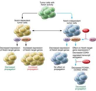
subclasses, according to an initial study based on gene expression profiling (23). It was immediately speculated that the proneural subgroup would be sensitive to GSI therapy, as this population of GBMs exhibits a higher level of Notch pathway activation than the other subgroups (23). Indeed, a recent study showed that GBM neurosphere cells with a strong proneural gene signature respond to GSI treatment (24). Although no stratified clinical trial to assess GSI for the treatment of proneural GBMs has been carried out, identification of the molecular mechanism of GSI resis-tance in GBM has the potential to help develop novel therapies for this deadly disease. In the current issue, Xie et al. elegantly demonstrate that a mediator of Notch signaling, RBPJ, is overexpressed in GSI-resistant BTICs preferentially in proneural GBMs and is required for BTIC propagation both in vitro and in vivo (25). The results of this study are of substantial clinical relevance, because a mechanism of GSI resistance in BTICs has been identi-fied, thereby providing a new approach to target GSI-resistant tumor cells in general.
The Notch signaling pathway
and RBPJ
Canonical Notch signaling is initiated when a Notch ligand, such as JAGGED (JAG1 or JAG2) or DELTA (DLL1, 3, or 4), binds to a NOTCH receptor (NOTCH1, 2, 3, or 4) on adjacent cells. In turn, ligand binding to the NOTCH receptor induces
γ-secretase–mediated proteolytic cleav-age of the NOTCH receptor at the trans-membrane domain to release NOTCH intracellular domain (NICD), which translocates into the nucleus, binds RBPJ protein complexes situated on DNA, recruits activators, and removes co-repressors to activate downstream target genes, such as HES and HEY family genes (26–28). In the absence of NICD, RBPJ associates with histone deacetylase–con-taining co-repressors, including SMRT, SHARP, CtBP, SKIP, and CIR, in closed
Related Article: p. 2757
Conflict of interest: The author has declared that no conflict of interest exists.
Reference information: J Clin Invest. 2016;126(7):2415–2418. doi:10.1172/JCI88619.
While most studies have shown that canonical Notch signaling activates tar-get genes through the DNA-binding RBPJ protein complex, Rbpj knockout mice do not have the same phenotype as Notch knockout mice (34). This discrepancy suggests that RBPJ can function in both Notch-dependent and -independent man-ners (refs. 35, 36, and Figure 1). In addi-tion, RBPJ generally represses target gene expression in the absence of NICD; there-fore, it is believed that knockout or knock-down of RBPJ could result in derepression of target genes (26, 36). Indeed, loss of RBPJ has been shown to induce expres-sion of several Notch target genes, either in the presence or absence of NICD, and increase tumorigenesis in breast cancer and Burkitt lymphoma cells (37). In con-trast, Xie et al. show that knockdown of RBPJ only derepresses a few Notch target genes, such as HES5, but induces apopto-sis in BTICs (25). One possible reason for these opposite effects from knockdown of RBPJ in tumor cells is that the breast can-cer cells and Burkitt lymphoma cells used were dependent on Notch signaling to grow (37), whereas the BTICs used by Xie et al. were already independent of Notch signaling for their growth (GSI-resistant cells) (Figure 1). Another possibility is that there could be different RBPJ binding landscapes across the genome and differ-ent Notch-dependdiffer-ent target genes in dif-ferent types of tumor cells.
RBPJ target genes
Xie et al. performed RNA-seq to examine changes in the transcriptional profile of BTICs in response to RBPJ knockdown or Notch signaling blockade with GSI (25). Only 10% to 15% of genes were commonly regulated by both RBPJ and Notch signal-ing, suggesting that RBPJ regulates BTIC propagation mostly through the regulation of Notch-independent genes, an observa-tion supported by the fact that propagaobserva-tion of GSI-treated BTICs is independent of Notch signaling. Xie et al. determined that tumorigenesis-associated genes FOXM1,
CCNA2 (cyclin A2), and KRAS are not
only exclusively regulated by RBPJ at the transcriptional level but also contain RBPJ binding sites at their promoter regions, sug-gesting that these genes are possible direct targets of RBPJ in BTICs (25). ChIP-PCR analysis confirmed that FOXM1, CCNA2, GSI resistance in GBMs has not been
care-fully investigated. Interestingly, Xie et al. found that GSI-resistant BTICs have ele-vated RBPJ expression compared to non-BTICs and that non-BTICs lose RBPJ expres-sion and stem cell markers upon their differentiation (ref. 25 and Figure 1). In addition, knockdown of RBPJ expression by shRNA decreased BTIC propagation in vitro and in vivo by inducing apoptosis (25). This reduction in BTIC propagation prolonged survival in mice bearing intra-cranial xenografts (25). Together, these results demonstrate that RBPJ is required for GSI-resistant BTIC propagation. chromatin to suppress gene transcription
(26–28). Binding of NICD to RBPJ dissoci-ates the co-repressor complex from RBPJ, and recruits mastermind-like proteins (MAMLs) and histone acetyltransfer-ases to the NICD-RBPJ complex, thereby remodeling chromatin to activate tran-scription of target genes (26–28). Active
NOTCH1 mutations have been found in
[image:3.585.38.367.53.365.2]about 60% of T cell acute lymphoblastic leukemias (T-ALLs) that respond to GSI treatment (29, 30). Subsequently, it has been shown that genetic and epigenetic alterations of tumor cells contribute to GSI resistance in T-ALL (31–33). However,
in these GSI-resistant tumor cells will decrease propagation by reducing expres-sion of genes that are regulated exclusively by RBPJ, but not Notch, including FOXM1,
CCNA2, and KRAS. In addition, the results
of Xie et al. demonstrated that these GSI-resistant tumor cells can be treated with the c-MYC inhibitor JQ1 or the CDK9 inhibitor dinaciclib (ref. 25 and Figure 1). As RBPJ is considered to be a housekeep-ing gene and is expressed in most cells in the body (39, 40), the potential toxicity of these approaches in normal cells will need to be closely watched during future studies aimed at evaluating the clinical application of RBPJ-targeting therapies.
Acknowledgments
This work was funded by grants from the NIH: R01CA148621 (to X. Fan) and R01CA163737 (to X. Fan).
Address correspondence to: Xing Fan, Associate Professor of Neurosurgery and Cell and Developmental Biology, Univer-sity of Michigan Medical School, Depart-ment of Neurosurgery, 109 Zina Pitcher Place, 5018 BSRB, Ann Arbor, Michigan 48109-2200, USA. Phone: 734.615.7266; E-mail: xingf@umich.edu.
1. Louis D, Ohgaki H, Wiestler O, Cavenee W.
WHO Classification of Tumours of the Central Nervous System. Lyon, France: IARC Press; 2007.
2. Wen PY, Kesari S. Malignant gliomas in adults.
N Engl J Med. 2008;359(5):492–507.
3. Furnari FB, et al. Malignant astrocytic glioma: genetics, biology, and paths to treatment. Genes
Dev. 2007;21(21):2683–2710.
4. Hemmati HD, et al. Cancerous stem cells can arise from pediatric brain tumors. Proc Natl Acad
Sci U S A. 2003;100(25):15178–15183.
5. Singh SK, et al. Identification of human brain tumour initiating cells. Nature. 2004;432(7015):396–401.
6. Kondo T, Setoguchi T, Taga T. Persistence of a small subpopulation of cancer stem-like cells in the C6 glioma cell line. Proc Natl Acad Sci U S A. 2004;101(3):781–786.
7. Galli R, et al. Isolation and characterization of tumorigenic, stem-like neural precur-sors from human glioblastoma. Cancer Res. 2004;64(19):7011–7021.
8. Bao S, et al. Glioma stem cells promote radioresis-tance by preferential activation of the DNA dam-age response. Nature. 2006;444(7120):756–760. 9. Liu G, et al. Analysis of gene expression and
chemoresistance of CD133+ cancer stem cells in
glioblastoma. Mol Cancer. 2006;5:67. 10. Piccirillo SG, et al. Bone morphogenetic
proteins inhibit the tumorigenic potential of human brain tumour-initiating cells. Nature.
located in the promoter region of RBPJ (25). Moreover, they confirmed that over-expression of c-MYC induces transcription of RBPJ, as detected by a luciferase report-er assay, and demonstrated direct c-MYC binding at the RBPJ promoter using ChIP-PCR. In addition, knockdown of MYC decreased RBPJ expression at the protein level. Taken together, these results dem-onstrate that c-MYC is one of the upstream regulators of RBPJ that directly regulates
RBPJ expression at the transcriptional
level. However, MYC has been shown to be a canonical Notch target in previous studies (41–43). Notch regulates c-MYC expression through RBPJ binding to both promoter and super-enhancer regions of
MYC (41–43). As the BTICs used by Xie
et al. have canonical Notch activity, albeit they were not dependent on Notch signal-ing to grow, it is unclear if canonical Notch signaling contributes to the expression of c-MYC in these cells. While GSI treatment indeed blocked NICD1 formation in BTICs (25), the effects of GSI on c-MYC expres-sion are not known. Nevertheless, Xie et al. found that blocking c-MYC expression with the selective bromodomain inhibitor JQ1 decreases BTIC propagation in vitro and in vivo, providing another potential therapeutic strategy for treating GSI-resis-tant GBMs (Figure 1).
Indication and future directions
Xie and colleagues have discovered that the MYC/RBPJ/CDK9 pathway is critical for BTIC self-renewal (25), results with clinical implications not only for GBM treatment, but also for cancer-targeting therapies in general, particularly for GSI-resistant tumors (Figure 1). Based on pre-vious reports and the current study by Xie et al., tumor cells with Notch activity can be roughly divided into Notch-dependent and Notch-independent classes (Figure 1). Notch-dependent tumor cells most likely will respond to GSI treatment; however, knockdown of RBPJ in Notch-dependent tumor cells will release repression of Notch target genes and may promote tumor growth. In contrast, for Notch-independent tumor cells, although GSI treatment can still block Notch signal-ing, this strategy will not inhibit growth of these cells. Therefore, Notch-indepen-dent tumor cells are considered to be GSI resistant. Knockdown of RBPJ expression and KRAS are indeed direct targets of RBPJ
and independent of Notch regulation in BTICs, suggesting that RBPJ regulates propagation of GSI-resistant BTICs at least in part through direct regulation of FOXM1,
CCNA2, and KRAS expression.
Furthermore, Xie et al. transduced HA-tagged RBPJ into BTICs and per-formed immunoprecipitation using an anti-HA antibody and carried out pro-teomic analysis of RBPJ binding proteins to identify RBPJ co-factors (25). CDK9 tightly bound to RPPJ and regulated tran-scription of RBPJ target genes, includ-ing FOXM1, CCNA2, and KRAS, through transcription elongation, a result that is consistent with previous studies show-ing that CDK9, unlike CDK8, is involved in RBPJ-regulated transcription indepen-dent of NICD (38). The study by Xie et al. indicates that CDK9 is only involved in transcription elongation; therefore, it remains unknown how RBPJ modulates the changes in chromatin conformation that are required to initiate transcription, as RBPJ generally binds to closed chroma-tin in its default state. Intereschroma-tingly, Xie et al. indeed found that the lysine-specific demethylase LSD1 is also associated with RBPJ and may be involved in transcription initiation (25). Further investigation of the mechanism of RBPJ-regulated, Notch-independent transcription initiation will be very interesting. Nevertheless, Xie et al. have now shown that CDK9 is also required for propagation of GBM BTICs through regulation of FOX1, CCNA2, and
KRAS, and that pharmacological inhibition
of CDK9 activity with the CDK9 inhibitor dinaciclib (or LY2857785) inhibits BTIC growth and prolongs survival in mice bear-ing intracranial GBM xenografts (ref. 25 and Figure 1). Together, these results of Xie and colleagues indicate that interfer-ing with RBPJ-regulated gene transcrip-tion with CDK9 inhibitors has potential as a clinically relevant therapeutic approach to treat GSI-resistant BTICs.
Regulation of RBPJ expression
RBPJ has been shown to function as a33. Knoechel B, et al. An epigenetic mechanism of resistance to targeted therapy in T cell acute lymphoblastic leukemia. Nat Genet. 2014;46(4):364–370.
34. Nakhai H, et al. Conditional ablation of Notch signaling in pancreatic development.
Develop-ment. 2008;135(16):2757–2765.
35. Fujimoto M, et al. RBP-J promotes neuronal differentiation and inhibits oligodendroglial development in adult neurogenesis. Dev Biol. 2009;332(2):339–350.
36. Tanigaki K, Honjo T. Two opposing roles of RBP-J in Notch signaling. Curr Top Dev Biol. 2010;92:231–252.
37. Kulic I, et al. Loss of the Notch effector RBPJ promotes tumorigenesis. J Exp Med. 2015;212(1):37–52.
38. Fryer CJ, White JB, Jones KA. Mastermind recruits CycC:CDK8 to phosphorylate the Notch ICD and coordinate activation with turnover.
Mol Cell. 2004;16(4):509–520.
39. Hamaguchi Y, et al. Biochemical and immuno-logical characterization of the DNA binding pro-tein (RBP-Jκ) to mouse J κ recombination signal sequence. J Biochem. 1992;112(3):314–320. 40. Kawaichi M, et al. Genomic organization of mouse
J κ recombination signal binding protein (RBP-Jκ) gene. J Biol Chem. 1992;267(6):4016–4022. 41. Weng AP, et al. c-Myc is an important direct
target of Notch1 in T-cell acute lympho-blastic leukemia/lymphoma. Genes Dev. 2006;20(15):2096–2109.
42. Herranz D, et al. A NOTCH1-driven MYC enhancer promotes T cell development, trans-formation and acute lymphoblastic leukemia.
Nat Med. 2014;20(10):1130–1137.
43. Yashiro-Ohtani Y, et al. Long-range enhancer activity determines Myc sensitivity to Notch inhibitors in T cell leukemia. Proc Natl Acad Sci
U S A. 2014;111(46):E4946–E4953.
glioma stem cells. Stem Cells. 2010;28(1):17–28. 22. Krop I, et al. Phase I pharmacologic and
phar-macodynamic study of the gamma secretase (Notch) inhibitor MK-0752 in adult patients with advanced solid tumors. J Clin Oncol. 2012;30(19):2307–2313.
23. Phillips HS, et al. Molecular subclasses of high-grade glioma predict prognosis, delineate a pat-tern of disease progression, and resemble stages in neurogenesis. Cancer Cell. 2006;9(3):157–173. 24. Saito N, et al. A high Notch pathway activation
predicts response to γ secretase inhibitors in pro-neural subtype of glioma tumor-initiating cells.
Stem Cells. 2014;32(1):301–312.
25. Xie Q, et al. RBPJ maintains brain tumor– initiating cells through CDK9-mediated transcriptional elongation. J Clin Invest. 2016;126(7):2757–2772.
26. Bray SJ. Notch signalling: a simple pathway becomes complex. Nat Rev Mol Cell Biol. 2006;7(9):678–689.
27. Kopan R, Ilagan MX. The canonical Notch sig-naling pathway: unfolding the activation mecha-nism. Cell. 2009;137(2):216–233.
28. Nam Y, Sliz P, Song L, Aster JC, Blacklow SC. Structural basis for cooperativity in recruitment of MAML coactivators to Notch transcription complexes. Cell. 2006;124(5):973–983. 29. Weng AP, et al. Activating mutations of NOTCH1
in human T cell acute lymphoblastic leukemia.
Science. 2004;306(5694):269–271.
30. Aster JC, Pear WS, Blacklow SC. Notch signaling in leukemia. Annu Rev Pathol. 2008;3:587–613. 31. Herranz D, et al. Metabolic reprogramming
induces resistance to anti-NOTCH1 therapies in T cell acute lymphoblastic leukemia. Nat Med. 2015;21(10):1182–1189.
32. Palomero T, et al. Mutational loss of PTEN induc-es rinduc-esistance to NOTCH1 inhibition in T-cell leukemia. Nat Med. 2007;13(10):1203–1210. 2006;444(7120):761–765.
11. Fan X, et al. Notch pathway inhibition depletes stem-like cells and blocks engraft-ment in embryonal brain tumors. Cancer Res. 2006;66(15):7445–7452.
12. Shen Q, et al. Endothelial cells stimulate self-renewal and expand neurogenesis of neural stem cells. Science. 2004;304(5675):1338–1340. 13. Yoon K, Gaiano N. Notch signaling in the
mamma-lian central nervous system: insights from mouse mutants. Nat Neurosci. 2005;8(6):709–715. 14. Louvi A, Artavanis-Tsakonas S. Notch signalling
in vertebrate neural development. Nat Rev
Neu-rosci. 2006;7(2):93–102.
15. Androutsellis-Theotokis A, et al. Notch signal-ling regulates stem cell numbers in vitro and in vivo. Nature. 2006;442(7104):823–826. 16. Ignatova TN, Kukekov VG, Laywell ED, Suslov
ON, Vrionis FD, Steindler DA. Human corti-cal glial tumors contain neural stem-like cells expressing astroglial and neuronal markers in vitro. Glia. 2002;39(3):193–206.
17. Purow BW, et al. Expression of Notch-1 and its ligands, Delta-like-1 and Jagged-1, is critical for glioma cell survival and proliferation. Cancer
Res. 2005;65(6):2353–2363.
18. Lee J, et al. Tumor stem cells derived from glioblastomas cultured in bFGF and EGF more closely mirror the phenotype and genotype of primary tumors than do serum-cultured cell lines. Cancer Cell. 2006;9(5):391–403. 19. Ranganathan P, Weaver KL, Capobianco AJ.
Notch signalling in solid tumours: a little bit of everything but not all the time. Nat Rev Cancer. 2011;11(5):338–351.
20. Fan X, et al. NOTCH pathway blockade depletes CD133-positive glioblastoma cells and inhibits growth of tumor neurospheres and xenografts.
Stem Cells. 2010;28(1):5–16.