function
Jeffrey B. Kopp
J Clin Invest. 2015;125(3):968-971. https://doi.org/10.1172/JCI80280.
Krüppel-like factors (KLFs) are zinc finger transcription factors that share homology in three C-terminal zinc finger domains. KLF family members are expressed in most if not all tissues and have diverse roles in organismal development and cell differentiation, function, and death. The glomerular podocyte is particularly sensitive to mitochondrial dysfunction, as seen in various genetic disorders manifesting as progressive glomerulosclerosis. In this issue of the JCI, Mallipattu and coworkers show that KLF6 expression is reduced in mouse and human glomerular disease. Podocyte-specific deletion of Klf6 expression in mice leads to mitochondrial dysfunction and apoptosis, followed by glomerulosclerosis. This is the first demonstration that defective transcriptional regulation of nuclear-encoded mitochondrial genes can result in experimental glomerular disease.
Commentary
Find the latest version:
Loss of Krüppel-like factor 6 cripples podocyte
mitochondrial function
Jeffrey B. Kopp
Kidney Diseases Branch, National Institute of Diabetes and Digestive and Kidney Diseases (NIDDK), NIH, Bethesda, Maryland, USA.
Krüppel and Krüppel-like
factors
The 1995 Nobel Prize in Physiology or Medicine was award to Edward B. Lewis, Christiane Nüsslein-Volhard, and Eric F. Wieschaus for their work on embryonic development of Drosophila melanogaster, particularly with regard to homeotic genes that regulate development in plants and animals. Perhaps best known among the homeotic genes are those that encode the homeobox (Hox and ParaHox) pro-teins, which regulate other gene families (1). In 1950, Gloor described a fly mutant that caused severe body malformation, which he named Krüppel, meaning “crip-ple” (2). Nüsslein-Volhard and Wieschaus showed that Krüppel mutations disrupt Drosophila body segmentation at very early stages of embryogenesis, leading to embryonic lethality (3).
Mammalian genomes contain many genes that encode orthologs of Drosophila Krüppel, termed Krüppel-like factors (KLFs). KLFs manifest amino acid homol-ogy to Krüppel in the three DNA-binding zinc fingers located at the C terminus
(Figure 1). The highly conserved zinc finger domains comprise three sets of Cys2/His2 amino acid residues located at the C terminus, which are also similar to three motifs in the transcription factor SP1 (Figure 1). The zinc finger motifs of members of the KLF and SP1 superfamily of transcriptional regulators bind GC-rich motifs in gene regulatory regions, and they act as generally weak activators and often as strong repressors of gene expression. KLFs bound to DNA attract partners. For group 2 KLFs such as KLF4, which are transcriptional activators, these may include CBP/p300 (4). For example, KLF4 binds a TGF-β transcriptional con-trol element located in the angiotensin II type 1 receptor (ATR1). In response to TGF-β signaling, SP1 dissociates from KLF4 and PPARγ binds KLF4, resulting in enhanced ATR1 transcription (5).
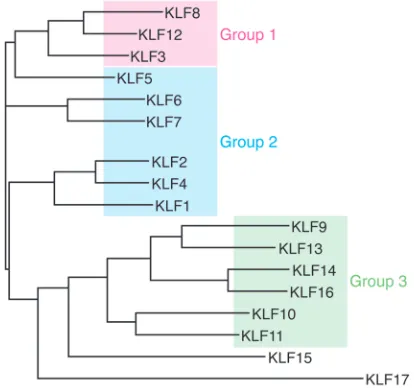
The human genome contains 17 KLF-encoding genes and can be classified into three phylogenetic groups that cor-relate with functional differences: group 1 KLFs are transcriptional repressors, group 2 KLFs are predominantly transcriptional
activators, and group 3 KLFs bind and potentiate the activity of the co-repres-sor SIN3A; KLF16 and -17 do not cluster with other group members (ref. 6 and Fig-ure 2). Expression of some KLFs is tissue restricted, such as KLF1 in hematopoietic cells, while others are widely expressed. This class of transcription factors has highly diverse roles that include regulat-ing cell growth, differentiation, and death; endothelial and epithelial function; fat and glucose metabolism; and mitochondrial function; among other effects (6). Shinya Yamanaka and colleagues have shown that KLF4 is one of a set of four transcription factors (the others being OCT4, SOX2, and MYC) that can reprogram adult cells to become induced pluripotent stem cells (7), a discovery that resulted in Yamanaka sharing the 2012 Nobel Prize in Physiology or Medicine with John Gurdon. As tran-scriptional factors, KLFs are present in the nucleus, but they are also present in the cytoplasm and have distinct roles there.
KLFs have diverse roles
in kidney physiology and
pathophysiology
Glomerular endothelial cells that are exposed in vitro to laminar shear stress express higher levels of KLF2, leading Slater et al. to suggest that this might contribute to the phenotype-stabilizing effects of shear stress on the vascular endothelium (8). Compared with WT mice, animals with a endothelial-specific deletion of Klf2 mani-fest a more severe streptozotocin-diabetic nephropathy, including more proteinuria, more glomerular hypertrophy, and evidence of more endothelial injury but interestingly also more severe podocyte injury (9). In a study of glomerular transcripts expressed in atypical hemolytic uremic syndrome, KLF4 expression was modestly elevated (10); on the other hand, KLF2 and KLF4 transcripts were reduced in glomerular transcripts from renal transplant–associated thrombotic microangiopathy (11), suggesting a complex picture for KLF-mediated gene expression.
Related Article: p. 1347
Conflict of interest: The author has declared that no conflict of interest exists. Reference information: J Clin Invest. 2015;125(3):968–971. doi:10.1172/JCI80280.
genetic disorders that alter mitochon-dria can affect all tissues. With regard to kidney phenotypes, mitochondrial dis-orders may manifest as proximal tubu-lar dysfunction with loss of electrolytes, glucose, amino acids, and low-molec-ular-weight proteins; tubulointerstitial disease and cystic kidney disease; and podocytopathy with subnephrotic or nephrotic proteinuria.
Further insight into the role of
KLF6 in podocyte biology
In this issue, Mallipattu and coworkers present three lines of evidence that sup-port a role for KLF6 in maintaining normal podocyte function and specifically link this to the ability of KLF6 to support podocyte mitochondrial function (22).
First, Mallipattu et al. surveyed renal expression of 9 KLFs in HIV-infected human podocytes and in HIV-transgenic mice (23) that manifest with HIV-associ-ated nephropathy (collapsing glomerulop-athy). It is currently thought that podocyte injury and loss lead to podocyte replace-ment from several sources, particularly parietal epithelial stem cells and cells of the renin lineage. A survey of the mRNA expression patterns of KLF family mem-bers revealed that only KLF6 expression was altered, with it being reduced in both HIV-infected podocytes and in HIV trans-genic mouse glomeruli.
Second, mice with podocyte-specific deletion of Klf6 manifested albuminuria but not glomerulosclerosis, as least within the observation period (22). Doxorubicin induces podocyte injury and results in glomerulosclerosis in susceptible mouse strains. Doxorubicin is also used in cancer chemotherapy, injuring or killing sensitive cells. While doxorubicin intercalates into DNA, the cytotoxic effects of this agent may be mediated by poisoning topoi-somerase II, which corrects DNA super-coils that otherwise would result in tangled DNA, thereby releasing torsional stress; therefore, the expected result of decreased topoisomerase function would be com-promised transcription (23). Doxorubic-in-sensitive mice that manifest podocyte injury have a mutation in the DNA repair gene Prkdc (24). Further, doxorubicin decreases intracellular ATP (by mecha-nisms that remain unclear), resulting in a compensatory increase in mitochondrial knockdown of KLF2 increased cell
prolif-eration (17). In cultured human podocytes, retinoids were shown to increase KLF15 expression, and KLF15 drove podocyte differentiation in vitro, binding to the pro-moter regions of nephrin and podocin as shown by ChIP assay (18). Further, Klf15-knockout mice manifest more podocyte injury in response to the podocyte toxins LPS and doxorubicin, suggesting a pheno-type-stabilizing role for KLF15.
KLFs and mitochondria
KLFs have previously been associated with regulation of mitochondria number and function. Mitochondria are heterogeneous and dynamic organelles, with properties that may differ within a cell and between cells. For example, mitochondria differ in membrane potential and metabolic activity and in their tendency to be located in isola-tion or in aggregates with other mitochon-dria. In cultured pulmonary artery smooth muscle cells, KLF5 drives survivin expres-sion, generates mitochondrial hyperpolar-ization, and protects against apoptotic cell death (19). In cultured neurites, KLF4 and KLF6 reduce mitochondrial complex size (isolated versus aggregated), and KLF6, but not KLF4, reduces mitochondrial den-sity. As KLF6 is associated with increased neurite growth, it has been suggested that smaller, less dense mitochondria may par-ticipate in neurite growth (20). Finally, in Caenorhabditis elegans, Klf3 is essential for the expression of enzymes involved in beta-oxidation of fatty acids in mitochon-dria and peroxisomes (21).
As mitochondria are present in all nucleated cells, it is not surprising that In contrast to the predominantly
endo-thelial expression pattern of KLF2 and KLF4, KLF5 is expressed in collecting duct epithelial cells and plays a role in inflam-mation and fibrosis. Mice haploinsufficient for Klf5 exhibit less renal injury following unilateral ureteral ligation and an apparent shift from M1 macrophages to M2 macro-phages, which would be compatible with a less fibrotic outcome (12).
During metanephric development, Klf6 is expressed in the Wolffian duct and its derivatives and in the ureteric bud and its branches, including the distal tubule, with limited glomerular staining seen in what was interpreted as mesangial cells (13). In diabetic Ren-2 rats on the other hand, Klf6 expres-sion is increased in proximal tubular cells, and overexpression of KLF6 in cultured proximal tubular cells increased TGF-β1 expression and promoted a phenotype suggestive of epithelial-mesenchymal transition (14). KLF12 is also expressed in the distal tubule, specifically in the inner medullary collecting tubule, and in vitro it was shown to drive expression of the urea transporter A-1 but not the epithelial sodium channel (ENaC) (15).
[image:3.585.46.374.60.160.2]KLF15 is decreased in the renal inter-stitium of 5/6 nephrectomized rats and has been suggested to play an antifibrotic role, as in vitro studies demonstrate that KLF15 represses TGF-β1 signaling via ERK/ MAPK and JNK/MAPK pathways (16). Two studies have addressed the role of KLF15 in glomerular cells. In the rat Thy1 nephritis model, glomerular KLF15 expression fell during the period of mesangial prolifera-tion, and in cultured rat mesangial cells,
Figure 1. The KLF family of transcription factors. (A) KLF family members all contain three highly
conserved zinc finger domains within their C terminus that bind to GC-rich regions in DNA. These three zinc finger domains insert into the major groove of the DNA helix. (B) KLF transcription factors
respiratory factor 1 (Nrf1) and mitochon-drial inner membrane protein (Mpv17); however, it remains to be determined whether these genes are directly or indi-rectly affected by KLF6 deficiency.
Mitochondria and podocyte
function
When one looks at a kidney biopsy under the electron microscope, it is apparent that the renal cells endowed with the most mitochondria are proximal tubular cells, which depend heavily on mitochon-drial oxidative phosphorylation to support sodium transport. Nevertheless, it is strik-ing that genetic disorders that affect mito-chondrial function often include podocyte dysfunction as part of a spectrum of cel-lular pathologies (Table 1). Mitochondrial function can be disrupted by mutations in nuclear-encoded genes and mitochondrial DNA–encoded genes, as has been the sub-ject of a recent comprehensive review (26). Within the kidney, mutations that affect mitochondrial-expressed genes tend to manifest primarily as tubular epithelial cell dysfunction or podocyte dysfunction.
The apparent high-energy require-ment of the podocyte is not well under-stood but has been remarked upon by several groups (27–29). Possible reasons include foot process motility, which is enabled by continuous remodeling of the actin cytoskeleton; uptake of filtered proteins; or other unknown mechanisms. Another link between energy shortage and podocyte injury is the observation that defective and damaged mitochon-dria are cleared by autophagy, a process linked to activation of inflammasomes and apoptosis (30). Therapeutic strate-gies to address podocyte mitochondrial dysfunction hold considerable promise. depletion of KLF6 was also associated
with increased levels of cleaved caspase-3 and -9, release of mitochondrial cyto-chrome c, and increased apoptosis.
Promoter analysis suggested that the human cytochrome c oxidase assembly (SCO2) had KLF6-binding sites, which was confirmed by ChIP analysis. Furthermore, SCO2 expression was reduced in podo-cytes from KLF6-deficient mice. In human podocytes, knockdown of SCO2 expres-sion increased cleaved caspase-9 levels and release of cytochrome c, adding some support to the proposal by Mallipattu and colleagues that SCO2 may be a key KLF6 target for mediating protection against mitochondrial injury (22). The expression of a number of other mitochondrial genes was also reduced in podocyte-specific Klf6-knockout mice, including nuclear oxidation, and this may impose
oxida-tive stress (25). This suggests that KLF6 contributes to progression of glomerular injury, at least in this model.
[image:4.585.99.306.60.254.2]Third, Mallipattu and colleagues developed further in vivo and in vitro evidence that suggests KLF6 is essential for normal mitochondrial function, par-ticularly in the setting of mitochondrial stress (22). Mice lacking Klf6 manifested dysmorphic mitochondria following dox-orubicin exposure, and KLF6 expression increased in WT mice and in cultured mouse podocytes exposed to doxoru-bicin, suggesting a role in protection against mitochondrial stress. In cultured human podocytes, KLF6 knockdown was associated with decreased mitochon-drial membrane potential, oxygen con-sumption rate, and ATP levels. Moreover,
Figure 2. KLF phyologenetic tree. Analysis was conducted with full-length protein sequences
using ClustalW to generate phylogenetic relationships between KLFs. Group 1 and 3 KLFs are tran-scriptional repressors, while group 2 KLFs are generally trantran-scriptional activators. Figure adapted with permission from the American Physiological Society (6).
Table 1. Overview of genetic mutations associated with podocyte dysfunction
Manifestation Nuclear genes Mitochondrial DNA-encoded genes
Tubular dysfunction is prominent, typically spares the podocyte OX-PHOS defects: mutations in the genes affecting complex III and IV are more common than complex I
Fatty acid metabolism defects: CPT1
mtDNA: often large deletions
Podocyte dysfunction is typically present Human: COQ10 synthesis — COQ2, COQ6,
COQ9, PDSS2, ADCK4
Mouse model: Mpv17, Klf6
MELAS: tRNA-Leu
proliferation of mesangial cells. Cell Physiol
Bio-chem. 2012;29(5):893–904.
18. Mallipattu SK, et al. Kruppel-like factor 15 (KLF15) is a key regulator of podocyte differenti-ation. J Biol Chem. 2012;287(23):19122–19135. 19. Courboulin A, et al. Kruppel-like factor 5
con-tributes to pulmonary artery smooth muscle pro-liferation and resistance to apoptosis in human pulmonary arterial hypertension. Respir Res. 2011;12:128.
20. Steketee MB, et al. Mitochondrial dynamics regulate growth cone motility, guidance, and neurite growth rate in perinatal retinal gan-glion cells in vitro. Invest Ophthalmol Vis Sci. 2012;53(11):7402–7411.
21. Zhang J, et al. Regulation of fat storage and reproduction by Kruppel-like transcription fac-tor KLF3 and fat-associated genes in Caenorhab-ditis elegans. J Mol Biol. 2011;411(3):537–553. 22. Mallipattu SK, et al. Krüppel-like factor 6
regu-lates mitochondrial function in the kidney. J Clin
Invest. 2015;125(3):1347–1361.
23. Yang F, Teves SS, Kemp CJ, Henikoff S. Doxoru-bicin, DNA torsion, and chromatin dynamics.
Biochim Biophys Acta. 2014;1845(1):84–89.
24. Papeta N, et al. Prkdc participates in mito-chondrial genome maintenance and prevents Adriamycin-induced nephropathy in mice. J Clin
Invest. 2010;120(11):4055–4064.
25. Strigun A, Wahrheit J, Niklas J, Heinzle E, Noor F. Doxorubicin increases oxidative metabolism in HL-1 cardiomyocytes as shown by 13C metabolic flux analysis. Toxicol Sci. 2012;125(2):595–606. 26. O’Toole JF. Renal manifestations of genetic
mitochondrial disease. Int J Nephrol Renovasc
Dis. 2014;7:57–67.
27. Imasawa T, Rossignol R. Podocyte energy metabolism and glomerular diseases. Int J
Bio-chem Cell Biol. 2013;45(9):2109–2118.
28. Muller-Deile J, Schiffer M. The podocyte power-plant disaster and its contribution to glomerulopathy. Front Endocrinol (Lausanne). 2014;5:209.
29. Yan K, et al. The struggle for energy in podo-cytes leads to nephrotic syndrome. Cell Cycle. 2012;11(8):1504–1511.
30. Fougeray S, Pallet N. Mechanisms and biological functions of autophagy in diseased and ageing kidneys. Nat Rev Nephrol. 2015;11(1):34–45. 31. Ashraf S, et al. ADCK4 mutations promote
steroid-resistant nephrotic syndrome through CoQ10 biosynthesis disruption. J Clin Invest. 2013;123(12):5179–5189.
32. Zhu C, et al. Mitochondrial dysfunction medi-ates aldosterone-induced podocyte damage: a therapeutic target of PPARγ. Am J Pathol. 2011;178(5):2020–2031.
33. Wang Z, et al. Pharmacological targeting of GSK3β confers protection against podocytopa-thy and proteinuria by desensitizing mitochon-drial permeability transition. Br J Pharmacol. 2015;172(3):895–909.
segmentation. Dev Biol. 1984;104(1):172–186. 4. Mas C, et. al. Structural and functional
charac-terization of an atypical activation domain in erythroid Krüppel-like factor (EKLF). Proc Natl
Acad Sci U S A. 2011;108(26):10484–10489.
5. Zhang XH, Zheng B, Gu C, Fu JR, Wen JK. TGF-β1 downregulates AT1 receptor expression via PKC-δ-mediated Sp1 dissociation from KLF4 and Smad-mediated PPAR-γ associa-tion with KLF4. Arterioscler Thromb Vasc Biol. 2012;32(4):1015–1023.
6. McConnell BB, Yang VW. Mammalian Krüppel-like factors in health and diseases. Physiol Rev. 2010;90(4):1337–1381.
7. Takahashi K, Yamanaka S. Induction of pluripo-tent stem cells from mouse embryonic and adult fibroblast cultures by defined factors. Cell. 2006;126(4):663–676.
8. Slater SC, et al. Chronic exposure to laminar shear stress induces Kruppel-like factor 2 in glo-merular endothelial cells and modulates interac-tions with co-cultured podocytes. Int J Biochem
Cell Biol. 2012;44(9):1482–1490.
9. Zhong F, et al. Reduced Kruppel-like factor 2 expression may aggravate the endothelial injury of diabetic nephropathy [published online ahead of print September 3, 2014]. Kidney Int. doi:10.1038/ki.2014.286.
10. Modde F, et al. Comprehensive analysis of glomerular mRNA expression of pro- and antithrombotic genes in atypical haemolyt-ic-uremic syndrome (aHUS). Virchows Arch. 2013;462(4):455–464.
11. Agustian PA, et al. Glomerular mRNA expression of prothrombotic and antithrombotic factors in renal transplants with thrombotic microangio-pathy. Transplantation. 2013;95(10): 1242–1248. 12. Fujiu K, Manabe I, Nagai R. Renal collecting
duct epithelial cells regulate inflammation in tubulointerstitial damage in mice. J Clin Invest. 2011;121(9):3425–3441.
13. Fischer EA, Verpont MC, Garrett-Sinha LA, Ronco PM, Rossert JA. Klf6 is a zinc finger protein expressed in a cell-specific manner during kidney development. J Am Soc Nephrol. 2001;12(4):726–735.
14. Holian J, et al. Role of Kruppel-like factor 6 in transforming growth factor-β1-induced epithelial-mesenchymal transition of prox-imal tubule cells. Am J Physiol Renal Physiol. 2008;295(5):F1388–F1396.
15. Suda S, Rai T, Sohara E, Sasaki S, Uchida S. Postnatal expression of KLF12 in the inner medullary collecting ducts of kidney and its trans-activation of UT-A1 urea transporter promoter. Biochem Biophys Res Commun. 2006;344(1):246–252.
16. Gao X, Wu G, Gu X, Fu L, Mei C. Kruppel-like factor 15 modulates renal interstitial fibrosis by ERK/MAPK and JNK/MAPK pathways regula-tion. Kidney Blood Press Res. 2013;37(6):631–640. 17. Hong Q, et al. Kruppel-like factor-15 inhibits the
For example, therapy with the electron transport chain component coenzyme Q10 (COQ10) can reduce progressive renal damage in individuals with muta-tions in genes that affect the COQ10 syn-thetic pathway, providing a strong ratio-nale for genetic testing of patients with nephrotic syndrome, particularly chil-dren, to identify individuals who could benefit from this therapy (31). Other new therapeutic approaches are also being explored. For example, PPARα protects podocytes from aldosterone-induced mitochondrial dysfunction (32). Glyco-gen synthetase kinase (GSK) 3β integrates signals that regulate mitochondrial pore transition, and a thiazolidinedione com-pound that inhibits GSK2β was shown to alleviate doxorubicin-induced podocyte injury in vitro and in vivo (33).
The study by Mallipattu and col-leagues gives one more example of why this is an exciting time in glomerular pathophysiology, as existing concepts are being remodeled by new findings obtained from human genetic and pathology stud-ies, animal and cell culture models, and systems biology approaches applied to large patient cohorts.
Acknowledgments
[image:5.585.222.516.60.683.2]This work was supported by the Intra-mural Research Program of the NIDDK, NIH. The author wishes to acknowledge the generosity of Beth McConnell and colleagues in allowing the adaptation of Figure 2, and the help of Jurgen Heymann with manuscript review.
Address correspondence to: Jeffrey Kopp, 10 Center Drive, 3N116, NIH, Bethesda, Maryland 20192-1268, USA. Phone: 301.594.3403; E-mail: [email protected].
1. Gehring WJ, Kloter U, Suga H. Evolution of the Hox gene complex from an evolutionary ground state. Curr Top Dev Biol. 2009;88:35–61. 2. Gloor H. Schädigungsmuster eines Letalfaktors
(Kr) von Drosophila melanogaster. Arch Jul Klaus
Stiftung. 1950;25:38–44.