Copyright © 2003, American Society for Microbiology. All Rights Reserved.
Optimization and Validation of Multilocus Sequence Typing for
Candida albicans
Arianna Tavanti,
1,2Neil A. R. Gow,
1Sonia Senesi,
2Martin C. J. Maiden,
3and Frank C. Odds
1*
Department of Molecular & Cell Biology, Institute of Medical Sciences, University of Aberdeen, Aberdeen AB25 2ZD,1and
Peter Medawar Building for Pathogen Research and Department of Zoology, University of Oxford, Oxford OX1 3SY,3
United Kingdom, and Dipartimento di Patologia Sperimentale, Biotecnologie Mediche, Infettivologia ed Epidemiologia, Universita` degli Studi di Pisa, Pisa 56127, Italy2
Received 20 March 2003/Returned for modification 5 May 2003/Accepted 7 May 2003
Multilocus sequence typing (MLST) was applied to 75 Candida albicans isolates, including 2 that were expected to be identical, 48 that came from diverse geographical and clinical sources, and 15 that were sequential isolates from two patients. DNA fragments (⬇500 bp) of eight genes encoding housekeeping functions were sequenced, including four that have been described before forC. albicansMLST, and four new gene fragments,AAT1a,AAT1b,MPI, andZWF1. In total, 87 polymorphic sites were found among 50 notionally different isolates, giving 46 unique sequence types, underlining the power of MLST to differentiate isolates for epidemiological studies. Additional typing information was obtained by detecting variations in size at the transcribed spacer region of the 25S rRNA gene and tests for homozygosity at the mating type-like (MTL) locus. The stability of MLST was confirmed in two sets of consecutive isolates from two patients. In each set the isolates were identical or varied by a single nucleotide. Reference strain SC5314 and a derived mutant, CAF2, gave identical MLST types. Heterozygous polymorphisms were found in at least one isolate for all but 16 (18.4%) of the variable nucleotides, and 35 (41%) of the 87 individual sequence changes generated nonsynonymous amino acids. Cloning and restriction digestion of a gene fragment containing heterozygous polymorphisms indicated that the heterozygosity was genuine and not the result of sequencing errors. Our data validate and extend previous MLST results for C. albicans, and we propose an optimized system based on sequencing eight gene fragments for routine MLST with this species.
The advent of high-throughput nucleotide sequencing tech-nology has provided numerous opportunities for routine and unambiguous microbial isolate characterization. In addition to providing accurate and portable information, techniques such as multilocus sequence typing (MLST) (19) permit epidemio-logical isolate characterization to be integrated with popula-tion and evolupopula-tionary studies. MLST is a generic technique that has been exploited for several bacterial species (5, 7, 8, 23), and a number of web-accessible databases are available that enable the rapid dissemination and comparison of isolate characterization data (http://www.mlst.net).
The particular advantages of MLST as a typing method are that DNA nucleotide sequences can be determined by auto-mated technology with minimal subjective interpretation of data such as exists in all methods dependent on phenotypic characteristics, fermentation profiles, and other qualitative comparators. In addition, MLST data from different sources can be archived and distributed electronically and interrogated and added to from distant locations to facilitate comparisons for global epidemiology and population studies. A web site (http://www.mlst.net/new/index.htm) has already been created for data archiving and analysis with six pathogenic bacteria.
Recently, Bougnoux et al. described an MLST system for the
opportunistic fungal pathogenCandida albicans(2). Many
ap-proaches to strain typing have been developed for this species (32), but no system has yet achieved universal acceptance.
Those based onC. albicansnucleotide sequences show greater
or lesser diversity of types depending on the extent of conser-vation of the target sequences. Typing based on the intergenic transcribed spacer sequences of genes encoding rRNA tend to differentiate isolates into a very small number of major sub-classes (22, 33), and similar conserved sequences have also been used for differentiation at the species level (4). By con-trast, DNA fingerprints revealed with oligonucleotide probes
for widely dispersed repeat sequences in the C. albicans
ge-nome show a great diversity of strain types (28, 29) and are even capable of revealing minor genomic adaptations to host microenvironments, a process known as microevolution (25, 31). MLST, based on allelic variation in the nonconserved portion of unrelated genes, aims to provide characterization that is sufficiently conservative to be robust and reproducible but provides levels of discrimination appropriate for the pur-poses both of investigation of clinically relevant problems, such as epidemic outbreaks of infection and resistance to antifungal agents, and of population analyses.
Most MLST schemes described to date have been for hap-loid microorganisms. The permanently diphap-loid chromosome
complement inC. albicansallows extra differentiation of
iso-lates in this species because MLST data for some isoiso-lates may show two bases at the same variable site, indicative of the presence of two diploid alleles in these diploid organisms (2).
To optimize and validate MLST for typingC. albicans, we
analyzed results with 75 C. albicansisolates for sequences of
* Corresponding author. Mailing address: Department of Molecular & Cell Biology, Institute of Medical Sciences, University of Aberdeen, Aberdeen AB25 2ZD, United Kingdom. Phone and fax: (44) 1224 273128. E-mail: [email protected].
3765
on May 15, 2020 by guest
http://jcm.asm.org/
the most discriminatory genes described by Bougnoux and colleagues (2) and of four other sequences chosen on the basis that the encoded enzymes were shown previously to be poly-morphic in multilocus enzyme electrophoresis analyses (26).
Our results allow us to propose an improved gene set forC.
albicansMLST, and we demonstrated that MLST data forC. albicans showing two bases at a single site indeed represent allelic heterozygosity and not sequencing errors. Finally, we included two non-MLST, DNA-based typing characters in our typing scheme. The first is the subdivision of isolate types as described by McCullough et al., based on sequence variation at the transcribed spacer locus of the 25S rRNA gene (22), which divides the species into three major subtypes of epidemiolog-ical significance (21). The second is the determination of ho-mozygosity or heterozygosity at the mating type-like (MTL) locus, originally described by Hull and Johnson (13), which is emerging as being associated with important properties such as antifungal resistance (27) and rapid phenotypic switching (17). We propose a unified scheme for gene sequence-based strain
typing of C. albicans that is portable, reproducible, and
dis-criminatory.
MATERIALS AND METHODS
Isolates.The 75C. albicansisolates were from our collection of pathogenic fungi. Sixteen were sequential oral isolates from a human immunodeficiency virus-positive patient and have been described previously (35). Eleven were sequential isolates from different anatomical sites of a patient undergoing che-motherapy for hematological malignancy (24). Each of these two sets of isolates was assumed to be epidemiologically related. Two of the isolates were SC5314, a strain widely used for molecular genetic studies, and CAF2, derived from
SC5314 by specific disruption of one copy of theURA3gene. This pair of strains
was included as a control to check for complete identity of sequences in strains that should not differ by MLST. Strain T26 was derived from SC5314 by a complex lineage that included a spontaneous mutation step (6) and was expected to be either identical or very similar to SC5314.
The remaining 45 isolates were chosen to represent different degrees of ge-netic and phenotypic diversity based on their date, anatomical site, and geo-graphical source of isolation. The set included 13 vaginal isolates originating from the United States in 1998, intended to represent isolates with a constant anatomical location. The set of 45 isolates plus SC5314, CAF2, T26, and one representative each from the two sequential series constituted our main set of 50 notionally diverse strain types. The yeasts were maintained on Sabouraud agar (Oxoid, Basingstoke, United Kingdom).
Choice of loci for MLST.Initially, 10 gene fragments were chosen: five that gave the greatest MLST discrimination in the hands of Bougnoux et al. (2) and
five that corresponded to a subset of the 13C. albicanshousekeeping genes that
were previously shown to be polymorphic in multilocus enzyme electrophoresis experiments (26). We reduced the set to a total of eight gene fragments that gave
good discrimination in pilot experiments with 20 isolates ofC. albicans(Table 1).
Use of these two sets of four gene fragments allowed us to make direct com-parisons of the results obtained from our full panel of isolates with those for the same four gene fragments already published (2). Primers were designed to amplify gene fragments of 450 to 750 bp and are also detailed in Table 1. The
primers 5⬘-ACTCAAGCTAGATTTTTGGC-3⬘(forward) and 5⬘-CAGCAACA
TGATTAGCCC-3⬘(reverse), which are specific for theAAT1aregion of the
AAT1gene upstream of the conserved region, were used for experiments to
investigate heterozygosity in MLST.
DNA extraction.Genomic DNA was extracted from yeasts grown in YPD broth, containing 2% glucose, 2% mycological peptone (Oxoid), and 1% yeast extract (Difco, Detroit, Mich.). Briefly, cells were harvested in stationary phase and lysed by vortexing the pellet for 3 min with 0.3 g of glass beads (0.45 to 0.52
mm in diameter; Sigma, St. Louis, Mo.) in 200l of buffer (100 mM Tris-HCl
[pH 8.0] containing 2% Triton X-100, 1% sodium dodecyl sulfate, and 1 mM
EDTA) and 200l of 1:1 (vol/vol) phenol-chloroform solution. After vortexing,
200l of TE (1 mM EDTA, 10 mM Tris-HCl, pH 8.0) was added to the lysate;
the mixture was microcentrifuged at full speed for 10 min, and the aqueous phase was transferred to a new tube. DNA was precipitated by addition of 1 ml of
TABLE 1. List of gene fragments used for C. albicans MLST, with details of primers Fragment Gene product Primer Amplicon size (bp) 5 ⬘ sequence start 3 ⬘ sequence start Sequenced fragment (bp) No. of variable sites Nucleo- tides Amino acids AAT1a Asparate aminotransferase Fwd 5 ⬘ -ACTCAAGCTAGATTTTTGGC-3 ⬘ 478 ATTGAA CGTTTC 373 7 1 Rev 5 ⬘ -CAGCAACATGATTAGCCC-3 ⬘ AAT1b Aspartate aminotransferase Fwd 5 ⬘ -ATGGCTTATCAAGGTTTTGC-3 ⬘ 491 TTGACTAA CTGGGACCA 349 6 1 Rev 5 ⬘ -GTGGCATAAACTGAATATTCG-3 ⬘ MPI Mannose phosphate isomerase Fwd 5 ⬘ -ACCAGAAATGGCCATTGC-3 ⬘ 486 TTTAAACC GGGAAGCA 375 11 5 Rev 5 ⬘ -GCAGCCATGCATTCAATTAT-3 ⬘ ZWF1 Glucose-6-phosphate dehydrogenase Fwd 5 ⬘ -GTTTCATTTGATCCTGAAGC-3 ⬘ 702 AAAACCAGG TTGAATTAC 491 8 2 Rev 5 ⬘ -GCCATTGATAAGTACCTGGAT-3 ⬘ CaVPS13 Vacuolar protein sorting protein Fwd 5 ⬘ -TCGTTGAGAGATAATCGACTT-3 ⬘ 741 CCTTGATATG CAAATCATGG 403 16 8 Rev 5 ⬘ -ACGGATGGATCTCCAGTCC-3 ⬘ CaADP1 ATP-dependent permease Fwd 5 ⬘ -GAGCCAAGTATGAATGATTTG-3 ⬘ 537 CACGTTGCAA GGAAATCCAA 443 15 8 Rev 5 ⬘ -TTGATCAACAAACCCGATAAT-3 ⬘ CaSYA1 Alanyl-RNA synthetase Fwd 5 ⬘ -AGAAGAATTGTTGCTGTTACTG-3 ⬘ 543 TAAATCTAAA AGCTGTATCA 391 13 4 Rev 5 ⬘ -GTTACCTTTACCACCAGCTTT-3 ⬘ CaRPN2 26S proteasome regulatory subunit Fwd 5 ⬘ -TTCATGCATGCTGGTACTAC-3 ⬘ 447 TTGGTCTAAA GTCTTTACGA 306 11 2 Rev 5 ⬘ -TAATCCCATACCCAAAGCAG-3 ⬘
on May 15, 2020 by guest
http://jcm.asm.org/
ethanol to the supernatant. Samples were centrifuged, and the pellet was
resus-pended in 400l of TE containing 10l of 10-mg/ml RNase (Sigma, St. Louis,
Mo.). The mixture was incubated for 1 h at 37°C, and then DNA was precipitated
with 2 volumes of isopropanol and 10l of 4 M ammonium acetate, dried, and
redissolved in 50l of TE, pH 8.0.
Amplification and nucleotide sequence determination.PCR assays were used
to amplify the gene fragments. Reaction volumes of 50l contained 100 ng of
genomic DNA, 2.5 U ofPfuDNA polymerase (Promega, Madison, Wis.), 5l of
10⫻buffer (supplied with the enzyme), 200M deoxynucleoside triphosphate
mix (Promega) and 10M each of the forward and reverse primers. A Flexigene
thermocycler (Techne, Cambridge, United Kingdom) was set up with a first cycle of denaturation for 2 min at 94°C, followed by 25 cycles of denaturation at 94°C for 1 min, annealing at 52°C for 1 min, elongation at 72°C for 1 min, and a final extension step of 10 min at 72°C. The amplified products were purified with a commercial PCR purification system (Wizard PCR preps DNA purification system; Promega, Southampton, United Kingdom). Both strands of purified gene fragments were sequenced on an ABI (Foster City, Iowa) 3700 DNA analyzer
with a 2.5M concentration of the same primers that were used in the PCR step.
The sequence data were coupled with DNAStar software. Heterozygosities were defined by the presence of two coincident, equivalently sized peaks in the for-ward and reverse sequence chromatograms. The one-letter code for nucleotides from the International Union of Pure and Applied Chemistry nomenclature was used to define the results.
Statistical analysis of MLST data.To determine similarities between MLST strain types, the nucleotides at all 87 polymorphic loci found for the eight gene fragments and 50 notionally diverse isolates were scored for each pair of isolates as 0 for identical nucleotides, 0.5 for heterozygous or homozygous pairs that shared one nucleotide, and 0.0 for identical nucleotides. A similarity matrix was generated by adding the 87 scores for each pair of isolates and dividing the result by 87. Each pair of isolates was therefore assessed by a similarity index between 0 (complete nonidentity) and 1.0 (complete identity). To represent the matrix in two dimensions, a single-linkage dendrogram was constructed by the unweighted pair group method with arithmetic mean with the aid of Mega software (http: //www.megasoftware.net/). The same software was used to construct a neighbor-joining tree.
To determine closely related genotypes within highly similar clusters, the data were subjected to a Burst analysis (http://www.mlst.net/Burst/burst.htm) that was devised originally for analysis of bacterial MLST data (9, 20). The software determines clonal complexes from the data, suggesting a consensus “ancestral” type which contains the most-represented identical loci for a subgroup of isolates and indicates variants differing at just one, two, or three loci. The results are displayed as concentric circles, with the consensus strain type in the center and each new circle indicating isolates that differ from the consensus sequence by one nucleotide for each circle. The default design of the software analyzes seven gene fragments for five groups. We set our data input to accommodate eight gene fragments and scrutinized the results with group settings of three, four, five, and six. The five-group and six-group settings gave identical and interpretable results.
The discriminatory power (D) of the MLST system was determined by the
formula of Hunter (14).
Investigation of heterozygous loci.The accuracy of determination of
heterozy-gosity was ascertained with experiments done with theAAT1afragment (478 bp)
fromC. albicans76/002, which showed four putatively heterozygous sites. It was
cloned in the pGEM-T Easy vector system (Promega). BecausePfupolymerase
generates blunt ends, the PCR product was A-tailed by incubating 5l of the
purified gene fragment at 70°C for 30 min in the presence of 1l of 10⫻buffer,
1l of 25 mM MgCl2, 1l of 10 mM dATP, and 5 U ofTaqpolymerase. The
DNA ligation reaction mix comprised a 10-l volume containing 1l of the
A-tailed PCR product, 1l of 50-ng/l pGEM-T vector, 5l of 2⫻rapid ligation
buffer (supplied with the enzyme), and 3 U of T4 DNA ligase (Promega), and the
mixture was incubated at 4°C overnight. Then 5l of ligation mix was used to
transform Escherichia coliXL-1-Blue competent cells, following the method
described by Hanahan (12). Transformed cells were subsequently plated on
Luria-Bertani plates with ampicillin, 5-bromo-4-chloro-3-indolyl--D
-galactopy-ranoside, and isopropylthiogalactopyranoside (IPTG) (12).
Plasmid DNA was extracted from six colonies ofE. colithat grew on this
medium with the Qiaprep Spin Miniprep kit (Qiagen, West Sussex, United Kingdom), following the manufacturer’s protocol. The presence of the expected gene fragments was separately checked by digesting the plasmid DNA obtained
from the six selected clones withEcoRI (New England Biolabs, Beverly, Mass.)
and by PCR amplifying theAAT1fragment from plasmid DNA. Plasmid DNA
obtained from the six clones was then sequenced as previously described, with a
5M concentration of the same primers that were used in the PCRs.
Second, we usedMspI to digest theAAT1afragment fromC. albicans76/002.
Sequence analyses had shown that the heterozygosities in this PCR product
should result in the creation of anMspI restriction site in one of the alleles at
polymorphic site 6. NoMspI restriction site was found anywhere else in the whole
gene fragment. The gene fragment was digested for 4 h at 37°C withMspI (New
England Biolabs) in a 30-l reaction volume containing 5l of the PCR product,
3l of 10⫻buffer 2 (supplied with the enzyme), and 1.5l of 20-U/lMspI.
Digestion products were loaded onto a 1.8% agarose gel containing ethidium
bromide (0.5g/ml). TAE (40 mM Tris acetate [pH 8.0], 1 mM EDTA) was used
as the running buffer, and a 100-bp DNA ladder (Promega) was used as molec-ular size markers. DNA bands were visualized by UV transillumination.
Additional strain typing characters.PCR for MTL status used the primers Fwd (5⬘-GAATTCACATCTGGAGGC-3⬘) and Rev (5⬘-CAAAGCAGCCAAC
TCAGG-3⬘) forMLT␣and Fwd (5⬘-ACCTGCATGAAGAAACAG-3⬘) and Rev
(5⬘-GTGGCTAGGTTGAATTTG-3⬘) forMTLa. Conditions were as described
above, but 50-l multi-PCR volumes contained 100 ng of genomic DNA, 2.5 U
ofTaqpolymerase (Promega), 5l of 10⫻magnesium-free buffer, 3l of 25 mM
MgCl2, 200M deoxynucleoside triphosphate mix, and 5M each of the forward
and reverse primers. PCR for the rRNA gene transcribed spacer region was done as previously described (22).
RESULTS
Optimization of MLST.A total of eight amplicons based on four previously used sequences and four new sequences were used for all MLST analyses (Table 1). For the four new gene
fragments (AAT1a toZWF1 in Table 1), sequences ranging
from 339 to 500 bp were obtained from nominal amplicon sizes of 478 to 702 bp. For the four gene fragments described pre-viously (2), we obtained sequences of comparable or greater length. Across all eight gene fragments sequenced, 6 to 16 polymorphic loci were found among our 75 test isolates. The sequence differences equated to one to seven polymorphic amino acid sites per gene fragment.
For the previously studied gene fragment derived from CaVPS13,we found a further four polymorphic sites upstream and one further site downstream of the portion of sequence
already published (2). For CaSYA1, an additional two
poly-morphic sites upstream of the published sequence were found,
and for CaRPN2 one more polymorphic site was revealed
downstream of the published sequence. The data for polymor-phic nucleotide sites in Table 1 for the four gene fragments already published are limited to the range of the published sequences and do not include these extra polymorphisms. The results show that the isolates that were investigated revealed two new polymorphic sites (positions 157 and 350) within the
published range forCaADP1and two (positions 32 and 307)
forCaSYA1.
Nucleotide polymorphisms and amino acid changes.To in-vestigate the impact of nucleotide polymorphisms on amino acid sequence, we mapped the triplet codons for each gene fragment, based on the genomic information available (from the Stanford [http://www-sequence.stanford.edu/group/candida/], Galar Fungail [http://www.pasteur.fr/recherche/unites/Galar _Fungail/], and Minneapolis [http://alces.med.umn.edu/bin
/genelist?genes]) for theC. albicansgenome databases. While
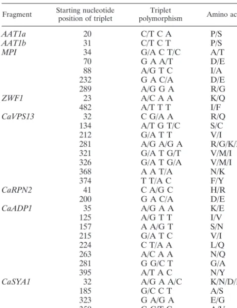
most of the polymorphisms were synonymous, 35 (40%) of the 87 individual changes recorded were nonsynonymous. Details of the alterations are shown in Table 2. Of the 35 amino acid changes, 19 were substantive changes, e.g., basic to acidic side chains, aliphatic to aromatic side chains, etc. In two cases, the change was between proline and serine.
The Appendix details the polymorphisms for the eight MLST gene fragments that were used in this study and the
on May 15, 2020 by guest
http://jcm.asm.org/
diversity of genotypes found based on these sequences (Table 1A). At some polymorphic loci, a majority of the 50 notionally diverse isolates had the same sequence, while others showed more interisolate diversity. The genotype numbers assigned in
the previous study were used forCaADP1, CaRPN2, CaSYA1,
and CaVPS13. Many new genotypes were determined in our set of isolates (Appendix) which had not been reported (2). Genotypes represented by large numbers of our isolates for each of the gene fragments shown in the Appendix were also represented by many isolates in the previous study. For
exam-ple, for CaADP1, 9 of our 50 isolates gave new strain types
(numbered from 17 upwards), forCaRPN2we found four new
types (17 to 20), represented by five isolates, forCaSYA1there
were 14 new types (14 and 27), each represented by a single
isolate, and forCaCAVPS1there were 20 new types (25 to 44),
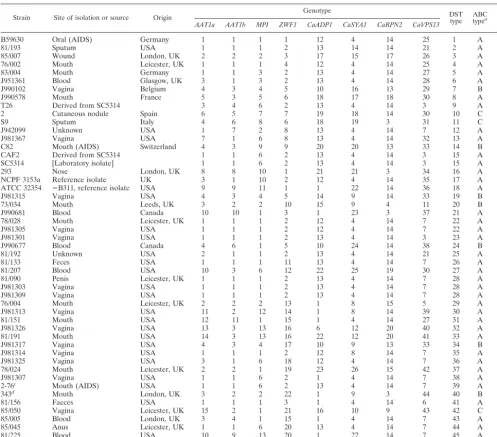
represented by 26 isolates. The MLST genotypes found in this study are summarized in Table 3. The results show a
discrim-inatory power ofD⫽0.996 (14).
Heterozygosity and MLST genotypes.The data in the Ap-pendix, like the equivalent data already published (2), show that sequence heterozygosity contributed considerably to the diversity of genotypes determined by MLST. For our panel of 50 C. albicans isolates (Table 3), a total of 87 polymorphic nucleotide sites were found across all eight gene fragments tested. For only 16 (18.4%) of these sites did the polymor-phisms consist entirely of homozygous nucleotide changes; the rest always included at least one example of heterozygosity at
the site. For each polymorphic site, only three sequence results were obtained: one of two bases or the heterozygous combi-nation of the same two bases. No polymorphic site resulted from more than two nucleotide changes. The heterozygous
PCR products obtained from the diploid genome ofC. albicans
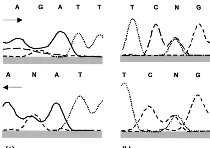
were presumed to arise from the coamplification of both al-leles, resulting in sequence profiles that showed two coincident peaks in the sequence chromatogram (Fig. 1). However, the high prevalence of apparent heterozygosity and the occasional ambiguity in double peaks (Fig. 1a) raised the possibility that sequencing errors rather than true heterozygosities generated patterns of this type.
Cloning and sequencing ofAAT1agene fragment.To inves-tigate further whether double peaks of the type shown in Fig. 1 represented true allelic heterozygosities or sequencing
er-rors, we chose the AAT1a gene fragment because sequence
analysis of the PCR product showed that the presumed
coam-plification of two different bases could create a unique MspI
restriction site in one of the alleles.
C. albicansisolate 76/002 had four potentially heterozygous
sites in theAAT1asequence (genotype 1 in the Appendix). At
position 40, the computer analysis of the sequence showed two equally sized peaks for adenine and guanine only for the re-verse direction (Fig. 1a), while the putative heterozygosity was detected by sequencing in both directions at nucleotide 124 (Fig. 1b). The putative heterozygosities at loci 7 and 89 were of the clear double peak variety shown in Fig. 1b. Six colonies of E. coli transformed with the cloned AAT1a PCR fragment were selected randomly for plasmid DNA extraction and se-quencing analysis. The results obtained showed that four of the six clones, each theoretically containing one of the two alleles of isolate 76/002, carried the bases G, A, A, and C at the four polymorphic sites, while for the other two clones the bases A G, G, and T were identified in the same nucleotide positions. Validation ofMspI polymorphism.One of the putative
se-quence heterozygosities (position 124, Y⫽C or T) observed in
the PCR product inC. albicans76/002 created anMspI
restric-tion site (C●CGG) in one of the alleles. Since noMspI
restric-tion sites were found anywhere else in the gene fragment, the
PCR product was digested withMspI. As shown in Fig. 2, the
digested products confirmed that the allele with theMspI
re-striction site gave the two predicted DNA fragments of 305 and 173 bp, while the other one remained undigested, as evidenced by the DNA band of 478 bp. Therefore, sequencing errors were unlikely to account for the polymorphisms observed in MLST for this diploid organism.
Additional strain typing characters.Among our 50 nomi-nally distinct isolates, four were homozygous at the MTL locus:
J990578, 85/005, and 81/225 were type a, while S9 was type␣.
The majority of the isolates (40 of 50) were genotype A by the transcribed spacer element PCR. There were seven type B strains and three type C strains. These characters are shown, together with the MLST data, in Table 3.
Epidemiological relationships of isolates typed.In Table 3 the diversity of genotypes detected across all eight MLST frag-ments is shown for all 50 isolates tested. For the four previ-ously studied gene fragments (2), the published genotype num-bers are used, and additional, higher numnum-bers are assigned for the novel genotypes that we found in this study (Table 3). The final column indicating the full range of diploid sequence
ge-TABLE 2. Changes in amino acid sequence resulting from nucleotide polymorphisms
Fragment Starting nucleotideposition of triplet polymorphismTriplet Amino acids
AAT1a 20 C/T C A P/S
AAT1b 31 C/T C T P/S
MPI 34 G/A C T/C A/T
70 G A A/T D/E
88 A/G T C I/A
232 G A C/A D/E
289 A/G G A R/G
ZWF1 23 A/C A A K/Q
482 A/T T T I/F
CaVPS13 32 C G/A A R/Q
134 A/T G T/C S/C
212 G/A T T V/I
281 A/G A/G A R/G/K/E
321 G/A T G/T V/M/I
326 G/A T G/A V/M/I
368 A A T/A N/K
374 T T/A C F/Y
CaRPN2 41 C A/G C H/R
200 G A C/A D/E
CaADP1 35 A/G A A K/E
125 A/G T T I/V
157 A A/G T S/N
215 G/A T C V/I
224 C T/A A L/Q
263 A/C A A N/Q
281 G G/C T G/A
395 A/T A C N/Y
CaSYA1 32 A/G A A/C K/N/D/E
185 G/C C T A/S
323 G A/G A E/G
350 G C/T C A/V
on May 15, 2020 by guest
http://jcm.asm.org/
[image:4.603.43.283.90.401.2]notypes (DSTs: the combination of genotypes from all individ-ual gene fragments) indicates that 46 unique types were found among the 50 isolates examined. Two identical DSTs were found for SC5314 and its derivative CAF2. An oral isolate, 78/028, from a healthy volunteer first cultured in 1978, and J981305, from a patient with vaginitis in the United States, were found to be identical to type 22. Three isolates, two from different U.S. patients with vaginitis and an isolate from a penis
obtained 17 years earlier in the United Kingdom, shared DST 28.
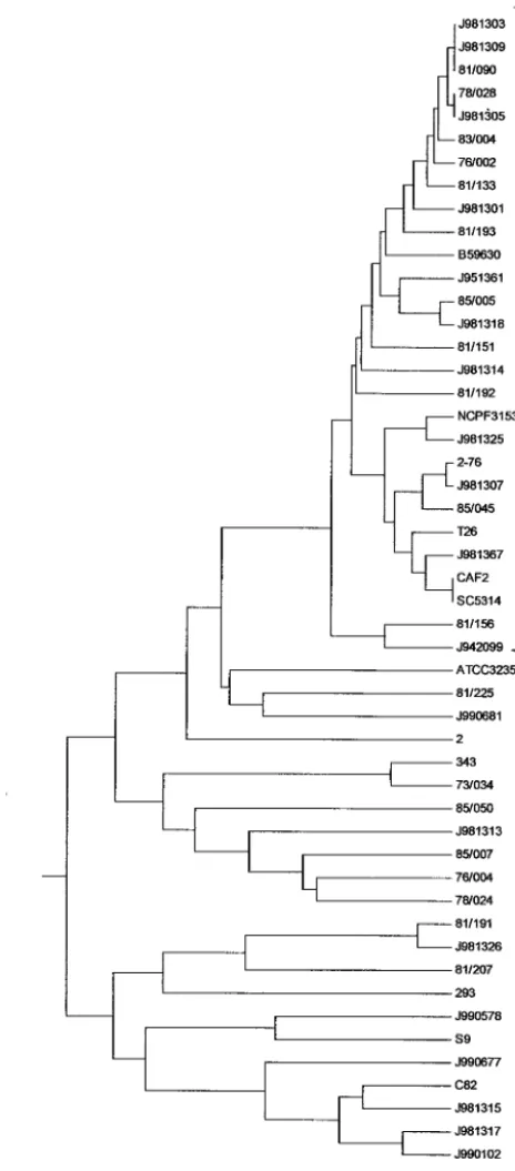
A UPGMA similarity dendrogram from the data for the 50 isolates tested was generated with Mega software (Fig. 3). The two pairs and triad of isolates with identical MLST types, together with a further 19 isolates, clustered in a single group
(bracketed in Fig. 3) with ⬎84% identity by this method of
analysis. The remaining isolates generally showed a higher level of diversity. Analysis of the strain types by means of a neighbor-joining tree similarly clustered the same 26 isolates bracketed in Fig. 3 into a single, highly related group. Nine of the 13 vaginal isolates obtained from the United States in 1998 fell into the highly similar clusters in both analyses, with the remainder showing little relation to each other.
[image:5.603.117.469.67.314.2]Although single-linkage cluster analysis of the type shown in Fig. 3 permits sorting of isolates into similar clusters, MLST data allow a more refined analysis of isolates based on the relationships between very closely related genotypes within clonal complexes. This model postulates a putative ancestral genotype from which other types have developed by mutation at just one or two loci (9, 20). Analysis with the Burst algorithm revealed three such clonal complexes for our isolates; however, two of the putative clonal complexes included just two or three isolates. The largest complex, which was therefore capable of structural analysis, is shown in Fig. 4. The complex was in fact composed of two related subcomplexes, which divided our main UPGMA cluster (Fig. 3) into three sets. The first com-prised isolates J981303, J981309, 81/190, 78/028, J981305, 83/ 004, 76/002, 81/133, J981301, 81/193, B59630, J981314, and 81/192; the second comprised 2-76, J981307, 85/045, T26,
[image:5.603.91.238.499.676.2]FIG. 1. Two examples of raw sequencing data for forward and reverse strands of PCR product fromC. albicansstrain 76/002,AAT1agene fragment, illustrating sequence results interpreted as heterozygosity. Solid line, adenine (A); long-dashed line, cytosine (C); short-dashed line, guanine (G); dotted line, thymine (T); N, site of putative heterozygosity because the peaks suggest that two nucleotide bases are present in the sequence. (a) Nucleotide 40; (b) nucleotide 124. Arrows indicate direction of sequencing.
FIG. 2. MspI restriction digestion ofAAT1aPCR product fromC.
albicans strain 76/002. Electrophoresis of the digest revealed three
bands (lane D), indicating the products predicted when two heterozy-gous sequences are treated with the enzyme and only one contains the restriction site. Lane M, 100-bp ladder.
on May 15, 2020 by guest
http://jcm.asm.org/
CAF2, and SC5314; and the third comprised the remainder of the isolates from the related cluster.
MLST patterns of sequential isolates from the same pa-tients.The 16 sequential isolates from an AIDS patient (35) all gave the same diploid sequence type (DST) by MLST. Of the 11 consecutive isolates from oral and fecal surveillance
cul-tures from a single patient undergoing chemotherapy for he-matological malignancy, nine gave one DST 40 and two gave a
DST that differed at a single polymorphic site in CaADP1,
which was heterozygous (A or G) in the nine isolates and homozygous (G) at this locus in two, suggesting that MLST was sufficiently sensitive to detect microsequence evolution within the clade.
DISCUSSION
The set of gene fragments we had chosen for MLST, based on multilocus enzyme electrophoresis data (26), differed from the set published by Bougnoux et al. (2), and the appearance of their publication therefore gave us the opportunity to validate the published data and to add our own MLST gene fragment set to facilitate the selection of an optimal set of gene
frag-ments that could be used routinely forC. albicansMLST. The
results of this study confirm that both the published set of gene fragments and those we chose were able to indicate genotypic diversity among isolates. The set of eight gene fragments listed in Table 1 all give high genotypic diversity with nonoverlapping results: 49 nonidentical isolates (SC5314 and CAF2 were ex-pected to be identical) gave 46 different diploid sequence types on the basis of these sequences (Table 3). This high level of differentiation reinforces the view of MLST as a highly dis-criminatory strain-typing procedure.
Data for highly related isolates show that MLST gives high
FIG. 3. Single-linkage dendrogram indicating the similarities of 50
[image:6.603.51.283.75.603.2]C. albicansisolates determined by MLST with eight gene fragments.
FIG. 4. Burst analysis showing 16 clonally related diploid sequence type numbers (see Table 3) derived from a subset of the isolates bracketed in Fig. 3. In each set of concentric circles, the central type has a common consensus sequence between isolates for the eight gene fragments tested; each surrounding circle indicates one level of change.
on May 15, 2020 by guest
http://jcm.asm.org/
reproducibility within and between laboratories. Isolate SC5314 was also tested by MLST in the study by Bougnoux and colleagues; the SC5314 genotypes found by them (2) were identical to those we determined with the same DNA frag-ments. CAF2, derived from SC5314 by deletion of one copy of
theURA3gene, also gave the same result. This consistency is
a demonstration of the power and reproducibility of MLST
applied toC. albicans. Unlike some typing systems, in which
reproducibility and discriminatory power are inversely related (14), our data show both 100% reproducibility and a discrim-inatory power of 0.996 (14). Moreover, sequential isolates from each of two patients gave identical MLST types in one instance and types that differed by only a single nucleotide in the second case. These findings demonstrate that MLST with the gene
fragments used in this study can recognize isolates ofC.
albi-cansthat are identical or nearly so and may be detecting minor
microevolutionary changes, known to occur in longitudinal studies with repeated isolates from the same patient (16, 25, 31).
[image:7.603.44.542.80.517.2]That such changes may occur even in laboratory isolates is exemplified by the finding of four nucleotide differences be-tween strain T26 and its parents SC5314 and CAF2. T26 was engineered from SC5314 by a series of changes that included gene disruptions and selection for spontaneous resistance to echinocandins (6, 15). We conclude that one or more of the steps in the lineage of T26 resulted in the small sequence differences detected in this study. All four nucleotide changes in strain T26 occurred in a single strand of the diploid DNA in
TABLE 3. Genotypes of 50C. albicansisolates analyzed by MLST
Strain Site of isolation or source Origin Genotype DSTtype ABCtypea MLTtypeb
AAT1a AAT1b MPI ZWF1 CaADP1 CaSYA1 CaRPN2 CaVPS13
B59630 Oral (AIDS) Germany 1 1 1 1 12 4 14 25 1 A Het
81/193 Sputum USA 1 1 1 2 13 14 14 21 2 A Het
85/007 Wound London, UK 2 2 2 3 17 15 17 26 3 A Het
76/002 Mouth Leicester, UK 1 1 1 4 12 4 14 25 4 A Het
83/004 Mouth Germany 1 1 3 2 13 4 14 27 5 A Het
J951361 Blood Glasgow, UK 3 1 3 2 13 4 14 28 6 A Het
J990102 Vagina Belgium 4 3 4 5 10 16 13 29 7 B Het
J990578 Mouth France 5 3 5 6 18 17 18 30 8 A a
T26 Derived from SC5314 3 4 6 2 13 4 14 3 9 A Het
2 Cutaneous nodule Spain 6 5 7 7 19 18 14 30 10 C Het
S9 Sputum Italy 4 6 8 6 18 19 3 31 11 C ␣
J942099 Unknown USA 1 7 2 8 13 4 14 7 12 A Het
J981367 Vagina USA 7 1 6 8 13 4 14 32 13 A Het
C82 Mouth (AIDS) Switzerland 4 3 9 9 20 20 13 33 14 B Het
CAF2 Derived from SC5314 1 1 6 2 13 4 14 3 15 A Het
SC5314 [Laboratory isolate] 1 1 6 2 13 4 14 3 15 A Het
293 Nose London, UK 8 8 10 1 21 21 3 34 16 A Het
NCPF 3153a Reference isolate UK 3 1 10 2 12 4 14 35 17 A Het
ATCC 32354 ⫽B311, reference isolate USA 9 9 11 1 1 22 14 36 18 A Het
J981315 Vagina USA 4 3 4 5 14 9 14 33 19 B Het
73/034 Mouth Leeds, UK 3 2 2 10 15 9 4 11 20 B Het
J990681 Blood Canada 10 10 1 3 1 23 3 37 21 A Het
78/028 Mouth Leicester, UK 1 1 1 2 12 4 14 7 22 A Het
J981305 Vagina USA 1 1 1 2 12 4 14 7 22 A Het
J981301 Vagina USA 1 1 1 2 13 4 14 3 23 A Het
J990677 Blood Canada 4 6 1 5 10 24 14 38 24 B Het
81/192 Unknown USA 2 1 1 2 13 4 14 21 25 A Het
81/133 Feces USA 1 1 1 11 13 4 14 7 26 A Het
81/207 Blood USA 10 3 6 12 22 25 19 30 27 A Het
81/090 Penis Leicester, UK 1 1 1 2 13 4 14 7 28 A Het
J981303 Vagina USA 1 1 1 2 13 4 14 7 28 A Het
J981309 Vagina USA 1 1 1 2 13 4 14 7 28 A Het
76/004 Mouth Leicester, UK 2 2 2 13 1 8 15 5 29 A Het
J981313 Vagina USA 11 2 12 14 1 8 14 39 30 A Het
81/151 Mouth USA 12 11 1 15 1 4 14 27 31 A Het
J981326 Vagina USA 13 3 13 16 6 12 20 40 32 A Het
81/191 Mouth USA 14 3 13 16 22 12 20 41 33 A Het
J981317 Vagina USA 4 3 4 17 10 9 13 33 34 B Het
J981314 Vagina USA 1 1 1 2 12 8 14 7 35 A Het
J981325 Vagina USA 3 1 6 18 12 4 14 7 36 A Het
78/024 Mouth Leicester, UK 2 2 1 19 23 26 15 42 37 A Het
J981307 Vagina USA 1 1 6 2 1 4 14 7 38 A Het
2-76c Mouth (AIDS) USA 1 1 6 2 13 4 14 7 39 A Het
343d Mouth London, UK 3 2 2 22 1 9 3 44 40 B Het
81/156 Faeces USA 1 1 1 3 1 4 14 6 41 A Het
85/050 Vagina Leicester, UK 15 2 1 21 16 10 9 43 42 C Het
85/005 Blood London, UK 3 4 1 15 1 4 14 7 43 A a
85/045 Anus Leicester, UK 1 1 6 20 13 4 14 7 44 A Het
81/225 Blood USA 10 9 13 20 1 27 14 7 45 A a
J981318 Vagina USA 3 4 1 2 1 4 14 6 46 A Het
aDetermined by PCR of transcribed spacer region of rDNA region.
bDetermined by multiplex PCR of MLT locus.
cFrom a series of 16 sequential isolates (35).
dFrom a series of 12 isolates from a single patient with a hematological malignancy.
on May 15, 2020 by guest
http://jcm.asm.org/
the AAT1 gene, since each involved loss of nucleotide het-erozygosity.
Among the amino acid changes resulting from 40% of the nucleotide polymorphisms, many involved switches between types of amino acid, including two instances of proline to serine (Table 2), which would be expected to effect significant alterations in secondary and higher peptide structures. If this level of sequence change is representative of variation for the
products of otherC. albicansgenes, it must be concluded that
the fungus is tolerant of the observed differences in protein structure. It is likely that many subtle phenotypic differences between strains may exist that have yet to be detected. The high level of genotype-phenotype differences possible between strains of a yeast species was indicated in a recent study based
on expression profiling with a fresh, wild-type isolate of
Sac-charomyces cerevisiaeand a laboratory-maintained isolate. This analysis showed that 1,500 of the 6,116 genes in the yeasts differed in levels of expression (3). To what extent the differ-ences in expression are reflected as differdiffer-ences in the genome sequence have yet to be determined.
The findings of this study indicate that the set of genes listed in Table 1 are adequate for high-quality strain typing by MLST. For population genetic analyses and other statistical
ap-proaches to the epidemiological study ofC. albicans, this larger
set of MLST fragments should represent a more discriminatory tool than the six-fragment set already proposed (2). It is nota-ble that the sequence differences between SC5314 and T26 would not have been detected with the published six-fragment set. These two strains are not the only examples of isolates whose types were indistinguishable by MLST with the four published gene fragments but could be distinguished by the new gene fragments. Conversely, some strains could be distin-guished by the published gene fragments but not by the new ones.
So far,C. albicansis the only example for which MLST has
been attempted with a species having a permanently diploid genome, where heterozygous alleles may occur. The frequency
with which heterozygous sites occur in MLST withC. albicans
is high, both in our own and in the previous study of MLST (2). The present study investigated the possibility that apparent heterozygosity may arise through sequencing artifacts and con-firmed it to be genuine. Heterozygosity in a diploid genome adds extra characters for strain discrimination over simple se-quence variation and will allow future analyses of population genetics research based on haplotypes. The relative frequen-cies of clonal reproduction and sexual or other recombina-tional events in natural populations of the fungus remain an open question (1, 10, 11, 16, 18, 26, 30, 34).
The addition of two non-MLST characters to aC. albicans
strain-typing scheme adds extra discriminatory detail. Data
from surveys based on the system that dividesC. albicansinto
three subtypes, A, B, and C, based on sequence differences in the transcribed spacer region in the gene encoding rRNA have already been used to demonstrate geographical differences in C. albicansisolate populations (21). This approach also allows
direct, unequivocal recognition of the species C. dubliniensis
(22). In common with McCullough et al. (21), we found most of our isolates were type A by rDNA PCR. Of note, all the isolates in the cluster of highly related strains (Fig. 3) were type
A. The level of discrimination possible by MLST clearly ex-ceeds that of ribosomal DNA typing, since we could distinguish 36 distinct strains among the 40 designated as type A. All the isolates of type B and type C strains could be differentiated by MLST.
Determination of mating type inC. albicansisolates is of
possible relevance to antifungal resistance (27) and to pheno-typic switching in this fungus (17). In common with Rustad and colleagues (27), we found that only a minority of isolates were
homozygous at MTL. They found 12 homozygous isolates
among 96 tested (12.5%); we found 4 among 50 (8%). Even if the frequency of homozygous mating types in the clinical
pop-ulation ofC. albicansis only on the order of 10%, this may be
adequate to explain small departures from clonality in the Hardy-Weinberg equilibrium analyses that have been de-scribed previously for this species (11, 26).
We conclude that MLST withC. albicansoffers an effective
system for epidemiological work with the species, that the high frequency of heterozygous sequences in the DNA regions cho-sen for MLST add extra information to MLST that is not available with haploid organisms, and that the creation of a central database for archiving of MLST data will enhance research based on strain typing. Although at present MLST is more likely to constitute a research rather than a reference tool, MLST has the advantage that it is scalable from a small number of isolates to many hundreds or even thousands of isolates by the exploitation of robotic DNA extraction and high-throughput nucleotide sequence determination technolo-gies. The application of high-throughput technology also leads to substantial reductions in the cost of isolate characterization. For example, in this study full MLST profiles were obtained for a consumables cost of approximately US$30 per isolate; how-ever, with recently developed DNA analyzers, a cost of less than US$15 per isolate is now attainable, with the prospect of further substantial reductions in cost in the near future. Such automation will not only reduce costs but also increase throughput of the method. At present, at least 24 isolates per week can be typed with all procedures done by hand, but automation of the processes will increase this number to hun-dreds per week.
Inclusion of theMTL status and genotype (A, B, or C) as
extra typing data further refines the ability of DNA-based
methods to distinguish isolates ofC. albicans. The findings of
this study and the previous investigation (2) show a high level of sequence variation in transcribed housekeeping genes
among isolates ofC. albicans. We are now establishing MLST
for otherCandidaspecies and investigating the frequencies of
sequence changes in isogenic strains ofC. albicansexposed to
various conditions in vitro and in vivo.
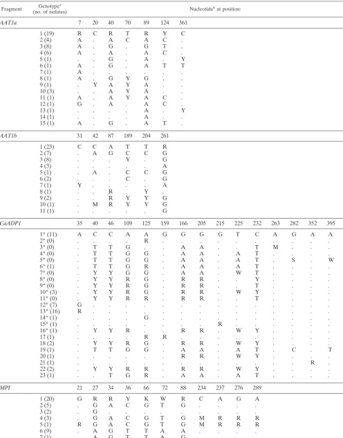
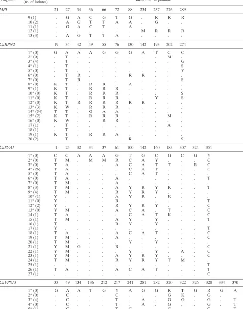
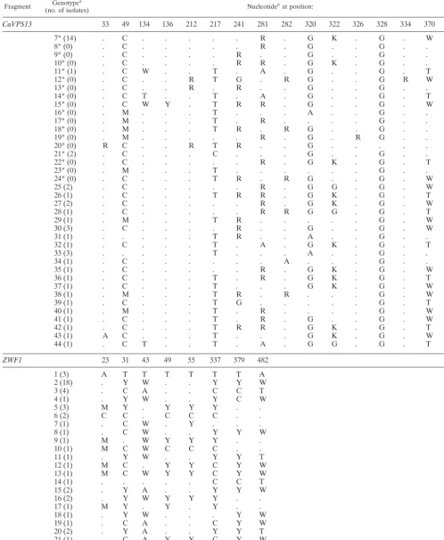
APPENDIX
The positions of polymorphic nucleotide sites identified in various gene fragments are shown in Table. A1. All the nucleotides present at each variable site are shown for genotype 1. For the other genotypes, only nucleotides that differ from those of genotype 1 are shown, and nucleotides identical to those in genotype 1 are indicated with a dot. The number of isolates from the set of 50 diverse isolates with the same genotype is shown in parentheses. The position of each polymor-phic site is indicated for each fragment.
on May 15, 2020 by guest
http://jcm.asm.org/
TABLE A1. Polymorphic sites
Fragment (no. of isolates)Genotypea Nucleotidebat position:
AAT1a 7 20 40 70 89 124 361
1 (19) R C R T R Y C
2 (4) A . A C A C .
3 (8) A . G . G T .
4 (6) A . A . A C .
5 (1) . . G . A . Y
6 (1) A . G . A T T
7 (1) A . . . .
8 (1) A . G Y G . .
9 (1) . Y A Y A . .
10 (3) . . A Y A . .
11 (1) A . A Y A C .
12 (1) G . A . A C .
13 (1) . . . . A . Y
14 (1) . . . . A . .
15 (1) A . G . A T .
AAT1b 31 42 87 189 204 261
1 (23) C C A T T R
2 (7) . A G C C G
3 (8) . . . Y . G
4 (3) . . . A
5 (1) . A . C C G
6 (2) . . . C . G
7 (1) Y . . . . A
8 (1) . . R . Y .
9 (2) . . R Y Y G
10 (1) . M R Y Y G
11 (1) . . . G
CaADP1 35 40 46 109 125 159 166 205 215 225 232 263 282 352 395
1* (11) A C C A A G G G G T C A G A A
2* (0) . . . . R . . . .
3* (0) . T T G . . A A . . T M . . .
4* (0) . T T G G . A A . A T . . . .
5* (0) . T T G G . A A . A T . S . W
6* (1) . T T G R . A A . A T . . . .
7* (0) . Y Y G G . A A . W T . . . .
8* (0) . Y Y R G . R R . . Y . . . .
9* (0) . Y Y R G . R R . . T . . . .
10* (3) . Y Y R G . R R . W Y . . . .
11* (0) . Y Y R R . R R . . T . . . .
12* (7) G . . . .
13* (16) R . . . .
14* (1) . . . . G . . . .
15* (1) . . . R . . . .
16* (1) . Y Y R . . R R . W Y . . . .
17 (1) . . . . R R . . . .
18 (2) . Y Y R G . R R . W Y . . . .
19 (1) . T T G G . A A . A T . C . T
20 (1) . . . R R . W Y . . . .
21 (1) . . . R .
22 (2) . Y Y R R . R R . W Y . . . .
23 (1) . . T G R . A A . A T . . . .
MPI 21 27 34 36 66 72 88 234 237 276 289
1 (20) G R R Y K W R C A G A
2 (5) . G A C G T G . . . .
3 (2) . G . . . .
4 (3) . G A C G T G M R R R
5 (1) R G A C G T G M R R R
6 (9) . A G T T A A . . . .
7 (1) . A G T T A G . . . .
8 (1) . G A C G T G A G A .
Continued on following page
on May 15, 2020 by guest
http://jcm.asm.org/
TABLE A1—Continued
Fragment (no. of isolates)Genotypea Nucleotidebat position:
MPI 21 27 34 36 66 72 88 234 237 276 289
9 (1) . G A C G T G . R R R
10 (2) . A G T T A A . G . .
11 (1) . G A C T . A . . . .
12 (1) . . . M R R R
13 (3) . A G T T A . . . . .
CaRPN2 19 34 42 49 55 76 130 142 193 202 274
1* (0) G A A A G G G A T C C
2* (0) . T . . . M .
3* (4) . T . . . G
4* (1) . T . . . S
5* (0) . T . . . Y
6* (0) . T R . . . R R . . .
7* (0) . T R . . . S
8* (0) K T . R R . A . . . .
9* (1) K T . R R R . . . . .
10* (0) K T . R R R . . . . S
11* (0) K T . R R R . . Y . S
12* (0) K T R R R R R R . . .
13* (3) K W . R R R . . . . .
14* (34) T T . G A A . . . . .
15* (2) K T . R R R . . . M .
16* (0) K W . . R R . . . . .
17 (1) . T . . . A .
18 (1) . T . . . .
19 (1) K T . R R A . . . . .
20 (2) . T . . . . R . . S
CaSYA1 1 25 32 34 37 61 100 142 160 185 307 324 351
1* (0) C C A A A G T G C G C G Y
2* (0) T M . M M R C A Y . . . C
3* (0) T A . . . A C A T T . R C
4* (26) T A . . . . C A T . . . C
5* (0) T A . . . . C A T . . . .
6* (0) T A . . . A . . . T
7* (0) T M . . . A . . . .
8* (3) T M . . . A Y R Y K . . T
9* (4) T M . . . R Y R Y . . . .
10* (1) Y . . . . A Y R . K . . .
11* (0) Y . . . . R . . . T
12* (2) Y . . . . R Y R Y . . . C
13* (0) Y M . . . A C A . T . . C
14 (1) T A . . . . C A T K . . C
15 (1) T M . . . A Y . Y . . . T
16 (1) T . . . . R Y . Y . . . .
17 (1) Y . . . T
18 (1) T A . . . A C A T . . . C
19 (1) T M . . . A . . . C
20 (1) T M . . . . Y . Y . . . .
21 (1) Y M G . . R . . . C
22 (1) Y M . . . . Y . Y . A . C
23 (1) Y M . . . A Y R Y . . . C
24 (1) T M . . . R Y R Y T M . .
25 (1) . . . T
26 (1) T A . . . A C A T . . . T
27 (1) . . . C
CaVPS13 33 49 134 136 212 217 241 281 282 320 322 326 328 334 370 375
1* (0) G A A T G Y A G G R T G R G A C
2* (0) . C . . . C . . . G K . G . . Y
3* (4) . C . . . T . A . G G . G . T T
4* (0) . C . . . T . A . G . . G . T .
5* (1) . C . . . T G . . G . . G . T .
6* (2) . C . . . R . G K . G . . Y
Continued on following page
on May 15, 2020 by guest
http://jcm.asm.org/
TABLE A1—Continued
Fragment (no. of isolates)Genotypea Nucleotidebat position:
CaVPS13 33 49 134 136 212 217 241 281 282 320 322 326 328 334 370 375
7* (14) . C . . . R . G K . G . W Y
8* (0) . C . . . R . G . . G . . .
9* (0) . C . . . . R . . G . . G . . .
10* (0) . C . . . . R R . G K . G . . Y
11* (1) . C W . . T . A . G . . G . T .
12* (0) . C . . R T G . R G . . G R W .
13* (0) . C . . R . R . . G . . G . . .
14* (0) . C T . . T . A . G . . G . T .
15* (0) . C W Y . T R R . G . . G . W .
16* (0) . M . . . T . . . A . . G . . .
17* (0) . M . . . T . R . . . . G . . .
18* (0) . M . . . T R . R G . . G . . .
19* (0) . M . . . R . G . R G . . .
20* (0) R C . . R T R . . G . . . .
21* (2) . C . . . C . . . G . . G . . .
22* (0) . C . . . R . G K . G . T Y
23* (0) . M . . . T . . . G . . .
24* (0) . C . . . T R . R G . . G . W .
25 (2) . C . . . R . G G . G . W Y
26 (1) . C . . . T R R . G K . G . T Y
27 (2) . C . . . R . G K . G . W .
28 (1) . C . . . R R G G . G . T .
29 (1) . M . . . T R . . . G . W .
30 (3) . C . . . . R . . G . . G . W .
31 (1) . . . T R . . A . . G . . .
32 (1) . C . . . T . A . G K . G . T T
33 (3) . . . T . . . A . . G . . .
34 (1) . C . . . A . . . G . . .
35 (1) . C . . . R . G K . G . W T
36 (1) . C . . . T . R . G K . G . T Y
37 (1) . C . . . T . . . G K . G . W Y
38 (1) . M . . . T R . R . . . G . W .
39 (1) . C . . . T G . . . G . T .
40 (1) . M . . . T . R . . . . G . W .
41 (1) . C . . . T . R . G . . G . W .
42 (1) . C . . . T R R . G K . G . T .
43 (1) A C . . . T . . . G K . G . W Y
44 (1) . C T . . T . A . G G . G . T .
ZWF1 23 31 43 49 55 337 379 482
1 (3) A T T T T T T A
2 (18) . Y W . . Y Y W
3 (4) . C A . . C C T
4 (1) . Y W . . Y C W
5 (3) M Y . Y Y Y . .
6 (2) C C . C C C . .
7 (1) . C W . Y . . .
8 (1) . C W . . Y Y W
9 (1) M . W Y Y Y . .
10 (1) M C W C C C . .
11 (1) . Y W . . Y Y T
12 (1) M C . Y Y C Y W
13 (1) M C W Y Y C Y W
14 (1) . . . C C T
15 (2) . Y A . . Y Y W
16 (2) . Y W Y Y Y . .
17 (1) M Y . Y . Y . .
18 (1) . Y W . . . Y W
19 (1) . C A . . C Y W
20 (2) . Y A . . Y Y T
21 (1) . C A Y Y C Y W
22 (1) C . . C C C . .
a*, genotype according to Bougnoux et al. (2).
bR, A or G; Y, C or T, K, G or T; M, A or C; S, G or C; and W, A or T.
on May 15, 2020 by guest
http://jcm.asm.org/
ACKNOWLEDGMENTS
For the pilot phase of this study, A. Tavanti was supported by the University of Pisa, and the study was generously supported by an unrestricted grant from Pfizer UK. Our MLST research is now sup-ported by grant 069615 from the Wellcome Trust. We also acknowl-edge the British Society for Antimicrobial Chemotherapy and the BBSRC for laboratory support.
We thank Amanda Davidson for excellent technical assistance; Merck, Inc., for strain T26; and the colleagues who have supplied clinical isolates ofC. albicansto our collection over the last 30 years.
REFERENCES
1. Arnavielhe, S., T. De Meeus, A. Blancard, M. Mallie, F. Renaud, and J. M. Bastide.2000. Multicentric genetic study ofCandida albicansisolates from non-neutropenic patients using multilocus enzyme electrophoresis typing:
population structure and mode of reproduction. Mycoses43:109–117.
2. Bougnoux, M.-E., S. Morand, and C. d’Enfert.2002. Usefulness of
multilo-cus sequence typing for characterization of clinical isolates ofCandida
albi-cans.J. Clin. Microbiol.40:1290–1297.
3. Brem, R. B., G. Yvert, R. Clinton, and L. Kruglyak.2002. Genetic dissection
of transcriptional regulation in budding yeast. Science296:752–755.
4. Chen, Y. C., J. D. Eisner, M. M. Kattar, S. L. Rassoulian-Barrett, K. LaFe, S. L. Yarfitz, A. P. Limaye, and B. T. Cookson.2000. Identification of medically important yeasts using PCR-based detection of DNA sequence polymorphisms in the internal transcribed spacer 2 region of the rRNA
genes. J. Clin. Microbiol.38:2302–2310.
5. Dingle, K. E., F. M. Colles, D. R. A. Wareing, R. Ure, A. J. Fox, F. E. Bolton, H. J. Bootsma, R. J. L. Willems, R. Urwin, and M. C. Maiden.2001.
Mul-tilocus sequence typing system forCampylobacter jejuni.J. Clin. Microbiol.
39:14–23.
6. Douglas, C. M., J. A. Dippolito, G. J. Shei, M. Meinz, J. Onishi, J. A. Marrinan, W. Li, G. K. Abruzzo, A. Flattery, K. Bartizal, A. Mitchell, and M. B. Kurtz.1997. Identification of theFKS1gene ofCandida albicansas the essential target of 1, 3--d-glucan synthase inhibitors. Antimicrob. Agents
Chemother.41:2471–2479.
7. Enright, M. C., N. P. J. Day, C. E. Davies, S. J. Peacock, and B. G. Spratt.
2000. Multilocus sequence typing for characterization of methicillin-resistant
and methicillin-susceptible clones ofStaphylococcus aureus.J. Clin.
Micro-biol.38:1008–1015.
8. Enright, M. C., B. G. Spratt, A. Kalia, J. H. Cross, and D. E. Bessen.2001.
Multilocus sequence typing ofStreptococcus pyogenesand the relationships
between emm type and clone. Infect. Immun.69:2416–2427.
9. Feil, E. J., and B. G. Spratt. 2001. Recombination and the population
structures of bacterial pathogens. Annu. Rev. Microbiol.55:561–590.
10. Forche, A., G. Schonian, Y. Graser, R. Vilgalys, and T. G. Mitchell.1999.
Genetic structure of typical and atypical populations ofCandida albicans
from Africa. Fungal Genet. Biol.28:107–125.
11. Graser, Y., M. Volovsek, J. Arrington, G. Schonian, W. Presber, T. G. Mitchell, and R. Vilgalys.1996. Molecular markers reveal that population
structure of the human pathogenCandida albicansexhibits both clonality
and recombination. Proc. Natl. Acad. Sci. USA93:12473–12477.
12. Hanahan, D.1983. Studies on transformation ofEscherichia coliwith
plas-mids. J. Mol. Biol.166:557–580.
13. Hull, C. M., and A. D. Johnson.1999. Identification of a mating type-like
locus in the asexual pathogenic yeastCandida albicans.Science285:1271–
1275.
14. Hunter, P. R.1991. A critical review of typing methods forCandida albicans
and their applications. Crit. Rev. Microbiol.17:417–434.
15. Kurtz, M. B., G. Abruzzo, K. Bartizal, J. A. Marrinan, W. Li, J. Milligan, K. Nollstadt, and C. M. Douglas.1996. Characterization of
echinocardin-resis-tant muechinocardin-resis-tants ofCandida albicans — genetic, biochemical, and virulence
studies. Infect. Immun.64:3244–3251.
16. Lockhart, S. R., J. J. Fritch, A. S. Meier, K. Schroppel, T. Srikantha, R. Galask, and D. R. Soll.1995. Colonizing populations ofCandida albicansare clonal in origin but undergo microevolution through C1 fragment
reorgani-zation as demonstrated by DNA fingerprinting and C1 sequencing. J. Clin.
Microbiol.33:1501–1509.
17. Lockhart, S. R., C. Pujol, K. J. Daniels, M. G. Miller, A. D. Johnson, M. A. Pfaller, and D. R. Soll.2002. InCandida albicans, white-opaque switchers
are homozygous for mating type. Genetics162:737–745.
18. Lott, T. J., and M. M. Effat.2001. Evidence for a more recently evolved clade
within aCandida albicansNorth American population. Microbiology147:
1687–1692.
19. Maiden, M. C. J., J. A. Bygraves, E. Feil, G. Morelli, J. E. Russell, R. Urwin, Q. Zhang, J. J. Zhou, K. Zurth, D. A. Caugant, I. M. Feavers, M. Achtman, and B. G. Spratt.1998. Multilocus sequence typing: a portable approach to the identification of clones within populations of pathogenic
microorgan-isms. Proc. Natl. Acad. Sci. USA95:3140–3145.
20. Maynard Smith, J., N. H. Smith, M. O’Rourke, and B. G. Spratt.1993. How
clonal are bacteria? Proc. Natl. Acad. Sci. USA90:4384–4388.
21. McCullough, M., K. V. Clemons, and D. A. Stevens.1999. Molecular
epide-miology of the global and temporal diversity ofCandida albicans. Clin.
Infect. Dis.29:1220–1225.
22. McCullough, M. J., K. V. Clemons, and D. A. Stevens.1999. Molecular and
phenotypic characterization of genotypicCandida albicanssubgroups and
comparison withCandida dubliniensisandCandida stellatoidea.J. Clin.
Mi-crobiol.37:417–421.
23. Nallapareddy, S. R., R. W. Duh, K. V. Singh, and B. E. Murray.2002.
Molecular typing of selectedEnterococcus faecalisisolates: Pilot study using
multilocus sequence typing and pulsed-field gel electrophoresis. J. Clin.
Microbiol.40:868–876.
24. Odds, F. C., C. C. Kibbler, E. Walker, A. Bhamra, H. G. Prentice, and P. Noone.1989. Carriage ofCandidaspecies andC. albicansbiotypes in patients undergoing chemotherapy or bone marrow transplantation for
haematologi-cal disease. J. Clin. Pathol.42:1259–1266.
25. Pujol, C., S. Joly, B. Nolan, T. Srikantha, and D. R. Soll.1999.
Microevo-lutionary changes inCandida albicansidentified by the complex Ca3
finger-printing probe involve insertions and deletions of the full-length repetitive
sequence RPS at specific genomic sites. Microbiology UK145:2635–2646.
26. Pujol, C., J. Reynes, F. Renaud, M. Raymond, M. Tibayrenc, F. Ayala, F. Janbon, M. Mallie, and J. Bastide.1993. The yeastCandida albicanshas a clonal mode of reproduction in a population of infected human
immunode-ficiency virus-positive patients. Proc. Natl. Acad. Sci. USA90:9456–9459.
27. Rustad, T. R., D. A. Stevens, M. A. Pfaller, and T. C. White.2002.
Homozy-gosity at theCandida albicansMTL locus associated with azole resistance.
Microbiology-SGM148:1061–1072.
28. Sadhu, C., M. J. McEachern, E. P. Rustchenko-Bulgac, J. Schmid, D. R. Soll, and J. B. Hicks.1991. Telomeric and dispersed repeat sequences inCandida
yeasts and their use in strain identification. J. Bacteriol.173:842–850.
29. Scherer, S., and D. A. Stevens.1988. ACandida albicansdispersed, repeated gene family and its epidemiological applications. Proc. Natl. Acad. Sci. USA
85:1452–1456.
30. Schonian, G., A. Forche, H. J. Tietz, M. Muller, Y. Graser, R. Vilgalys, T. G. Mitchell, and W. Presber.2000. Genetic structure of geographically different
populations ofCandida albicans.Mycoses43:51–56.
31. Schroppel, K., M. Rotman, R. Galask, K. MAC, and D. R. Soll.1994.
Evolution and replacement ofCandida albicansstrains during recurrent
vaginitis demonstrated by DNA fingerprinting. J. Clin. Microbiol.32:2646–
2654.
32. Soll, D. R.2000. The ins and outs of DNA fingerprinting the infectious fungi.
Clin. Microbiol. Rev.13:332–370.
33. Tamura, M., K. Watanabe, Y. Mikami, K. Yazawa, and K. Nishimura.2001.
Molecular characterization of new clinical isolates ofCandida albicansand
C. dubliniensisin Japan: analysis reveals a new genotype ofC. albicanswith
group I intron. J. Clin. Microbiol.39:4309–4315.
34. Tibayrenc, M.1997. AreCandida albicansnatural populations subdivided?
Trends Microbiol.5:253–257.
35. White, T. C.1997. Increased mRNA levels ofERG16, CDR, andMDR1
correlate with increases in azole resistance inCandida albicansisolates from
a patient infected with human immunodeficiency virus. Antimicrob. Agents
Chemother.41:1482–1487.