0095-1137/05/$08.00⫹0 doi:10.1128/JCM.43.9.4402–4406.2005
Copyright © 2005, American Society for Microbiology. All Rights Reserved.
Performances of VITEK 2 Colorimetric Cards for Identification of
Gram-Positive and Gram-Negative Bacteria
Fre
´de
´ric Wallet, Caroline Loı¨ez, Emilie Renaux, Nadine Lemaitre, and Rene
´ J. Courcol*
Laboratoire de Bacte´riologie-Hygie`ne, Centre Hospitalier Universitaire, Lille, France
Received 15 April 2005/Returned for modification 16 June 2005/Accepted 22 June 2005
The purpose of this study was to evaluate the new VITEK 2 identification cards that use colorimetric reading to identify gram-positive and gram-negative bacteria (GP and GN cards, respectively) in comparison to fluorimetric cards (ID-GPC and ID-GNB, respectively). A total of 580 clinical isolates and stock collection strains belonging to 116 taxa were included in the study. Of the 249 gram-positive strains tested with both the ID-GPC and GP cards, 218 (87.5%) and 235 (94.4%) strains were correctly identified (to the genus and species level), respectively. Of the 331 gram-negative strains tested with the ID-GNB and GN cards, 295 (89.1%) and 321 (97%) strains were correctly identified, respectively. Another focus of the study was to apply the percent-ages of correct identifications obtained in this study to the list of bacteria isolated in our laboratory (32,739 isolates) in the year 2004. We obtained 97.9% correct identifications with the colorimetric cards and 93.9% with fluorescent cards.
The correct and rapid identification of gram-negative and gram-positive bacteria in clinical microbiology is the first step in the interpretation of antimicrobial susceptibility tests for correct treatment of patients (8). Although the VITEK 2 sys-tem (bioMe´rieux, Marcy l’Etoile, France) combined with the ID-GNB and ID-GPC cards allowed an identification within 3 h using fluorescence reading, the weakness of this system was the breadth of its identification database, especially for non-fermenting bacilli, such as Pseudomonas spp. and Acineto-bacter, and for gram-positive cocci, such asStreptococcaceae(2, 5). New cards (GP and GN cards) will soon be available that use colorimetric reading. These cards are suited to the VITEK 2 system, improving the identification of nonfermenting bac-teria and gram-positive cocci (3, 4). The aims of this study were (i) to evaluate the performances of the new colorimetric cards in comparison to fluorimetric cards (ID-GNB, ID-GPC) to identify 580 clinical isolates and stock collection strains belong-ing to 116 taxa and (ii) to determine the accuracy obtained with both readings by applying the percentages of correct identifi-cations obtained in this study with the colorimetric and fluo-rescent cards to the list of bacteria isolated in our laboratory in the year 2004.
MATERIALS AND METHODS
Strains.A total of 580 strains were tested, consisting of 331 gram-negative
bacilli and 249 gram-positive cocci belonging to 68 and 48 taxa, respectively. Clinical isolates were collected over 6 months from nonconsecutive patient cultures and selected either to obtain around 20 strains of the most frequently isolated species or to be in agreement with the distribution of isolates annually recovered in the laboratory. In order to have an idea of the performance of testing for the most rarely isolated species, a panel of 181 microorganisms was selected from the laboratory stock collection.
Identification protocol.All isolates were cultured onto Columbia agar with 5%
horse blood (18 to 24 h at 35°C) to ensure purity and viability. Stock strains were
subcultured twice. Microorganisms were tested separately with two VITEK 2 instru-ments: the first for fluorescence reading, the second for colorimetric reading. The new VITEK 2 cards and the upgraded VITEK 2 were previously described (3). Both systems were used according to the recommendations of the manufacturer. Bacterial suspensions were made in 0.45% sodium chloride solution and adjusted to a Mc-Farland standard of 0.50 to 0.63 by using a Densicheck system (bioMe´rieux). Iden-tical inocula of each strain were tested in parallel using fluorimetric cards (ID-GNB, ID-GPC) and colorimetric cards (GN, GP) according to the manufacturer’s instruc-tions. When the identification results were different between the fluorimetric and the colorimetric cards, the strain was retested with the both methods. In case of persis-tent discrepancy, the strain was identified with API strips (ID-32 Staph, Rapid ID-32 Strept, ID-32 E, ID-32 GN, API 20NE; bioMe´rieux) to resolve the identity of the strain. When there was a mismatch between identifications obtained with both the VITEK 2 cards and the API strips, isolate identification was determined by DNA sequencing of the 16S rRNA (Microseq 500; Applera, Foster City, Calif.) (1, 10) and/orsodA(9), and/orrpoB(7) gene.
Quality controls.Fifteen strains were used as quality controls every 2 months
during the evaluation. For gram-positive bacteria, the strains were
Staphylococ-cus saprophytiStaphylococ-cusATCC BAA 750,S. aureussubsp. aureusATCC 29213,Kocuria
kristinaeATCC BAA 752,Listeria monocytogenesATCC BAA 751,Streptococcus
thermophilusATCC 19258 T,S. sciuriATCC 29061,Enterococcus casseliflavus
ATCC 700327, andS. equisubsp.zooepidemicusATCC 43079. For gram-nega-tive quality control, the strains wereKlebsiella oxytocaATCC 700324,
Acineto-bacter baumanniiBAA 747,Enterobacter cloacaeATCC 700323,Ochrobactrum
anthropiATCC BAA 749,Proteus vulgarisATCC 6380,Shigella sonneiATCC
25931, andStenotrophomonas maltophiliaATCC 17666. Each quality control strain was tested with both the VITEK 2 fluorimetric system and the VITEK 2 colorimetric system.
Data analysis.Results were separated into four groups: first-choice
identifi-cation (i.e., the determination of the genus-and-species level was the same with both systems and was given as excellent, very good, good, or acceptable); low discrimination (the determination resulted in a choice among two or four species with different values of T indexing needing a few supplemental procedures such as oxidase, motility, indole, pigmentation, or hemolysis testing to determine the correct identification); misidentification (the determination resulted in incorrect identification); and no identification (the determination resulted in doubtful, unacceptable, or unreliable identification). Correct identification was defined as the association of first-choice identification and low discrimination. Results were expressed in numbers and percentages.
Two supplemental analyses of the data were carried out. The first analysis was performed to determine the levels of accuracy obtained with the two systems by comparing the bacterial species identified in the year 2004 in our laboratory and isolated five times or more to the bacterial species tested in the study. The second analysis was to perform the same evaluation with the 17 gram-negative rods most frequently recovered from blood cultures in the microbiology laboratories of 33 French university hospitals (11).
* Corresponding author. Mailing address: Laboratoire de Bacte ´ri-ologie-Hygie`ne, Hoˆpital A. Calmette, F-59037 Lille Cedex, France. Phone: 33 320 444 908. Fax: 33 320 444 895. E-mail: rcourcol@chru -lille.fr.
4402
on May 15, 2020 by guest
http://jcm.asm.org/
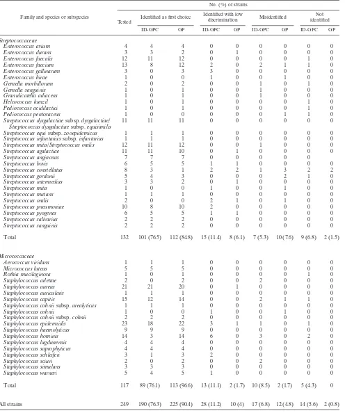
TABLE 1. Identification of gram-positive bacteria by fluorimetric and colorimetric VITEK 2 cards
Family and species or subspecies
No. (%) of strains
Tested
Identified as first choice Identified with low
discrimination Misidentified
Not identified ID-GPC GP ID-GPC GP ID-GPC GP ID-GPC GP
Streptococcaceae
Enterococcus avium 4 4 4 0 0 0 0 0 0
Enterococcus durans 3 3 2 0 1 0 0 0 0
Enterococcus faecalis 12 11 12 0 0 0 0 1 0
Enterococcus faecium 13 8 12 2 0 2 1 1 0
Enterococcus gallinarum 3 0 3 3 0 0 0 0 0
Enterococcus hirae 1 0 0 1 0 0 1 0 0
Gemella morbillorum 2 0 2 0 0 1 0 1 0
Gemella sanguinis 1 0 1 0 0 1 0 0 0
Granulicatella adiacens 1 0 1 0 0 1 0 0 0
Helcococcus kunzil 1 0 1 0 0 0 0 1 0
Pediococcus acidilactici 1 0 1 0 0 0 0 1 0
Pediococcus pentosaceus 1 0 0 0 0 0 1 1 0
Streptococcus dysgalactiaesubsp.dysgalactiae/ Streptococcus dysgalactiaesubsp.equisimilis
11 11 11 0 0 0 0 0 0
Streptococcus equisubsp.zooepidemicus 1 1 1 0 0 0 0 0 0 Streptococcus infantariussubsp.infantarius 1 1 1 0 0 0 0 0 0 Streptococcus mitis/Streptococcus oralis 12 11 12 0 0 1 0 0 0
Streptococcus agalactiae 11 11 10 0 1 0 0 0 0
Streptococcus anginosus 7 7 7 0 0 0 0 0
Streptococcus bovis 6 5 5 1 1 0 0 0 0
Streptococcus constellatus 8 3 1 2 2 1 3 2 2
Streptococcus gordonii 5 4 3 0 0 0 2 1 0
Streptococcus intermedius 3 3 2 0 1 0 0 0 0
Streptococcus mitis 1 0 0 1 0 0 1 0 0
Streptococcus mutans 1 1 1 0 0 0 0 0 0
Streptococcus oralis 2 0 0 2 1 0 1 0 0
Streptococcus pneumoniae 10 8 10 2 0 0 0 0 0
Streptococcus pyogenes 6 5 5 1 1 0 0 0 0
Streptococcus salivarius 2 2 2 0 0 0 0 0 0
Streptococcus sanguinis 2 2 2 0 0 0 0 0 0
Total 132 101 (76.5) 112 (84.8) 15 (11.4) 8 (6.1) 7 (5.3) 10 (7.6) 9 (6.8) 2 (1.5)
Micrococcaceae
Aerococcus viridans 1 1 1 0 0 0 0 0 0
Micrococcus luteus 5 5 5 0 0 0 0 0 0
Rothia mucilaginosa 1 0 1 0 0 0 0 1 0
Staphylococcus arlettae 2 0 2 0 0 2 0 0 0
Staphylococcus aureus 21 21 20 0 1 0 0 0 0
Staphylococcus auricularis 1 1 1 0 0 0 0 0 0
Staphylococcus capitis 15 12 14 0 0 2 1 1 0
Staphylococcus cohniisubsp.urealyticus 1 1 1 0 0 0 0 0 0
Staphylococcus cohnii 1 0 0 1 0 0 1 0 0
Staphylococcus cohniisubsp.cohnii 2 2 2 0 0 0 0 0 0
Staphylococcus epidermidis 23 18 22 3 1 1 0 1 0
Staphylococcus haemolyticus 9 9 9 0 0 0 0 0 0
Staphylococcus hominis 14 3 14 6 0 3 0 2 0
Staphylococcus lugdunensis 4 4 4 0 0 0 0 0 0
Staphylococcus saprophyticus 4 4 4 0 0 0 0 0 0
Staphylococcus schleiferi 3 1 3 2 0 0 0 0 0
Staphylococcus sciuri 2 0 2 0 0 2 0 0 0
Staphylococcus simulans 3 3 3 0 0 0 0 0 0
Staphylococcus warneri 5 4 5 1 0 0 0 0 0
Total 117 89 (76.1) 113 (96.6) 13 (11.1) 2 (1.7) 10 (8.5) 2 (1.7) 5 (4.3) 0
All strains 249 190 (76.3) 225 (90.4) 28 (11.2) 10 (4) 17 (6.8) 12 (4.8) 14 (5.6) 2 (0.8)
VOL. 43, 2005 PERFORMANCES OF VITEK 2 COLORIMETRIC CARDS 4403
on May 15, 2020 by guest
http://jcm.asm.org/
Family and species or subspecies
No. (%) of strains
Tested
Identified as first choice Identified with low
discrimination Misidentified Not identified ID-GPC GP ID-GPC GP ID-GPC GP ID-GPC GP
Fermenting
Aeromonas hydrophila/Aeromonas caviae 5 3 5 2 0 0 0 0 0
Citrobacter amalonaticus 1 1 1 0 0 0 0 0 0
Citrobacter braakii 2 0 2 2 0 0 0 0 0
Citrobacter freundii 6 6 6 0 0 0 0 0 0
Citrobacter koseri 11 11 11 0 0 0 0 0 0
Citrobacter youngae 1 1 1 0 0 0 0 0 0
Enterobacter aerogenes 18 17 17 1 0 0 0 0 1
Enterobacter amnigenus 1 0 1 1 0 0 0 0 0
Enterobacter asburiae 2 2 2 0 0 0 0 0 0
Enterobacter cloacae 15 10 14 4 0 1 1 0 0
Enterobacter gergoviae 1 1 1 0 0 0 0 0 0
Enterobacter sakazakii 1 1 1 0 0 0 0 0 0
Escherchia coli 26 24 25 1 1 1 0 0 0
Escherichia hermannii 2 2 2 0 0 0 0 0 0
Escherichia vulneris 1 0 1 0 0 1 0 0 0
Hafnia alvei 7 7 7 0 0 0 0 0 0
Klebsiella oxytoca 11 11 10 0 1 0 0 0 0
Klebsiella pneumoniaesubsp.pneumoniae 12 10 11 2 1 0 0 0 0 Klebsiella pneumoniaesubsp.ozaenae 1 1 1 0 0 0 0 0 0
Klebsiellaspp. 1 0 1 1 0 0 0 0 0
Leclercia adecarboxylata 1 1 1 0 0 0 0 0 0
Moellerella wisconsensis 1 1 1 0 0 0 0 0 0
Morganella morganii 11 11 11 0 0 0 0 0 0
Pantoea agglomerans 3 3 3 0 0 0 0 1 0
Pantoeaspp. 2 0 2 1 0 0 0 0 0
Pasteurella aerogenes 1 1 1 0 0 0 0 0 0
Pasteurella multocida 6 4 6 1 0 1 0 0 0
Pasteurella pneumotropica 2 2 2 0 0 0 0 0 0
Proteus mirabilis 17 17 17 0 0 0 0 0 0
Proteus vulgarisgroup/Proteus penneri 7 7 7 0 0 0 0 0 0
Providencia rettgeri 1 1 1 0 0 0 0 0 0
Providencia stuartii 11 11 11 0 0 0 0 0 0
Rahnella aquatilis 1 1 1 0 0 0 0 0 0
Salmonellagroup 12 9 12 3 0 0 0 0 0
Serratia liquefaciensgroup 1 1 1 0 0 0 0 0 0
Serratia marcescens 15 15 15 0 0 0 0 0 0
Serratia proteamaculans 1 0 1 1 0 0 0 0 0
Shigellagroup 3 1 3 2 0 0 0 0 0
Shigella sonnei 5 5 5 0 0 0 0 0 0
Vibrio alginolyticus 1 1 1 0 0 0 0 0 0
Yersinia enterocoliticagroup 13 11 13 1 0 0 0 1 0
Yersinia pseudotuberculosis 3 2 2 0 0 0 0 1 1
Total 243 213 (87.7) 237 (97.5) 23 (9.5) 3 (1.2) 4 (1.6) 1 (0.4) 3 (1.2) 2 (0.8)
Nonfermenting
Achromobacter xylosoxidanssubsp.denitrificans 1 0 0 0 1 0 0 1 0 Achromobacter xylosoxidanssubsp.xylosoxidans 7 0 6 0 1 5 0 2 0
Acinetobacter baumannii 8 6 8 2 0 0 0 0 0
Acinetobacter haemolyticus 4 0 4 0 0 3 0 1 0
Acinetobacter lwoffii 2 0 2 2 0 0 0 0 0
Acinetobacterspp. 5 0 1 2 1 0 3 3 0
Bordetella bronchiseptica 4 0 3 3 0 0 1 1 0
Bordetella trematum 1 0 0 0 1 1 0 0 0
Burkholderia cepacia 8 7 8 1 0 0 0 0 0
Chryseobacterium indologenes 2 2 2 0 0 0 0 0 0
Chryseobacterium meningosepticum 1 1 1 0 0 0 0 0 0
Comamonas testosteroni 1 0 1 0 0 0 0 1 0
Delftia acidovorans 1 0 1 1 0 0 0 0 0
Moraxella osloensis 1 0 0 1 1 0 0 0 0
Ochrobactrum anthropi 1 1 1 0 0 0 0 0 0
Oligella ureolytica 2 0 2 0 0 0 0 2 0
Pseudomonas aeruginosa 13 12 13 0 0 0 0 1 0
Continued on facing page
4404
on May 15, 2020 by guest
http://jcm.asm.org/
[image:3.585.49.541.82.717.2]RESULTS AND DISCUSSION
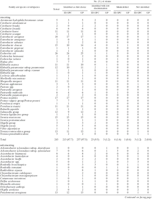
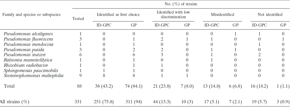
Of the 249 gram-positive strains tested with both the ID-GPC and GP cards and the 331 gram-negative strains tested with both the ID-GNB and GN cards, 218 (87.5%), 235 (94.4%), 295 (89.1%), and 321 (97%) were correctly identified (to the genus or species level), respectively (Tables 1 and 2). A total of 33 bacteria remained unidentified with the fluorimetric cards, whereas the colorimetric cards did not give an identifi-cation for five strains. Regardless of their origin (combined stock collection and clinical isolates), fermenting gram-nega-tive bacteria were correctly identified with both ID-GN and GN cards (97.2% and 98.7%, respectively). In contrast, non-fermenting gram-negative bacteria were better identified with GN cards (92.1%) than with ID-GN cards (67%). Gram-pos-itive bacteria were better identified with colorimetric cards than with fluorimetric cards. ForStreptococcaceae, readings of fluorimetric and colorimetric cards gave 87.9% and 90.9% correct identifications, respectively. ForMicrococcaceae, these readings were 87.2% and 98.3%, respectively. Our results ob-tained with gram-negative bacteria were for the most part in agreement with those reported by Funke and Funke-Kissling (3). Testing 511 fermenting and 144 nonfermenting gram-neg-ative bacilli, these authors (3) obtained slightly better results with the GN cards than we did in the present study (99.5% and 98.7%, respectively). Recently, in another study focusing on gram-positive bacteria, Funke and Funke-Kissling (4) obtained correct identification ofStreptococcaceae(217 strains) and Mi-crococcaceae(147 strains) for 99.1% and 99.3% of these bac-teria, respectively, whereas the results were 90.9% and 98.3% in our study. It should be noticed that the results reported by Funke and Funke-Kissling (3, 4) appeared better but that the numbers of taxa tested (13, 18, and 12 for Micrococcaceae, Streptococcaceae, and nonfermenting bacilli, respectively) were lower than in our study, except for fermenting bacteria (42 taxa in both studies). In fact, as Funke and Funke-Kissling have previously claimed to have done (3, 4), we have selected rare bacteria isolated in routine practice and some of them, such as S. constellatus or S. gordonii, while infrequently isolated in routine testing, were isolated in large numbers in our study (for
example, eight strains of S. constellatus instead of one in Funke’s study).
The GP and GN identification cards contain new tests (16 and 21 tests for GP and GN, respectively) allowing an improve-ment of the VITEK 2 databases; in fact, 57 and 38 new species of gram-positive cocci and gram-negative bacilli were added to the database. Of these, seven gram-positive species (eight strains) were tested and only one strain ofPediococcus pen-tosaceus was not correctly identified. Of the 17 new gram-negative species tested (36 strains), only four species (four strains) were not correctly identified:Bordetella bronchiseptica, Pseudomonas alcaligenes,and P. putidawere misidentified (one strain each), and one strain ofP. fluorescenswas not identified. Thus, the misidentification percentages obtained with the col-orimetric cards ranged from 2.1% to 4.8%, results which were slightly better than those obtained with the fluorescent cards (5.1% to 6.8%). These misidentified bacteria were observed only with Streptococcus spp. such as S. constellatus (three strains) or S. gordonii(two strains) or nonfermenting gram-negative bacilli such asAcinetobacterspp. belonging to geno-mospecies 1, 2, 3, or 13 (A. calcoaceticus/A. baumannii com-plex). Compared to previous study results (2, 5, 6), the database was enlarged and improved, especially for nonfer-menting bacilli and Micrococcaceae. The database was also enlarged to include some gram-positive bacilli such as Erysip-elothrix rhusiopathiaeand six species of theListeriagenus.
[image:4.585.44.547.80.267.2]The second aim of this study was to evaluate the perfor-mance of the new colorimetric cards in routine practice. Thus, we applied the percentages of correct identifications obtained in this study with the colorimetric and fluorescent cards to the list of bacteria isolated in our laboratory in the year 2004. From the species included in the database of the VITEK 2, 71 species were selected, representing 32,739 bacteria isolated five times or more in 2004. An overall correlation of 97.9% correct iden-tifications for gram-positive and gram-negative bacteria was obtained, whereas it was equal to 93.9% with fluorescent cards. The same determination was performed with a selection of 17 gram-negative taxa isolated more frequently in 33 French uni-versity hospitals (11). Identification with colorimetric cards
TABLE 2—Continued
Family and species or subspecies
No. (%) of strains
Tested
Identified as first choice Identified with low
discrimination Misidentified Not identified ID-GPC GP ID-GPC GP ID-GPC GP ID-GPC GP
Pseudomonas alcaligenes 1 0 0 0 0 0 1 1 0
Pseudomonas fluorescens 3 0 1 2 1 1 0 0 1
Pseudomonas mendocina 1 0 1 0 0 0 0 1 0
Pseudomonas putida 3 0 2 2 0 1 1 0 0
Pseudomonas stutzeri 6 0 6 3 0 1 0 2 0
Ralstonia mannitolilytica 1 0 1 0 0 1 0 0 0
Rhizobium radiobacter 1 0 1 1 0 0 0 0 0
Sphingomonas paucimobilis 1 1 1 0 0 0 0 0 0
Stenotrophomonas maltophilia 9 8 8 1 1 0 0 0 0
Total 88 38 (43.2) 74 (84.1) 21 (23.8) 7 (8.0) 13 (14.8) 6 (6.8) 16 (18.2) 1 (1.1)
All strains (%) 331 251 (75.8) 311 (94) 44 (13.3) 10 (3) 17 (5.1) 7 (2.1) 19 (5.7) 3 (0.9)
VOL. 43, 2005 PERFORMANCES OF VITEK 2 COLORIMETRIC CARDS 4405
on May 15, 2020 by guest
http://jcm.asm.org/
gave an overall identification to the species level of 99.7%, whereas it was equal to 95.9% with fluorescent cards.
The VITEK 2 system, equipped for colorimetric reading of the new GP and GN cards, keeps the advantages of the VITEK 2 (2, 5, 6, 8), i.e., reliable identification, fully automated incu-bation and interpretation, and minimal supplemental testing required. The results provided by the colorimetric VITEK 2 may be considered accurate due to the improvement and the extension of its database, mainly for nonfermenting bacteria and Strepto-coccaceae. In this study, the identifications of bacteria were pro-vided between 5.2 h (fermenting bacteria) and 6.7 h (nonferment-ing bacteria), which was slightly greater than the time required for reading with fluorescent cards. However, the results were always provided within a day. In conclusion, the results obtained in this study demonstrate the good performances of the new VITEK 2 cards, allowing their use in routine practice with a highly accept-able level of identification accuracy.
ACKNOWLEDGMENTS
We thank bioMe´rieux for kindly providing the VITEK 2 system and Genevie`ve Bossy and Marie-Christine Saccomani for their technical assistance.
REFERENCES
1.Fontana, C., M. Favaro, M. Pelliccioni, E. S. Pistoia, and C. Favalli.2005.
Use of the MicroSeq 500 16S rRNA gene-based sequencing for identification
of bacterial isolates that commercial automated systems failed to identify correctly. J. Clin. Microbiol.43:615–619.
2.Funke, G., D. Monnet, C. deBernardis, A. von Graevenitz, and J. Freney.
1998. Evaluation of the VITEK 2 system for rapid identification of medically relevant gram-negative rods. J. Clin. Microbiol.36:1948–1952.
3.Funke, G., and P. Funke-Kissling.2004. Evaluation of the new VITEK 2
card for identification of clinically relevant gram-negative rods. J. Clin. Microbiol.42:4067–4071.
4.Funke, G., and P. Funke-Kissling.2005. Performance of the new VITEK 2
card for identification of medically relevant gram-positive cocci in a routine clinical laboratory. J. Clin. Microbiol.43:84–88.
5.Jossart, M. F., and R. J. Courcol.1999. Evaluation of an automated system
for identification of Enterobacteriaceae and nonfermenting bacilli. Eur. J. Clin. Microbiol. Infect. Dis.18:902–907.
6.Ling, T. K. W., P. C. Tam, Z. K. Liu, and A. F. B. Cheng.2001. Evaluation
of VITEK 2 rapid identification and susceptibility testing system against gram-negative clinical isolates. J. Clin. Microbiol.39:2964–2966.
7.Mollet, C., M. Drancourt, and D. Raoult.1997.rpoBsequence analysis as a
novel basis for bacterial identification. Mol. Microbiol.26:1005–1011.
8.O’Hara, C. M., M. P. Weinstein, and J. M. Miller. 2003. Manual and
automated systems for detection and identification of microorganisms, p. 185–207.InP. R. Murray, E. J. Baron, J. H. Jorgensen, M. A. Pfaller, and R. H. Yolken (ed.). Manual of clinical microbiology, 8th ed. American Society for Microbiology, Washington, D.C.
9.Poyart, C., G. Quesne, C. Boumaila, and P. Trieu-Cuot.2001. Rapid and
accurate species-level identification of coagulase-negative staphylococci by using thesodAgene as a target. J. Clin. Microbiol.39:4296–4301.
10.Rantakokko-Jalava, K., S. Nikkari, J. Jalava, E. Eerola, M. Skurnik, O.
Meurman, O. Ruuskanen, A. Alanen, E. Kotilainen, P. Toivanen, and P.
Kotilainen.2000. Direct amplification of rRNA genes in diagnosis of
bacte-rial infections. J. Clin. Microbiol.38:32–39.
11.Rot, P., J. P. Mamet, and V. Goulet.1990. Releve´ des bacte´ries isole´es dans
les he´mocultures en 1987 et 1988. Bulletin Epide´miologique Hebdomadaire
33:142–143.