0095-1137/06/$08.00⫹0 doi:10.1128/JCM.00517-06
Copyright © 2006, American Society for Microbiology. All Rights Reserved.
Evaluation of the SPF
10
-INNO LiPA Human Papillomavirus (HPV)
Genotyping Test and the Roche Linear Array HPV Genotyping Test
Dennis van Hamont,
1,2Maaike A. P. C. van Ham,
2Judith M. J. E. Bakkers,
1Leon F. A. G. Massuger,
2and Willem J. G. Melchers
1*
Department of Medical Microbiology, Nijmegen University Centre for Infectious Diseases, Radboud University Nijmegen Medical Centre,
Nijmegen, The Netherlands,1and Department of Obstetrics and Gynaecology, Radboud University Nijmegen Medical Centre,
Nijmegen, The Netherlands2
Received 10 March 2006/Returned for modification 9 May 2006/Accepted 19 June 2006
The need for accurate genotyping of human papillomavirus (HPV) infections is becoming increasingly important, since (i) the oncogenic potential among the high-risk HPV genotypes varies in the pathogenesis of cervical cancer, (ii) monitoring multivalent HPV vaccines is essential to investigate the efficiency of the vaccines, and (iii) genotyping is crucial in epidemiologic studies evaluating HPV infections worldwide. Various genotyping assays have been developed to meet this demand. Comparison of different studies that use various HPV genotyping tests is possible only after a performance assessment of the different assays. In the present study, the SPF10LiPA version 1 and the recently launched Roche Linear Array HPV genotyping assays are
compared. A total of 573 liquid-based cytology samples were tested for the presence of HPV by a DNA enzyme immunoassay; 210 were found to be positive for HPV DNA and were evaluated using both genotyping assays (163 with normal cytology, 22 with atypical squamous cells of undetermined significance, 20 with mild/ moderate dysplasia, and 5 with severe dysplasia). Comparison analysis was limited to the HPV genotype probes common to both assays. Of the 160 samples used for comparison analysis, 129 (80.6%) showed absolute agreement between the assays (concordant), 18 (11.2%) showed correspondence for some but not all genotypes detected on both strips (compatible), and the remaining 13 (8.2%) samples did not show any similarity between the tests (discordant). The overall intertest comparison agreement for all individually detectable genotypes was considered very good ( value, 0.79). The genotyping assays were therefore highly comparable and reproducible.
Molecular and epidemiologic studies have shown that a per-sistent infection with high-risk human papillomavirus (HPV) is the most important risk factor for both cervical cancer and its precursors (9, 11, 29, 33). Approximately 40 different HPV types can infect the mucosa of the anogenital tract. Based on their carcinogenicities, these anogenital HPV types have been subdivided into low-risk HPV (lr-HPV) types, probable high-risk HPV (hr-HPV) types, and hr-HPV types (27), although some controversy remains regarding the probable high-risk genotypes (30). Almost all squamous cell cervical cancers worldwide harbor hr-HPV types (36). Moreover, high-risk HPV DNA can be detected in 74% of the premalignant low-grade cervical intraepithelial neoplasia (CIN) lesions and ap-proximately 84% of the high-grade CIN lesions (25). Conse-quently, the efficacy of population-based screening programs solely using cervical cytology could benefit from adding hr-HPV testing (32). Accordingly, many ongoing international research projects assess the feasibility of introducing hr-HPV tests in available routine screening.
For these screening purposes, several tests have been devel-oped in order to distinguish high-risk HPV infections from no HPV infection. Among these are the signal amplification
method Hybrid Capture II (hc2) (Digene Corp., Gaithersburg, Maryland) and the recently developed target amplification method Roche AMPLICOR HPV test (Roche Molecular Sys-tems, Inc., Branchburg, NJ) (35). Although both tests are com-mercially available and Conformite´ Europe´enne (CE) marked, hc2 is currently the only FDA-registered HPV screening assay (7). Both tests differentiate between an infection with one or more of 13 hr-HPV genotypes (genotypes 16, 18, 31, 33, 35, 39, 45, 51, 52, 56, 58, 59, and 68) and no hr-HPV infection—an “hr-HPV plus/minus” screening. Although these tests are not designed to detect the recently described probable hr-HPV or any lr-HPV infection, some cross-reactivity outside of the spec-trum of 13 hr-HPV genotypes has been reported for the hc2 assay (5). Neither the hc2 nor the AMPLICOR HPV assay allows the identification of specific genotypes (26), nor do they have the ability to identify infections involving multiple geno-types.
However, recent studies have provided evidence for a dif-ference in oncogenic potential between the different hr-HPVs (6), arguing for the importance of HPV genotyping in addition to the “hr-HPV plus/minus” screening. Outside of the clinical setting, HPV genotyping is a key characteristic of studies eval-uating the epidemiology of HPV infections worldwide. Al-though a number of HPV genotyping assays have been used in such studies, a reliable comparison between the diagnostic and epidemiological data generated is difficult, since data on the intertest comparisons between the different genotyping assays are limited.
* Corresponding author. Mailing address: Department of Medical Microbiology (internal postal code 699), Nijmegen Centre for Molec-ular Life Sciences, Nijmegen University Centre for Infectious Dis-eases, Radboud University Nijmegen Medical Centre, P.O. Box 9101, 6500 HB Nijmegen, The Netherlands. Phone: 31 (0)24 3614356. Fax: 31 (0)24 3540216. E-mail: [email protected].
3122
on May 16, 2020 by guest
http://jcm.asm.org/
The SPF10-INNO LiPA assay is capable of amplifying up to
43 different genotypes and providing type-specific genotype information for 25 different HPV genotypes simultaneously, has been extensively tested, and has proven to be highly sen-sitive and specific (15, 25). The Roche Linear Array (LA) HPV genotyping test (Roche Molecular Systems, Inc., Branchburg, NJ) is a recently launched new HPV genotyping assay able to genotype 37 HPV types, concurrently assessing human -glo-bin. The full spectrum of HPV genotypes amplified by the PGMY primer system (13) used in the Roche Linear Array HPV genotyping test has not been assessed beyond the 37 genotypes probed. In essence, both assays could be used for genotyping analysis.
This study was designed to compare these two well-known and commonly used commercially available genotyping assays with HPV DNA-positive samples.
MATERIALS AND METHODS
Cervical scrapes were obtained from 573 women attending the Department of Gynaecology for routine cervical screening. Specimens were collected using the Cervex-Brush (Rovers Medical Devices B.V., Oss, The Netherlands) and processed using a liquid-based cytology medium (ThinPrep; Cytyc Corp., Marlborough, MA) that provides monolayer distribution for cytological assessment. Moreover, it offers the opportunity to isolate DNA for various HPV detection assays. This method has received U.S. FDA approval for clinical use (20, 31).
Specimen preparation.For isolation of DNA from cervical scrapes in liquid-based cytology medium, the MagNAPure LC isolation station (Roche Diagnos-tics GmbH, Roche Applied Science, Mannheim, Germany) was used; 200l of material was isolated using the MagNA Pure LC Total Nucleic Acid Isolation kit (Roche Diagnostics GmbH, Roche Molecular Biochemicals, Mannheim Ger-many), as described by the manufacturer. With each set of 28 cervical-scrape samples, four negative controls (distilled water) were used to monitor the DNA isolation procedure and to assess contamination. Nucleic acid was resuspended in a final volume of 50l; 10l was used for each of the various PCR analyses.
SPF10-INNO LiPA HPV detection and genotyping (DNA enzyme
immunoas-say [DEIA] and LiPA). (i) PCR amplification of HPV DNA.Broad-spectrum HPV DNA amplification was performed using a short-PCR-fragment assay (SPF10HPV PCR; Labo Bio-Medical Products B.V., Rijswijk, The Netherlands). This assay amplifies a 65-bp fragment of the L1 open reading frame and allows detection of at least 43 different HPV types (16, 25). The SPF10PCR system was used in a final reaction volume of 50l containing 10l of the isolated DNA sample and 40l of the PCR mixture, which contained 10 mmol/liter Tris-HCl (pH 9.0), 50 mmol/liter KCl, 2.0 mmol/liter MgCl2, 0.1% Triton X-100, 0.01% gelatin, 200mol/liter of each deoxynucleoside triphosphate (dATP, dCTP, dGTP, and dTTP), 15 pmol each of the forward and reverse primers tagged with biotin at the 5⬘end, and 1.5 units of AmpliTaq Gold (Applied Biosystems, Foster City, CA). Activation of AmpliTaq Gold for 9 min at 94°C, was followed by 40 cycles of 30 s at 94°C, 45 s at 52°C, and 45 s at 72°C, with a final extension of 5 min at 72°C. Appropriate negative and positive controls were used to monitor the performance of the PCR method in each experiment.
(ii) HPV detection by DEIA.The presence of HPV DNA was determined by hybridization of SPF10amplimers to a mixture of general HPV probes recogniz-ing a broad range of high-risk, low-risk, and possible high-risk HPV genotypes in a microtiter plate format, as described previously (15, 25). All HPV DNA-positive samples (by SPF10DEIA) were genotyped using the INNO-LiPA HPV genotyping assays and the Roche Linear Array HPV genotyping test as described below. Twenty randomly selected DEIA-negative samples that had previously tested negative by the Roche AMPLICOR HPV test (35) were also assessed using both genotyping assays.
(iii) HPV genotyping by reverse hybridization using the INNO-LiPA HPV genotyping system.The 28 oligonucleotide probes that recognize 25 different types (Table 1) were tailed with poly(dT) and immobilized as parallel lines to membrane strips (Labo Bio-Medical Products B.V., Rijswijk, The Netherlands). The HPV genotyping assay was performed as described previously (15). The LiPA strips were manually interpreted using the reference guide provided.
The samples that tested positive using the DNA enzyme immunoassay but that showed no results on the LiPA strip were considered to be HPV X type, i.e., genotypes not available on the LiPA strip.
Linear Array HPV genotyping test.The LA HPV genotyping test (Roche Molecular Systems, Inc., Branchburg, NJ) is a new qualitative in vitro test for the determination of 37 anogenital HPV DNA genotypes (Table 1). The LA test was applied to all samples that tested positive for HPV by DEIA and to 20 randomly selected DEIA-negative samples.
[image:2.585.298.540.80.505.2](i) PCR amplification of HPV DNA.The LA test uses biotinylated PGMY primers to amplify a 450-bp fragment within the polymorphic L1 region of the HPV genome. The PGMY amplification system has been described previously (13). The PGMY primers are present in the “master mixture” (containing buffer, nucleotides [dATP, dCTP, dGTP, and dUTP], MgCl2, and⬍0.02% AmpliTaq Gold DNA polymerase) and amplify HPV DNA from 37 HPV genotypes, in-cluding 13 high-risk types (Table 1). Amplicons incorporate dUTP, allowing the use of AmpErase enzyme (uracilN-glycosylase), which is included in the master mixture to prevent PCR carryover contamination. Capture probe sequences are located in polymorphic regions of L1 bound by these primers. An additional primer pair targets the human-globin gene (268-bp amplicon) to provide a control for cell adequacy, extraction, and amplification.
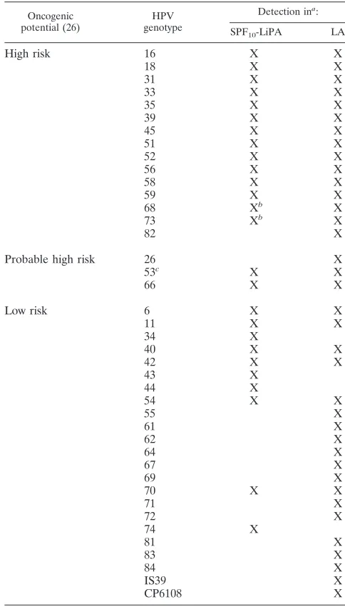
TABLE 1. Distribution of HPV genotypes in the LiPA and LA assays
Oncogenic potential (26)
HPV genotype
Detection ina:
SPF10-LiPA LA
High risk 16 X X
18 X X
31 X X
33 X X
35 X X
39 X X
45 X X
51 X X
52 X X
56 X X
58 X X
59 X X
68 Xb X
73 Xb X
82 X
Probable high risk 26 X
53c X X
66 X X
Low risk 6 X X
11 X X
34 X
40 X X
42 X X
43 X
44 X
54 X X
55 X
61 X
62 X
64 X
67 X
69 X
70 X X
71 X
72 X
74 X
81 X
83 X
84 X
IS39 X
CP6108 X
aX, detected.
bLiPA does not distinguish between HPV 68 and HPV 73, since both types are
detected by a single probe.
cThe oncogenic potential of HPV 53 is controversial (30).
on May 16, 2020 by guest
http://jcm.asm.org/
PCR was performed in a final reaction volume of 100l, containing 50l HPV master mixture, 40l PCR water, and 10l isolated DNA. The mixture was incubated for 2 min at 50°C and for 9 min at 95°C, followed by 40 cycles of 30 seconds at 95°C, 1 min at 55°C, and 1 min at 72°C, with a final extension at 72°C lasting from 10 min to a maximum of 1 h. The provided HPV-positive and -negative controls were used with each set of 10 samples to assess the perfor-mance of the reaction.
(ii) Hybridization and detection.Following amplification, the HPV and hu-man-globin amplicons were denatured by immediately adding 100l denatur-ation solution to each PCR tube. Hybridizdenatur-ation and HPV genotyping were performed as described by the manufacture (Roche Molecular Systems, Inc., Branchburg, NJ). The strips were manually interpreted using the Linear Array HPV reference guide, by reading the individual types down the length of the strip. Samples that were both SPF10DEIA and LA-globin positive yet were not reactive to any of the genotype probes on the LA strip were considered “LA negative.”
Design of the study.Previously, the samples had been assessed in an analysis comparing only high-risk HPV types detected by the Roche AMPLICOR HPV test and the INNO-LiPA HPV detection and genotyping assay (35). Since the present study compares two genotyping assays, only the DEIA HPV-positive samples and 20 randomly selected DEIA (and Roche AMPLICOR) HPV-neg-ative samples were assessed. In order to have the most accurate comparison between the two genotyping tests, only the HPV genotypes identified by both assays (i.e., lr-HPV 6, 11, 40, 42, 54, and 70; possible hr-HPV 53 and 66; and hr-HPV 16, 18, 31, 33, 35, 39, 45, 51, 52, 56, 58, and 59) were considered for direct comparison of the individual HPV genotypes (Table 1). These will be referred to as assay-common genotypes. High-risk HPV genotypes 68 and 73 were not taken into account for individual comparison, since these types are identified by a single probe in the LiPA assay and thus cannot be distinguished. Moreover, the classification of HPV 53 as possibly high risk is currently disputed. When comparing the two genotyping assays, results were termed concordant, compatible, or discordant based on the following definitions. If the analyses yielded identical assay-common genotypes in both tests, the results were termed concordant. Results were termed compatible if one or more additional assay-common genotypes were not detected by either of the assays. Genotyping results were termed discordant if there were no similarities in the assay-common geno-types between the two tests. Assay results for HPV genogeno-types uniquely identified by each of the two assays (i.e., assay-unique HPV genotypes 34, 43, 44, and 74 detected only by LiPA and the assay-unique HPV genotypes 26, 55, 61, 62, 64, 67, 69, 71, 72, 81, 82, 83, 84, IS39, and CP6108 detected solely by the LA test) were not considered in determining concordant, compatible, or discordant status.
From all compatible and discordant samples, a reextracted DNA sample was randomly retested in a blind approach in a discrepancy analysis using both genotyping assays. Eleven concordant samples (six single infections, four double infections, and one triple infection) and six double-negative (i.e., DEIA-positive, LiPA X-type, and LA-negative) samples were used as positive and negative controls for both inter- and intra-assay performance control.
All HPV tests were performed by investigators unaware of the results of the comparative HPV detection or genotyping tests.
Statistics.All data were analyzed using SPSS version 12.0.1. for Windows. Agreement was measured by absolute agreement and Cohen’s kappa statistics, a measure of the agreement between two methods that is in excess of that due to chance.
RESULTS
In total, 218 of the 573 DNA samples tested positive by SPF10DEIA. These were considered suitable for analysis using
the SPF10LiPA and LA HPV genotyping assays. Eight samples
were excluded from further analysis: four showed negative -globin results in the LA test, and for four other samples, insufficient material was available to perform adequate assess-ments. Twenty randomly selected DEIA-negative control sam-ples were negative in both genotyping assays and were thus not taken into consideration for further analysis. Of the 210 DEIA-positive samples, 163 (77.6%) indicated normal cytology. Atyp-ical squamous cells of undetermined significance (ASCUS) were detected in 22 samples (10.5%), mild/moderate dysplasia was observed in 20 samples (9.5%), and 5 samples (2.4%) showed severe dysplasia.
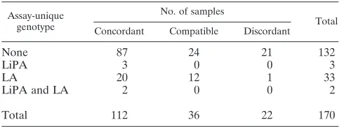
Of the 210 DEIA-positive samples tested using both geno-typing assays, 40 samples were excluded, since one of the tests was negative whereas the comparative test detected an assay-unique genotype or LA was negative and LiPA showed an X type (Table 2).
In 132 of the remaining 170 samples, all detected genotypes could have been identified by both assays. Of the samples harboring only assay-common genotypes, 87/132 (65.9%) were concordant, 24 (18.2%) were compatible, and 21 (15.9%) showed discordant results (Table 3). Finally, in 38 cases, assay-unique genotypes were detected in addition to assay-common genotypes. Of these samples, 25 (65.8%) had concordant re-sults, 12 (31.6%) were compatible, and one (2.6%) was discor-dant. In the final analysis of 170 samples, these 38 samples were retained. The additional assay-unique genotypes found in these 38 samples were not taken into consideration. The out-comes of the concordant, compatible, and discordant cases are described in detail below.
Concordant cases.Of the 112 concordant cases (25 with and 87 without assay-unique genotypes), 69 (61.6%) contained a single HPV genotype and the remaining 43 samples contained multiple genotypes. Thirty-two samples (28.6%) harbored two different genotypes, eight samples (7.1%) contained three HPV genotypes, and three samples (2.7%) contained four ge-notypes. One or more high-risk genotypes were detected in 86.6% (97/112) of these samples, whereas seven samples (6.3%) contained only low-risk genotypes and eight samples (7.1%) also harbored probable hr-HPV genotypes.
[image:3.585.300.545.89.180.2]Compatible cases. All 36 compatible cases were multiple infections. The LiPA assay did not detect a total of 41 geno-types in 30 separate clinical samples. In 23 cases, 1 type was missed; in 5 cases, 2 types were missed; and in 2 cases, 4 types
TABLE 2. Distribution of 40 excluded samples that either showed only assay-unique genotypes or were HPV DNA positive but
genotype negative (i.e., LiPA X type)
LA result
No. of samples with indicated result by SPF10-LiPA
Total LiPA X
type
Assay-unique genotype
Negative 9 7 16
Assay-unique genotype 24 0 24
Total 33 7 40
TABLE 3. Overview of the 170 included samples with assay-common genotypes
Assay-unique genotype
No. of samples
Total Concordant Compatible Discordant
None 87 24 21 132
LiPA 3 0 0 3
LA 20 12 1 33
LiPA and LA 2 0 0 2
Total 112 36 22 170
on May 16, 2020 by guest
http://jcm.asm.org/
[image:3.585.43.282.98.186.2]were missed (13 low-risk, 1 possible high-risk, and 27 high-risk genotypes were not detected by the LiPA test). The Linear Array assay, on the other hand, did not detect 12 genotypes in eight separate samples. In six cases, 1 type was missed; in one
case, 2 types were missed; and in one case, 4 types were missed (2 low-risk, 1 possible high-risk, and 9 high-risk HPV types). Table 4 gives an overview of the individual types that were not detected. Fifteen of the 16 cases in which LiPA missed an hr-HPV type were samples infected with multiple hr-HPV types that tested positive for another high-risk type, which was also detected in the LA.
Discordant samples.In 22 (12.9%) of the 170 samples con-sidered, no similarity was observed between the genotypes found in the two tests. These were predominantly single infec-tions. An overview of the individual discordant cases is given in Table 4. Twenty-seven genotypes were discrepant between the two assays in 22 different samples. The LA test did not detect 13 hr-HPV, 5 probable hr-HPV, and 3 lr-HPV types that were found to be positive in the LiPA assay. The LiPA assay, on the other hand, failed to detect two risk, one probable high-risk, and three low-risk types, which were all found to be positive on the LA strip.
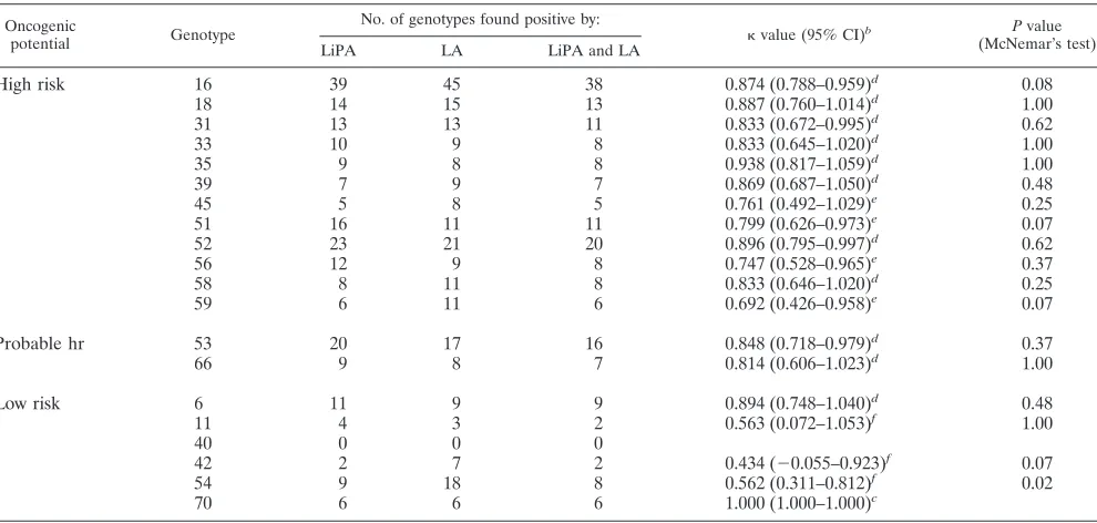
The genotypes that were detectable by both assays among all 170 samples (112 concordant, 36 compatible, and 22 discor-dant) were individually compared, as summarized in Table 5. The overall strength of agreement between the two assays for
the individual genotypes was considered good ( ⫽ 0.792).
[image:4.585.42.284.81.348.2]Although HPV 16 was detected in 45 samples using the LA test and in 39 samples using the LiPA, agreement between the tests was considered very good, with avalue of 0.874. The agree-ment between the two assays for the other high-risk and prob-able high-risk genotypes varied between “good” and “very good.” The agreement between the two tests for the low-risk genotypes was “moderate” to “perfect.” The agreement for
TABLE 4. Overview of the 36 compatible and 22 discordant samples
Oncogenic
potential Genotype
No. of specific genotypes not detected
Compatible samples
Discordant samples
LiPA LA LiPA LA
High risk 16 7 1
18 2 1
31 2 2
33 1 2
35 1
39 3
45 2 1
51 1 4
52 1 1 2
56 3 1 1
58 3
59 5
68/73 1 1 2
Probable hr 53 1 1 3
66 1 2
Low risk 6 2
11 1 2
42 4 1
54 8 2 1
[image:4.585.46.541.440.676.2]Total 41 12 6 21
TABLE 5. Kappa values andPvalues by McNemar’s test for individual HPV genotypes detectable by both assaysa
Oncogenic
potential Genotype
No. of genotypes found positive by:
value (95% CI)b Pvalue
(McNemar’s test)
LiPA LA LiPA and LA
High risk 16 39 45 38 0.874 (0.788–0.959)d 0.08
18 14 15 13 0.887 (0.760–1.014)d 1.00
31 13 13 11 0.833 (0.672–0.995)d 0.62
33 10 9 8 0.833 (0.645–1.020)d 1.00
35 9 8 8 0.938 (0.817–1.059)d 1.00
39 7 9 7 0.869 (0.687–1.050)d 0.48
45 5 8 5 0.761 (0.492–1.029)e 0.25
51 16 11 11 0.799 (0.626–0.973)e 0.07
52 23 21 20 0.896 (0.795–0.997)d 0.62
56 12 9 8 0.747 (0.528–0.965)e 0.37
58 8 11 8 0.833 (0.646–1.020)d 0.25
59 6 11 6 0.692 (0.426–0.958)e 0.07
Probable hr 53 20 17 16 0.848 (0.718–0.979)d 0.37
66 9 8 7 0.814 (0.606–1.023)d 1.00
Low risk 6 11 9 9 0.894 (0.748–1.040)d 0.48
11 4 3 2 0.563 (0.072–1.053)f 1.00
40 0 0 0
42 2 7 2 0.434 (⫺0.055–0.923)f 0.07
54 9 18 8 0.562 (0.311–0.812)f 0.02
70 6 6 6 1.000 (1.000–1.000)c
aThe results for 112 concordant, 36 compatible, and 22 discordant samples after initial analysis are shown. bCI, confidence interval.
cStrength of agreement considered perfect. dStrength of agreement considered very good. eStrength of agreement considered good. fStrength of agreement considered moderate.
on May 16, 2020 by guest
http://jcm.asm.org/
HPV 54 was moderate, since LiPA and LA shared 8 samples harboring the low-risk genotype whereas LA detected it in 10 additional samples. Also, the agreement for lr-HPVs 11 and 42 was moderate, while HPV 70 was detected in equal amounts by both assays. Low-risk HPV 40 was not detected in either of the tests; thus, no agreement could be calculated. The difference in detection of lr-HPV 54 was statistically significant (P⬍ 0.05; McNemar’s test). Although the differences for hr-HPV 16, 51, and 59 and lr-HPV 42 between the assays were large, they were considered not quite statistically significant (P⬎0.07; McNemar’s test). In the individual comparison of the other genotypes, no statistically significant differences were detected.
Discrepancy analysis.The compatible (n⫽36) and discor-dant (n⫽22) samples were reanalyzed using the two
[image:5.585.49.540.81.549.2]genotyp-ing assays in a discrepancy analysis. DNA was reextracted from these 58 compatible/discordant samples. As interassay test controls, 11 previously concordant (6 single and 5 multiple infections) and 6 previously double-negative samples (LiPA X type and LA negative) were also included; these samples were used for method performance assessment only. All 6 double-negative samples remained double-negative, and all 11 concordant samples appeared identical in both second genotyping assays. These internal controls were not further considered in the discrepancy analysis. Of the 58 discrepant samples, 10 were -globin negative by the Linear Array and were also negative by LiPA. Of these 10 samples, 6 had been concordant and 4 had been discordant; these 10 samples were excluded from the discrepancy analysis. The crude initial and discrepancy analysis
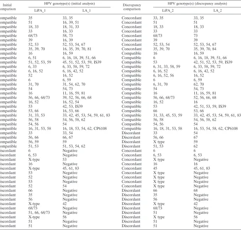
TABLE 6. All genotyping and comparison results for the 35 initially compatible and discordant samples assessed by discrepancy analysis
Initial comparison
HPV genotype(s) (initial analysis) Discrepancy
comparison
HPV genotype(s) (discrepancy analysis)
LiPA_1 LA_1 LiPA_2 LA_2
Compatible 35 33, 35 Concordant 33, 35 33, 35
Compatible 51 16, 39, 51 Concordant 51 51
Compatible 18, 33 18, 31, 33 Concordant 18, 33 18, 33
Compatible 33 16, 33 Concordant 33 33
Compatible 68/73 58, 73 Concordant 68/73 73
Compatible 39 16, 39 Concordant 39 39
Compatible 52, 53 52, 53, 54, 67 Concordant 52, 53, 54 52, 53, 54, 67
Compatible 35, 39, 70 16, 35, 39, 70, 81 Concordant 35, 39, 70 35, 39, 70, 84
Compatible 16 16, 59 Compatible 16 16, 59
Compatible 6, 51 6, 16, 18, 39, 51, 66 Compatible 6 6, 16, 18, 39, 51, 66
Compatible 51, 52, 53, 59 45, 51, 52, 53, 59, IS39 Compatible 53 42, 51, 52, 53, 59, IS39 Compatible 6, 33 6, 33, 58, 59, 72 Compatible 6, 31, 33, 58, 59 6, 33, 58, 59, 72
Compatible 6, 16, 52 6, 16, 42, 52 Compatible 6, 16, 52 6, 16, 42, 52
Compatible 52 16, 52 Compatible 6, 16, 52, 56 16, 52
Compatible 6 6, 59 Compatible 6 6, 59
Compatible 31, 70 31, 54, 62, 70 Compatible 6, 31, 70 62, 70
Compatible 54 54, 73 Compatible 54 54, 73
Compatible 16 11, 16, 59, 81 Compatible 16 11, 16, 59, 81
Compatible 56, 66, 68/73 39, 52, 56, 66, 68 Compatible 56, 66, 68/73 52, 56, 66, 68
Compatible 16, 52 16, 52, 54 Compatible 16, 52 16
Compatible 53 42, 53, IS39 Compatible 53 42, 51, 53, 59, IS39
Compatible 53, 66 16, 53, 66 Compatible 66 53, 66
Compatible 31, 33, 53 33, 42, 45, 53, 54, 59, 61, 83 Compatible 31, 33, 45, 53, 59 33, 42, 45, 53, 54, 59, 61, 83
Compatible 56, 58 54, 56, 58, 62 Compatible 56, 58 54, 56, 58, 62
Compatible 54, 56 54 Compatible 54, 56 54
Compatible 16, 31, 53, 58 16, 18, 53, 54, 62, CP6108 Compatible 16, 18, 31, 53, 58 16, 53, 54, 58, 62, CP6108
Compatible 33 33, 54 Discordant 33 54
Compatible 56, 66 66, 67 Discordant 56, 66 67
Compatible 56, 59 59 Discordant X type 59
Compatible 51, 53 51, 53, 54, 62 Discordant 51, 53 62
Discordant 6 Negative Concordant 6 6
Discordant 6, 53 Negative Concordant 6, 53 6, 53
Discordant X-type 53 Concordant X type Negative
Discordant 16 Negative Concordant 16 16
Discordant X-type 45, 61, 83 Concordant 45 45, 61, 83
Discordant 53 Negative Concordant X type Negative
Discordant 52 Negative Concordant X type Negative
Discordant 53 Negative Concordant X type Negative
Discordant 52 54 Concordant X type Negative
Discordant 66 Negative Discordant 66 68
Discordant 35 Negative Discordant 35 Negative
Discordant 56 Negative Discordant 56 Negative
Discordant X-type 42 Discordant X type 42
Discordant 68/73 Negative Discordant 68/73 Negative
Discordant 51, 66, 68/73 Negative Discordant 51 Negative
Discordant X-type 56 Discordant X type 56
Discordant 51 Negative Discordant 51 Negative
Discordant 51 Negative Discordant 51 Negative
on May 16, 2020 by guest
http://jcm.asm.org/
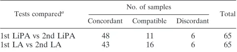
results for the remaining 48 samples are shown in Table 6. Of the 30 compatible samples from the initial analysis, 18 re-mained compatible after discrepancy analysis, while 8 ap-peared concordant and 4 discordant in a comparison of the second genotyping assays. Of the 18 discordant samples from the first test run, 9 remained discordant in the second analyses between LiPA and LA, whereas 4 appeared genotype concor-dant and 5 were concorconcor-dant as LiPA X type, LA negative. Thus, comparing the second LiPA and LA tests yielded 17 concordant, 18 compatible, and 13 discordant results.
Intra-assay comparisons taking these 48 samples and the 17 control samples in both initial and discrepancy analyses into account showed highly comparable results for the two assays (Table 7).
In conclusion, of the 160 samples considered for final anal-ysis, 80.6% (129/160) showed identical results, 11.2% (18/160) appeared compatible, and 13 samples (8.2%) were discordant.
DISCUSSION
Based on this study, we can conclude that the SPF10-INNO
LiPA and the Linear Array HPV genotyping assays are highly congruent for the genotypes detectable in both assays. More-over, the manageabilities of both the SPF10-INNO LiPA and
the Linear Array assays are highly comparable, as are to a large extent the total run times required for both assays, including for amplification and preparation of all of the reagents.
Generally, a separate screening is needed preceding geno-typing in order to assess a sample’s HPV DNA positivity, i.e., an HPV plus/minus screening. An advantage of the LiPA is the use of the same amplicon for both detection of 43 different lr-, probable hr-, and hr-HPV genotypes and genotyping of 25 different HPVs. For the LA, a prescreening test with the PGMY primers is available using a generic HPV probe labeled with digoxigenin in a microtiter plate-based assay, as recently described (18). Without the need for further amplification, this amplicon can be directly used for the Linear Array genotyping assay. However, the efficiency of such a combination has not been studied. The recently launched HPV Roche AMPLICOR test for HPV plus/minus screening is not meant for an LA screen. It could also be used as a pretest, but the assay detects only high-risk HPV types (35).
In the initial comparison, i.e., prior to the discrepancy anal-ysis, LiPA did not detect 27 high-risk genotypes in 30 compat-ible cases. Evidently, all the cases involved were multiple in-fections, i.e., containing two or more HPV types. Apparently, if an infection encompasses multiple genotypes, the SPF10
-INNO LiPA assay is less sensitive than the LA. After finding analogous results using the LiPA assay, Van Doorn et al.
propounded the idea of PCR competition between genotypes in mixed infections and suggested a combined testing algo-rithm using broad-spectrum and type-specific PCRs for HPV 16 and HPV 18 (L. J. van Doorn, A. C. Molijn, B. Kleter, W. G. V. Quint, and B. Colau, Abstr. 22nd IPV Conf., abstr. N-01, 2005). The complexity of assessing multiple genotypes was addressed previously (34). Amplification and identification of two genotypes present in equimolar amounts are likely pos-sible. However, “primer competition” between genotypes might occur if one genotype is present in molar excess, out-competing the other (34). In the present study, this was dem-onstrated by the samples harboring multiple infections that were not identically genotyped by both assays. Also, LA de-tected hr-HPV 16 in seven samples that were LiPA HPV 16 negative; after the second LA, however, five samples no longer showed HPV 16. Moreover, in a previous study Van Doorn and colleagues detected HPV 16 and HPV 18 using type-specific PCR in samples negative for these genotypes (but not for other genotypes) using general primer sets (34). In the present study we observed similar results (data not shown). Although the viral load was not determined in the present study, low-copy-number samples have previously shown more discrepancies in intralaboratory and interlaboratory compari-sons (17).
The LA assay is unable to distinguish hr-HPV 52 from other high-risk genotypes (33, 35, and 58). This could be inconve-nient in future studies using the Linear Array, since hr-HPV 52 is prevalent in approximately 5% of the HPV-positive women with normal cytology (8) and causes 2.2% of all cervical can-cers (27). In 19 samples from the present study, hr-HPV 52 positivity could not be excluded based on LA genotyping. However, in these cases, the comparative LiPA tests did not detect this specific genotype. Two samples were considered Linear Array HPV 52 positive based on the LiPA results.
[image:6.585.43.284.99.152.2]Among the 22 discordant cases, the number of hr-HPV genotypes detected by the Linear Array was not higher than the number detected by LiPA. All but three of these samples were single infections, predominately HPV 33, HPV 51, and HPV 52. A higher inclusivity level has been observed for some high- and low-risk HPV genotypes, particularly hr-HPV 33 and hr-HPV 56, when the PGMY amplification system is used (see the product insert for the CE-marked Linear Array HPV geno-typing test, European market). The inclusivity level equates to the lowest concentration (copies/ml) that shows a 100% posi-tive hit rate in a replicate of six tests or the concentration that is the probit-predicted 95% positive hit rate. This could explain some of the differences between the two assays observed in our study. Thus, the LA seems to be less sensitive than the LiPA if a sample has a single infection with some specific HPV geno-types that are poorly amplified by PGMY. Even though the majority of samples were cytologically classified as normal, proper HPV assessment, including genotyping, remains essen-tial, particularly for healthy women with normal cytology (35), especially since Wallin and colleagues observed a strong con-cordance between the HPV type found in baseline smears with normal cytology and the eventual type found in histological samples of invasive cancers (37). In the present study, hr-HPV 51 was missed by LA in four of the discordant cases; this genotype accounted for approximately 0.9% of all squamous cell cervical cancers in previous studies (27). Curiously, the
TABLE 7. Intra-assay comparison overview of the 65 samples reanalyzed in the discrepancy analysis, including the 17
control samples concordant in all four assays
Tests compareda No. of samples
Total Concordant Compatible Discordant
1st LiPA vs 2nd LiPA 48 11 6 65
1st LA vs 2nd LA 43 16 6 65
a
1st, initial comparison; 2nd, discrepancy comparison.
on May 16, 2020 by guest
http://jcm.asm.org/
inclusivity level for HPV 51 is lower than the level for HPV 16 using PGMY primers, suggesting highly sensitive detection (see the product insert for the CE-marked Linear Array HPV genotyping test, European market). The observed difference in HPV 51 detection between the two assays thus cannot be explained by a lower efficiency of the Linear Array PGMY primer.
After discrepancy analysis of the compatible and discordant cases, both LiPA and LA detected more concordance (Table 6). Some previously undetected genotypes, for example, ap-peared in the second test run, and vice versa. This could be due to low copy numbers or to sampling, as DNA reextracts were used for the analysis. Also, it could possibly indicate the sug-gested competition between genotypes present in more or less molar excess. However, results from a discrepancy analysis should generally be handled with care and interpreted care-fully. Discrepancy analyses are not perfect, since an analysis is easily biased in favor of the new test, and hard and fast rules do not exist (23). Moreover, the interpretation of results that cannot be dichotomized (i.e., concordant, compatible, and dis-cordant) is less straightforward.
Failing to detect genotypes will lead to underestimation of the prevalence of certain genotypes and will cause false-nega-tive results. Studies concerning (i) the epidemiology of HPV, (ii) HPV vaccination/surgical treatment trials, and (iii) cervical cancer screening and triage, especially, will be negatively af-fected by this. In epidemiologic studies, genotyping is compul-sory in order to evaluate type-specific HPV DNA prevalence among infected women (3), to assess geographic heterogeneity in HPV type distributions (8), and to study type-specific HPV concordance between sexual partners (4). The importance of suitable algorithms for HPV detection and genotyping, in ad-dition to the introduction of type-specific antiviral therapies or monovalent vaccines, was already addressed by Koutsky and colleagues (19). Moreover, current extensive trials testing mul-tivalent vaccines, comprising multiple commonly occurring HPV types, demand accurate, unequivocal, and sensitive meth-ods and algorithms detecting and specifically genotyping HPV (14, 19, 22). These algorithms are also compulsory for clinical trials monitoring surgical treatment of HPV-induced CIN le-sions (24, 26) or monitoring persistent infections in consecutive smears, because persistence has been identified as an impor-tant risk factor (10, 28). Finally, according to Snijders and colleagues, adding general hr-HPV testing could be beneficial for the efficacy of the population-based screening programs for cervical cancer (32). Castle and colleagues, however, observed that ASCUS women infected with hr-HPV 16 had a 2-year cumulative absolute risk for developing CIN of at least grade 3 of 32.5% compared to the 8.4% risk of developing CIN of at least grade 3 for other high-risk HPV types (6). This underlines the potential importance of assessing the specific genotype causing the HPV infection. Triaging patients using cytology and genotyping assays might have a cost benefit over cytology combined with hr-HPV testing alone. The existence of triage management of ASCUS women in the United States depends solely on an accurate genotyping test (1). Both tests assessed in the present study could be suitable as triage tests.
In addition to accurate genotyping, the appropriate detec-tion of multiple infecdetec-tions seems to be an important applica-tion of tests when they are implemented into any format of
population-based screening for the prevention of cervical can-cer, especially since the presence of multiple human papillo-mavirus genotypes in a single sample—suggesting repetitive exposure—is suspected to be associated with an increased risk for progressive disease (2). Moreover, mixed infections appear to be more frequent than previously suspected; 35% of the HPV-positive samples and more than 50% of human immu-nodeficiency virus-positive women are infected with multiple HPV types (12, 21). Multiple infections were less prevalent in cervical carcinomas (15).
In conclusion, the two genotyping assays are handled equally well and have been shown to be highly comparable. All of the HPV genotypes detected in either one or both of the assays, regardless of the analytical or clinical sensitivity and specificity of the tests, should not be trivialized, since their natural be-haviors and cancerous potentials in both single and mixed infections remain ambiguous.
ACKNOWLEDGMENTS
This study was supported by The Netherlands Organization for Health Research and Development, ZonMw grant 2200.0147. Roche Molecular Systems provided Linear Array HPV genotyping tests and detection reagents, and Delft Diagnostic Laboratory provided the SPF10-INNO LiPA genotyping test and detection reagents.
REFERENCES
1.The ALTS Group.2000. Human papillomavirus testing for triage of women with cytologic evidence of low-grade squamous intraepithelial lesions: base-line data from a randomized trial. J. Natl. Cancer Inst.92:397–402. 2.Bachtiary, B., A. Obermair, B. Dreier, P. Birner, G. Breitenecker, T. H.
Knocke, E. Selzer, and R. Potter.2002. Impact of multiple HPV infection on response to treatment and survival in patients receiving radical radiotherapy for cervical cancer. Int. J. Cancer102:237–243.
3.Baseman, J. G., and L. A. Koutsky.2005. The epidemiology of human papillomavirus infections. J. Clin. Virol.32(Suppl. 1):S16–S24.
4.Bleeker, M. C., C. J. Hogewoning, J. Berkhof, F. J. Voorhorst, A. T. Hes-selink, P. M. van Diemen, A. J. van den Brule, P. J. Snijders, and C. J. Meijer.2005. Concordance of specific human papillomavirus types in sex partners is more prevalent than would be expected by chance and is associ-ated with increased viral loads. Clin. Infect. Dis.41:612–620.
5.Castle, P. E., M. Schiffman, R. D. Burk, S. Wacholder, A. Hildesheim, R. Herrero, M. C. Bratti, M. E. Sherman, and A. Lorincz.2002. Restricted cross-reactivity of hybrid capture 2 with nononcogenic human papillomavirus types. Cancer Epidemiol. Biomark. Prev.11:1394–1399.
6.Castle, P. E., D. Solomon, M. Schiffman, and C. M. Wheeler.2005. Human papillomavirus type 16 infections and 2-year absolute risk of cervical pre-cancer in women with equivocal or mild cytologic abnormalities. J. Natl. Cancer Inst.97:1066–1071.
7.Castle, P. E., C. M. Wheeler, D. Solomon, M. Schiffman, and C. L. Peyton.
2004. Interlaboratory reliability of Hybrid Capture 2. Am. J. Clin. Pathol.
122:238–245.
8.Clifford, G. M., S. Gallus, R. Herrero, N. Munoz, P. J. Snijders, S. Vac-carella, P. T. Anh, C. Ferreccio, N. T. Hieu, E. Matos, M. Molano, R. Rajkumar, G. Ronco, S. de Sanjose, H. R. Shin, S. Sukvirach, J. O. Thomas, S. Tunsakul, C. J. Meijer, and S. Franceschi.2005. Worldwide distribution of human papillomavirus types in cytologically normal women in the Inter-national Agency for Research on Cancer HPV prevalence surveys: a pooled analysis. Lancet366:991–998.
9.Cuschieri, K. S., H. A. Cubie, M. W. Whitley, G. Gilkison, M. J. Arends, C. Graham, and E. McGoogan.2005. Persistent high risk HPV infection asso-ciated with development of cervical neoplasia in a prospective population study. J. Clin. Pathol.58:946–950.
10.Cuschieri, K. S., M. J. Whitley, and H. A. Cubie.2004. Human papilloma-virus type specific DNA and RNA persistence—implications for cervical disease progression and monitoring. J. Med. Virol.73:65–70.
11.Cuzick, J., G. Terry, L. Ho, T. Hollingworth, and M. Anderson.1994. Type-specific human papillomavirus DNA in abnormal smears as a predictor of high-grade cervical intraepithelial neoplasia. Br. J. Cancer69:167–171. 12.Goncalves, M. A., E. Massad, M. N. Burattini, and L. L. Villa.1999.
Rela-tionship between human papillomavirus (HPV) genotyping and genital neo-plasia in HIV-positive patients of Santos City, Sao Paulo, Brazil. Int. J. STD AIDS10:803–807.
13.Gravitt, P. E., C. L. Peyton, T. Q. Alessi, C. M. Wheeler, F. Coutlee, A.
on May 16, 2020 by guest
http://jcm.asm.org/
Hildesheim, M. H. Schiffman, D. R. Scott, and R. J. Apple.2000. Improved amplification of genital human papillomaviruses. J. Clin. Microbiol.38:357– 361.
14.Harper, D. M., E. L. Franco, C. Wheeler, D. G. Ferris, D. Jenkins, A. Schuind, T. Zahaf, B. Innis, P. Naud, N. S. De Carvalho, C. M. Roteli-Martins, J. Teixeira, M. M. Blatter, A. P. Korn, W. Quint, and G. Dubin.
2004. Efficacy of a bivalent L1 virus-like particle vaccine in prevention of infection with human papillomavirus types 16 and 18 in young women: a randomised controlled trial. Lancet364:1757–1765.
15.Kleter, B., L. J. van Doorn, L. Schrauwen, A. Molijn, S. Sastrowijoto, J. ter Schegget, J. Lindeman, B. ter Harmsel, M. Burger, and W. Quint.1999. Development and clinical evaluation of a highly sensitive PCR-reverse hy-bridization line probe assay for detection and identification of anogenital human papillomavirus. J. Clin. Microbiol.37:2508–2517.
16.Kleter, B., L. J. van Doorn, J. ter Schegget, L. Schrauwen, K. van Krimpen, M. Burger, B. ter Harmsel, and W. Quint.1998. Novel short-fragment PCR assay for highly sensitive broad-spectrum detection of anogenital human papillomaviruses. Am. J. Pathol.153:1731–1739.
17.Kornegay, J. R., M. Roger, P. O. Davies, A. P. Shepard, N. A. Guerrero, B. Lloveras, D. Evans, and F. Coutlee.2003. International proficiency study of a consensus L1 PCR assay for the detection and typing of human papillo-mavirus DNA: evaluation of accuracy and intralaboratory and interlabora-tory agreement. J. Clin. Microbiol.41:1080–1086.
18.Kornegay, J. R., A. P. Shepard, C. Hankins, E. Franco, N. Lapointe, H. Richardson, and F. Coutlee.2001. Nonisotopic detection of human papillo-mavirus DNA in clinical specimens using a consensus PCR and a generic probe mix in an enzyme-linked immunosorbent assay format. J. Clin. Micro-biol.39:3530–3536.
19.Koutsky, L. A., K. A. Ault, C. M. Wheeler, D. R. Brown, E. Barr, F. B. Alvarez, L. M. Chiacchierini, and K. U. Jansen.2002. A controlled trial of a human papillomavirus type 16 vaccine. N. Engl. J. Med.347:1645–1651. 20.Lee, K. R., R. Ashfaq, G. G. Birdsong, M. E. Corkill, K. M. McIntosh, and
S. L. Inhorn.1997. Comparison of conventional Papanicolaou smears and a fluid-based, thin-layer system for cervical cancer screening. Obstet. Gynecol.
90:278–284.
21.Levi, J. E., B. Kleter, W. G. Quint, M. C. Fink, C. L. Canto, R. Matsubara, I. Linhares, A. Segurado, B. Vanderborght, J. E. Neto, and L. J. van Doorn.
2002. High prevalence of human papillomavirus (HPV) infections and high frequency of multiple HPV genotypes in human immunodeficiency virus-infected women in Brazil. J. Clin. Microbiol.40:3341–3345.
22.Mahdavi, A., and B. J. Monk.2005. Vaccines against human papillomavirus and cervical cancer: promises and challenges. Oncologist10:528–538. 23.McAdam, A. J.2000. Discrepant analysis: how can we test a test? J. Clin.
Microbiol.38:2027–2029.
24.Meijer, C. J., P. J. Snijders, and A. J. van den Brule.2000. Screening for cervical cancer: should we test for infection with high-risk HPV? CMAJ
163:535–538.
25.Melchers, W. J., J. M. Bakkers, J. Wang, P. C. de Wilde, H. Boonstra, W. G. Quint, and A. G. Hanselaar.1999. Short fragment polymerase chain reaction reverse hybridization line probe assay to detect and genotype a broad
spec-trum of human papillomavirus types. Clinical evaluation and follow-up. Am. J. Pathol.155:1473–1478.
26.Molijn, A., B. Kleter, W. Quint, and L. J. van Doorn.2005. Molecular diagnosis of human papillomavirus (HPV) infections. J. Clin. Virol.32(Suppl. 1):S43–S51. 27.Munoz, N., F. X. Bosch, S. de Sanjose, R. Herrero, X. Castellsague, K. V. Shah, P. J. Snijders, and C. J. Meijer.2003. Epidemiologic classification of human papillomavirus types associated with cervical cancer. N. Engl. J. Med.
348:518–527.
28.Nobbenhuis, M. A., T. J. Helmerhorst, A. J. van den Brule, L. Rozendaal, F. J. Voorhorst, P. D. Bezemer, R. H. Verheijen, and C. J. Meijer.2001. Cytological regression and clearance of high-risk human papillomavirus in women with an abnormal cervical smear. Lancet358:1782–1783. 29.Remmink, A. J., J. M. Walboomers, T. J. Helmerhorst, F. J. Voorhorst, L.
Rozendaal, E. K. Risse, C. J. Meijer, and P. Kenemans.1995. The presence of persistent high-risk HPV genotypes in dysplastic cervical lesions is asso-ciated with progressive disease: natural history up to 36 months. Int. J. Cancer61:306–311.
30.Schiffman, M., M. J. Khan, D. Solomon, R. Herrero, S. Wacholder, A. Hildesheim, A. C. Rodriguez, M. C. Bratti, C. M. Wheeler, and R. D. Burk.
2005. A study of the impact of adding HPV types to cervical cancer screening and triage tests. J. Natl. Cancer Inst.97:147–150.
31.Sherman, M. E., M. H. Schiffman, A. T. Lorincz, R. Herrero, M. L. Hutchinson, C. Bratti, D. Zahniser, J. Morales, A. Hildesheim, K. Helgesen, D. Kelly, M. Alfaro, F. Mena, I. Balmaceda, L. Mango, and M. Greenberg.1997. Cervical specimens collected in liquid buffer are suitable for both cytologic screening and ancillary human papillomavirus testing. Cancer81:89–97.
32.Snijders, P. J., A. J. van den Brule, and C. J. Meijer.2003. The clinical relevance of human papillomavirus testing: relationship between analytical and clinical sensitivity. J. Pathol.201:1–6.
33.Steenbergen, R. D., J. de Wilde, S. M. Wilting, A. A. Brink, P. J. Snijders, and C. J. Meijer.2005. HPV-mediated transformation of the anogenital tract. J. Clin. Virol.32(Suppl. 1):S25–S33.
34.van Doorn, L. J., W. Quint, B. Kleter, A. Molijn, B. Colau, M. T. Martin, I. Kravang, N. Torrez-Martinez, C. L. Peyton, and C. M. Wheeler.2002. Geno-typing of human papillomavirus in liquid cytology cervical specimens by the PGMY line blot assay and the SPF10) line probe assay. J. Clin. Microbiol.
40:979–983.
35.van Ham, M. A., J. M. Bakkers, G. K. Harbers, W. G. Quint, L. F. Massuger, and W. J. Melchers.2005. Comparison of two commercial assays for detec-tion of human papillomavirus (HPV) in cervical scrape specimens: validadetec-tion of the Roche AMPLICOR HPV test as a means to screen for HPV geno-types associated with a higher risk of cervical disorders. J. Clin. Microbiol.
43:2662–2667.
36.Walboomers, J. M., M. V. Jacobs, M. M. Manos, F. X. Bosch, J. A. Kummer, K. V. Shah, P. J. Snijders, J. Peto, C. J. Meijer, and N. Munoz.1999. Human papillomavirus is a necessary cause of invasive cervical cancer worldwide. J. Pathol.189:12–19.
37.Wallin, K. L., F. Wiklund, T. Angstrom, F. Bergman, U. Stendahl, G. Wadell, G. Hallmans, and J. Dillner.1999. Type-specific persistence of human papil-lomavirus DNA before the development of invasive cervical cancer. N. Engl. J. Med.341:1633–1638.