R E S E A R C H
Open Access
Tracing the expression of circular RNAs in
human pre-implantation embryos
Yujiao Dang
1†, Liying Yan
1,2†, Boqiang Hu
1†, Xiaoying Fan
1, Yixin Ren
1,2, Rong Li
1,2, Ying Lian
1,2, Jie Yan
1,2,
Qingqing Li
1, Yan Zhang
1,2, Min Li
1,2, Xiulian Ren
1,2, Jin Huang
1,2, Yuqi Wu
1,2, Ping Liu
1,2, Lu Wen
1, Chen Zhang
1,
Yanyi Huang
1,4,7*, Fuchou Tang
1,3,4,5*and Jie Qiao
1,2,4,6*Abstract
Background:PolyA–RNAs have not been widely analyzed in human pre-implantation embryos due to the scarcity of materials. In particular, circular RNA (circRNA), a novel type of polyA–RNA, has not been characterized during human pre-implantation development.
Results:We systematically analyze polyA+ messenger RNAs (mRNAs) and polyA–RNAs in individual human oocytes and pre-implantation embryos using SUPeR-seq. Wede novoidentify 10,032 circRNAs from 2974 hosting genes. Most of these circRNAs are developmentally stage-specific and dynamically regulated. Many of them are maternally expressed, implying their potentially important regulatory functions in oogenesis and the formation of totipotent zygotes. Comparison between human and mouse embryos reveals both high conservation and clear distinction between these two species. Human pre-implantation embryos generate more types of circRNA
compared with mouse embryos and this is associated with a striking increase of the length of the circRNA flanking introns in humans. We also perform RNAde novoassembly and identify novel transcript units, many of which are potentially novel long non-coding RNAs.
Conclusions:This study reports the first analysis of the whole transcriptome comprising both polyA+ mRNAs and polyA–RNAs including circRNAs during human pre-implantation development. It provides an invaluable resource for analyzing the unique function and complex regulatory mechanisms of circRNAs during this process.
Keywords:SUPeR-seq, circular RNA, human pre-implantation embryos, mRNA quantification, RNAde novoassembly
Background
The analysis of gene expression dynamics is important to elucidate the molecular mechanisms regulating the developmental processes of human early embryos. We recently analyzed the transcriptome profiles of human pre-implantation embryos at the single-cell level [1]. However, the oligo-d(T) primers used in our previous work only allowed us to detect the polyA+ messenger
RNAs (mRNAs), leaving the polyA– RNAs largely
unknown.
A specific type of polyA– RNA, circular RNA (cir-cRNA), has recently emerged as a large class of
non-coding RNAs in eukaryotic cells [2–4]. The circular transcripts can consist of back-spliced exons [5], introns as ciRNAs [6], or both exons and introns as EIciRNAs [7]. CircRNAs may play important roles—for example, acting as microRNA sponges [8, 9], competing with lin-ear splicing [10], or interacting with the U1 snRNP to regulate gene expression [7]—during several biological processes. The genomic features that promote circRNA biogenesis, such as inverted repeats in the flanking in-trons [11], longer flanking inin-trons [12], and canonical splicing sites [10], have been investigated in vitro and in vivo. CircRNAs have been identified in many tissues across different species. A recent study of circRNAs in the mammalian brain shows that they are significantly conserved in expression patterns and sequences [13, 14]. To fully reveal a more complete landscape of individual embryo transcriptome, including the newly discovered
* Correspondence:yanyi@pku.edu.cn;tangfuchou@pku.edu.cn;jie.qiao@263.net
†Equal contributors 1
Biodynamic Optical Imaging Center & Department of Obstetrics and Gynecology, College of Life Sciences, Third Hospital, Peking University, Beijing 100871, China
Full list of author information is available at the end of the article
a method that can detect both polyA+ mRNAs and polyA– RNAs in a single embryo is needed. However, conventional RNA-sequencing (RNA-seq) methods for polyA–RNAs requires a large amount of starting material and is unsuitable for such scarce and precious samples, and the current single-cell RNA-seq methods are incap-able of capturing polyA–RNA species due to the usage of oligo dT as the reverse transcription primers [15–18].
Recently, we have developed a novel single-cell RNA-seq technique, SUPeR-RNA-seq [19], which can detect both polyA+ mRNAs and polyA– RNAs from a single mam-malian cell. This novel method has been successfully ap-plied for investigating polyA–RNAs including circRNAs during mouse pre-implantation development [19]. Here, we apply SUPeR-seq to systematically analyze the tran-scriptomes of individual human pre-implantation embryos. We have identified a total of 10,032 exonic circRNAs from 2974 hosting genes in human pre-implantation embryos, including a large proportion of circRNA host-ing genes of mouse pre-implantation embryos. In addition, based on spike-ins, we quantitatively calculated the total copy number of mRNAs in each oocyte or em-bryo and analyzed the differential expressed genes (DEG) during human pre-implantation development with RPKM normalized by the mRNA content. A total of 5573 maternal genes and 7427 zygotically activated genes during the major wave of the maternal zygotic transition were identified. Based on the DEG analysis, among 2974 circRNA hosting genes, over half of them (1554) were maternal genes and 851 were zygotic genes. This is the first analysis of the whole transcriptome com-prised of both polyA+ mRNAs and polyA–RNAs includ-ing circRNAs in human pre-implantation embryos.
Results and discussion
Global expression dynamics of RefSeq genes during human pre-implantation development
To determine the expression dynamics of the complete transcriptome, including polyA+ mRNAs and polyA– RNAs, during human pre-implantation development, we sequenced individual cells and embryos at seven con-secutive stages (mature oocytes, zygotes, 2-cell, 4-cell, and 8-cell embryos, morulae, and blastocysts) using SUPeR-seq, a single-cell RNA-seq technique we devel-oped recently [19]. Specifically, we profiled the blasto-cysts at the early blastocyst, blastocyst, and hatched blastocyst stages and analyzed a total of 29 oocytes and embryos (Fig. 1a; Additional file 1: Table S1). Principal component analysis (PCA) showed that the embryos at the same developmental stage clustered together prop-erly and the embryos at different stages separated from each other as expected (Fig. 1b). The analysis of global Pearson correlation coefficients showed a similar pattern
replicates at the same stage are high (Additional file 2: Figure S1A, in the red box). To further verify the stability of the SUPeR-seq method, we also compared the External RNA Controls Consortium (ERCC) RNAs spiked in these samples. The average Pearson correlation coefficients of ERCC between different samples was 0.94, verifying that the SUPeR-seq method is reproducible for measuring gene expression of individual human pre-implantation embryos (Additional file 2: Figure S1B). The proportion of du-plicated pair-end reads in our SUPeR-seq data is low (Additional file 2: Figure S1C).
Next, we estimated the total transcript number and their dynamic changes by using external spike-ins. Con-sidering that the commonly used ERCC spike-ins have low capture efficiency (Additional file 2: Figure S2A), which may be due to their shorter polyA tails (≈20 nt) [20, 21], we added an additional set of RGC-A80 spike-ins for normalization which have 80 nt polyA tails closed to the polyA tail length of the endogenous mRNA mole-cules (see “Methods”). The accuracy of this algorithm of mRNA quantification was validated by droplet digital polymerase chain reaction (ddPCR) [22, 23] (Additional file 2: Figure S2B). We estimated that a human oocyte expressed an average of 66 million copies of mRNA, which decreased to 29 million after fertilization (Fig. 2a, Additional file 2: Figure S2C–E and Table 1). The lowest total copy number of the mRNAs in each embryo was achieved at the 8-cell stage (average 15.7 million). After this stage, the mRNAs gradually increased in number due to glo-bal zygotic activation [24]. In the hatched blastocyst stage, each embryo contained 150 million copies of mRNAs.
Analysis of the circRNAs in human pre-implantation embryos by SUPeR-seq
The circRNA is a new class of polyA– RNAs that has potentially important functions in a variety of biological processes. Using CIRCexplorer, a recently developed software [12], we extracted back-spliced ordering reads
from the reads unmapped to the hg19 reference genome. These candidate back-spliced junction reads were then used to annotate exonic circRNAs with precise splice sites linked downstream of the donor exon and up-stream of the acceptor exon with at least two back-spliced reads in an individual embryo (see “Methods”).
A
B
C
Fig. 1Morphology of human early embryos and global expression pattern of RefSeq genes during human pre-implantation development.a
Microscopy imagesof human mature oocyte and pre-implantation embryos at zygote (2PN), 2-cell, 4-cell, 8-cell, morula, early blastocyst, blastocyst, and hatched blastocyst stages. Scale bar, 50um.bPCAof the transcriptome of single embryos during human pre-implantation development.c
[image:3.595.59.540.87.599.2]A
B
[image:4.595.58.539.87.542.2]Fig. 2mRNA quantification and analysis of differential expressed genes.amRNA copy numbers in each individual embryo during human pre-implantation development were estimated by the algorithm of ERCC/RGC-A80 normalization. The calculation process is described in the section of“Methods.”bClusters of differential expressed genes by normalized RPKM accounting the total mRNA content in each sample during pre-implantation development. The first cluster includes 5573 maternal genes, such as ZP family genes, TET3, and GDF9. These 7427 genes from the 2–5 clusters which are activated from the 4-cell stage during the subsequent developmental stages are defined as zygotic genes. The representative GO terms of each cluster and correspondingPvalue are shown at theright panel
Table 1mRNA and circRNA quantification by ERCC/RGC-A80 normalization algorithm
Stage Oocyte Zygote 2-cell 4-cell 8-cell Morula Early blastocyst Blastocyst Hatched blastocyst Average mRNA copy number (106) 66.4 29.0 24.6 17.0 15.7 38.1 45.9 104.5 150.7
[image:4.595.57.539.633.731.2]We identified a total of 10,032 exonic circRNAs derived from 2974 hosting genes in human pre-implantation embryos. To validate the strategy for identifying the cRNA, we verified the back-spliced sites from five cir-cRNAs identified in human embryonic stem cells (hESCs) by Sanger sequencing (Additional file 2: Figure S3A and Additional file 4: Table S3). These five circRNA candidates were also resistant to RNase R treatment, which validated their circularized characteristics (Add-itional file 2: Figure S3B and Add(Add-itional file 4: Table S3). The abundance of circRNAs dynamically changes be-tween 28,774 and 190,008 copies per embryo during hu-man pre-implantation development (see “Methods” and Table 1). More than half (56 %) of the hosting genes pro-duce multiple circRNA isoforms as “hot-spot” genes (Fig. 3a) [14] and the average expression level for each type of circRNA in each embryo is 92 copies. CSPP1 (centrosome and spindle pole associated protein 1), which has been reported to host a high expression level of circRNAs in porcine embryonic brain tissues [14], produces the highest number of different types of cir-cRNA transcripts during human pre-implantation devel-opment (n= 46). Previous studies have shown that circRNAs are usually excluded from the first and last exons of their hosting genes [12] and our results showed that this is also true in human pre-implantation em-bryos: 10,026 (99.9 %) of 10,032 circRNAs have no asso-ciation with the first or last exons of their hosting genes (Fig. 3b). We then manually examined the six (0.1 %) ex-ceptional circRNAs that appeared to include the first exon and determined that at least five of them had sev-eral reads that mapped upstream of the first exon, im-plying that those originally annotated first exons may not be real first exons. For example, FAT3, which has also been identified as a circRNA hosting gene in an-other cell line [28] that modulates the extracellular space surrounding axons during embryonic development, seems to produce circRNA containing its first exon. Nevertheless, we detected tens of reads spanning the re-gion upstream of the annotated first exon boundary of FAT3 (Fig. 3c). This confirms the importance of flanking introns for circularization [12, 29, 30]. The majority of circRNAs are composed of multiple exons and the max-imum number of exons in a circRNA is 56 (Fig. 3d).
The median length of exons of circRNAs is in the range of 124–227 bp and the longest exons are present in single-exon circRNAs. This observation indicates that a minimum length, approximately 200 bp of hosting RNA, is needed to form a circRNA [12] (Fig. 3e). We observed that the length of introns flanking the cir-cRNAs is prominently longer than control introns (up-stream flanking intron: median 8.7 kb; down(up-stream flanking intron: median 7.5 kb, all introns: median 1.6 kb, which is consistent with previous studies [10, 12, 31]
(Additional file 2: Figure S3C). While the density of the Alu repeat elements in the flanking introns of circRNAs is similar to control introns (Additional file 2: Figure S3D), the number of Alu elements in the flanking in-trons is significantly higher than that in control inin-trons (median 4 and 5 versus 1, Fig. 3f ), which is consistent with previous findings that Alu element probably pro-mote exon circularization via RNA pairing across flank-ing introns [12, 29, 32].
Expression patterns of circRNAs during human pre-implantation development
Next, we evaluated the ratio of circular transcripts to all transcripts of a given hosting gene by calculating the ra-tio of back-spliced reads to the total reads mapping to each junction site. In human mature oocytes, circRNAs accounted for on average about 9 % of all transcripts from a hosting gene. This ratio is relatively stable before the 4-cell stage. From the 8-cell stage, the proportion of circRNAs gradually increases, reaching 25 % at the mor-ula stage (Fig. 4a). Thus, circRNAs constitute a signifi-cant proportion of hosting gene expression. While the circRNA/hosting gene transcripts ratio is on average ap-proximately 10 %, some circRNAs are expressed at levels even higher than their linear counterparts during pre-implantation development. Five representative genes (PRDM2, SETD2 [5], MLLT3, MLLT4, KIT) are shown in Additional file 2: Figure S4A. These genes participate in different important processes, such as histone methy-lation and transcriptional regumethy-lation.
Comparing the percentage of circRNA transcripts for all hosting genes (Fig. 4a), the increase is sharper and the percentage after the 8-cell stage is higher. These results reflect that circRNAs are more resistant to the global degradation of maternal RNA compared with the linear transcripts during the MZT process [1, 24]. In addition, we made a comparison between the cir-cRNA relative abundance and their hosting gene ex-pression levels. Irrespective of the type of hosting gene, a negative relationship between the logarithm of the CLR value and the hosting gene expression was observed at all time points during human pre-implantation development (Additional file 2: Figure S4B), consistently with the previous finding in neur-onal development [13].
Furthermore, we separated all 2974 circRNA hosting genes during human pre-implantation development to ten clusters according to their expression pattern (Add-itional file 2: Figure S4C and Add(Add-itional file 5: Table S4). The circRNAs with high CLR values at early stages were mainly enriched for Gene Ontology (GO) terms such as “chromosome organization” and “transcription.” Cir-cRNAs with specifically high CLRs in morula stage embryos are mainly enriched for GO terms including “cell cycle” and “nuclear division.” The corresponding expression levels of these hosting genes are shown in Additional file 2: Figure S4D, with the same clustering manner. In summary, these results showed that more than half of the circRNA hosting genes in human pre-implantation embryos are maternal genes (Fig. 4b, c) and these circRNAs are more resistant to the maternal linear RNA decay machineries than the corresponding linear transcripts (Fig. 4a, 4d and Additional file 2: Figures S4C, D).
Comparative analysis of human and mouse circRNAs
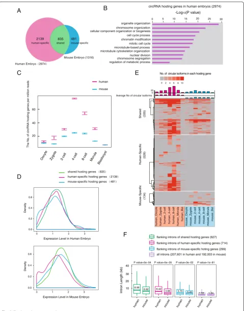
To gain insight into the evolution of circRNAs, we com-pared the circRNAs of human pre-implantation embryos with those identified in our previous mouse study [19]. Of all the 1316 circRNAs hosting genes identified in mouse pre-implantation embryos, 835 (63 % of 1316) also generate circRNAs in human embryos, indicating
that the circRNA production is in general conserved be-tween human and mouse (Fig. 5a). Of the 2926 circRNA hosting genes identified in human H9 embryonic stem cells (ESCs) in a previous study using a different method (Additional file 6: Table S5) [12], 1388 (47 % of 2926) were found to generate circRNAs in human early em-bryos (Additional file 2: Figure S5A). GO analysis against the background linear RNA expression showed that the circRNAs hosting genes in human embryos are enriched for "genes of organelle organization", "chromosome organization", "cell cycle process", and "regulation of meta-bolic process", which is similar to those in mouse embryos and H9 cells (Fig. 5b, and Additional file 2: Figure S5B, C). These results indicate that circRNAs are generated by a highly conserved set of genes in both human and mouse.
An interesting finding is that human-specific circRNAs hosting genes prominently outnumber the mouse-specific ones (human versus mouse: 2139 versus 481, Fig. 5a, Additional file 2: Figure S5D). To exclude the ef-fect of different sequencing depth, we subsampled the human and mouse sequencing data to the same depth and obtained a similar result (human versus mouse: 795 versus 285, Additional file 2: Figure S5E). The human early embryos showed a higher number of circRNA host-ing genes as well as more types of circRNA transcripts than the mouse embryos from oocyte to morula stages, reaching the highest level at the 4-cell stage (Fig. 5c and Additional file 2: Figure S5F). Comparing expressions of the species-specific and shared genes showed that, while species-shared circRNAs hosting genes are generally expressed in both human and mouse embryos, a portion of species-specific circRNA hosting genes are solely expressed in the corresponding species (Fig. 5d). This in-dicates that the species-specific circRNAs are partially due to the differential expression of their hosting genes be-tween human and mouse pre-implantation embryos.
To investigate whether there were factors other than differential hosting gene expression leads to a species dif-ference of circRNA, we compared the circRNAs derived from the hosting genes that are highly expressed in both human and mouse embryos (RPKM >10, Additional file 2: Figure S5G). The result showed that the human-specific (See figure on previous page.)
A
B
C
D
[image:8.595.57.540.88.690.2]circRNA hosting genes still outnumber the mouse-specific ones (human versus mouse: 526 versus 134, Fig. 5e) and also in the subsampling data (human versus mouse: 232 versus 104, Additional file 2: Figure S5H). Since the flanking intron has been shown to play an important role in cir-cRNA generation, we calculated the length of introns flank-ing these circRNAs. Notably, we found that the introns flanking the species-shared or human-specific circRNAs are significantly longer than their mouse counterparts (P <0.001, Fig. 5f). In particular, the introns flanking the human-specific circRNAs in the human genome are about 1.7-fold longer than their mouse counterparts (human ver-sus mouse: median 6.42 verver-sus 3.86 kb, P= 5E-5, Fig. 5f). On the contrary, the length of the introns flanking the mouse-specific circRNAs showed only a mild difference be-tween the two species (human versus mouse: median 3.7 versus 3.45 kb,P= 0.03).
Together, these results demonstrated that circRNAs in human pre-implantation embryos are more complex compared with those in mouse embryos, which may be partially due to the increase in intron length during evo-lution of the human genome.
Analysis of the novel linear transcripts in human pre-implantation embryos by SUPeR-seq
Finally, we performed RNAde novoassembly and identi-fied 2322 novel candidate transcript units and 10,084 candidate isoforms by excluding known genes in RefSeq genes, Noncode V4.0 long non-coding RNA (lncRNA) databases [33], and novel lncRNAs reported in our pre-vious study [1] (Additional file 7: Table S6). These de novo transcripts were rigorously filtered: each transcript comprised at least two exons, was located >10 kb away from the known genes in the human genome, and was longer than 500 bp [1, 19, 34]. The average length of these candidates was 1052 bp and they were usually shorter than 2 kb (Additional file 2: Figure S6A). These novel genes have longer introns than other known genes and half of them consist of two exons (Additional file 2: Figures S6B and S6C). Furthermore, their average expres-sion levels are higher than those of the genes identified in the Noncode database or our previous work using the single-cell RNA-seq technique that only detected polyA+ mRNAs (Fig. 6a), implying that these novel transcripts are
polyA–or with short polyA tail. These novel genes were classified into distinct categories that may function in spe-cific stages during pre-implantation development (Fig. 6b). The expression levels of novel transcripts in each develop-mental stage increased before the 8-cell stage and then de-creased (Fig. 6c). This pattern was similar to that of the novel genes discovered in mouse embryos using SUPeR-seq [19]. The conservation level (calculated with the metric ω) [35] of these novel transcripts was similar to those of the known lncRNAs and novel transcripts that we detected previously. In addition, these novel transcript candidates were less conserved than protein-coding exons but more conserved than the introns of protein-coding genes (Additional file 2: Figure S6D). CPC (Coding Poten-tial Calculator) [36] analysis revealed that among these 2322 novel genes, 89.9 % (2087) produced transcripts without significant coding potential, indicating that they were potential novel lncRNAs.
Conclusions
Overall, our investigation of the transcriptomic land-scape of human pre-implantation development by SUPeR-seq identified abundant circRNAs and revealed dynamic gene expression changes during human pre-implantation development. A large number of circRNAs are transcribed from maternal genes, most of which are present before fertilization, and persisted during pre-implantation development possibly due to their resistant to the maternal mRNA degradation process. Compared with mouse, human circRNAs are proved to have both conservation and an increase in complexity, pointing to their conserved and specific roles during human pre-implantation development. In sum, our data provide an invaluable resource for investigating their functions in the future.
Methods
Embryo collection
The oocytes and embryos for this study were donated from female volunteers who provided informed consent. After ICSI (intracytoplasmic sperm injection), embryos were cultured in G1.3 medium (Vitrolife, Sweden) covered with mineral oil (Sigma, 6 % CO2). Oocytes, zygotes, and
(See figure on previous page.)
Fig. 4CircRNA and mRNA expression pattern during human pre-implantation development.aThe pattern of circRNAs percentage in their hosting genes changes during pre-implantation development based on the ratio of back-spliced reads in total reads at each junction locus, the equation is: back-spliced reads/(back-spliced reads + forward-spliced reads).bComparison of the expression levels of circRNA hosting genes and other coding genes in each stage during pre-implantation development. The expression levels of the hosting genes only include linear transcripts by excluding circular transcripts (RPKMdiscounted= RPKM×(1-circRNA ratio in Fig. 4a). The expression levels of circRNA hosting genes are higher than those of other
2-cell-stage embryos were collected at the appropriate time during embryonic development.
Embryos at the 4-cell and 8-cell stages were thawed immediately after removal from liquid nitrogen as de-scribed previously [1]. The embryos were cultured in G2 medium (Vitrolife) continuously to obtain morulae and early blastocysts, blastocysts, and hatched blastocysts.
Each selected oocyte or embryo was transferred drop wise to the acidic solution to remove the zona pellucida by mouth pipette. Then, the embryo or oocyte was washed gently several times before being transferred to lysis buffer.
To obtain ICM and TE transcriptome information, we isolated these compartments from each other by laser cutting. This process was executed carefully to retain all cells in the ICM with minimal laser damage.
Single embryo transcriptome amplification
The RNAs in individual oocytes or embryos were reverse transcribed and amplified using the SUPeR-seq method we recently developed. Briefly, after cell lysis, RNAs with or without polyA were reverse transcribed with T15N6 primer [19] using Super Script III (Invitrogen). After re-verse transcription, unreacted primer was digested by ExoSAP-IT (USB) and RNA was degraded by RNase H (Invitrogen). Then, a polyA tail was added to the first strand cDNA at its 3′ end by terminal deoxynucleotidyl transferase (Invitrogen). Thus, the second strand cDNA could be synthesized by a primer with a poly T and an anchor sequence. The double-stranded cDNAs were then amplified by primers with the two anchor sequences for 20 + 10 cycles.
Before single-cell RNA amplification, we quantitatively added spike-in RNAs, as ERCC RNA Spike-In Mix1 (Ambion) and RGC-A80 to the lysis mixture. The spike-in RNAs were used for quality control and mRNA quantification.
RNA-seq library construction and sequencing
After the single-cell cDNAs were amplified with the SUPeR-seq method, we sheared approximately 200 ng of purified cDNA products into fragments of 150–350 bp
using the Covaris S2 system. The fragmented DNA was subjected to end-repair, dA-tail, adaptor ligation, and 10–12 cycles of PCR amplification using the TruSeq DNA library preparation kit (Illumina).
SUPeR-seq data processing and validation of circRNA candidates
The sequenced raw data were first cleaned to remove low-quality reads (reads with more than 50 % of the bases with quality value ≤5 and >10 % of the bases un-determined). The adaptor sequences, poly (A) 24/(T) 24 sequences and sequences with >80 % AT bases were trimmed. To detect circular reads using CIRCexplorer, the trimmed data were aligned to an hg19 reference using the two-step approach recommended by https:// github.com/YangLab/CIRCexplorer/ [12].
The mapped reads in the first step were considered linearly mapped reads. For linearly mapped reads, HTSeq [38] was used to count the unique mapped reads to each gene to estimate the abundance of the transcripts (shown as RPKM) and define differentially expressed genes. We used a GTF file combined with hg19 RefSeq genes in the UCSC Genome Browser, the NONCODE V4.0 database, and the genes from a former study previ-ously reported by our lab [1] to identify non-coding genes as well as 92 exogenous ERCC spike-in RNAs and RGC-A80 information. After HTSeq, unannotated reads were used to assemble novel genes. The potential novel tran-scripts were identified based on three criteria. First, the expression level (RPKM) of candidate transcripts was >0.5 and the RPKM in every replicate was >0.25. Second, the potential novel transcript was at least 10 kb away from any known genes. Third, the potential novel gene had at least two exons, and the total length for all exons was >500 bp. The coding potential for novel genes was estimated using the Coding Potential Calculator (http://cpc.cbi.pku.edu.cn) [36].
In the second step, the unmapped reads from the first step were mapped to the genome using TopHat-Fusion [12, 39]. CIRCexplorer was used to detect circular reads. (See figure on previous page.)
Fig. 5Comparative analysis of human and mouse circRNAs.aHigh conservation of circRNA hosting genes between human and mouse pre-implantation embryos. TheVenn diagramshows that the majority of genes that express circRNAs in mouse also produce circular transcripts in human embryos.bGO analysis of the top enriched terms of the circRNA hosting genes in human pre-implantation embryos.cThebox plotsshow the numbers of expressed circRNA hosting genes in human embryos (inred) and mouse embryos (inblue), during pre-implantation development, and the number was normalized to the total mapped reads (in millions) after subsampling the sequencing data.dExpression level of circRNA hosting genes in human embryo and mouse embryo. All circRNA hosting genes are plotted according to the value of log10of (max RPKM during pre-implantation) in
human and mouse embryo, respectively.eHeat mapof circularized level of highly expressed circRNA hosting genes (RPKM >10) during human and mouse pre-implantation development. The numbers of circular isoforms from each hosting gene were presented and thecolumnson the top of the
heat mapshow the average number of circular isoforms from each hosting gene who were producing circular transcripts at this stage. The number of circRNA isoforms is frequently higher than that in mouse. The shared, human-specific, and mouse-specific hosting genes are presented, respectively.f
Because we used pair-end sequencing data, which pro-vides more reliable results for circular regions, CIRCex-plorer was modified to ensure that each circular read had a back-spliced read across two exon junctions in the same gene and the other read from a pair-end reads was linearly aligned between the two exons. Finally, the ratio of circular to linear transcripts was estimated by the back-spliced reads over the step1 mapped reads at each junction locus.
We verified five circRNA candidates in hESCs. The total RNA was extracted from 1 million hESCs, then the total RNA (2 ug) was treated with RNase R (Epicentre) or nuclease-free water as mock control at 37 °C for 15 min. After being reverse transcribed with random primers, the cDNAs were used as qPCR templates to compare the different effects of RNase R treatment be-tween the linear transcripts and circRNA candidates. The hESCs total RNA was subjected to RT-PCR and
A
B
[image:12.595.61.538.87.527.2]Sanger sequencing to verify the back-spliced sites of cir-cRNA candidates at single-base resolution.
Estimation of technical error
We first merged the counts of reads mapped to the 92 exogenous ERCC spike-in RNAs for each sample. Then, the ERCC expression level (RPKM) matrix was calcu-lated using the total mapped reads and the length of each spike-in molecule. Only ERCC with RPKM ≥1 in more than two samples was considered. ERCC with RPKM <1 was excluded for further analysis. The tech-nical error was then estimated using the Pearson correl-ation between samples.
Correlation analysis for RNA-seq data and hierarchy clustering, PCA
The correlation between samples was calculated using the RefSeq gene expression level (RPKM) matrix with the parameter use =“pairwise.complete.obs,” method
=“pearson” using an in house-developed R script. Based
on the correlation matrix, ward distance was used when performing hierarchy clustering. PCA was also per-formed using the FactoMineR package in the CRAN R program based on the same expression level matrix.
Quantification of total transcripts copy number
The spike-ins of RGC-A80 included three species of in vitro transcribed RNA molecules (RFP, GFP, and CRE) which had 80 nt polyA tails and mixed as the molecule ratio of RFP: GFP: CRE being 100: 10: 1. By using ddPCR (BioRad, QX200), the capture efficiency of RGC-A80 was verified as about three times higher than ERCCs in SUPeR-seq (Additional file 2: Figure S2A). Therefore, addition of the RGC-A80 spike-ins can par-tially overcome the low capture efficiency of the ERCC spike-ins and achieve a more accurate estimation of the total transcript content in each sample.
For the ERCC/RGC-A80 normalization algorithm, firstly, linear regression was applied to fit the data points between the RPKM value of the 92 exogenous ERCC spike-in RNAs (log10-transformed RPKM) in the SUPeR-seq dataset and the number of molecules per lysis reaction (log10-transformed attomole) (Additional file 2: Figure S2C). Only ERCC species whose molecules >0.001 atto-mole were retained in the regression. The linear regression equations for each sample were then applied to the RPKM value of all RefSeq genes and summed up as the ERCC-based total mRNA copy number. Then, the total mRNA copy number was also calculated from each molecule spe-cies from spiked RGC-A80 as the ratio of the total RPKM to the RGC-A80 RPKM multiply the spiked molecules and then averaged out (Additional file 2: Figure S2D). Lastly, the final total mRNA copy number was obtained by fitting the RGC-A80-based and the ERCC-based values
in all 29 samples to a linear regression model (Additional file 2: Figure S2E).
We also estimated the copy number of circRNAs in each oocyte and embryo during human pre-implantation development. Firstly, we calculated the RPKM of cir-cRNA as junction reads/(circir-cRNA length × total mapped reads). And the circRNA length = (length of reads-25 bp) × 2, as 25 bp is the segment length of Tophat. This means that for reads of 100 bp in length, a back-splicing event can be detected by reads mapping up to 75 bp away in each direction, as 75 bp × 2 = 150 bp in length. Then we could calculate the copy number of circRNAs = (sum of circRNAs’ RPKM)/(sum of Refseq genes’ RPKM) × (copy number of mRNA).
Copy number quantification with ddPCR
For validation of the algorithm of mRNA quantification in SUPeR-seq by ERCC and RGC-A80, the total RNAs were extracted from ~100,000 hESCs and SUPeR-seq was performed in three technical replicates for 1 ng total RNA in each replicate. The rest of the RNAs were reverse transcribed to cDNAs to examine the copy number of 44 genes in 1 ng RNAs by ddPCR in two replicates. The primers for ddPCR are listed in Additional file 8: Table S7.
Differentially expressed genes identified based on normalized RPKM
Differential gene expression analysis across all samples was performed using the DESeq2 package in the Bioconductor R program [40], which is based on the negative binomial distribution model. Raw read counts calculated by HTSeq were normalized by a set of size factors accounting for both the sequencing depth and the mRNA quantity in each sample. For a strict definition of differential expressed genes, the RefSeq genes expressed in at least one of the samples with normalized RPKM≥1 were used for the analysis.
Comparison of circRNAs with hESC data and mouse pre-implantation embryo data
The hosting gene and read counts of the circRNA matrix were merged from the pair-end checked CIRCex-plorer results. For each sample, circRNAs with read depths <2 were discarded. The hosting gene of circRNAs in H9 cell line were filtered according to the gene list provided in a former study [12] and are listed in the Supplemental Information Table. We also subjected the sequencing data of mouse early embryo cell data in our published work [19] to the same approach. The 20 bp at each site of the back-spliced junction of mouse cir-cRNAs were converted from mm10 to hg19 using the
liftover tool from UCSC utilities with the parameter
-minMatch = 0.5, which enabled comparison with
cing data from human and mouse pre-implantation em-bryos to the same depth as 9 millions reads per stage and kept the same number of developmental stages side by side for analysis. Venn diagrams were used to show the shared gene lists and GO analysis for each portion of the Venn diagram was performed using the GOstat package in the Bioconductor R program [41]. For GO analysis of the circRNA hosting genes in human pre-implantation embryos, all genes expressed above 1 RPKM were set as the background. The combined gene list of circRNA hosting genes in human and mouse em-bryos was set as the background for the comparison be-tween these two species. The combined gene list of circRNA hosting genes in human embryos and H9 cells was set as the background for the comparison between these two cell sources.
Additional files
Additional file 1: Table S1.Sequencing statistics summary of samples analyzed in this study. (XLS 65 kb)
Additional file 2:Figure S1.Reproducibility of the SUPeR-seq method. Figure S2.mRNA copy numbers quantified by ERCC/RGC-A80 normalization algorithm.Figure S3.Feature analysis of the flanking in-trons of circRNAs.Figure S4.Expression pattern of circRNAs and the hosting genes during human pre-implantation development.Figure S5. Comparison of circRNA hosting genes.Figure S6. De novoassembled transcripts in the human pre-implantation embryos. (PDF 23698 kb)
Additional file 3: Table S2.Normalized RPKM of RefSeq gene. Corresponding to Fig. 2b, the 5573 RefSeq genes clustered in group 1 were defined as maternal genes and the 7427 genes in groups 2 to 5 transcribedde novoat later stages from the late 4-cell stage were defined as zygotically activated genes. (XLSX 1359 kb)
Additional file 4: Table S3.Sequencing statistics and circRNA candidates of hESCs in this study. (XLS 77 kb)
Additional file 5: Table S4.CLR value of circRNA during human pre-implantation development. The log2-transformed values of CLR for all circRNA hosting genes at all developmental stages were listed. NAN means this hosting gene have not circRNA reads at this stage. (XLSX 245 kb)
Additional file 6: Table S5.Circular RNA detected in human embryos and list of circRNA hosting genes in H9 Cell. The circRNA hosting genes in H9 cell line were filtered as the reads counts >1 according to the Supplemental Table in a previously published paper [12]. (XLS 1349 kb)
Additional file 7: Table S6.A total of 2322 novel assembled gene RPKM cluster and transcripts locus information. (XLS 4066 kb)
Additional file 8: Table S7.Sequences of RGC-A80 and the primers used in ddPCR and circRNAs validation. The sequences of RGC-A80, the primers used in ddPCR for detecting the copy number of 44 genes, and the primers of circRNAs validation are listed. (DOCX 19 kb)
Abbreviations
circRNA, Circular RNA; CLR, Circular to linear reads ratio; ddPCR, Droplet digital polymerase chain reaction; DEG, Differential expressed genes; ERCC, External RNA Controls Consortium; FDR, False positive rate; GO, Gene ontology; hESCs, Human embryonic stem cells; lncRNA, long non-coding RNA; MZT, Maternal to zygotic transition; PCA, Principal component analysis; RGC-A80, Mixture of RFP, GFP, CRE with 80 nt polyA tails; RPKM, Reads per kilobase of exon model per million mapped reads; RT, Reverse transcription; SUPeR-seq, Single cell universal poly(A)-independent RNA sequencing
Institute of Genomics for providing the platform for ddPCR experiments. And we would like to thank the Computing Center for Life Science, Peking University, as part of the analysis was performed on this computing platform.
Funding
This work was supported by the Beijing Municipal Science and Technology Commission (D151100002415000) and the National Natural Science Foundation of China (81521002, 31522034, 31230047, 31571544, 31440063, 81561138005, and 31322037).
Availability of data and material
All SUPeR-seq data in this study are available in GEO under accession number: GSE71318.
Authors’contributions
FT, YaH, and JQ designed the study. YD and LYAN conducted the main experiments. BH was in charge of all the bioinformatic analyses. XF and QL were involved in the molecular biology experiments. YR, RL, YL, JY, YZ, ML, XR, JH, YW, and YP were involved in the embryo collection and culture. YD, LYAN, BH, LW, YaH, FT, and JQ wrote the paper with help from all the authors. All authors read and approved the final manuscript.
Author information
Scripts for the pipeline are available at GitHub (https://github.com/ hubqoaing/TanglabCircularRNAPipeline).
Competing interests
The authors declare that they have no competing interests.
Ethics approval and consent to participate
This study was conducted following the guidelines from the Declaration of Helsinki. And this study has been approved by the Reproductive Study Ethics Committee of Peking University Third Hospital (Research license 2012SZ015). All of the oocytes and embryos were collected voluntarily with written informed consent signed by the donor couples. The informed consent confirmed that the donor couples were voluntarily donating oocytes and embryos (including sperm) for research on human early embryonic transcriptome. All of the oocytes and embryos were obtained from the donors at the Center for Reproductive Medicine in Peking University Third Hospital using standard clinical protocols [37].
Author details
1Biodynamic Optical Imaging Center & Department of Obstetrics and Gynecology, College of Life Sciences, Third Hospital, Peking University, Beijing 100871, China.2Key Laboratory of Assisted Reproduction, Ministry of Education, Beijing 100191, China.3Ministry of Education Key Laboratory of Cell Proliferation and Differentiation, Beijing 100871, China.4Peking-Tsinghua Center for Life Sciences, Peking University, Beijing 100871, China.5Center for Molecular and Translational Medicine, Peking University Health Science Center, Beijing 100191, China.6Beijing Key Laboratory of Reproductive Endocrinology and Assisted Reproduction, Beijing, China.7College of Engineering, Peking University, Beijing 100871, China.
Received: 13 August 2015 Accepted: 24 May 2016
References
1. Yan L, Yang M, Guo H, Yang L, Wu J, Li RR, Liu P, Lian Y, Zheng X, Yan J, Huang J, Li M, Wu X, Wen L, Lao K, Qiao J, Tang F. Single-cell RNA-Seq profiling of human preimplantation embryos and embryonic stem cells. Nat Struct Mol Biol. 2013;20:1131–9.
2. Jeck WR, Sharpless NE. Detecting and characterizing circular RNAs. Nature Biotechnology. 2014;32:453–61.
3. Salzman J, Chen RE, Olsen MN, Wang PL, Brown PO. Cell-type specific features of circular RNA expression. PLoS Genet. 2013;9:e1003777. 4. Suzuki H, Zuo Y, Wang J, Zhang MQ, Malhotra A, Mayeda A. Characterization of
5. Salzman J, Gawad C, Wang PL, Lacayo N, Brown PO. Circular RNAs are the predominant transcript isoform from hundreds of human genes in diverse cell types. PLoS One. 2012;7:e30733.
6. Zhang Y, Zhang X, Chen T, Xiang J, Yin Q-F, Xing Y, Zhu S, Yang L, Chen L-L. Circular intronic long noncoding RNAs. Mol Cell. 2013;51:792–806. 7. Li Z, Huang C, Bao C, Chen L, Lin M, Wang X, Zhong G, Yu B, Hu W, Dai L,
Zhu P, Chang Z, Wu Q, Zhao Y, Jia Y, Xu P. Exon-intron circular RNAs regulate transcription in the nucleus. Nat Struct Mol Biol. 2015;22:256–64. 8. Hansen TB, Jensen TI, Clausen BH, Bramsen JB, Finsen B, Damgaard CK,
Kjems J. Natural RNA circles function as efficient microRNA sponges. Nature. 2013;495:384–8.
9. Memczak S, Jens M, Elefsinioti A, Torti F, Krueger J, Rybak A, Maier L, Mackowiak SD, Gregersen LH, Munschauer M, Loewer A, Ziebold U, Landthaler M, Kocks C, Noble F, Rajewsky N, le Noble F, Rajewsky N. Circular RNAs are a large class of animal RNAs with regulatory potency. Nature. 2013;495:333–8.
10. Ashwal-Fluss R, Meyer M, Pamudurti NRR, Ivanov A, Bartok O, Hanan M, Evantal N, Memczak S, Rajewsky N, Kadener S. circRNA biogenesis competes with pre-mRNA splicing. Mol Cell. 2014;56:55–66.
11. Dubin RA, Kazmi MA, Ostrer H. Inverted repeats are necessary for circularization of the mouse testis Sry transcript. Gene. 1995;167:245–8. 12. Zhang X, Wang H-B, Zhang Y, Lu X, Chen L-L, Yang L. Complementary
sequence-mediated exon circularization. Cell. 2014;159:1–14.
13. Rybak-Wolf A, Stottmeister C, Glažar P, Jens M, Pino N, Giusti S, Hanan M, Behm M, Bartok O, Ashwal-Fluss R, Herzog M, Schreyer L, Papavasileiou P, Ivanov A, Öhman M, Refojo D, Kadener S, Rajewsky N. Circular RNAs in the mammalian brain are highly abundant, conserved, and dynamically expressed. Mol Cell. 2015;58:870–85.
14. Venø MT, Hansen TB, Venø ST, Clausen BH, Grebing M, Finsen B, Holm IE, Kjems J. Spatio-temporal regulation of circular RNA expression during porcine embryonic brain development. Genome Biol. 2015;16:245. 15. Tang F, Lao K, Surani MA. Development and applications of single-cell
transcriptome analysis. Nat Methods. 2011;8:S6–11.
17. Picelli S, Björklund ÅK, Faridani OR, Sagasser S, Winberg G, Sandberg R. Smart-seq2 for sensitive full-length transcriptome profiling in single cells. Nat Methods. 2013;10:1096–8.
18. Hashimshony T, Wagner F, Sher N, Yanai I. CEL-Seq: Single-cell RNA-Seq by multiplexed linear amplification. Cell Rep. 2012;2:666–73.
19. Fan X, Zhang X, Wu X, Guo H, Hu Y, Tang F, Huang Y. Single-cell RNA-seq transcriptome analysis of linear and circular RNAs in mouse preimplantation embryos. Genome Biol. 2015;16:148.
20. Stegle O, Teichmann S, Marioni JC. Computational and analytical challenges in single-cell transcriptomics. Nat Rev Genet. 2015;16(3):133–45.
21. Su Z,Łabaj PP, Li S, Thierry-Mieg J, Thierry-Mieg D, Shi W, Wang C, Schroth GP, Setterquist R a, Thompson JF, Jones WD, Xiao W, Xu W, Jensen R V, Kelly R, Xu J, Conesa A, Furlanello C, Gao H, Hong H, Jafari N, Letovsky S, Liao Y, Lu F, Oakeley EJ, Peng Z, Praul C a, Santoyo-Lopez J, Scherer A, Shi T, et al. A comprehensive assessment of RNA-seq accuracy, reproducibility and information content by the Sequencing Quality Control Consortium. Nat Biotechnol. 2014;32:903–14.
22. Baker M. Digital PCR, hits its stride. Nat Methods. 2012;9:541–4. 23. Pinheiro LB, Coleman VA, Hindson CM, Herrmann J, Hindson BJ, Bhat S,
Emslie KR. Evaluation of a droplet digital polymerase chain reaction format for DNA copy number quantification. Anal Chem. 2012;84:1003–11. 24. Piras V, Tomita M, Selvarajoo K. Transcriptome-wide variability in single
embryonic development cells. Sci Rep. 2014;4:7137.
25. Vassena R, Boué S, González-Roca E, Aran B, Auer H, Veiga A, Izpisua Belmonte JC. Waves of early transcriptional activation and pluripotency program initiation during human preimplantation development. Development. 2011;138:3699–709.
26. Niakan KK, Han J, Pedersen R, Simon C, Pera RR. Human pre-implantation embryo development. Development. 2012;139:829–41.
27. Dobson AT, Raja R, Abeyta MJ, Taylor T, Shen S, Haqq C, Reijo Pera RA. The unique transcriptome through day 3 of human preimplantation development. Hum Mol Genet. 2004;13:1461–70.
28. Szabo L, Morey R, Palpant NJ, Wang PL, Afari N, Jiang C, Parast MM, Murry CE, Laurent LC, Salzman J. Statistically based splicing detection reveals neural enrichment and tissue-specific induction of circular RNA during human fetal development. Genome Biol. 2015;16:126.
29. Lasda E, Parker R. Circular RNAs: diversity of form and function. RNA. 2014;20:1829–42.
30. Petkovic S, Muller S. RNA circularization strategies in vivo and in vitro. Nucleic Acids Res. 2015;43:2454–65.
31. Westholm JO, Miura P, Graveley BR, Lai EC, Westholm JO, Miura P, Olson S, Shenker S, Joseph B, Sanfilippo P. Genome-wide analysis of drosophila circular RNAs reveals their structural and sequence properties and age-dependent neural accumulation. Cell Rep. 2014;9:1966–80. 32. Chen L-L, Yang L. Regulation of circRNA biogenesis. RNA Biol.
2015;12:381–8.
33. Liu C, Bai B, Skogerbø G, Cai L, Deng W, Zhang Y, Bu D, Zhao Y, Chen R. NONCODE: An integrated knowledge database of non-coding RNAs. Nucleic Acids Res. 2005;33:D112–5.
34. Xie W, Schultz MD, Lister R, Hou Z, Rajagopal N, Ray P, Whitaker JW, Tian S, Hawkins RD, Leung D, Yang H, Wang T, Lee AY, Swanson SA, Zhang J, Zhu Y, Kim A, Nery JR, Urich MA, Kuan S, Yen C, Klugman S, Yu P, Suknuntha K, Propson NE, Chen H, Edsall LE, Wagner U, Li Y, Ye Z, et al. Resource epigenomic analysis of multilineage differentiation of human embryonic stem cells. Cell. 2013;153:1134–48.
35. Garber M, Guttman M, Clamp M, Zody MC, Friedman N, Xie X. Identifying novel constrained elements by exploiting biased substitution patterns. Bioinformatics. 2009;25:54–62.
36. Kong L, Zhang Y, Ye Z, Liu X, Zhao S, Wei L. CPC: assess the protein-coding potential of transcripts using sequence features and support vector machine. Nucleic Acids Res. 2007;35:345–9.
37. Guo H, Zhu P, Yan L, Li R, Hu B, Lian Y, Yan J, Ren X, Lin S, Li J, Jin X, Shi X, Liu P, Wang X, Wang W, Wei Y, Li X, Guo F, Wu X, Fan X, Yong J, Wen L, Xie SX, Tang F, Qiao J. The DNA methylation landscape of human early embryos. Nature. 2014;511:606–10.
38. Anders S, Pyl PT, Huber W. HTSeq–a Python framework to work with high-throughput sequencing data. Bioinformatics. 2015;31:166–9. 39. Trapnell C, Pachter L, Salzberg SL. TopHat: Discovering splice junctions with
RNA-Seq. Bioinformatics. 2009;25:1105–11.
40. Love MI, Huber W, Anders S. Moderated estimation of fold change and dispersion for RNA-Seq data with DESeq2. Genome Biol. 2014;15:550. 41. Beißbarth T, Speed TP. GOstat: Find statistically overrepresented Gene
Ontologies with a group of genes. Bioinformatics. 2004;20:1464–5.
• We accept pre-submission inquiries
• Our selector tool helps you to find the most relevant journal
• We provide round the clock customer support
• Convenient online submission
• Thorough peer review
• Inclusion in PubMed and all major indexing services
• Maximum visibility for your research
Submit your manuscript at www.biomedcentral.com/submit