Various Techniques for Classifying Brain
Tumor
Sangeeta Sehrawat1, Ritu Khatri2
1M.Tech student C.S.E. Dept., H.C.E, Sonepat, Haryana, India 2Astt. Prof. C.S.E. Dept., H.C.E, Sonepat, Haryana, India
Abstract –Image processing for medical images has been grown rapidly for the purpose of enhancing the security, content verification and robustness. Even then because of requirement of high accuracy it is the open research area. A tumor is the name for a neoplasm or a solid lesion formed by an abnormal growth of cells (termed neoplastic) which looks like a swelling. Tumor is not synonymous with cancer. A tumor can be benign, pre-malignant or malignant, whereas cancer is by definition malignant.In this paper, we are discussing various neural network techniques used recently for classifying the brain tumor in medical science.
Keywords— Image Processing, Artificial Intelligence (AI), Artificial Neural Network (ANN), Magnetic Resonance Imaging(MRI), Fuzzy C-means (FCM).
I. INTRODUCTION
Image processing is a method to transform an image into digital form and perform some operations on it, in order to get an enhanced image or to extract some useful information from it. It is a type of signal dispensation in which input is image, like video frame or photograph and output may be image or characteristics associated with that image. Usually Image Processing system includes treating images as two dimensional signals while applying already set signal processing methods to them.
Image processing basically includes the three steps. These three steps includes importing the image, processing the image by compressing it and enhancing the image, and the last and third step ends to the image analysis which is the output obtained on the basis of analysis. Image processing is rapidly growing technology today, having its application in various aspects of business, medical science, research area within engineering and computer science disciplines too.Figure1 shows that the presence of tumor in the brain.
A. Brain Tumor Categories
Brain Tumor is categorized into three parts:
a. Benign Tumor
b. Malignant
c. Premalignant
[9]
Benign Tumor:Benign tumor is such a tumor that does not have the malignant properties of a cancer. Therefore, by definition, it does not grow in an unlimited, aggressive manner, does not invade surrounding tissues, and does not spread to non-adjacent tissues (metastasize). Some examples of benign tumors include moles and uterine fibroids.
Malignant Tumor: The word malignant is developed from the
word Malignancy (from the Latin roots mal- = "bad" and -ignis = "fire"). Malignancy is the susceptibility of a medical condition, especially tumors, to become progressively worse and to finally result in death. The term is most familiar as a description of cancer.
Premalignant Tumor: It is a precancerous condition (or
premalignant condition) which is such a disease, syndrome, or finding that, if left untreated, it may lead to cancer. It is a generalized state associated with a significantly increased risk of cancer.
B. Image Processing Applications
The purpose of image processing is divided into 5 groups. They are:
[image:3.612.354.550.112.264.2]1. Visualization: Visualization is a technique which is used for communication with the help of message by creating diagrams, animations or images through visual imagery in an effective way to communicate both abstract and as well as concrete ideas since the dawn of man. In medical science, the MIPAV (Medical Image Processing, Analysis, and Visualization) application enables quantitative analysis and visualization of medical images of numerous modalities like PET (Positron Emission Tomography), MRI (Magnetic Resonance Imaging), CT (Computed Tomography) or Microscopy.
Figure 2[10]. MRI brain tumor image.
2. Image Sharpening and Restoration: The term Image
sharpening refers to the enhancement of edges definition in an image. The image may be from a scanner or a digital camera which require more sharpening , but it cannot correct a severely blurred image. It increases the image contrast.
Image Restoration is the operation of taking a corrupted or noisy image and estimating the clean original image. Degradation or corruption may be in form of a noise , camera misfocus or motion blur. The purpose of image restoration is to
“compensate for” or “undo” defects which degrade an image.
3. Image Retrieval: Basically Image Retrieval is used to seek for the image of interest. Image Retrieval in medical applications (IRMA) is a cooperative project of the department of diagnostic radiology, the department of medical informatics, division of medical image processing and the chair of computer science VI at the Aachen University of technology (RWTH Aachen).
4. Measurement of pattern:The idea of Pattern Measurement
measures various objects in an image. In medical science basically pattern measurement is used to detect the movement of patient under treatment and finding the standard deviation of the image from image histograph and keeping it as a template at the initial state. First a point can be marked on the patient’s body and then get the co-ordinates of the mark, and then comparing the co-ordinates with the next successive images.
5. Image Recognition : Image Recognition basically
methods in biology and medicine over the past decade. This has led to a huge growth in the application of digital processing techniques for solving medical problems. Therefore a lot of research work is done in present to improve the imperfect image material.
C. Artificial Neural Networks for Medical Images
Computers having the ability to duplicate the functions of the human brain are known as Artificially Intelligent Systems. The people, procedures, software,hardware, and knowledge required to develop the computer systems and machines to demonstrate the features and characteristics of intelligence.
Artificial Intelligence (AI) is such a branch of computer science which aims to create it and known as intelligence of machines. It may also referred as the study and design of intelligent agents. Neural Network (NN) is a computer architecture in which processors are connected in a manner suggestive of connections between neurons.
It can be learn by trial and error any neuron network or nuclei that function together to perform some functions in the body.
In this paper we are discussing various approaches that have been proposed for medical images and these approaches take robustness into consideration. In Section II, ANN schemes for medical images are described and then conclusions drawn from the study have been given in next Section.
II. DIFFERENT ANN SCHEMES FOR MEDICAL
IMAGES
Artificial neural networks comprises of many non - linear computational elements which operates in parallel and arranged in patterns reminiscent of biological neural nets. There exist two modes of training for neural networks, namely, supervised and unsupervised. Earlier researchers have suggested many interesting findings with the supervised and unsupervised neural networks.
Much work has been done in the area of artificial neural network but still there is more work that need to be done. In this section, the methods for the schemes that have been proposed for medical image are discussed and the advantages and problems related to each research is also discussed.
A lot of work has been already done in artificial intelligence and neural networks for medical images are a topic of interest so that patient’s problem can be easily detected. This section
includes some ANN schemes which are designed for medical images.
Scheme 1:Magnetic Resonance Spectroscopic Imaging (MRSI)
is a non-invasive technique[1] for using biochemical fingerprint of tissue composition. To determine the type of abnormality before biopsy or surgery motivated development and application of MRSI, it is necessary to differentiate the normal and abnormal tissues.
There are various reasons that make the brain easy to examine with MRSI as compare to other organs. This scheme presents the proposed method and result for analysis of brain spectra of brain patients with its three types (Malignant Glioma, Astrocytoma, and Oligodendroglia). Steps followed in this scheme are as follows:
a. First of all, two denoising methods to end up the existence of noise are used, namely, wavelets and wavelets packets. All distortions were removed almost.
b. Next step go for preprocessing to prepare the signals for feature extraction by applying different methods like noise reduction using wavelets and wavelets packets.
c. After extracting features, Artificial Neural Network is used to find out the abnormal spectra and the type of abnormality present.
d. Then, evaluation is performed using simulated MRSI data and biopsy results and clinical data.
Fig. 3[2]: MRSI Brain Tumor Characterization for scheme 1
Scheme 2: Two AI techniques are mostly used now a days for biological computational applications. These are Artificial Neural Networks (ANN) and Fuzzy systems.
Fuzzy theory requires expertise knowledge to guarantee high accuracy while ANN is less accurate. Both methodologies
comprises some advantages and disadvantages that’s why it is important to compare and contrast these two techniques. In this scheme both are analyzed in context of MR brain tumor image segmentation. This scheme employs a sequential approach for the image classification system. Steps followed in this scheme are as follows:
a. For this work, images used are the real time abnormal MR brain images. These MRI Images are then normalized to gray scale values from 0 to 1 to make feature extraction simpler.
b. From these normalized images some features are extracted.
c. Then from that image, significant features are selected by using an optimized algorithm that is Genetic Algorithm(GA).
d. Now the next step leads to image segmentation. Two techniques used for that purpose. One of the technique
is based on Linear Vector Quantization (LVQ) network and the other is based on Fuzzy C-means (FCM) algorithm which belongs to the category of fuzzy systems.
e. An extensive analysis and comparison is performed in terms of segmentation, efficiency and convergence time period. Experimental results show promising results for the neural classifier over the fuzzy classifier in terms of the performance measures.
The neural and the fuzzy techniques, both are employed for brain tumor image segmentation on the real time image dataset. The performance measures which are used in this work are like Segmentation efficiency and convergence time period. Segmentation efficiency may be defined as the ratio of the pixels (correctly classified pixels) to the total number of pixels. Convergence time period may be defined as the total time period required for training and testing process. Figure. 4 shows the overall procedure for analysis used in this scheme.
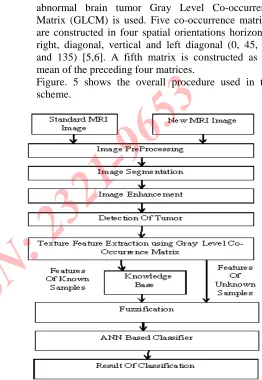
[image:5.612.66.259.117.373.2] [image:5.612.332.573.361.638.2]Scheme 3: A Brain Cancer Detection and Classification System has been designed and developed in this scheme. The work done involved processing of MRI images for detection which are affected by brain cancer and then classification on different types of brain tumor. In this scheme, the system uses computer based procedures to detect tumor blocks or lesions and ANN for classifying the type of tumor in MRI images of different patients with strocytoma type of brain tumors. Steps followed in this scheme are as follows:
a. First of all Image processing is done by taking a new MRI image or a standard MRI Image with the help of different techniques ( Histogram Equalization, Thresholding, and Sharpening Filter etc.) to isolate the tumor region from the rest of the image. Then textural features are extracted using Gray Level Co-occurrence Matrix. These extracted features are then stored in the knowledge base.
b. Next step followed is Image Segmentation which is a process of subdividing an image into its constituent parts. The level to which the subdivision should be carried depends on the problem being solved and should stop when the edge of the tumor is able to be detected.
The main motive of segmentation is to separate the tumor from its background.Thresholding has been used for segmentation because it is most suitable for present application in order to obtain a binarized image having two values i.e. 0 (representing the background) and 1 (representing the tumor) [7].
c. After segmentation, enhancement of image is required to increase the contrast between the whole brain and tumor. For image enhancement process, sharpening filter is applied to the digitized MRI which results in noticeable enhancement in image contrast. The dilation operator is used for filling the broken gaps at the edges and to have continuities at the boundaries. To fill the close contours, a filling operator is used. Then after filling operation, centroids are calculated to localize the regions and finally extracted regions used for extraction of massive region in given MRI image.
d. Next step involve extracting the important features which provide the details like property of texture, and are stored in knowledge base. These features are compared with the features of unknown sample Image for classification. To distinguish between normal and
abnormal brain tumor Gray Level Co-occurrence Matrix (GLCM) is used. Five co-occurrence matrices are constructed in four spatial orientations horizontal, right, diagonal, vertical and left diagonal (0, 45, 90, and 135) [5,6]. A fifth matrix is constructed as the mean of the preceding four matrices.
Figure. 5 shows the overall procedure used in this scheme.
Fig. 5[4]: Classification of Brain Cancer Using Artificial Neural Network
Scheme 4:One more scheme to classify brain tumor is by using
Probabilistic Neural Network (PNN). In this scheme, the principal component analysis (PCA) is used as a feature extraction algorithm and Probabilistic Neural Network (PNN).
The principal component analysis (PCA) is one of the most successful techniques that have been used in image recognition and compression. The purpose of PCA is to reduce the large dimensionality of the data. PNN with image and data processing techniques are used for classification.
[image:6.612.318.580.112.492.2]retaining time, very small training time and robustness to weight changes. Figure. 6 shows the overall procedure used in this scheme. There are six stages involved from input to output in this scheme. Stages are:
a. Basically, the first stage is Image Processing system in which image acquisition, image enhancement are to do. But here, these two steps are skipped and all the images are collected from available resource.
b. This scheme requires conversion of an image into such a format that is capable of being manipulated by the computer. Therefore, collected MR images are converted into matrices form by using MATLAB which is the 2ndstage of the scheme.
c. In the third stage the data is obtained in the form which is compatible with PNN for classification purpose. d. Then in the next stage, PNN is used to classify the MR
images.
e. In the fifth stage, output is obtained.
f. Lastly, in sixth stage, performance based on the result will be analyzed at the end of the development phase.
Fig.6 [7]: Probabilistic Neural Network for Brain Tumor Classification
Scheme 5:Image processing plays an important role in medical
diagnosis. In this scheme, a brain tumor detection method based on cellular neural networks (CNNs) is explained. Radiologists will also evaluate the grey scale MRI images to examine the location of tumor present in the brain. In Reality, this procedure is very time and energy consuming. So to overcome this problem, an automated detection method for brain tumor namely, CNN is developed. In 1988, Cellular Neural Network (CNN) was proposed by Leon O. Chua and Lin Yang. It was used in image, video signal processing, robotic, biological visions and high brain functions [8]. CNN may be defined as any spatial arrangement of locally-coupled cells, where each cell is a
dynamical system which works on an input, gives an output, and evolves a state according to some prescribed dynamical laws. Interconnections among cells in CNN are local, it means each processing unit can interact directly only with the neighboring cells, which are located within a prescribed sphere of influence [8].In this method output of desired image can easily be performed by using the template in the CNN simulator. Therefore, many templates were combined in one to obtain an accurate result so that radiologists will get help in detecting the tumor from brain images easily. In this scheme, an algorithm using CNN to detect brain tumor is designed. The flow chart of the new designed algorithm is shown in Figure 7.
Some MRI images were gathered from KPJ Penang Specialist.10 images were used to simulate with the proposed algorithm. However, this algorithm still has its weakness on the area where the skull (white ring) unable to filter with CNN template. To prevail over this problem, a C or C++ coding is used to filter it.
[image:7.612.338.566.338.630.2]III. CONCLUSION
This paper outlined the brief introduction on classification of brain tumor in medical images. Till now various classification schemes have been proposed. Here, we focus on to make a note of various approaches used in the classification schemes of brain tumor , that are applicable to digital medical images. Various Schemes have been developed for detecting the presence of brain tumor in the medical images.
Schemes which are main focused in this paper are: MRSI characterization using wavelets packets for feature extraction or denoising and multilayer perceptron neural network for classification, classification using ANN and Fuzzy system with the help of LVQ based segmentation and FCM based segmentation and then performing evaluation and comparison process, classification with GLCM( Gray Level Co- Occurrence Matrix) and fuzzification using ANN based classifier, detection of brain tumor in medical images with the help of PCA technique and PNN classifier, detecting the brain tumor with the help of cellular neural networks(CNN).
The classification schemes for medical images are developed for detecting the presence of tumor in medical image. Some schemes can be further improved by trying different techniques for feature extraction and by using different types of classifiers.
IV. References
[1] D. Sridhar, “Brain Tumor Classification U sing Discrete Cosine Transform and Probabilistic Neural Network”, in proceedings of IEEE International Conference on Signal Processing, Image Processing and Pattern Recognition [ICSIPR], 2013
[2]Azadeh Yazdan-Shahmorad,Hamid Soltanian-Zadeh,Reza A. Zoroofi, “MRSI Brain Tumor Characterization Using Wavelet and Wavelet Packets Feature Spaces and Artificial Neural Networks”, Proceedings of the 26th Annual International
Conference of the IEEE EMBS San Francisco, CA, USA •
September 1-5, 2004
[3] D.Jude Hemanth, C.Kezi Selva Vijila and J.Anitha ,
“Comparative Analysis of Neural Model and Fuzzy Model for MR Brain Tumor Image Segmentation”,in proceedings of IEEE
2009 World Congress on Nature & Biologically Inspired
Computing (NaBIC 2009), 2009.
[4] Dipali M. Joshi, Dr.N. K. Rana and V. M. Misra., “Classification of Brain Cancer Using Artificial Neural
Networkx”, 2nd International Conference on Electronic
Computer Technology (ICECT 2010), 2010.
[5] Mohd Fauzi Othman and Mohd Ariffanan Mohd Basri,
“Reversible Medical Image Watermarking For Tamper Detection and Recovery”, Second International Conference on Intelligent Systems, Modelling and Simulation , 2011
[6] Azian Azamimi Abdullah, Bu Sze Chize and Yoshifumi Nishio ,“Implementation of An Improved Cellular Neural Network Algorithm For Brain Tumor Detection”, in proceedings of International Conference on Biomedical Engineering (ICoBE),27-28 February 2012,Penang, 2012. [7] Mathew C. Clark, Lawrence O. Hall, “Automatic tumor
segmentation using Knowledge based techniques,” IEEE
transaction on medical imaging, Vol 17, No. 2, April 1998.
[8] Leon O.Chua and Tamas Roska. (2002). Cellular Neural Network and Visual Computing, Foundation and Applications, 1st Edition, Cambridge University Press, United Kingdom.
![Figure 2[10]. MRI brain tumor image.](https://thumb-us.123doks.com/thumbv2/123dok_us/8587614.862883/3.612.354.550.112.264/figure-mri-brain-tumor-image.webp)
![Fig. 3[2]: MRSI Brain Tumor Characterization for scheme 1](https://thumb-us.123doks.com/thumbv2/123dok_us/8587614.862883/5.612.332.573.361.638/fig-mrsi-brain-tumor-characterization-scheme.webp)
![Fig.6[7]:Probabilistic Neural Networkfor Brain Tumor ClassificationScheme 5: Image processing plays an important role in medicaldiagnosis](https://thumb-us.123doks.com/thumbv2/123dok_us/8587614.862883/7.612.338.566.338.630/probabilistic-neural-networkfor-brain-classificationscheme-processing-important-medicaldiagnosis.webp)
