Detecting
Staphylococcus aureus
Virulence and Resistance Genes: a
Comparison of Whole-Genome Sequencing and DNA Microarray
Technology
Lena Strauß,aUlla Ruffing,bSalim Abdulla,cAbraham Alabi,d,eRuslan Akulenko,fMarcelino Garrine,gAnja Germann,h Martin Peter Grobusch,e,iVolkhard Helms,fMathias Herrmann,bTheckla Kazimoto,cWinfried Kern,jInácio Mandomando,g Georg Peters,kFrieder Schaumburg,kLutz von Müller,b* Alexander Mellmanna
Institute of Hygiene, Westphalian Wilhelms-University, Münster, Germanya; Institute of Medical Microbiology and Hygiene, University of Saarland, Homburg, Germanyb; Ifakara Health Research and Development Centre, Bagamoyo, Tanzaniac; Albert Schweitzer Hospital, Lambaréné, Gabond; Institute of Tropical Medicine, Eberhard Karls University, Tübingen, Germanye; Center for Bioinformatics, University of Saarland, Saarbrücken, Germanyf; Manhiça Health Research Centre, Manhiça, Mozambiqueg; Fraunhofer Institute for Biomedical Engineering, Sulzbach, Germanyh; Center of Tropical Medicine and Travel Medicine, Department of Infectious Diseases, Division of Internal Medicine, Amsterdam Medical Center, University of Amsterdam, Amsterdam, The Netherlandsi; Division of Infectious Diseases, Department of Medicine, University of Freiburg, Freiburg, Germanyj; Institute of Medical Microbiology, Westphalian Wilhelms-University, Münster, Germanyk
Staphylococcus aureus
is a major bacterial pathogen causing a variety of diseases ranging from wound infections to severe
bacte-remia or intoxications. Besides host factors, the course and severity of disease is also widely dependent on the genotype of the
bacterium. Whole-genome sequencing (WGS), followed by bioinformatic sequence analysis, is currently the most extensive
genotyping method available. To identify clinically relevant staphylococcal virulence and resistance genes in WGS data, we
de-veloped an
in silico
typing scheme for the software SeqSphere
ⴙ(Ridom GmbH, Münster, Germany). The implemented target
genes (n
ⴝ
182) correspond to those queried by the Identibac
S. aureus
Genotyping DNA microarray (Alere Technologies, Jena,
Germany). The
in silico
scheme was evaluated by comparing the typing results of microarray and of WGS for 154 human
S.
au-reus
isolates. A total of 96.8% (n
ⴝ
27,119) of all typing results were equally identified with microarray and WGS (40.6% present
and 56.2% absent). Discrepancies (3.2% in total) were caused by WGS errors (1.7%), microarray hybridization failures (1.3%),
wrong prediction of ambiguous microarray results (0.1%), or unknown causes (0.1%). Superior to the microarray, WGS enabled
the distinction of allelic variants, which may be essential for the prediction of bacterial virulence and resistance phenotypes.
Multilocus sequence typing clonal complexes and staphylococcal cassette chromosome
mec
element types inferred from
mi-croarray hybridization patterns were equally determined by WGS. In conclusion, WGS may substitute array-based methods due
to its universal methodology, open and expandable nature, and rapid parallel analysis capacity for different characteristics in
once-generated sequences.
S
taphylococcus aureus
is a Gram-positive facultative pathogenic
bacterium that is responsible for a high percentage of
hospital-and community-acquired infections worldwide. An infection
with
S. aureus
may manifest itself in a broad variety of diseases,
ranging from rather harmless local skin infections to severe
bac-teremia or intoxications (
1
). This extensive spectrum of virulence
is owed, in part, to the bacterium’s individual equipment with
virulence factors. Analyzing these virulence factors is difficult
be-cause purified staphylococcal toxins do not essentially be-cause
dis-tinctive symptoms when administered in the absence of the
bac-terium, and the specific knockout of single virulence factors does
not necessarily reduce the bacterial virulence (
2
). Thus, it seems
that the combination of different virulence factors, their
regula-tion and transcripregula-tion, and their allelic variants play a crucial role
in determining the eventually expressed virulence phenotype.
Therefore, it is important to determine not only the presence or
absence of single key factors, such as, e.g., Panton-Valentine
leu-cocidin (PVL) or certain enterotoxins, but to obtain a
comprehen-sive picture of the exact allelic variants of as many
virulence-asso-ciated genes and their regulatory systems as possible. With regard
to treatment, it is also essential to know whether the bacterium is
resistant to one or multiple antimicrobial agents. For
S. aureus,
especially the methicillin resistance status (methicillin-resistant
S.
aureus
[MRSA] phenotype) and the identification of the
respon-sible resistance-conferring mobile genetic element
(staphylococ-cal cassette chromosome
mec
element [SCCmec]) are of interest.
In addition to this essential information for patient treatment, it is
also important to determine the bacterium’s clonal lineage in
or-der to trace its spread over time and space. One of the most
fre-quently used molecular methods to determine clonal lineage is
Received13 November 2015Returned for modification14 December 2015
Accepted21 January 2016
Accepted manuscript posted online27 January 2016
CitationStrauß L, Ruffing U, Abdulla S, Alabi A, Akulenko R, Garrine M, Germann A, Grobusch MP, Helms V, Herrmann M, Kazimoto T, Kern W, Mandomando I, Peters G, Schaumburg F, von Müller L, Mellmann A. 2016. DetectingStaphylococcus aureusvirulence and resistance genes: a comparison of whole-genome sequencing and DNA microarray technology. J Clin Microbiol 54:1008 –1016.
doi:10.1128/JCM.03022-15.
Editor:S. S. Richter
Address correspondence to Alexander Mellmann, [email protected].
*Present address: Lutz von Müller, Institute of Laboratory Medicine, Microbiology and Hygiene, Christophorus Hospital Dülmen, Dülmen, Germany.
Supplemental material for this article may be found athttp://dx.doi.org/10.1128 /JCM.03022-15.
Copyright © 2016, American Society for Microbiology. All Rights Reserved.
on May 16, 2020 by guest
http://jcm.asm.org/
multilocus sequence typing (MLST), which classifies the isolates
into sequence types (STs) and clonal complexes (CCs) (
3
,
4
).
Many different single PCRs or phenotypic tests are available to
obtain information about the different genetic features of
S.
au-reus
isolates. More extensive information about the bacterial
ge-notype can be obtained by DNA microarrays, which allow the
parallel identification of a variety of genes. One of them is the
commercial Identibac
S. aureus
Genotyping DNA microarray
(Al-ere Technologies, Jena, Germany), which queries 191 unique
staphylococcal genes and automatically deduces the CC of the
isolate and, if present, the SCCmec
type (
5–9
). Its targets were
selected either to encode clinically relevant information or to be of
use for typing purposes (
5
). Since the costs of whole-genome
se-quencing (WGS) of prokaryotes have been dramatically dropping
during the last few years, WGS is on its way to replace DNA
mi-croarrays in clinical and biological laboratories (
10
). Hence, the
amount of complete bacterial genomes available in public
databases like NCBI GenBank (
http://www.ncbi.nlm.nih.gov
/GenBank/
) is growing rapidly, providing a massive amount of
genomic information. However, sequence analysis, i.e., the
extrac-tion of relevant target gene sequences in WGS raw data, is not trivial
and usually requires extensive knowledge in bioinformatics. In order
to bridge this gap between available sequence raw data and precise
genomic information with regard to applied (clinical) questions,
we developed an easy-to-use WGS
in silico
typing scheme that, for
the moment, queries the same target genes as the Alere Identibac
DNA microarray. The commercial software SeqSphere
⫹(Ridom
GmbH, Münster, Germany) was used for sequence analysis. In the
future, this
in silico
typing scheme may be extended to other genes
not present in the microarray; however, for a first accuracy
vali-dation of the new typing scheme, we assessed the concordance of
the two methods.
MATERIALS AND METHODS
Bacterial isolates.The analyzed study set contained 154 epidemiologi-cally unrelatedS. aureusisolates (145 methicillin-susceptibleS. aureus
[MSSA] isolates and 9 MRSA isolates) collected from healthy volunteers and various clinical specimens from six different hospitals in Germany
(Münster [n⫽19], Freiburg [n⫽25], and Homburg [n⫽22]) and
sub-Saharan Africa (Gabon [n⫽35], Mozambique [n⫽17], Tanzania
[n⫽36]) between 2010 and 2012 in the framework of the African-Ger-man Network on Staphylococci (11) (www.African-German-Staph.net). The isolates originated from diverse human samples, including asymp-tomatic nasal colonization (n⫽78), wound infections (n⫽64), and bacteremia (n⫽12). For clinical isolates, the inclusion criterion was com-munity onset of disease, i.e., the samples were takenⱕ48 h after admis-sion. For nasal isolates, the inclusion criteria were (i) no hospitalization in the past 4 weeks, (ii) no antibacterial treatment in the past 4 weeks, and (iii) no antituberculous treatment in the past 4 weeks. No further exclu-sion criteria were applied. Until used for WGS, the bacteria were stored at⫺70°C.
Microarray genotyping and data processing.The 154 isolates were at first genotyped using the IdentibacS. aureusGenotyping DNA microarray (hereafter referred to as “microarray”; Alere Technologies GmbH, Jena, Germany). The laboratory protocol was executed according to the man-ufacturer’s instructions; subsequent data analysis was performed with the software Iconoclust (version 3.2.r1; Alere Technologies). This software automatically assigns the targets to the categories “positive” (present), “negative” (absent), or “ambiguous” based on the intensity of the repre-senting spot in relation to the background using predefined thresholds. As recently described (7), targets that were determined as ambiguous (n⫽
2,788, 5.4%) were replaced by present or absent according to predictions
made with latent factor models (LFM) (12,13). Briefly, the LFM method reconstructs missing entries in a data matrix based on the entries in neigh-boring fields of the involved columns and rows. The accuracy of this approach was tested by a bootstrap approach (14) as follows: First, 5% of randomly selected entries that were known to be positive or negative were removed from the data set. This fraction corresponds to the typical num-ber of targets typed as ambiguous in the microarray experiments (see above). Then, these missing entries were predicted using LFM and were compared to the original values. As a result, LFM yielded an accuracy of 97% against the original values. Thus, the error rate of predicted values can be estimated as about 3%.
The microarray includes 334 oligonucleotide probes in total, covering various genes for clinically relevant features and clonal lineage typing. The typing results of probes that covered different allelic variants of the same gene were summarized within one single result; if one of multiple probes for a single gene was positive, the gene was regarded as present. This summary resulted in 191 unique targets (101 virulence/persistence genes, 60 resistance genes, 15 regulatory genes, and 6 genes for species identifi-cation; see Table SA1 in the supplemental material). For comparison with WGS, the following nine unique targets were excluded from analysis.
At first, the hypothetical proteins Q2FXC0, Q2YUB3, Q7A4X2, and
Q9XB68were excluded because it was impossible to find homologous sequences in NCBI GenBank using the applied criteria (Fig. 1) due to unspecified or ambiguous nomenclature. Moreover, targetshsdS1,hsdS2,
hsdS3, and hsdSx were excluded due to insufficient annotation in GenBank and to high sequence diversity resulting in inconsistent groups (see Fig. SA1 in the supplemental material). Finally, the targetspawas excluded because it was already implemented in SeqSphere⫹asspatyping (15). In total, the typing results of 182 unique targets were used for com-parison with the obtained WGS typing results.
Whole-genome sequencing, genome analysis, and data processing.
Staphylococcal chromosomal DNA was extracted using the MagAttract HMW DNA kit (Qiagen, Hilden, Germany) according to the manufac-turer’s instructions for Gram-positive bacteria. Whole-genome shotgun
sequencing of approximately 1 ng DNA and subsequentde novoassembly
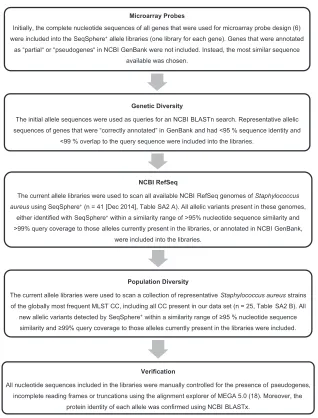
of the obtained reads were conducted as described previously (15,16). The software SeqSphere⫹version 2.3 beta (Ridom GmbH) was used for bioin-formatic sequence analysis. The complete coding sequences of the 182 queried target genes were searched within the genomes using a similarity search based on the BLAST algorithm (17) implemented in SeqSphere⫹. These nucleotide sequences were stored within so-called FASTA allele libraries and represented each target’s allelic diversity. They were grouped into four task templates, representing the functional categories of identi-fication, regulation, resistance, and virulence. These task templates are implemented in SeqSphere⫹and are accessible for all SeqSphere⫹users; the FASTA raw files can be found in the supplemental material. The cre-ation of allele libraries is essential for typing success; our study’s approach is shown inFig. 1. All allelic nucleotide sequences of each gene were aligned and were visually inspected for the presence of pseudogenes or incomplete open reading frames using MEGA 5.0 (18).
Targets were regarded as present if they were found in the genome within a range ofⱖ95% sequence identity andⱖ99% query overlap to any of the nucleotide sequences stored in the allele library. In case of multiple matches for one target, the best match was chosen automatically. Targets that were only found partially in the genomes due to location on a cropped contig were not included; however, their cropped status was noted for further analyses. Internal stop codons, frame shifts, or nucleotide ambi-guities were set to cause a “failed target” typing result. New alleles were assigned if the identified sequence wasⱖ95% identical andⱖ99% over-lapping to any of the allele sequences already present in one of the allele libraries. For the analysis of typing concordance of microarray and WGS, only the binary information gene present or gene absent was used. Cropped targets were regarded as absent, while failed targets were re-garded as present. Further analysis of the functionality of different allelic variants, including failed targets (pseudogenes), was not conducted in this
on May 16, 2020 by guest
http://jcm.asm.org/
study. The allelic variants of the 13 genescoa,fnbB,fnbA,clfA,clfB,cna,
ebh,vwb,sdrC,sdrD,sdrE,map(syn.,eap), andhysAwere highly variable between all examined isolates. Due to their low nucleotide sequence sim-ilarity to sequences already present in the allele libraries (⬍95% identity
and⬍99% overlap), they were regarded as absent by SeqSphere⫹. In
order to prevent such false-negative results, so-called “indicator targets” were designed for these genes that comprised partial nucleotide sequences (50 to 250 bp) of conserved regions of the genes. They enable the detection of the particular target gene but not the exact allelic determination.
Comparison of typing results and examination of discrepancies.
The concordance of typing results obtained by the microarray and WGS, respectively, was quantified by comparing the respective presence/ab-sence determination of the analyzed targets. This resulted in four catego-ries: (i) target present in WGS and microarray, (ii) target present in WGS and absent in microarray, (iii) target absent in WGS and present in mi-croarray, and (iv) target absent in WGS and microarray. Targets that were only detected with one of the typing methods (categories ii and iii) were regarded as discrepant and investigated in further detail (Fig. 2).
In order to detect false-negative results caused byde novoassembly
errors, reference mapping was performed using CLC Genomics Work-bench version 8 (Qiagen CLC bio, Aarhus, Denmark). The reads of all 154 samples were mapped against an artificial reference sequence comprising the representative nucleotide sequences of all investigated target genes, using default parameters with exception of length fraction (“0.8”) and nonspecific match handling (“Ignore”). Variable and diverse genes were represented by the nucleotide sequences of various alleles, and rather con-served genes were represented by only one allelic variant. Individual allelic sequences were separated by a sequence of 250 Ns (i.e., any base). The obtained bam files were analyzed with SeqSphere⫹analogous to thede novoassembled ace files by performing a sequence similarity search for the 182 targets. Moreover, it was checked whether targets that were not
iden-tified in thede novoassembled or reference-mapped genomes had been
partially identified on cropped contigs. Thus, the final WGS data set com-prised four categories: (i) target present inde novoassembly, (ii) target absent inde novoassembly but detected by reference mapping, (iii) target absent inde novoassembled and reference-mapped genomes but partially identified on a cropped contig, and (iv) target not identified at all in whole-genome data. Similarly, information about initially obtained
am-FIG 1Workflow of allele library creation. For each target gene, a FASTA allele library must be defined that includes a variety of nucleotide sequences that represent the allelic diversity of this gene. Only those sequences that are present in these libraries or differ within a defined similarity range can be found in the assembled genomes by gene-by-gene comparison.
on May 16, 2020 by guest
http://jcm.asm.org/
[image:3.585.134.452.61.479.2]biguous microarray typing results (determined by Iconoclust software) was checked in order to identify discrepant typing results caused by mis-predictions of the LFM. This resulted once more in four categories: (i) target detected as positive (present), (ii) target detected as ambiguous and predicted to be present by LFM, (iii) target detected as ambiguous and predicted to be absent by LFM, and (iv) target not detected (absent). In the following, these two data sets were compared in order to identify the causes of discrepant typing results (Fig. 2).
Discrepant typing results that were positively identified with WGS but that were negative in the microarray were always assigned to be typed false negatively by the microarray (or predicted false negatively by LFM). This was done because the extensive review and verification of implemented WGS allele libraries was assumed to prevent false-positive identifications inde novoassembled WGS data. Equally, targets that were positively iden-tified in the reference-mapped genomes and with the microarray, but not in thede novoassembled genomes, were in principle regarded as typed false negatively by WGS due tode novoassembly errors. The remaining discrepancies were resolved manually based on their most probable bio-logical explanation and the following assumptions: (i) All targets used for species identification should be positive; (ii)agrandcapgenes should be present completely but only in one variant; (iii) targetshld,saeS,sarA,
vraA,ebpS,icaC,isdA,clfA,hla,map,setB1,setB2,setB3,splA,splB, and
splPshould be present in all samples; (iv) operons likebla(blaI,blaZ,
blaR), arginine catabolic mobile elements (ACMEs) (arcA,acrB,arcC,
arcD), or theegccluster (sei,seg,sem,sen,seo,seu) should be present completely; (v) single SCCmec-associated genes (likeugpQ,pls, orxylR) were only regarded as potentially positive if amecAgene was present in the respective sample; (vi) leukotoxins (lukE and lukD, hlgB and hlgC,
lukF-PVandlukS-PV,lukMandlukF-PV83,lukXandlukY) andmerAand
merBshould only be present in pairs; and (vii)setCandfibwere regarded as present or absent depending on their CC (forfib, only ST152 was neg-ative in the used set of isolates;setCwas not detected in isolates of CC30 and ST152 [as described in reference5]). Discrepancies insslgenes were resolved according to the expected CC patterns described by Monecke et al. in 2008 (5). In cases in which a target was detected in the
reference-mapped genomes, but not in thede novoassembled genomes or with the
microarray, their nucleotide sequences and assembly results were investi-gated in further detail in order to verify the correctness of the reference mapping result. If the reference mapping result was considered correct, a false-negative result was attested for WGS and microarray or LFM typing. The same was true for targets located on a cropped contig if there was a biological reason to assume the presence of this target. Remaining dis-crepancies were checked by target-specific PCRs or phenotypic tests. These included PCRs forqacC(19,20),sdrC,sdrD,sdrE, (21)fnbB(22),
sasG(23),etB,sea,seb,sed,seg,seh(24), andhlb(25) and phenotypic resistance tests for the presence offosB(fosfomycin resistance) andcat
(chloramphenicol resistance) in accordance with EUCAST clinical break-points (version 5.0). Discrepancies that still remained unsolved after this were categorized as unknown.
Determination of MLST CCs and SCCmectypes.In addition to mo-lecular characterization regarding the presence or absence of specific vir-ulence and resistance genes, the microarray is also able to infer the isolates’ MLST CCs from the obtained hybridization patterns (5). Moreover, if present, the SCCmectype is similarly deduced. From WGS data, we ex-tracted the sequences of the seven genes comprising the allelic profile of theS. aureusMLST scheme and queried them against theS. aureusMLST database (http://saureus.mlst.net) in order to assign the classical STin silico. The respective CC was calculated using the eBURST algorithm
FIG 2Decision tree for the explanation of discrepancies. Genes that were only detected with one of the applied typing methods (WGS or microarray) in one sample were regarded as discrepant. In order to find out which method was causative for the discrepancy, the depicted decision criteria were applied.
on May 16, 2020 by guest
http://jcm.asm.org/
[image:4.585.114.479.66.382.2](http://saureus.mlst.net/eburst/). SCCmec elements were determined using presence/absence detection of nomenclature-relevant genes
(based onhttp://www.sccmec.org/Pages/SCC_TypesEN.htmland
ref-erences26and27).
Nucleotide sequence accession number.All generated raw reads were
submitted to the European Nucleotide Archive (http://www.ebi.ac.uk
/ena/) under the study accession number PRJEB11627.
RESULTS
In this study, the whole-genome sequences of 154 diverse
S. aureus
isolates were determined and analyzed with regard to 182 targets
[image:5.585.47.543.87.297.2]relevant for clonal lineage typing and staphylococcal virulence
and resistance, resulting in 28,028 individual typing results. The
results of WGS typing were compared to those obtained with the
microarray (
Tables 1
and
2
; see also Table SA3 in the supplemental
material). A total of 96.8% of these 28,028 individual typing
re-sults (n
⫽
27,119) were identically assigned to be either “present”
(n
⫽
11,374; 40.6%) or “absent” (n
⫽
15,745; 56.2%) by
microar-ray and WGS. Hence, 3.2% (n
⫽
909) of all typing results were
only positive in one typing approach and were thus regarded as
discrepant. Discrepant results were related to errors caused by
TABLE 1The percentage of concordantly typed (WGS and microarray identify a gene as present or absent, respectively) and discrepantly typed results (either only WGS or only microarray identifies a gene as present) for each functional target categoryaResult category Result caused by
Result by functional category of genes (no.)
Total no.
Total, %
Identification Regulation Resistance Virulence
Concordant
(n⫽27,119; 96.8 %)
Positive Microarray and WGS (de novo) 829 990 1,060 8,495 11,374 40.6
Negative Microarray and WGS (de novo) 0 1,159 8,100 6,486 15,745 56.2
Discrepant
(n⫽909; 3.2 %)
False positive Microarray Mishybridizations 0 78 21 103 202 0.7
LFM Misprediction 0 17 2 9 28 0.1
False negative Microarray Polymorphisms 0 3 14 140 157 0.6
LFM Misprediction 0 0 0 5 5 ⬍0.1
WGS Assembly error 88 42 16 164 310 1.1
Cropped contig 1 12 15 28 56 0.2
Not sequenced or aberrant allele
6 9 8 100 123 0.4
Unknown 0 0 4 24 28 0.1
Total no. of typing results 924 2,310 9,235 15,554 28,028 100
aComparison of typing concordance of WGS typing and the traditional DNA microarray. In total, 182 targets in 154 samples were analyzed, resulting in 28,028 individual typing
results. The discrepant results are subdivided by false-positive and false-negative typing results and different causes of error. Results that were assumed to be false negatively typed by the two methods are not considered in this table (n⫽4).
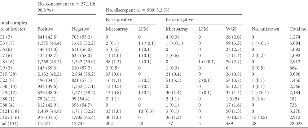
TABLE 2Samples grouped by their clonal complexes and the concordant and discrepant WGS and microarray typing results calculated for each CC (percentages per CC given in parentheses)a
Clonal complex (no. of isolates)
No. concordant (n⫽27,119;
96.8 %) No. discrepant (n⫽909; 3.2 %)
No. unknown Total no.
Positive Negative
False positive False negative
Microarray LFM Microarray LFM WGS
CC1 (7) 541 (42.5) 703 (55.2) 0 0 4 (0.3) 0 26 (2.0) 0 1,274
CC5 (17) 1,375 (44.4) 1,615 (52.2) 2 (0.1) 1 (⬍0.1) 1 (⬍0.1) 0 99 (3.2) 1 (⬍0.1) 3,094
CC6 (6) 448 (41.0) 611 (56.0) 5 (0.5) 1 (0.1) 0 0 27 (2.5) 0 1,092
CC7 (6) 423 (38.7) 633 (58.0) 11 (1.0) 1 (0.1) 7 (0.6) 0 15 (1.4) 2 (0.2) 1,092
CC8 (16) 1,258 (43.2) 1,542 (53.0) 38 (1.3) 3 (0.1) 0 1 (⬍0.1) 70 (2.4) 0 2,912
CC9 (2) 143 (39.3) 210 (57.7) 2 (0.5) 0 1 (0.3) 0 6 2 (0.5) 364
CC15 (28) 2,152 (42.2) 2,864 (56.2) 33 (0.6) 0 21 (0.4) 0 26 (0.5) 0 5,096
CC22 (8) 496 (34.1) 831 (57.1) 16 (1.1) 5 (0.3) 51 (3.5) 2 (0.1) 54 (3.7) 1 (0.1) 1,456
CC30 (13) 937 (39.6) 1,352 (57.1) 13 (0.5) 6 (0.3) 0 0 55 (2.3) 3 (0.1) 2,366
CC45 (12) 829 (38.0) 1,271 (58.2) 17 (0.8) 1 (0.3) 30 (1.4) 2 (0.1) 33 (1.5) 1 (⬍0.1) 2,184
CC80 (1) 75 (41.2) 99 (54.4) 2 (1.1) 0 2 (1.1) 0 1 (0.5) 3 (1.6) 182
CC88 (4) 312 (42.9) 398 (54.7) 0 0 1 (0.1) 0 17 (1.6) 0 728
CC121 (18) 1,469 (44.8) 1,711 (52.2) 33 (1.0) 10 (0.3) 3 (0.1) 0 50 (1.5) 0 3,276
CC152 (16) 916 (31.5) 1,905 (65.4) 30 (1.0) 0 36 (1.2) 0 10 (0.3) 15 (0.5) 2,912
Total (154) 11,374 15,745 202 28 157 5 489 28 28,028
aComparison of typing concordance of WGS typing and the traditional DNA microarray. In total, 182 targets in 154 samples were analyzed, resulting in 28,028 individual typing
results. The discrepant results are subdivided by false-positive and false-negative typing results and different causes of error. Results that were assumed to be false negatively typed by the two methods are not considered in this table (n⫽4). WGS results were summarized in one column irrespective of the cause of error.
on May 16, 2020 by guest
http://jcm.asm.org/
[image:5.585.47.544.490.697.2]WGS (n
⫽
489; 1.7%), to errors caused by the microarray (n
⫽
359; 1.3%), or to errors caused by wrong ambiguity predictions by
LFM (n
⫽
33; 0.1%). In 28 cases (0.1%), the cause of error
re-mained unknown. All WGS errors were caused by false-negative
detection of targets in the
de novo
assembled genomes. They were
subdivided into three distinct sources of error: (i) failures of
de
novo
assembly (target was identified in reference-mapped
ge-nome), (ii) insufficient genome coverage during shotgun
se-quencing (target was identified partially on a cropped contig), and
(iii) either insufficient genome coverage or missing reference
al-lele/sequence similarity below the applied thresholds (not
de-tected in WGS data at all although it should be present based on
biological assumptions) (
Tables 1
and
2
). Microarray errors can
be either false positive or false negative; false-positive results were
presumably caused by mismatch hybridizations of similar probes
with the same amplicon; false-negative results were presumably
caused by polymorphisms in the gene sequence that prevented
binding to the probe or primer. Errors of LFM were of a statistical
nature and corresponded to the expected statistical error
calcu-lated from the total number of ambiguous microarray typing
re-sults (n
⫽
2,788; 5.4%) and the experimentally determined LFM
error rate of 3% (
Tables 1
and
2
). Very few discrepancies could not
be resolved, even after traditional single-target PCRs or
phenotyp-ing (n
⫽
28; 0.1%; targets
fnbB
[n
⫽
3],
fosB
[n
⫽
3],
ugpQ
[n
⫽
1],
ssl10
[n
⫽
2],
ssl01
[n
⫽
2],
ssl04
[n
⫽
1],
ssl06
[n
⫽
1], and
lukY
[n
⫽
15]). All of them were identified with the microarray but not
with WGS. We found that
fnbB
(n
⫽
3) has a high intrinsic allelic
sequence diversity, which may have prevented the PCR primers of
the applied diagnostic PCRs from binding (
19
,
20
).
Ssl10
could not
be resolved because all other
ssl
genes were absent in the respective
samples, and thus they did not correspond to the data provided by
Monecke et al. (
5
). The remaining discrepancies in
ssl
genes could
not be resolved because the respective samples belonged to clonal
complexes that were not present in the data of Monecke et al. (
5
)
(CC80 and CC9). No PCR was available for
ugpQ. Target
lukY
was
recognized in 15 of 16 CC152 isolates by the microarray but not by
WGS.
lukY
is one of the targets that does thus not follow a
univer-sal nomenclature in NCBI, which hampers the identification of
homologous sequences according to the applied criteria (
Fig. 1
).
CC152 was generally found to possess differing alleles compared
to those of other CCs; thus, it is possible that
lukY
was present in a
new allelic variant in these samples but was not found by
SeqSphere
⫹due to low sequence similarity. However, it is
ex-pected that
lukY
only occurs in combination with
lukX, which was
not detected with microarray or with WGS in CC152; this was due
either to sequence diversity of CC152 or to real absence. All other
examined isolates from other CCs possessed
lukX
and
lukY. Thus,
it could not be decided whether
lukY
was detected false positively
by the microarray or false negatively by WGS. Phenotypic testing
for fosfomycin resistance according to EUCAST breakpoints
re-sulted in a susceptible phenotype for all three isolates. However,
detailed analysis showed that all other isolates from Münster that
harbored the
fosB
gene were fosfomycin susceptible according to
EUCAST breakpoints. For the remaining 90
fosB-positive isolates
present in our data set, no phenotypic antibiotic resistance testing
results were available. Thus, it could not be decided based on the
phenotypic data whether
fosB
should be present or not.
The rate of discrepant results (n
⫽
909 in total) differed
be-tween the four functional gene categories of (i) species
identifica-tion (n
⫽
95), (ii) regulation (n
⫽
161), (iii) resistance (n
⫽
80),
and (iv) virulence (n
⫽
573) (
Table 1
). Discrepancies in targets of
(i) species identification were mainly caused by misassemblies of
23S rRNA sequences (identified in reference-mapped genomes
but not in
de novo
assembled genomes in 83 of 154 samples).
Discrepant typing results in (ii) regulatory targets were in large
part either caused by mishybridizations between
agr-I
and
agr-IV
sequences in the microarray (n
⫽
78) and related LFM
mispredic-tions (n
⫽
17) or by WGS nondetections (n
⫽
63). The
concor-dance of typing results for (iii) resistance targets was generally very
high, with only few errors in the two methods. Microarray and
WGS typing discrepancies in (iv) virulence genes were either
caused by WGS nondetection of staphylococcal enterotoxins or
superantigens (n
⫽
169) or by microarray errors caused by
poly-morphisms in probe/primer binding sequences (n
⫽
104), for
example, in CC22 (setB1-B3,
sdrM,
ssl11,
hlIII,
ebh), CC45 (setB1,
ssl03), CC152 (icaC,
hl), and CC7 (ssl11). In four cases, the target
was initially concordantly rated as negative by microarray and
WGS but was detected in the reference-mapped genomes. After a
manual investigation of the reference mapping result, it was
de-cided that these targets had obviously been rated as false negative
with microarray and WGS. This occurred in targets
indicator-fnbB,
ermC,
icaC, and
tetK.
Table 2
shows the typing results per
CC. CC22 contained the highest number of discrepancies (n
⫽
129, 8.8% of all typing results within CC22); however, in general,
the discrepancies were equally distributed across all CCs (
Table 2
).
Finally, all CCs and SCCmec
types were identically determined
by microarray and WGS typing; however, WGS typing was more
detailed regarding clonal lineage typing by determining the ST in
contrast to the CC (see Table SA3 in the supplemental material).
Nine previously undescribed STs of five different CCs were
iden-tified in seven German and two Tanzanian isolates. The novel
allelic profiles were submitted to the
S. aureus
MLST database
(
http://saureus.mlst.net/
) and were assigned to STs 3196 to 3204
(see Table SA3).
DISCUSSION
We successfully established and evaluated a novel easy-to-use
in
silico
typing method for the detection of 182 unique target genes in
staphylococcal WGS data. Validation of the
in silico
typing was
performed by comparing the obtained results to those of an
estab-lished microarray that queries the same targets. Overall, there was
a high concordance between the two methods; only 3.2% (n
⫽
909) of the 28,028 total typing results were discrepant.
The microarray was assumed to have produced erroneous
typ-ing results in 1.3% of all cases. False-negative nondetections of
particular targets were observed especially in samples of CC22,
CC45, CC152, and CC7. They were presumably caused by
non-binding of the sample amplicon to the microarray’s probe or
primer oligonucleotide due to polymorphisms in the respective
target gene (for the exemplary misdetection of
icaC
in CC152, see
Fig. SA2 in the supplemental material). CC22 and CC45 are
known to have diverse alleles compared to those of other
pan-demic lineages like CC1, CC8, and CC5 and are thus prone to yield
“aberrant,” “irregular,” or “weak” signals in microarray typing (
5
,
6
). CC152 is a clonal lineage frequently found in West and Central
Africa and the Balkan region but is rarely isolated in the rest of the
world (
28
). It was described as an “aberrant and isolated strain”
(
5
), thus causing some problems in microarray as well as in WGS
typing due to diverse alleles (
6
). CC7 isolates were only rarely
detected in previous microarray evaluation studies (
6
), which
on May 16, 2020 by guest
http://jcm.asm.org/
dicates that maybe not the entire genetic diversity of this CC can
be considered for microarray probe design. False-positive results
occurred between highly similar probe and amplicon sequences,
e.g., between
agrI
and
agrIV. In these cases, WGS typing was much
more precise. Typing errors of the microarray can be generally
explained by the fact that the microarray is a closed system; only
those targets can be detected whose PCR-amplified region is
com-plementary to one of the available oligonucleotide probes. This is
a limitation that the creators of the microarray are well aware of
and that cannot be prevented in this kind of hybridization
proce-dure. Although the microarray’s probe set is constantly curated,
its expansion is rather time-consuming and complicated because
one always has to consider potential cross-reactions with other
probes and targets. In contrast, WGS allele libraries may be more
easily extended as soon as a novel genetic variant is found (with the
sequence databases growing rapidly). However, the diversification
of WGS allele libraries also involves the risk of blurring frontiers
between different, though related, genes, e.g., between paralogs.
Evolution is a gradual process, and new genes evolve from older
ones; thus, there is always a gray zone between two distinct genes,
depending on their divergence time. In some cases, it is impossible
(or very difficult) to decide which sequence is still an allelic variant
of a gene A and which should already be assigned to gene B. Every
nucleotide sequence identified within a similarity range of
ⱖ
95%
identity and
ⱖ
99% overlap to any allele within the library will
automatically be detected as a new allele of the respective target,
although it may actually belong to another, highly similar gene.
This was, for example, observed for targets
blaI
and
mecI. Thus,
careful curation of the libraries is needed in order to prevent
false-positive results in WGS typing.
Furthermore, 1.8% of all typing results were regarded to have
been falsely rated as negative by WGS (n
⫽
489). The majority of
these errors were caused by identified assembly failures (n
⫽
310),
which were attested if a target was identified in the
reference-mapped genome but not in the
de novo
assembled data. These
failures predominantly occurred in genes or operons that exist in
multiple copies per genome (e.g., 23S rRNA) or share too much
nucleotide sequence similarity among each other (e.g.,
enterotox-ins,
ssl
genes). The remaining errors were presumably caused by
incomplete genome coverage of shotgun sequencing (
⬎
97%
in-stead of 100%) (
29
), which implies the possibility to lose some
sequence information. In some cases, at least parts of the target
gene can be identified on a cropped contig (n
⫽
56). The
remain-ing 121 typremain-ing results that were supposed to be present were not
detected with any of the WGS analyses (de novo
assembly,
refer-ence mapping, cropped contigs). For these typing results, it is also
possible that the respective target was not detected in the genomes
because it is present in an allelic variant that has
⬍
95% identity to
alleles implemented in the respective allele library. This may be the
case especially for genes with nonuniversal nomenclature like, for
example, the
ssl
genes;
setB1,
setB2, and
setB3; or
lukX
and
lukY.
Four cases were identified by reference mapping in which the
mi-croarray and the
de novo
assembled WGS produced false-negative
typing results. It cannot be excluded that there are more
double-negative results, which are also double-negative in the reference mapping
and thus may not be detected in our approach. These technical
limitations (incomplete genome coverage and failures of
de novo
assembly) are likely to be overcome in the future with the
devel-opment of third-generation single molecule sequencing
tech-niques that provide longer sequence reads, which can then bridge
these repetitive structures and lead to a correct assembly (
30
).
Moreover, assembly errors may also be overcome by direct
map-ping of reads to reference sequences of genes of interest as was
performed, for example, by Zhang et al. in a recent study (
31
).
False-positive results did not occur in
de novo
WGS typing because
all allelic sequences were checked manually with a BLASTn and
BLASTx search if they corresponded to “correctly” annotated
genes in the NCBI nucleotide database. However, the reference
mapping was shown to provide false-positive typing results for
target
hlb-intact because both the 3
=
and the 5
=
end of the
phage-disrupted gene mapped to the intact
hlb
reference sequence.
Compared to WGS with its irreproachable advantages, the
mi-croarray is still a valuable tool to examine the
S. aureus
gene
rep-ertoire. Nevertheless, it is very likely that the anticipated gap
be-tween technological profiles and development/curating of the two
techniques will soon result in the replacement of array-based
genotyping methods by WGS in the majority of laboratories due
to its universal methodology and open system. Moreover,
se-quencing costs are constantly dropping and are already
compara-ble to the costs for microarray analysis. In contrast to other
mo-lecular typing methods, WGS typing allows for the infinite use of
once generated raw data for many different studies and for the
easy combination of accessory typing results with
epidemiologi-cally relevant typing data like core genome MLST (cgMLST) (
32
).
A major benefit of WGS typing is that genes can be discriminated
into allelic variants, which is of particular interest for virulence
and resistance genes. Minor changes in the DNA sequence may
result in major changes in the expressed protein and thus in the
respective phenotype. This also relates to the easy detection of
truncated genes or pseudogenes using our
in silico
typing method,
which may either result in nonfunctional proteins or may present
new functional variants with new phenotypic properties. The
im-portance of allele typing is exemplified by the phenotyping and
genotyping of
fosB
in this study. Unfortunately, due to repetitive
regions and high diversity among different alleles, not all targets
can be distinguished into allelic variants, including the
staphylo-coccal surface proteins Ebh, Map (syn., Eap), Coa, Cna, SdrC,
SdrD, SdrE, ClfA, ClfB, FnbA, FnbB, and Vwb, which influence
the bacterium’s interaction with the host (
33
,
34
). Different allelic
variants likely represent specific adaptations to the human
im-mune system, and ongoing coevolution probably caused the
ex-tensive number of detectable diverse allelic variants. It is
impor-tant to note that it cannot be ruled out that certain targets were not
found with the microarray or with the WGS typing scheme. This
may be, for example, the case for
hlgC
in CC152 (n
⫽
16) because
this target was identified in samples of all other clonal lineages and
does usually occur in combination with
hlgB, whose presence was
identically identified with microarray and WGS in CC152
sam-ples.
In conclusion, our WGS typing scheme reliably identified the
presence of 182 clinically relevant genes in WGS data, including,
for example, toxic shock toxin, Panton-Valentine leucocidin, or
methicillin resistance. The number of investigated targets is easily
and infinitely expandable and is not limited to the targets used in
the Identibac microarray. Indeed, some targets of the microarray
can be excluded for future WGS analyses, for example, 23S rRNA
for species identification. On the other hand, other targets like
dfrG,
mecC, or
speG
should prospectively be included in our
in
silico
typing scheme in order to expand the covered clinical
char-acteristics. With few exceptions only, all targets can be
on May 16, 2020 by guest
http://jcm.asm.org/
nated into different allelic variants, enabling a detailed analysis of
disease-associated factors in the future. Based on such analysis, we
envision a risk assessment for every clinical
S. aureus
isolate based
on the association of specific
S. aureus
genotypes with specific
human disease progressions.
ACKNOWLEDGMENTS
We thank the members of the African-German StaphNet consortium, including Pedro Alonso (Barcelona, Spain), Alexander W. Friedrich (Groningen, The Netherlands), Jonas Hofmann-Eifler (Freiburg, Ger-many), Peter Kremsner (Tübingen, GerGer-many), Sabine Schubert (Hom-burg, Germany), Marcel Tanner (Basel, Switzerland), Hagen von Briesen (St. Ingbert, Germany), Delfino Vubil (Manhiça, Mozambique), and Laura Wende (Homburg, Germany). We thank Ursula Keckevoet, Isabell Höfig, Thomas Böking, and Stefan Bletz (Institute of Hygiene, Münster, Germany) and Martina Schulte (Institute of Medical Microbiology, Mün-ster, Germany) for performing the sequencing and molecular tests. More-over, we thank Stefan Monecke (Dresden, Germany) for help with the interpretation of microarray data and probe design.
Support by the Münster Graduate School of Evolution (MGSE) to Lena Strauß is gratefully acknowledged.
We declare no conflicts of interest.
FUNDING INFORMATION
Deutsche Forschungsgemeinschaft (DFG) provided funding to Alexander Mellmann under grant number Me 3205/4-1. Deutsche Forschungsge-meinschaft (DFG) provided funding to Mathias J. Herrmann under grant number He1850/9-1. Deutsche Forschungsgemeinschaft (DFG) provided funding to Winfried V. Kern under grant number Ke 700/2-1. Deutsche Forschungsgemeinschaft (DFG) provided funding to Georg Peters under grant number Ei 247/8-1.
The funders had no role in study design, data collection and interpreta-tion, or the decision to submit the work for publication.
REFERENCES
1.Lowy FD.1998.Staphylococcus aureusinfections. N Engl J Med339:520 – 532.http://dx.doi.org/10.1056/NEJM199808203390806.
2.Bukowski M, Wladyka B, Dubin G.2010. Exfoliative toxins of Staphy-lococcus aureus. Toxins2:1148 –1165.http://dx.doi.org/10.3390/toxins 2051148.
3.Maiden MC, Bygraves JA, Feil E, Morelli G, Russell JE, Urwin R, Zhang Q, Zhou J, Zurth K, Caugant DA, Feavers IM, Achtman M, Spratt BG.
1998. Multilocus sequence typing: a portable approach to the identifica-tion of clones within populaidentifica-tions of pathogenic microorganisms. Proc Natl Acad Sci U S A95:3140 –3145.http://dx.doi.org/10.1073/pnas.95.6 .3140.
4.Enright MC, Day NP, Davies CE, Peacock SJ, Spratt BG.2000. Multi-locus sequence typing for characterization of methicillin-resistant and methicillin-susceptible clones ofStaphylococcus aureus. J Clin Microbiol
38:1008 –1015.
5.Monecke S, Slickers P, Ehricht R.2008. Assignment ofStaphylococcus aureusisolates to clonal complexes based on microarray analysis and pat-tern recognition. FEMS Immunol Med Microbiol53:237–251.http://dx .doi.org/10.1111/j.1574-695X.2008.00426.x.
6.Monecke S, Coombs G, Shore AC, Coleman DC, Akpaka P, Borg M, Chow H, Ip M, Jatzwauk L, Jonas D, Kadlec K, Kearns A, Laurent F, O’Brien FG, Pearson J, Ruppelt A, Schwarz S, Scicluna E, Slickers P, Tan HL, Weber S, Ehricht R.2011. A field guide to pandemic, epidemic and sporadic clones of methicillin-resistantStaphylococcus aureus. PLoS One6:e17936.http://dx.doi.org/10.1371/journal.pone.0017936. 7.Ruffing U, Akulenko R, Bischoff M, Helms V, Herrmann M, von Müller
L.2012. Matched-cohort DNA microarray diversity analysis of methicillin sensitive and methicillin resistantStaphylococcus aureusisolates from hos-pital admission patients. PLoS One7:e52487.http://dx.doi.org/10.1371 /journal.pone.0052487.
8.Candès EJ, Recht B.2009. Exact matrix completion via convex
opti-mization. Found Comput Math9:717–772.http://dx.doi.org/10.1007
/s10208-009-9045-5.
9.Koren Y, Bell R, Volinsky C.2009. Matrix factorization techniques for recommender systems. Computer8:30 –37.
10. Efron B, Gong G.1983. A leisurely look at the bootstrap, the jackknife, and cross-validation. Am Stat 37:36 – 48. http://dx.doi.org/10.1080 /00031305.1983.10483087.
11. Hamed M, Nitsche-Schmitz DP, Ruffing U, Steglich M, Dordel J, Nguyen D, Brink J-H, Chhatwal GS, Herrmann M, Nübel U, Helms V, von Müller L.2015. Whole genome sequence typing and microarray profiling of nasal and blood stream methicillin-resistantStaphylococcus aureusisolates: clues to phylogeny and invasiveness. Infect Genet Evol
36:475– 482.http://dx.doi.org/10.1016/j.meegid.2015.08.020.
12. Shittu AO, Oyedara O, Okon K, Raji A, Peters G, von Müller L, Schaumburg F, Herrmann M, Ruffing U.2015. An assessment on DNA microarray and sequence-based methods for the characterization of me-thicillin-susceptibleStaphylococcus aureusfrom Nigeria. Front Microbiol
6:1160.
13. Didelot X, Bowden R, Wilson DJ, Peto TE, Crook DW.2012. Trans-forming clinical microbiology with bacterial genome sequencing. Nat Rev Genet13:601– 612.http://dx.doi.org/10.1038/nrg3226.
14. Herrmann M, Abdullah S, Alabi A, Alonso P, Friedrich AW, Fuhr G, Germann A, Kern WV, Kremsner PG, Mandomando I, Mellmann AC, Pluschke G, Rieg S, Ruffing U, Schaumburg F, Tanner M, Peters G, von Briesen H, von Eiff C, von Müller L, Grobusch MP.2013. Staphylococ-cal disease in Africa: another neglected ‘tropiStaphylococ-cal’ disease. Future Microbiol
8:17–26.http://dx.doi.org/10.2217/fmb.12.126.
15. Bletz S, Mellmann A, Rothgänger J, Harmsen D.2015. Ensuring back-wards compatibility: traditional genotyping efforts in the era of whole genome sequencing. Clin Microbiol Infect21:347.e1–347.e4.
16. Ruppitsch W, Pietzka A, Prior K, Bletz S, Fernandez HL, Allerberger F, Harmsen D, Mellmann A.2015. Defining and evaluating a core genome multilocus sequence typing scheme for whole-genome sequence-based typing ofListeria monocytogenes. J Clin Microbiol53:2869 –2876.http://dx .doi.org/10.1128/JCM.01193-15.
17. Altschul SF, Gish W, Miller W, Myers EW, Lipman DJ.1990. Basic local alignment search tool. J Mol Biol215:403– 410.http://dx.doi.org/10.1016 /S0022-2836(05)80360-2.
18. Tamura K, Peterson D, Peterson N, Stecher G, Nei M, Kumar S.2011. MEGA5: molecular evolutionary genetics analysis using maximum likeli-hood, evolutionary distance, and maximum parsimony methods. Mol Biol Evol28:2731–2739.http://dx.doi.org/10.1093/molbev/msr121. 19. Noguchi N, Hase M, Kitta M, Sasatsu M, Deguchi K, Kono M.1999.
Antiseptic susceptibility and distribution of antiseptic-resistance genes in methicillin-resistantStaphylococcus aureus. FEMS Microbiol Lett172:
247–253.http://dx.doi.org/10.1111/j.1574-6968.1999.tb13475.x. 20. Mayer S, Boos M, Beyer A, Fluit AC, Schmitz FJ.2001. Distribution of
the antiseptic resistance genesqacA,qacBandqacCin 497 methicillin-resistant and -susceptible European isolates ofStaphylococcus aureus. J Antimicrob Chemother47:896 – 897.http://dx.doi.org/10.1093/jac/47.6 .896.
21. Xue H, Lu H, Zhao X.2011. Sequence diversities of serine-aspartate repeat genes amongStaphylococcus aureusisolates from different hosts presumably by horizontal gene transfer. PLoS One6:e20332.http://dx.doi .org/10.1371/journal.pone.0020332.
22. Paniagua-Contreras G, Monroy-Pérez E, Gutiérrez-Lucas R, Sainz-Espuñes T, Bustos-Martínez J, Vaca S.2014. Genotypic characterization of methicillin-resistantStaphylococcus aureusstrains isolated from the an-terior nares and catheter of ambulatory hemodialysis patients in Mexico. Folia Microbiol (Praha) 59:295–302. http://dx.doi.org/10.1007/s12223 -013-0300-4.
23. Roche FM, Massey R, Peacock SJ, Day NP, Visai L, Speziale P, Lam A, Pallen M, Foster TJ.2003. Characterization of novel LPXTG-containing proteins ofStaphylococcus aureusidentified from genome sequences. Mi-crobiology149:643– 654.http://dx.doi.org/10.1099/mic.0.25996-0. 24. Becker K, Roth R, Peters G.1998. Rapid and specific detection of
toxi-genicStaphylococcus aureus: use of two multiplex PCR enzyme immuno-assays for amplification and hybridization of staphylococcal enterotoxin genes, exfoliative toxin genes, and toxic shock syndrome toxin 1 gene. J Clin Microbiol36:2548 –2553.
25. van Wamel WJ, Rooijakkers SH, Ruyken M, van Kessel KP, van Strijp
JA.2006. The innate immune modulators staphylococcal complement
inhibitor and chemotaxis inhibitory protein ofStaphylococcus aureusare
on May 16, 2020 by guest
http://jcm.asm.org/
located on beta-hemolysin-converting bacteriophages. J Bacteriol188:
1310 –1315.http://dx.doi.org/10.1128/JB.188.4.1310-1315.2006. 26. International Working Group on the Classification of Staphylococcal
Cassette Chromosome Elements (IWG-SCC). 2009. Classification of staphylococcal cassette chromosomemec(SCCmec): guidelines for re-porting novel SCCmecelements. Antimicrob Agents Chemother53:4961– 4967.http://dx.doi.org/10.1128/AAC.00579-09.
27. Hiramatsu K, Ito T, Tsubakishita S, Sasaki T, Takeuchi F, Morimoto Y, Katayama Y, Matsuo M, Kuwahara-Arai K, Hishinuma T, Baba T.2013. Genomic basis for methicillin resistance inStaphylococcus aureus. Infect Chemother45:117–136.http://dx.doi.org/10.3947/ic.2013.45.2.117. 28. Schaumburg F, Alabi AS, Peters G, Becker K.2014. New epidemiology
ofStaphylococcus aureusinfection in Africa. Clin Microbiol Infect20:589 – 596.http://dx.doi.org/10.1111/1469-0691.12690.
29. Jünemann S, Sedlazeck FJ, Prior K, Albersmeier A, John U, Kalinowski J, Mellmann A, Goesmann A, von Haeseler A, Stoye J, Harmsen D.
2013. Updating benchtop sequencing performance comparison. Nat Bio-technol31:1148.
30. Clarke J, Wu HC, Jayasinghe L, Patel A, Reid S, Bayley H. 2009. Continuous base identification for single-molecule nanopore DNA
se-quencing. Nat Nanotechnol4:265–270.http://dx.doi.org/10.1038/nnano .2009.12.
31. Zhang S, Yin Y, Jones MB, Zhang Z, Deatherage Kaiser BL, Dinsmore BA, Fitzgerald C, Fields PI, Deng X.2015.Salmonellaserotype determi-nation utilizing high-throughput genome sequencing data. J Clin Micro-biol53:1685–1692.http://dx.doi.org/10.1128/JCM.00323-15.
32. Leopold S, Goering RV, Witten A, Harmsen D, Mellmann A.2014. Bacterial whole-genome sequencing revisited: portable, scalable, and stan-dardized analysis for typing and detection of virulence and antibiotic re-sistance genes. J Clin Microbiol52:2365–2370.http://dx.doi.org/10.1128 /JCM.00262-14.
33. Clarke SR, Foster SJ.2006. Surface adhesins ofStaphylococcus aureus. Adv Microb Physiol51:187–224.http://dx.doi.org/10.1016/S0065-2911(06) 51004-5.
34. Lindsay JA, Moore CE, Day NP, Peacock SJ, Witney AA, Stabler RA, Husain SE, Butcher PD, Hinds J.2006. Microarrays reveal that each of the ten dominant lineages ofStaphylococcus aureushas a unique combi-nation of surface-associated and regulatory genes. J Bacteriol188:669 – 676.http://dx.doi.org/10.1128/JB.188.2.669-676.2006.