0095-1137/11/$12.00 doi:10.1128/JCM.02636-10
Copyright © 2011, American Society for Microbiology. All Rights Reserved.
Design and Performance of the CDC Real-Time Reverse Transcriptase
PCR Swine Flu Panel for Detection of 2009 A (H1N1)
Pandemic Influenza Virus
䌤
†‡
Bo Shu,
1Kai-Hui Wu,
1Shannon Emery,
1Julie Villanueva,
1Roy Johnson,
1,2Erica Guthrie,
1LaShondra Berman,
1Christine Warnes,
1Nathelia Barnes,
1,2Alexander Klimov,
1and Stephen Lindstrom
1*
Virus Surveillance and Diagnostic Branch, Influenza Division, Centers for Disease Control and Prevention,1and Battelle,2Atlanta, Georgia
Received 28 December 2010/Returned for modification 9 February 2011/Accepted 10 May 2011
Swine influenza viruses (SIV) have been shown to sporadically infect humans and are infrequently identified by the Influenza Division of the Centers for Disease Control and Prevention (CDC) after being received as unsubtypeable influenza A virus samples. Real-time reverse transcriptase PCR (rRT-PCR) procedures for detection and characterization of North American lineage (N. Am) SIV were developed and implemented at CDC for rapid identification of specimens from cases of suspected infections with SIV. These procedures were utilized in April 2009 for detection of human cases of 2009 A (H1N1) pandemic (pdm) influenza virus infection. Based on genetic sequence data derived from the first two viruses investigated, the previously developed rRT-PCR procedures were optimized to create the CDC rRT-PCR Swine Flu Panel for detection of the 2009 A (H1N1) pdm influenza virus. The analytical sensitivity of the CDC rRT-PCR Swine Flu Panel was shown to be 5 copies of RNA per reaction and 10ⴚ1.3⬃ⴚ0.7
50% infectious doses (ID50) per reaction for cultured viruses.
Cross-reactivity was not observed when testing human clinical specimens or cultured viruses that were positive for human seasonal A (H1N1, H3N2) and B influenza viruses. The CDC rRT-PCR Swine Flu Panel was distributed to public health laboratories in the United States and internationally from April 2009 until June 2010. The CDC rRT-PCR Swine Flu Panel served as an effective tool for timely and specific detection of 2009 A (H1N1) pdm influenza viruses and facilitated subsequent public health response implementation.
Swine influenza is a common contagious respiratory disease of pigs caused by influenza A viruses (6, 11). The first swine influenza virus (SIV), also referred to as classical SIV H1N1 virus, was isolated in the United States in 1930 and circulated in pigs for at least 60 years (22, 24). From 1997 to 1998, triple-reassortant SIV (tr-SIV) H1N1, H1N2, H3N2, and H2N3 subtypes containing gene segments from human, avian, and swine influenza viruses were found to be circulating in North American (N. Am) swine populations (13, 14, 29, 33). Genetic analysis showed that, in particular, the nucleoprotein (NP) gene of tr-SIVs was inherited from classical SIV, that the origins of the hemagglutinin (HA) and neuraminidase (NA) genes differed, and that those genes were most closely related to genes of classical SIV (H1N1), and recently isolated human (H1N2 and H3N2) or avian (H2N3) viruses (17, 30, 33) (Fig. 1). These findings provide compelling evidence that tr-SIVs have an enhanced ability to acquire novel surface glycopro-teins, thus causing public health concerns because of their potential pandemic threat (26).
Although SIVs regularly cause illness in pigs and do not
commonly infect humans, sporadic cases of human infection have been reported in many countries (10, 18, 20, 23, 31). Between 1990 and 2010, a total of 26 cases of human infections with SIVs were detected in the United States, most of which were isolated single cases of infection, with a history of direct or indirect exposure to pigs prior to onset of illness. In two cases, limited human-to-human transmission could not be ex-cluded (3, 4, 23). Because sporadic human infections of SIVs were detected through passive surveillance for human influ-enza, for which only a small subset of influenza A-positive samples are comprehensively analyzed, it was unclear how many human cases of SIV were occurring undetected. Thus, real-time reverse transcriptase PCR (rRT-PCR) procedures were designed and optimized at the Influenza Division of the Centers for Disease Control and Prevention (CDC) in 2008 to allow for rapid, sensitive, and specific detection of these cases (25).
In April 2009, the first human cases of 2009 A (H1N1) pandemic (pdm) influenza were detected using the CDC rRT-PCR procedures originally designed to detect human cases of tr-SIV infection (12, 25). This novel influenza virus, first found in the United States and Mexico (2, 7, 9, 19), had never been previously isolated. Complete genome analysis determined that the 2009 A (H1N1) pdm influenza virus inherited its genes from an N. Am tr-SIV H1N1 virus and a Eurasian “avian-like” SIV (2, 7, 9, 19) (Fig. 1).
This paper describes the development and analytical perfor-mance of the CDC rRT-PCR Swine Flu Panel, based on the initial CDC rRT-PCR procedures for detection of N. Am tr-SIV. In response to the influenza pandemic of 2009, the CDC
* Corresponding author. Mailing address: Virus Surveillance and Diagnostic Branch, Influenza Division, Centers for Disease Control and Prevention, 1600 Clifton Road, MS-G16, Atlanta, GA 30329. Phone: (404) 639-1587. Fax: (404) 639-0080. E-mail: SLindstrom@cdc .gov.
† Supplemental material for this article may be found at http://jcm .asm.org/.
䌤Published ahead of print on 18 May 2011.
‡ The authors have paid a fee to allow immediate free access to this article.
2614
on May 16, 2020 by guest
http://jcm.asm.org/
rRT-PCR Swine Flu Panel and required reagents and supplies were distributed globally and implemented for rapid testing of specimens for the 2009 A (H1N1) pdm influenza virus.
MATERIALS AND METHODS
Influenza viruses and clinical specimens.Influenza viruses tested in this study were grown to high titers in either Madin-Darby canine kidney (MDCK) cells or embryonated chicken eggs (ECE) (27). Infectious virus in culture supernatants or allantoic fluids was measured by determination of the number of 50% tissue
culture infectious doses per milliliter (TCID50/ml) or the number of 50% egg
infectious doses per milliliter (EID50/ml), respectively (21).
Clinical specimens (nasal washes, nasal swabs, nasopharyngeal swabs, throat swabs, and lower respiratory tract specimens) included in this study were re-ceived from U.S. state and local public health laboratories as well as foreign laboratories between April 2009 and June 2010.
RNA extraction.Viral RNA was extracted from 100l of virus isolates or
clinical specimens and eluted in 100-l volumes by the use of a MagNA Pure
Compact RNA isolation kit (RNA_Tissue_V3_1 protocol) and a MagNA Pure Compact instrument (Roche Applied Science), according to the manufacturer’s instructions.
CDC rRT-PCR Swine Flu Panel primers and probes.The CDC rRT-PCR Swine Flu Panel includes four oligonucleotide primer and probe sets (universal influenza A [InfA], swine influenza A [swInfA], swine H1 [swH1], and RNase P [RP]) designed for the detection and characterization of 2009 A (H1N1) influ-enza viruses in human specimens. The assay was designed such that all three assays (InfA, swInfA, and swH1) must be positive to indicate detection of N. Am SIV H1N1 or 2009 A (H1N1) pdm virus. A result where only one or two of the three assays are positive would be inconclusive and would require further char-acterization. The InfA assay was designed for universal detection of the matrix (M) gene of all influenza A viruses. The swInfA assay was designed for universal detection of the NP gene of all N. Am SIV subtypes and for differentiation of the SIV subtypes from those of human seasonal influenza A (H1N1) and A (H3N2) viruses. The swH1 assay was designed to specifically detect the HA gene of classical SIV or N. Am tr-SIV bearing classical H1 subtype as well as 2009 A (H1N1) pdm influenza virus and differentiate them from those of human sea-sonal influenza A (H1N1). The RP assay detects the human RNase P gene and is used with human clinical specimens to measure the quality of the specimens as well as to indicate that nucleic acid was extracted adequately from the clinical specimen (8).
Oligonucleotide primers and probes were designed based on available nucle-otide sequence data from the GenBank database of the National Center for Biotechnology Information (NCBI), NIH, the influenza sequence database of Los Alamos National Laboratories (LANL), and the Global Initiative on Sharing Avian Influenza Data (GISAID). Nucleotide BLAST search (NCBI) analysis was used to verify primer and probe sequence specificity and avoid potential non-specific reactivity. The primers and probes were designed to have annealing temperatures of approximately 60°C and 70°C, respectively, using PrimerExpress 3.0 software (Applied Biosystems, Foster City, CA). Dual-labeled hydrolysis
probes were labeled at the 5⬘end with the reporter dye 6-carboxyfluorescein
(FAM) and quenched with Blackhole Quencher 1 (BHQ1) either at the 3⬘end
or internally at a modified “T” residue with Spacer3 (3-Sp3) at the 3⬘end to
prevent probe extension byTaq polymerase (Biosearch Technologies, Inc.).
Primers and dually labeled TaqMan hydrolysis probes were synthesized by
Bio-search Technologies, Inc. (Novato, CA) (see Table S1 in the supplemental material) (32).
rRT-PCR conditions.Reaction conditions for rRT-PCR were based on the Food and Drug Administration (FDA)-cleared CDC Human Influenza Virus Real-time RT-PCR Detection and Characterization Panel (CDC rRT-PCR Flu Panel) (1). The CDC rRT-PCR Flu Panel contains the same primer and probe set for detection of the influenza A virus matrix gene as the CDC rRT-PCR Swine Flu Panel. In order to determine the optimal annealing and extension temperatures, thermal gradient analysis was performed in triplicate using the Bio-Rad CFX96 system and extracted RNA from 2009 A (H1N1) pdm influenza virus strain A/California/07/2009 (Table 1; see also Fig. S1 in the supplemental material). Thermocycling rRT-PCR conditions were as follows: 50°C for 30 min,
Taqactivation for 2 min at 95°C, and 45 cycles of 95°C for 15 s and an
annealing-extension step with temperatures ranging from 50 to 65°C for 30 s. All three assays showed comparable levels of performance, with annealing temperatures ranging from 50°C to 60°C. The final reaction annealing temperature was set to 55°C, which is 5°C below the maximum optimal annealing temperature (60°C) used to accommodate potential nucleotide mismatches in the primer-probe re-gions due to virus evolution.
PCR parameters of the CDC rRT-PCR Swine Flu Panel were optimized using Invitrogen SuperScript III Platinum One-Step quantitative RT-PCR kits (Life Technologies) and Stratagene Mx3005P, Bio-Rad CFX96, and Applied Biosys-tems (AB) 7500 Fast Dx real-time PCR sysBiosys-tems. All rRT-PCRs were performed
at a total reaction volume of 25l. Final primer and probe reaction
concentra-tions were 0.8M and 0.2M, respectively. All analytical performance data and
clinical specimen data were collected using an AB 7500 Fast Dx Real-Time PCR instrument.
RESULTS
[image:2.585.114.471.67.176.2]CDC rRT-PCR Swine Flu Panel reaction efficiencies. Reac-tion efficiencies of the primer and probe sets of InfA, swInfA, and swH1 assays were determined by testing a 5-fold serial
FIG. 1. Schematic diagram representing the genotypes of N. Am tr-SIV H1N1, H1N2, and H3N2 subtypes and 2009 A (H1N1) pdm influenza viruses. Gray, yellow, green, and pink gene segments represent genes originating from human seasonal, avian, classical swine, and Eurasian swine influenza viruses, respectively.
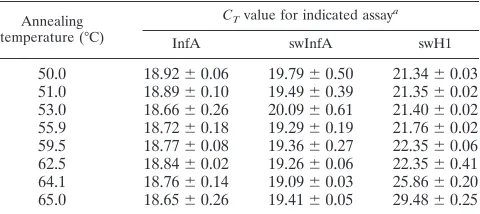
TABLE 1. Determination of optimal annealing temperature
Annealing temperature (°C)
CTvalue for indicated assaya
InfA swInfA swH1
50.0 18.92⫾0.06 19.79⫾0.50 21.34⫾0.03 51.0 18.89⫾0.10 19.49⫾0.39 21.35⫾0.02 53.0 18.66⫾0.26 20.09⫾0.61 21.40⫾0.02 55.9 18.72⫾0.18 19.29⫾0.19 21.76⫾0.02 59.5 18.77⫾0.08 19.36⫾0.27 22.35⫾0.06 62.5 18.84⫾0.02 19.26⫾0.06 22.35⫾0.41 64.1 18.76⫾0.14 19.09⫾0.03 25.86⫾0.20 65.0 18.65⫾0.26 19.41⫾0.05 29.48⫾0.25
a
Values represent meanCTvalues⫾SD (n⫽3) of the results with annealing
temperature ranging from 50°C to 65°C. Thermal gradient analysis was per-formed using viral RNA of 2009 A (H1N1) pdm influenza virus (A/California/ 07/2009) and a Bio-Rad CFX96 real-time PCR detection system.
on May 16, 2020 by guest
http://jcm.asm.org/
[image:2.585.301.541.584.691.2]dilution series of viral RNA of 2009 A (H1N1) pdm influenza virus strain A/California/07/2009 in duplicate. The resulting threshold cycle (CT) values were plotted versus relative RNA
concentration values, and linear regression analysis was ap-plied to determine the slopes. The reaction efficiencies of the InfA, swInfA, and swH1 assays were thereby estimated to be 95.7% (R2⫽ 0.996), 94.3% (R2 ⫽ 0.976), and 99.5% (R2 ⫽
0.994), respectively (see Fig. S2 in the supplemental material).
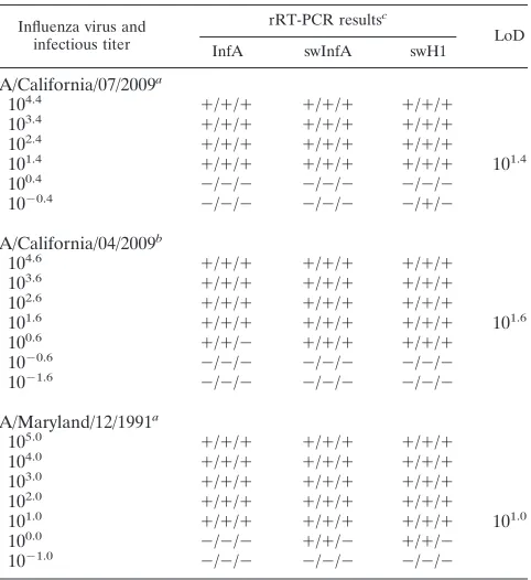
Analytical sensitivity.The limits of detection (LoD) of the InfA, swInfA, and swH1 assays were determined by analyzing a 10-fold dilution series of grown influenza viruses [classical SIV H1N1 strain A/Maryland/12/1991 and two 2009 A (H1N1) pdm influenza virus strains, A/California/04/2009 and A/Cali-fornia/07/2009]. A 10-fold dilution series was tested in tripli-cate for each extracted virus strain (Table 2). The LoD of the CDC rRT-PCR Swine Flu Panel was determined according to the lowest concentration at which all three assays (InfA, swInfA, and swH1) gave positive results. The LoD was deter-mined to be a virus concentration of 101.0⬃1.6ID
50/ml for the
three viruses tested. This correlates to 10⫺1.3⬃⫺0.7 ID 50 per
reaction (5.0l/reaction). Similarly, quantified synthetic RNA of 2009 A (H1N1) pdm influenza virus (Armored RNA Quant; AsuraGen, Inc.) was tested in duplicate (Table 3) to determine the minimum RNA copy number detectable by the assay. The LoD of the InfA, swInfA, and swH1 assays was found to be 5 copies of RNA per reaction. The cutoffCTvalue for the CDC Swine Flu Panel was determined to be⬍38 for domestic hu-man diagnostic testing purposes under conditions of FDA Emergency Use Authorization in the United States. This cutoff
value is based upon data from LoD analysis that are consistent with the cutoff value of ⬍38 previously established for the CDC rRT-PCR Flu Panel (1).
Analytical specificity and inclusivity. Assay specificity was demonstrated by testing 10 2009 A (H1N1) pdm influenza viruses, propagated in either MDCK cell culture or ECE. Vi-ruses were diluted to approximately 10-fold above the limit of detection of the assay (approximately 102to 103ID
50/ml), and
the extracted RNA was tested in triplicate. As expected, all 10 viruses tested positive in the InfA, swInfA, and swH1 assays at low virus concentrations (Table 4).
To assess the performance of the CDC rRT-PCR Swine Flu Panel with various SIVs isolated from pigs and humans, ex-tracted RNAs from cultured SIVs were tested (Table 5). Two classical SIV H1N1 subtype viruses and five N. Am tr-SIV H1N1 subtype viruses gave positive results in the InfA, swInfA, and swH1 assays. One N. Am tr-H1N2 subtype virus (whose HA originated from a human influenza virus) and one N. Am tr-H3N2 subtype virus were positive in the InfA and swInfA assays and negative in the swH1 assay. One Eurasian-lineage SIV H1N1 subtype virus was InfA positive and swInfA and swH1 negative. All test results were 100% concordant with the expected results.
[image:3.585.43.284.89.352.2]Analytical specificity and exclusivity. In order to demon-strate the absence of cross-reactivity with other common hu-man respiratory pathogens, exclusivity testing was performed
TABLE 2. Assay limit of detection (LoD) with 2009 A (H1N1) pdm and classical swine influenza viruses
Influenza virus and infectious titer
rRT-PCR resultsc
LoD
InfA swInfA swH1
A/California/07/2009a
104.4 ⫹/⫹/⫹ ⫹/⫹/⫹ ⫹/⫹/⫹ 103.4 ⫹/⫹/⫹ ⫹/⫹/⫹ ⫹/⫹/⫹ 102.4 ⫹/⫹/⫹ ⫹/⫹/⫹ ⫹/⫹/⫹ 101.4 ⫹/⫹/⫹ ⫹/⫹/⫹ ⫹/⫹/⫹ 101.4 100.4 ⫺/⫺/⫺ ⫺/⫺/⫺ ⫺/⫺/⫺ 10⫺0.4 ⫺/⫺/⫺ ⫺/⫺/⫺ ⫺/⫹/⫺
A/California/04/2009b
104.6 ⫹/⫹/⫹ ⫹/⫹/⫹ ⫹/⫹/⫹ 103.6 ⫹/⫹/⫹ ⫹/⫹/⫹ ⫹/⫹/⫹ 102.6 ⫹/⫹/⫹ ⫹/⫹/⫹ ⫹/⫹/⫹ 101.6 ⫹/⫹/⫹ ⫹/⫹/⫹ ⫹/⫹/⫹ 101.6 100.6 ⫹/⫹/⫺ ⫹/⫹/⫹ ⫹/⫹/⫹ 10⫺0.6 ⫺/⫺/⫺ ⫺/⫺/⫺ ⫺/⫺/⫺ 10⫺1.6 ⫺/⫺/⫺ ⫺/⫺/⫺ ⫺/⫺/⫺
A/Maryland/12/1991a
105.0 ⫹/⫹/⫹ ⫹/⫹/⫹ ⫹/⫹/⫹ 104.0 ⫹/⫹/⫹ ⫹/⫹/⫹ ⫹/⫹/⫹ 103.0 ⫹/⫹/⫹ ⫹/⫹/⫹ ⫹/⫹/⫹ 102.0 ⫹/⫹/⫹ ⫹/⫹/⫹ ⫹/⫹/⫹ 101.0 ⫹/⫹/⫹ ⫹/⫹/⫹ ⫹/⫹/⫹ 101.0 100.0 ⫺/⫺/⫺ ⫹/⫹/⫺ ⫹/⫹/⫺ 10⫺1.0 ⫺/⫺/⫺ ⫺/⫺/⫺ ⫺/⫺/⫺
aData represent EID
50/ml values.
b Data represent TCID
50/ml values.
[image:3.585.293.543.90.171.2]c⫹, positive rRT-PCR result;⫺, negative rRT-PCR result.
TABLE 3. Assay limit of detection determined using quantified RNAa
No. of RNA copies
CTvalues for duplicate assays
InfA swInfA swH1
500 31.30/31.71 33.17/33.56 33.14/33.45 50 34.54/34.49 37.44/37.46 37.05/37.12 5 37.88/38.04 39.54/40.98 39.06/40.32
1 ⫺/⫺ ⫺/⫺ ⫺/⫺
0.1 ⫺/⫺ ⫺/⫺ ⫺/⫺
aThe RNA material关Armored RNA Quant Flu A (H1N1) 2009; received
from AsuraGen, Inc.兴included HA, NP, and M gene region sequences derived
from 2009 A (H1N1) pdm influenza virus.⫺, negative rRT-PCR results.
TABLE 4. Analytical specificity (inclusivity) testing with 2009 A (H1N1) pdm influenza viruses (n⫽3)
Influenza virus Infectious
titera
AvgCTvalue for
indicated assayb
InfA swInfA swH1
A/Mexico/4108/2009 2.5c 33.46 34.88 32.89
A/California/08/2009 2.2c 29.75 30.59 28.93
A/California/07/2009 2.4c 34.16 35.05 33.48
A/California/04/2009 1.9d 32.26 32.35 32.05
A/Texas/48/2009 2.0d 32.20 32.01 32.23
A/Washington/29/2009 2.5d 30.58 31.21 30.76
A/South Carolina/18/2009 2.6d 25.44 28.77 27.45
A/New York/18/2009 2.7c 27.95 30.55 29.83
A/England/195/2009 2.0d 26.92 29.74 30.66
A/North Carolina/39/2009 2.7d 27.23 31.32 29.38
a
Values represent adjusted virus titers after dilution of cultured viruses.
b
Data represent meanCTvalues (n⫽3).
c
Data represent EID50/ml values.
d
Data represent TCID50/ml values.
on May 16, 2020 by guest
http://jcm.asm.org/
[image:3.585.301.541.557.692.2]by examining 10 contemporary human seasonal influenza A (H1N1), A (H3N2), and B viruses grown to high virus titers in either ECE or MDCK cells (see Table S2 in the supplemental material). All influenza A viruses were InfA positive and swInfA and swH1 negative, as expected. All influenza B viruses were negative with all three assays. Analytical specificity was further demonstrated by testing 34 noninfluenza virus strains and bacterial organisms commonly present in the nasopharynx region of the human respiratory tract. Cross-reactivity was not observed with any of the noninfluenza organisms tested at high titers (see Table S3 in the supplemental material). Extracted RNA from non-SIV animal influenza viruses were also tested at high virus titers (see Table S4 in the supplemental material). As expected, all non-SIV animal influenza viruses tested were InfA positive and swH1 negative, although some viruses were positive in the swInfA assay. However, as not all three assays were positive for any non-SIV animal influenza virus, such results would be considered inconclusive and require further testing.
Monitoring the clinical performance of the CDC rRT-PCR Swine Flu Panel.A set of 688 clinical specimens received by the CDC from May 2009 through June 2010 from U.S. public health and international laboratories, including 131 seasonal influenza A (H1N1), 360 seasonal influenza A (H3N2), 92 influenza B, and 105 influenza A- and B-negative samples, was used for monitoring clinical performance. All specimens from the set were swInfA and swH1 negative when the CDC rRT-PCR Swine Flu Panel was used.
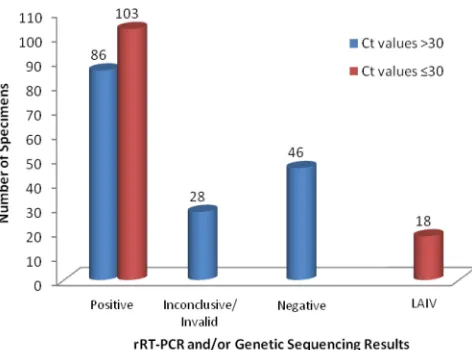
A total of 607,344 respiratory specimens were tested for the presence of influenza virus by U.S. public health laboratories from 2 May 2009 through 6 February 2010 (12). In order to monitor the performance of the CDC rRT-PCR Swine Flu Panel, U.S. public health laboratories were instructed to send human clinical specimens (nasal washes, nasal swabs, nasopha-ryngeal swabs, throat swabs, and lower respiratory tract spec-imens) with inconclusive test results to the CDC Influenza
Division for further testing and characterization (Fig. 2, CDC Influenza Division test results). Of 281 clinical specimens re-ceived due to inconclusive results, 160 specimens (59%) had InfA assay CT values higher than 30 when tested at CDC,
indicating low viral concentrations near the limit of detection of the assay (average InfACTvalue⫽34.0, standard deviation
[SD]⫽2.2). Of those 160 specimens tested at CDC, 86 (54%) were found to be positive for 2009 A (H1N1) pdm influenza virus, 26 were inconclusive, 2 were invalid (influenza virus and RP markers all tested negative), and 46 were negative. The remaining 121 specimens received due to inconclusive results with InfA testing had CT values ⱕ 30, indicating that virus
titers in these specimens were well above the limit of detection of the assay. Of those 121 speciments, 103 (85%) were found to be positive for 2009 A (H1N1) pdm influenza virus by rRT-PCR or genetic sequence characterization. Eighteen spec-imens received due to inconclusive results were positive for multiple influenza viruses, suggesting coinfection with two or more influenza virus strains. Upon further genetic character-ization, 12 of those 18 specimens contained seasonal trivalent live attenuated influenza virus vaccine (LAIV) and 6 were found to be positive for 2009 A (H1N1) pdm LAIV. Specimens containing 2009 A (H1N1) pdm LAIV that are tested using the CDC rRT-PCR Swine Flu Panel demonstrate inconclusive re-sults that are positive for InfA and swH1 and negative for swInfA.
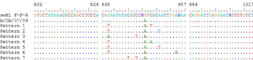
[image:4.585.42.283.90.262.2]A number of human clinical specimens received due to in-conclusive results that were positive for InfA and swInfA but negative for swH1 were subsequently confirmed as positive for 2009 A (H1N1) pdm influenza virus by bidirectional genetic sequence analysis. Comparison of the nucleotide sequences of the HA gene determined directly from clinical specimens con-firmed three nucleotide mismatches within the probe region of the swH1 assay (Fig. 3). However, a consistent pattern of conserved nucleotide mutations whose circulation persisted could not be identified. In fact, among the 7,122 HA genes of 2009 A (H1N1) pdm influenza viruses available from the
FIG. 2. CDC evaluation of inconclusive specimens (n⫽281) sub-mitted by U.S. public health laboratories. Results obtained by the CDC Influenza Division for 160 specimens with InfACTvalues⬎30 and 121
specimens withCTvaluesⱕ30 confirmed by the CDC rRT-PCR Swine
[image:4.585.302.538.502.677.2]Flu Panel and/or genetic sequence characterization are shown. TABLE 5. Analytical specificity (inclusivity) testing with
swine influenza virusesa
Influenza virus Subtype
CTvalue for indicated assay
InfA swInfA swH1
Classical SIV
A/swine/Indiana/1726/1988 H1N1 20.10 22.41 24.31 A/Maryland/12/1991 H1N1 17.28 17.18 18.41
N. Am tr-SIV
A/Iowa/01/2006 H1N1 25.66 26.30 30.06
A/Illinois/09/2007 H1N1 31.22 31.51 31.11
A/Ohio/01/2007 H1N1 24.28 24.42 25.21
A/Missouri/04/2006 H1N1 19.97 19.58 22.18 A/Texas/14/2008 H1N1 22.12 22.04 23.45 A/Michigan/09/2007b H1N2 29.17 31.31 —c
A/Iowa/16/2009b H3N2 17.00 17.35 —
Eurasian SIV
A/swine/Italy/711/2006 H1N1 17.78 — —
aThe tested viruses were isolated from swine as well as human cases of swine
origin influenza virus infection.
bH1N2 and H3N2 influenza viruses possessing HA and NA genes originating
from human seasonal influenza viruses.
c—, negative result.
on May 16, 2020 by guest
http://jcm.asm.org/
GISAID databases as of 30 August 2010, only 8 (0.11%) vi-ruses with such genetic changes were identified.
DISCUSSION
The CDC rRT-PCR Swine Flu Panel is a highly sensitive and specific assay for detection and characterization of N. Am tr-SIV containing H1 HA subtype as well as 2009 A (H1N1) pdm influenza viruses. The assay was shown to be highly sen-sitive and detected as low as 101.0⬃1.6 ID
50/ml of 2009 A
(H1N1) pdm or classical tr-SIV H1N1 viruses, which corre-sponds to approximately 10⫺1.3⬃⫺0.7ID
50per reaction.
Simi-larly, the assay was shown to detect as few as 5 copies/reaction of 2009 A (H1N1) pdm influenza virus RNA. Also, the CDC rRT-PCR Swine Flu Panel was demonstrated to be highly specific for characterization of N. Am SIV H1 subtype as well as 2009 A (H1N1) pdm influenza viruses. As expected, all tested classical SIV H1N1 and N. Am tr-SIV H1N1 viruses were positive with all three assays, while tested N. Am tr-SIV H1N2 and N. Am tr-SIV H3N2 viruses were InfA and swInfA positive but swH1 negative, since both of the latter virus sub-types possess HA antigens inherited from human seasonal viruses (13, 14, 30).
The CDC rRT-PCR Swine Flu Panel did not show any cross-reactivity among all three assays with common non-in-fluenza A virus respiratory pathogens, innon-in-fluenza B cultured viruses, or influenza B virus-positive clinical specimens. Cross-reactivity was not observed in the swInfA and swH1 assays used in testing human seasonal influenza A (H1N1 and H3N2) viruses. No cross-reactivity was observed in the swH1 assay when testing for highly pathogenic avian influenza (HPAI) H5N1 viruses and other animal influenza viruses of different subtypes. However, the swInfA assay demonstrated cross-re-activity with HPAI H5N1 viruses and other non-SIV animal influenza A viruses tested in this study due to similarities of primer and probe sequences to those of some of these viruses. Since none of the non-SIV animal influenza virus tested were positive for all three assays on the CDC rRT-PCR Swine Flu Panel, these results would be considered inconclusive and re-quire further genetic analysis. Conclusive positive results were not observed with a broad range of influenza viruses of non-swine origin as well as with other respiratory pathogens, thus demonstrating 100% analytical specificity of the CDC rRT-PCR Swine Flu Panel.
Soon after the first confirmation (15 April 2009) of human
infection with 2009 influenza A (H1N1) pdm virus, the CDC rRT-PCR Swine Flu Panel was cleared (27 April 2009) by the U.S. Food and Drug Administration (FDA) with an Emer-gency Use Authorization (EUA), and on 29 April 2009, testing procedures were posted on the website of the World Health Organization (WHO) (32). FDA clearance in 2008 (1) of the CDC rRT-PCR Flu Panel, a diagnostic assay that utilizes iden-tical procedures and instrumentation, facilitated expeditious EUA clearance of the CDC rRT-PCR Swine Flu Panel. The CDC rRT-PCR Flu Panel, including rRT-PCR primers, probes, and controls, had been distributed since 2008 to sup-port influenza surveillance and pandemic preparedness, allow-ing public health laboratories to detect seasonal influenza A (H1N1), A (H3N2), and HPAI A (H5N1) viruses (12). Ac-cordingly, U.S. public health laboratories as well as many for-eign laboratories have received training on use of the CDC rRT-PCR Flu Panel. Rapid deployment of the CDC rRT-PCR Swine Flu Panel to domestic and international laboratories facilitated the timely detection of 2009 A (H1N1) pdm influ-enza viruses, thus prompting state, local, and foreign govern-ments to enact public health response activities. From 1 May 2009 until the declaration of the end of the emergency in the United States (23 June 2010), more than 2,100 kits were dis-tributed to hundreds of U.S. public health laboratories and international National Influenza Centers (NICs).
[image:5.585.81.500.66.166.2]It is well established that influenza viruses evolve rapidly and are capable of adapting and generating diversity through ge-netic mutation and gege-netic reassortment, particularly following the emergence of a novel pandemic strain in humans (15, 16, 28). As the mutation rate and reassortment capabilities of the 2009 A (H1N1) pdm influenza viruses were initially unknown, it was imperative to monitor the performance of the CDC rRT-PCR Swine Flu Panel during the pandemic to determine the effect of any genetic changes due to mutation or reassort-ment that might affect assay performance. Therefore, U.S. public health laboratories were advised to submit any clinical specimens with inconclusive results to the CDC Influenza Di-vision for further testing and characterization. Among those submitted, six human specimens contained 2009 H1N1 LAIV, giving positive test results by the InfA and swH1 assays but negative results by the swInfA assay. This reactivity was ex-pected, since the 2009 H1N1 LAIV is a reassortant virus that contains six internal gene segments derived from the cold-adapted master donor virus, A/Ann Arbor/6/1960 (H2N2), and
FIG. 3. Nucleotide sequence alignment of swH1 primer and probe regions from specimens and of viruses positive for 2009 A (H1N1) pdm influenza virus that tested negative by the swH1 assay. Only locations where nucleotide differences were observed are indicated. The numbers of specimens identified with each pattern of nucleic acid differences were as follows: for pattern 1, 19; for pattern 2, 2; for pattern 3, 1; for pattern 4, 1; for pattern 5, 1; for pattern 6, 1; and for pattern 7, 1. The primer and probe locations are indicated according to the HA gene coding domain sequence of 2009 A (H1N1) pdm influenza virus strain A/California/07/2009 (FJ966974).
on May 16, 2020 by guest
http://jcm.asm.org/
the HA and NA gene segments from the recommended vac-cine virus A/California/07/2009 (H1N1) pdm (5).
Genetic analysis of clinical specimens received during the pandemic by the CDC due to inconclusive results obtained at local laboratories revealed some viruses with various patterns of three mutations in the binding region of the swH1 probe that caused decreased reactivity of the swH1 assay. However, viruses with similar mutations accounted for only 0.11% of sequences submitted to the GISAID database as of September 2010, thus indicating that the vast majority of 2009 A (H1N1) pdm influenza viruses were detectable by all three assays in the CDC rRT-PCR Swine Flu Panel.
The CDC rRT-PCR Swine Flu Panel served as an effective emergency tool throughout the pandemic for rapid detection of 2009 A (H1N1) pdm virus in many countries and was es-sential for emergency response implementation. Efforts to fur-ther update and optimize procedures and reagents for detec-tion of 2009 A (H1N1) pdm influenza viruses in human clinical samples are still necessary in order to address the potential risk of emerging variants with similar reactivity characteristics, as well as to eliminate cross-reactivity of the swInfA assay with avian and animal viruses.
ACKNOWLEDGMENTS
We thank the United States Public Health Laboratories as well as international laboratories participating in the WHO Global Influenza Surveillance Network for sharing materials and genetic sequence in-formation used in this study. We also thank our colleagues from the Virus Surveillance and Diagnosis Branch and the Influenza Sequenc-ing Activity, Influenza Division, CDC, for their help and cooperation. The findings and conclusions in this report are those of the authors and do not necessarily represent the views of the Centers for Disease Control and Prevention.
REFERENCES
1.CDC.2008. 510(k) Summary for Centers for Disease Control and Prevention
human influenza virus real-time RT-PCR detection and characterization panel. CDC, Atlanta, GA. http://www.accessdata.fda.gov/cdrh_docs/pdf8 /k080570.pdf.
2.CDC.2009. Swine influenza A (H1N1) infection in two children—Southern
California, March–April 2009. MMWR Morb. Mortal. Wkly. Rep.58:400–
402.
3.CDC.2009. Update: influenza activity—United States, 2009–2010. Morb.
Mortal. Wkly. Rep.59:901–908.
4.CDC.2010. The two cases of novel influenza A virus infection reported to
CDC during 2010 were identified as swine influenza A (H3N2) virus and are unrelated to the 2009 pandemic influenza A (H1N1) virus. Morb. Mortal.
Wkly. Rep.59:1457–1470.
5.Chan, W., H. Zhou, G. Kemble, and H. Jin.2008. The cold adapted and temperature sensitive influenza A/Ann Arbor/6/60 virus, the master donor virus for live attenuated influenza vaccines, has multiple defects in
replica-tion at the restrictive temperature. Virology380:304–311.
6.Dacso, C. C., et al.1984. Sporadic occurrence of zoonotic swine influenza
virus infections. J. Clin. Microbiol.20:833–835.
7.Dawood, F. S., et al.2009. Emergence of a novel swine-origin influenza A
(H1N1) virus in humans. N. Engl. J. Med.360:2605–2615.
8.Emery, S. L., et al.2004. Real-time reverse transcription-polymerase chain
reaction assay for SARS-associated coronavirus. Emerg. Infect. Dis.10:311–
316.
9.Garten, R. J., et al.2009. Antigenic and genetic characteristics of
swine-origin 2009 A (H1N1) influenza viruses circulating in humans. Science325:
197–201.
10.Gaydos, J. C., et al. 1977. Swine influenza A at Fort Dix, New Jersey (January–February 1976). II. Transmission and morbidity in units with cases.
J. Infect. Dis.136(Suppl.):S363–S368.
11.Hinshaw, V. S., W. J. Bean, Jr., R. G. Webster, and B. C. Easterday.1978. The prevalence of influenza viruses in swine and the antigenic and genetic
relatedness of influenza viruses from man and swine. Virology84:51–62.
12.Jernigan, D. B., et al.2011. Detecting 2009 pandemic influenza A (H1N1) virus infection: availability of diagnostic testing led to rapid pandemic
re-sponse. Clin. Infect. Dis.52(Suppl. 1):S36–S43.
13.Karasin, A. I., S. Carman, and C. W. Olsen.2006. Identification of human H1N2 and human-swine reassortant H1N2 and H1N1 influenza A viruses
among pigs in Ontario, Canada (2003 to 2005). J. Clin. Microbiol.44:1123–
1126.
14.Karasin, A. I., et al.2000. Genetic characterization of H3N2 influenza viruses isolated from pigs in North America, 1977–1999: evidence for wholly
human and reassortant virus genotypes. Virus Res.68:71–85.
15.Kawaoka, Y., S. Krauss, and R. G. Webster.1989. Avian-to-human trans-mission of the PB1 gene of influenza A viruses in the 1957 and 1968
pan-demics. J. Virol.63:4603–4608.
16.Lindstrom, S. E., N. J. Cox, and A. Klimov.2004. Genetic analysis of human H2N2 and early H3N2 influenza viruses, 1957–1972: evidence for genetic
divergence and multiple reassortment events. Virology328:101–119.
17.Ma, W., et al.2007. Identification of H2N3 influenza A viruses from swine in
the United States. Proc. Natl. Acad. Sci. U. S. A.104:20949–20954.
18.Myers, K. P., C. W. Olsen, and G. C. Gray.2007. Cases of swine influenza in
humans: a review of the literature. Clin. Infect. Dis.44:1084–1088.
19.Neumann, G., T. Noda, and Y. Kawaoka.2009. Emergence and pandemic
potential of swine-origin H1N1 influenza virus. Nature459:931–939.
20.Newman, A. P., et al.2008. Human case of swine influenza A (H1N1) triple
reassortant virus infection, Wisconsin. Emerg. Infect. Dis.14:1470–1472.
21.Reed, L. J., and H. Muench.1938. A simple method of estimating fifty
percent endpoints. Am. J. Hyg.27:493–497.
22.Sheerar, M. G., B. C. Easterday, and V. S. Hinshaw.1989. Antigenic
con-servation of H1N1 swine influenza viruses. J. Gen. Virol.70(Pt. 12):3297–
3303.
23.Shinde, V., et al.2009. Triple-reassortant swine influenza A (H1) in humans
in the United States, 2005–2009. N. Engl. J. Med.360:2616–2625.
24.Shope, R. E.1931. The etiology of swine influenza. Science73:214–215. 25.Shu, B., et al.2009. Universal detection of swine influenza viruses and
specific discrimination of swine H1 influenza viruses by real-time PCR assay, abstr. TP-15. Abstr. 25th Annu. Clin. Virol. Symp., Florida, April 2009. 26.Smith, G. J., et al.2009. Origins and evolutionary genomics of the 2009
swine-origin H1N1 influenza A epidemic. Nature459:1122–1125.
27.Szretter, K. J., A. L. Balish, and J. M. Katz.2006. Influenza: propagation, quantification, and storage. Curr. Protoc. Microbiol. Chapter 15, Unit 15G 1. 28.Vijaykrishna, D., et al.2010. Reassortment of pandemic H1N1/2009
influ-enza A virus in swine. Science328:1529.
29.Vincent, A. L., et al.2006. Evaluation of hemagglutinin subtype 1 swine
influenza viruses from the United States. Vet. Microbiol.118:212–222.
30.Webby, R. J., K. Rossow, G. Erickson, Y. Sims, and R. Webster.2004. Multiple lineages of antigenically and genetically diverse influenza A virus
co-circulate in the United States swine population. Virus Res.103:67–73.
31.Wentworth, D. E., et al.1994. An influenza A (H1N1) virus, closely related to swine influenza virus, responsible for a fatal case of human influenza.
J. Virol.68:2051–2058.
32.World Health Organization (WHO).30 April 2009. CDC protocol of real-time RT-PCR for influenza H1N1. World Health Organization, Geneva, Switzerland. http://www.who.int/csr/resources/publications/swineflu/realtimeptpcr /en/index.html.
33.Zhou, N. N., et al.2000. Emergence of H3N2 reassortant influenza A viruses
in North American pigs. Vet. Microbiol.74:47–58.