R E S E A R C H A R T I C L E
Open Access
Longitudinal assessment of cyst-like lesions of
the knee and their relation to radiographic
osteoarthritis and MRI-detected effusion and
synovitis in patients with knee pain
Daichi Hayashi
1*, Frank W Roemer
1, Zineb Dhina
1, C Kent Kwoh
2,3, Michael J Hannon
2, Carolyn Moore
4,5,
Ali Guermazi
1Abstract
Introduction:The purpose of the present study was to determine the prevalence of cystic lesions and cyst-like bursitides in subjects with frequent knee pain and to assess their relation to radiographic osteoarthritis (OA) severity; to describe bilaterality and size fluctuation of the lesions over 6 months; and to assess relations between the prevalence of synovium-lined lesions communicating with the joint capsule and severity of magnetic
resonance imaging (MRI)-detected effusion and synovitis.
Methods:One hundred and sixty-three subjects (total 319 knees) aged 35 to 65 with chronic, frequent knee pain were included. Imaging with 3 Tesla MRI was performed at baseline and 6-month follow-up with the same protocols as those used in the Osteoarthritis Initiative. Severity of radiographic OA was assessed using the Kellgren-Lawrence grade (0 to 4). Severity of effusion and synovitis was graded 0 to 3 based on the Whole Organ Magnetic Resonance Imaging Score system. The associations of cysts and cyst-like bursitides and severity of radiographic OA, MRI-detected effusion and synovitis were analyzed using logistic regression controlling for clustering by person. The Wilcoxon signed-rank test was used to determine whether there was a significant change in the size of lesions between baseline and follow-up.
Results:At least one lesion (any type) was present in 222 (70%) knees. The most prevalent lesions were popliteal cysts (40%, 128/319), followed by subgastrocnemius bursitis (15%, 49/319) and proximal tibiofibular joint cysts (8%, 26/319). Bilateral lesions were seen in 49% of the subjects. Only popliteal cysts and subgastrocnemius bursitis showed a significant change in size (P< 0.001). No trend was observed between prevalence of any of the cyst-like lesions analyzed and the increasing radiographic OA severity. Increasing prevalence of subgastrocnemius bursitis was associated with increasing severity of effusion (P= 0.0072) and synovitis (P= 0.0033).
Conclusions:None of the cyst-like lesions analyzed seems to be a marker of radiographic OA severity in knees with chronic frequent pain. Subgastrocnemius bursitis may be used as a marker of effusion/synovitis severity. Bilateral cyst-like lesions are relatively commonly observed in people with chronic knee pain.
Introduction
Fluid-equivalent lesions of the knee joint consist of a variety of pathologies ranging from benign intra-articular fluid collections to those associated with inflammatory or
degenerative arthritis, infection and malignancy [1-4]. These pathologies have historically been detected by arthrography [5] and ultrasound [6], and are often seen on routine magnetic resonance imaging (MRI) scans. MRI has emerged as the technique of choice for characterizing the nature of these lesions, which show fluid-equivalent hypointensity on T1-weighted images and hyperintensity on T2-weighted images. MRI also
* Correspondence: Daichi.Hayashi@bmc.org
1Quantitative Imaging Center, Department of Radiology, Boston University
School of Medicine, FGH Building 3rd Floor, 820 Harrison Avenue, Boston, MA 02118, USA
Full list of author information is available at the end of the article
enables determination of their anatomical relationship with the joint and other surrounding tissues [4,7].
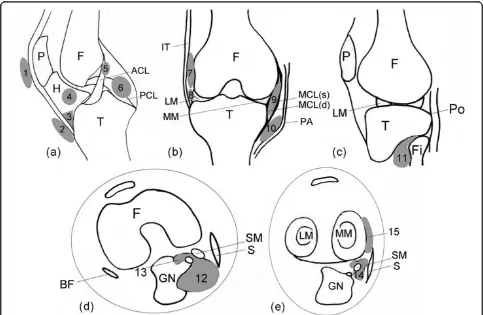
Detailed descriptions and illustrations of cyst-like lesions - that is, cysts and discrete fluid collections within bursae (bursitides) - have been published pre-viously [4,7,8]. Bursae are synovium-lined structures usually not easily detected by any imaging method. Inflammation may result in a cyst-like appearance, how-ever, due to accumulation of fluid within the bursa and thickening of the synovial membrane [8]. Locations of various bursae and cystic lesions of the knee are shown schematically in Figure 1.
To date, the prevalence of cysts and bursitides of the knee with or without symptoms has only been examined in a limited number of studies, which yielded discordant data [9,10]. Anserine, iliotibial, semi-membranosus
medial collateral ligament (SM-MCL), and deep infrapa-tellar bursitides have each been shown to be more com-mon in patients with knee osteoarthritis (OA) and pain in some studies [9,11]. In one study, however, deep infrapatellar bursitis showed an unusually high preva-lence (41%) in asymptomatic knees [12]. Published information on the prevalence of bilateral cysts and bur-sitides of the knee in the setting of OA is limited [13].
[image:2.595.56.539.303.618.2]The aim of our study was to assess the prevalence of cysts and cyst-like bursitides of the knee in a cohort of subjects with frequent knee pain and to examine their relation with radiographic OA severity. We also aimed to assess relations between synovium-lined cyst-like lesions communicating with the joint capsule and the severity of MRI-detected synovitis and effusion. Further-more, we wished to evaluate whether the occurrence of
these lesions was a bilateral phenomenon, and to describe their size changes over 6 months.
Materials and methods
Study sample
Subjects included in the present study were participants in the Joints On Glucosamine (JOG) cohort. The JOG study is a 6-month, double-blind, randomized controlled trial to examine the efficacy of oral glucosamine supple-mentation. Two hundred and one participants, aged 35 to 65 with mild to moderate chronic, frequent knee pain (Western Ontario and McMaster Universities score ≥ 25), were recruited at the University of Pittsburgh, PA, USA. Institutional Review Board approval and writ-ten informed consent from all participants were obtained for the present study.
Types of fluid-filled lesions of the knee
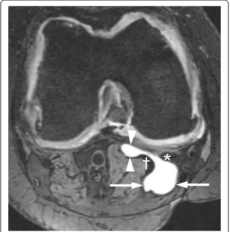
Fifteen types of lesions were studied, including popliteal cysts (Figure 2), proximal tibiofibular joint (PTFJ) cysts, medial and lateral meniscal cysts, and anterior and pos-terior cruciate ligament (ACL and PCL) and Hoffa’s fat pad ganglion cysts. In addition, the following bursitides with cyst-like appearances were included: anserine, prepatellar, superficial and deep infrapatellar, iliotibial,
SM-MCL, medial collateral ligament, and subgastrocne-mius bursitides [7] (Figures 1 and 2).
Radiographic assessment of osteoarthritis
Radiographic images of the knee were obtained using a standard radiographic technique according to published guidelines [14]. Radiographs were read according to the Kellgren-Lawrence (K-L) grading scheme [15] by one expert musculoskeletal radiologist (AG) with 11 years of experience in K-L grading of knee radiographs. A ran-dom set of 30 radiographs (60 knees) were re-read to calculate the intraobserver reliability for the K-L grading of the knee, and the weighted kappa value was 0.89.
Magnetic resonance imaging acquisition
Of 201 subjects in the JOG cohort, 38 were excluded because only a baseline MRI scan was available (24 sub-jects) or complete follow-up MRI assessment was not possible due to poor acquisition quality (14 subjects). Thus, 319 (161 left and 158 right) knees of 163 subjects were included in the present study.
MRI of each knee was performed using a 3 Tesla MRI (Siemens Trio, Erlangen, Germany) at baseline and at 6 months. The identical protocol used for the Osteoarthritis Initiative was applied in the JOG study, excluding the Fast Low Angle Shot sequence and the Multi-Echo Spin Echo T2 mapping sequence. Details of the full Osteoarthritis Initiative pulse sequence protocol and the sequence parameters have been pub-lished [16]. The protocol included the sagittal triplanar three-dimensional Dual Echo at Steady State sequence (slice thickness = 0.7 mm, interslice gap = 0 mm, repe-tition time = 16.3 ms, echo time = 4.7 ms, flip angle = 25°, field of view = 140 mm × 140 mm, matrix = 384 × 307 pixels, echo train length = 1, number of slices = 35, bandwidth = 185 Hz/pixel, number of excitations = 1, anterior/posterior phase encoding axis, acquisition time = 10 minutes 23 seconds) and the sagittal intermediate-weighted fat-suppressed sequence (slice thickness = 3 mm, interslice gap = 0 mm, repetition time = 30 ms, echo time = 3,200 ms, flip angle = 180°, field of view = 160 mm × 160 mm, matrix = 313 × 448 pixels, echo train length = 5, number of slices = 37, bandwidth = 248 Hz/pixel, number of excitations = 1, anterior/posterior phase encoding axis, acquisition time = 4 minutes 42 seconds). Axial and coronal images were reformatted from the sagittal three-dimensional Dual Echo at Steady State images.
Magnetic resonance imaging assessment
[image:3.595.56.292.424.663.2]One musculoskeletal radiologist (ZD) with 15 years of clinical experience, blinded to clinical data but not to the timepoint of the MRI examination, reviewed all images. The images were evaluated for the presence or
absence of cystic lesions at baseline and at 6 months. Measurements of the largest diameter of each lesion were made to the nearest millimeter using a manual caliper on a standard DICOM viewer (eFilm Worksta-tion, version 2.0.0; Merge Healthcare, Milwaukee, WI, USA). For the cystic lesions, unidimensional measure-ment was made based on the assumption that the size of a lesion increases or decreases in the same direction for all three axes - see Revised Evaluation Criteria in Solid Tumors (RECIST) (version 1.1) for size mea-surement of solid tumors [17] (in RECIST 1.1, cystic tumors are also considered a measurable lesion). The longest diameter of each lesion was measured on the sequence that showed the lesion in its largest dimension using the same plane (axial or sagittal) at baseline and at follow-up.
Of the lesions listed earlier, it was only possible to make meaningful size measurements on popliteal cysts, meniscal cysts, anterior cruciate ligament and PCL ganglion cysts, prepatellar bursitis, superficial infrapatel-lar bursitis and subgastrocnemius bursitis. These lesions had approximately circular or elliptical shape and their shape remained similar at two timepoints, enabling us to make meaningful size comparisons based on the assumption described earlier. However, it was not possi-ble to measure the size of other lesions due to their completely irregular or undefinable shape. For these lesions, only the presence or the absence was noted. Development of a new lesion at follow-up was treated as an increase in size, and complete resolution of a pre-existing lesion at follow-up was treated as a decrease in size for the purpose of our analysis.
Severity of synovitis and effusion at baseline was semi-quantitatively assessed according to the Whole Organ Magnetic Resonance Imaging Score (WORMS) system [18], taking into account all available sequences. Synovi-tis was graded 0 to 3 at infrapatellar and intercondylar sites (0, none; 1, mild; 2, moderate; 3, severe), and the maximum grade at either site was recorded. Effusion was also graded 0 to 3 (0, none; 1, ≤33% of maximum potential distention of the synovial cavity; 2, 33 to 66% of maximum potential distention; 3,≥66% of maximum potential distention).
Intraobserver reliability for the detection of cystic lesions and their size measurement was assessed on a random sample of 20 knee MRI scans with all acquired sequences, and was excellent (= 0.88 for detection of cystic lesions; intraclass correlation coefficient for their size measurement = 0.94). Assessment of interobserver reliability was performed on a random sample of 50 knees by a second expert musculoskeletal radiologist (AG) with 11 years of experience in semiquantitative analysis of knee OA. Agreement for detection of cystic lesions was high ( = 1.00), and the intraclass
correlation coefficient for size measurements on the same sample was 0.99.
Definition of significant change of size
During our intraobserver and interobserver reliability exercises, it was noted that the maximum difference between two readings for the measurement of the same lesion (either at baseline or at follow-up) was 4 mm for the popliteal cyst and 1 mm for other measurable lesions. We thus considered that any changes within these ranges might be attributable to measurement error. We defined the significant change of size to be≥5 mm for popliteal cyst and≥2 mm for other measurable lesions.
Statistical analysis
The K-L grading was used to categorize knees according to the severity of radiographic OA. When there were eight or more of a particular type of lesion, the associa-tion between K-L grade and the prevalence of those lesions was tested using logistic regression controlling for clustering by person [19,20]. For cyst-like lesions that are lined by synovium and known to have a com-munication with the joint capsule [4] (that is, popliteal cysts, subgastrocnemius bursitis and PTFJ cysts), we also analyzed whether there was a linear trend between their prevalence and increasing severity of MRI-detected effusion and synovitis using logistic regression control-ling for clustering by person.
Significance in associations between prevalence of cyst-like lesions and categorical/continuous variables was determined using Fisher’s Exact Test for gender, and attest for age and body mass index. These analyses were also performed only for those lesions observed in eight or more knees.
Finally, the Wilcoxon signed-rank test was used to determine whether there was a significant change in the size of lesions between baseline and follow-up.
All statistical calculations were performed using SAS® software (version 9.1 for Windows; SAS Institute, Cary, NC, USA), except for the logistic regression that was performed using STATA (version 11.0; Statacorp LP, College Station, TX, USA).
Results
Subject characteristics
PCL ganglion cysts. Nine subjects (82%) who had PCL ganglion cysts were women, whereas 66 (43%) of those without the lesion were women (P = 0.024).
Characteristics of subjects who were excluded from the present study were similar to those included in terms of baseline presence of radiographic OA (that is, proportion of K-L grade ≥2 was 59% vs. 59%) and the presence of effusion (WORMS grade≥1, 41% vs. 44%), but those excluded had lower prevalence of synovitis than those included (WORMS grade≥1, 37% vs. 62%; P= 0.01, Fisher’s exact test).
Prevalence of cyst-like lesions and their relation to radiographic osteoarthritis
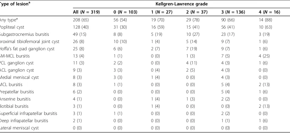
Table 1 summarizes the prevalence of all lesions. At least one fluid-filled lesion of any type was present in 208 knees (65%). The most prevalent type was popliteal cysts (Figure 2), found in 128 knees (40%). Subgastroc-nemius bursitis, PTFJ cysts and Hoffa’s fat pad ganglion cysts were observed in 49 knees (15%), 26 knees (8%) and 25 knees (8%), respectively. Nineteen (39%) subgas-trocnemius bursitides co-existed with a communicating popliteal cyst (Figure 2). Other types such as SM-MCL bursitis, ACL and PCL ganglion cysts, and medial meniscal cysts were observed less frequently. Superficial and deep infrapatellar, prepatellar, anserine, and ilioti-bial bursitides were rarely observed but seemed to be more common in knees with OA than in those without OA. No lateral meniscal cysts were detected in any
knee at baseline but one lesion had appeared at 6-month follow-up.
Of the 319 knees, 103 (32%) were K-L grade 0, 27 (9%) were K-L grade 1, 37 (12%) were K-L grade 2, 136 (43%) were K-L grade 3, and 16 (5%) were K-L grade 4 (Table 1). A linear trend was found between increasing prevalence of fluid-filled lesions of any type and increas-ing K-L grade (K-L 0 = 54%, K-L 1 = 70%, K-L 2 = 78%, K-L 3 = 66% and K-L 4 = 88%;P = 0.014). This trend, however, did not extend to any individual type of lesions.
Trend between prevalence of synovium-lined cyst-like lesions and severity of effusion and synovitis
Increasing prevalence of subgastrocnemius bursitis was associated with increasing severity of effusion (P = 0.0072) and synovitis (P = 0.0060) after controlling for clustering by person (Table 2). For popliteal cysts and PTFJ cysts, such a trend was not observed.
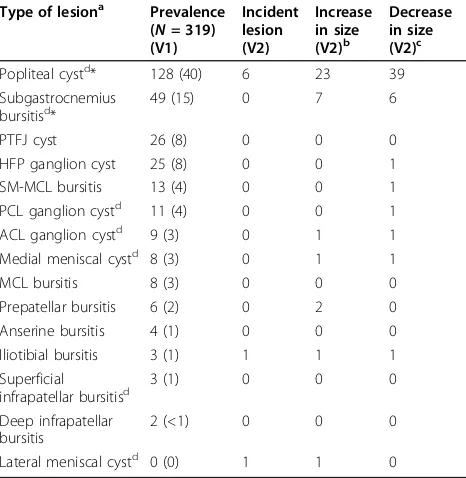
Size changes of cyst-like lesions over 6 months
[image:5.595.59.538.476.697.2]Changes in the size of the lesion were significant only for popliteal cysts and subgastrocnemius bursitides (P< 0.001 for both) (Table 3). Of the 128 popliteal cysts identified at baseline, 95 (74%) exhibited a change in size over 6 months. Twenty-three increased in size (including the development of six new lesions) and 39 decreased in size. Of the 49 subgastrocnemius bursi-tides, 13 (27%) exhibited a significant change in size
Table 1 Distribution of prevalent cysts and bursitides according to baseline Kellgren-Lawrence grades of radiographic osteoarthritis
Type of lesiona Kellgren-Lawrence grade
All (N= 319) 0 (N= 103) 1 (N= 27) 2 (N= 37) 3 (N= 136) 4 (N= 16)
Any type* 208 (65) 56 (54) 19 (70) 29 (78) 90 (66) 14 (88)
Popliteal cyst 128 (40) 31 (30) 16 (59) 15 (41) 56 (41) 10 (63)
Subgastrocnemius bursitis 49 (15) 8 (8) 5 (19) 10 (27) 23 (17) 3 (19)
proximal tibiofemoral joint cyst 26 (8) 10 (10) 1 (4) 5 (14) 9 (7) 1 (6)
Hoffa’s fat pad ganglion cyst 25 (8) 6 (6) 2 (7) 7 (19) 9 (7) 1 (6)
SM-MCL bursitis 13 (4) 1 (1) 0 (0) 1 (3) 7 (5) 4 (25)
PCL ganglion cyst 11 (3) 2 (2) 0 (0) 4 (11) 4 (3) 1 (6)
ACL ganglion cyst 9 (3) 3 (3) 0 (4) 2 (5) 4 (3) 0 (0)
Medial meniscal cyst 8 (3) 3 (3) 1 (4) 0 (0) 4 (3) 0 (0)
MCL bursitis 8 (3) 1 (1) 0 (0) 0 (0) 5 (4) 2 (13)
Prepatellar bursitis 6 (2) 0 (0) 0 (0) 0 (0) 5 (4) 1 (6)
Anserine bursitis 4 (1) 0 (0) 1 (4) 1 (3) 2 (2) 0 (0)
Iliotibial bursitis 3 (1) 0 (0) 1 (4) 0 (0) 0 (0) 2 (13)
Superficial infrapatellar bursitis 3 (1) 1 (1) 0 (0) 0 (0) 2 (2) 0 (0)
Deep infrapatellar bursitis 2 (1) 0 (0) 0 (0) 0 (0) 1 (1) 1 (6)
Lateral meniscal cyst 0 (0) 0 (0) 0 (0) 0 (0) 0 (0) 0 (0)
Data presented as number of lesions, either grand total or subtotal for each type of cystic lesions (percentage of knees that had a cystic lesion out of total 319 knees surveyed).N, number of knees that belong to each Kellgren-Lawrence grade; SM-MCL, semi-membranosus medial collateral ligament; PCL, posterior cruciate ligament; ACL, anterior cruciate ligament; MCL, medial collateral ligament.a
over 6 months (seven increased and six decreased). The remaining lesions did not show notable fluctuation in size.
Bilaterality
Seventy-six (49%) of the 156 subjects with readings for both knees exhibited at least one fluid-filled lesion (of any type) in both knees (Table 4). Bilateral popliteal cysts were observed in 34 subjects (22%) and bilateral subgas-trocnemius bursitis in 11 subjects (7%). Bilaterality was observed to a lesser extent for PTFJ cysts, Hoffa’s fat pad ganglion cysts, medial meniscal cysts and SM-MCL bursitis.
Discussion
Our study showed that popliteal cysts, subgastrocnemius bursitis, PTFJ cysts and Hoffa’s fat pad ganglion cysts were a common finding in painful knees. Other cyst-like lesions were infrequently observed (prevalence <5%). A linear trend can be shown between increasing preva-lence of fluid-filled lesions of any type and increasing severity of radiographic OA, as assessed by K-L grade. No such trend, however, was demonstrated for indivi-dual type of lesions. An increasing prevalence of subgas-trocnemius bursitis was associated with increasing severity of both effusion and synovitis, while that of popliteal and PTFJ cysts was not.
[image:6.595.56.289.110.271.2]We observed that a large number of popliteal cysts changed in size at 6-month follow-up. These are the most frequently encountered cystic lesions of the knee, with a prevalence of 9% among older individuals with asymptomatic OA [10] and 33% for those with sympto-matic OA [10]. Our finding most probably reflects the fact that the popliteal cyst is not a closed structure; that is, the cyst has a communication with the knee joint capsule [21,22], and the amount of fluid it con-tains may be affected by the degree of joint effusion and the intra-articular pressure. The increased pressure is thought to enlarge the popliteal cyst [23], and
Table 2 Distribution of prevalent cysts according to baseline severity of effusion and synovitis
Type of lesion WORMS grade
0 1 2 3
Effusion N=
179
N= 80 N= 50
N= 10
Popliteal cyst 74 (41) 34 (43) 15 (30) 5 (50)
Subgastrocnemius bursitis* 20 (11) 16 (20) 8 (16) 5 (50) Proximal tibiofemoral joint
cyst
13 (7) 7 (9) 4 (8) 2 (20)
Synovitis N=
121 N= 128
N= 61
N= 9
Popliteal cyst 41 (33) 59 (46) 25 (41) 3 (33)
Subgastrocnemius bursitis** 11 (9) 21 (16) 15 (25) 2 (22) Proximal tibiofemoral joint
cyst
8 (7) 12 (9) 4 (7) 2 (22)
Data presented as number of each type of lesion (percentage). WORMS, Whole Organ Magnetic Resonance Imaging Score;N, number of knees that belong to each WORMS grade. Significant increasing trend observed in subgastrocnemius bursitis: *P= 0.0072, **P= 0.0060.
Table 3 Comparison of the prevalence of cysts and bursitides at baseline and at 6-month follow-up
Type of lesiona Prevalence (N= 319) (V1) Incident lesion (V2) Increase in size (V2)b Decrease in size (V2)c
Popliteal cystd* 128 (40) 6 23 39
Subgastrocnemius
bursitisd* 49 (15) 0 7 6
PTFJ cyst 26 (8) 0 0 0
HFP ganglion cyst 25 (8) 0 0 1
SM-MCL bursitis 13 (4) 0 0 1
PCL ganglion cystd 11 (4) 0 0 1
ACL ganglion cystd 9 (3) 0 1 1
Medial meniscal cystd 8 (3) 0 1 1
MCL bursitis 8 (3) 0 0 0
Prepatellar bursitis 6 (2) 0 2 0
Anserine bursitis 4 (1) 0 0 0
Iliotibial bursitis 3 (1) 1 1 1
Superficial
infrapatellar bursitisd 3 (1) 0 0 0
Deep infrapatellar bursitis
2 (<1) 0 0 0
Lateral meniscal cystd 0 (0) 1 1 0
Data presented as number of lesions, either grand total or subtotal for each type of cystic lesion (percentage of knees that had a cystic lesion out of total 319 knees surveyed). V1, visit at baseline; V2, visit at 6-month follow-up;N= total number of knees; PTFJ, proximal tibiofibular joint; HFP, Hoffa’s fat pad; SM-MCL, semi-membranosus medial collateral ligament; PCL, posterior cruciate ligament; ACL, anterior cruciate ligament; MCL, medial collateral ligament.a
In order of descending frequency at baseline.b
The count for increase in size includes incident lesions seen at V2.c
[image:6.595.305.540.112.221.2]The count for decrease in size includes lesions present at V1 but absent at V2.dMeaningful size measurements were performed in these lesions only. A significant size change was defined to be≥5 mm change for popliteal cysts and≥2 mm for other lesions. For lesions without size measurements, increase in size means appearance of a new lesion at V2 and decrease in size means complete resolution of a pre-existing lesion at V2. *Significant rate of change in size between baseline and follow-up (P< 0.001, by Wilcoxon signed-rank test).
Table 4 Prevalence of bilateral cysts and bursitides at baseline
Type of lesiona Bilateral
(N= 156)
Any type 76 (49)
Popliteal cyst 34 (22)
Subgastrocnemius bursitis 11 (7)
proximal tibiofibular joint cyst 5 (3)
Hoffa’s fat pad ganglion cyst 2 (1)
Medial meniscal cyst 1 (1)
Semi-membranosus medial collateral ligament bursitis 1 (1)
Data presented as number of subjects with bilateral cystic lesions, either grand total or subtotal for each type of cystic lesions (%).N, number of subjects.a
[image:6.595.57.290.364.603.2]decreased pressure to reduce the size. The presence of popliteal cysts has been reported to be associated with joint effusion [21], and the presence of joint effusion is correlated with synovitis in knee OA [24]. We there-fore expected to see a higher prevalence of popliteal cysts with higher effusion and synovitis grades on MRI scan. This trend was not observed, however, and the prevalence of poplitial cysts was high (30 to 50%) at all grades of effusion or synovitis (including grade 0) in our study. These findings suggest that the presence of popliteal cysts is equally common irrespective of effu-sion or synovitis status, and thus the presence of popli-teal cysts on its own may not act as a marker of effusion/synovitis severity in subjects with chronic knee pain.
Another lesion that showed a significant change in size was subgastrocnemius bursitis. This bursa is located deep in the medial gastrocnemius muscle and com-monly communicates with the semi-membranosus med-ial gastrocnemius bursa [25]. Subgastrocnemius bursa is therefore commonly seen to contain fluid on MRI scan-ning in patients with a popliteal cyst [21,26]. In contrast to popliteal cysts, there was a linear trend between the increasing prevalence of subgastrocnemius bursitis and the severity of effusion and synovitis. This discrepancy can be attributed to the fact that subgastrocnemius bur-sitis has a lower prevalence in knees with low grades of effusion or synovitis; that is, the prevalence of subgas-trocnemius bursitis was similar to that of popliteal cysts in knees with grade 3 effusion (50% vs. 50%) or synovitis (22% vs. 33%), but was much lower in knees with grade 0 effusion (11% vs. 41%) or synovitis (9% vs. 33%). In contrast to popliteal cysts, therefore, subgastrocnemius bursitis may serve as an indirect marker for severity of synovitis and effusion.
Some authors treat subgastrocnemius bursitis as part of a popliteal cyst due to the presence of a communica-tion between them, but our data imply that these two lesions should be treated as separate entities because of a discrepancy in their relation to increasing severity of effusion and synovitis. In the present study, although 19 knees had both of these lesions at the same time, simul-taneous changes in size in the same direction rarely occurred (5%, 1/19). This may be due to the fact that the fluid can move between the two lesions depending on the patient’s position or on the status of muscle con-traction/relaxation.
There is conflicting evidence regarding the association of popliteal cysts with OA. Chatzopoulos and colleagues reported a higher prevalence of popliteal cysts (37%, 72/ 195) in knees with OA than in those without OA (2%, 1/54) [6]. In contrast, Tschirch and colleagues reported a 25% (26/102) prevalence of popliteal cysts in knees without OA, and suggested that the prevalence was
similar in knees with or without OA [12]. Our results seem to be in general agreement with those of the latter study, but our reported prevalence is higher in knees without OA (30%, 31/103 knees with K-L grade 0). This variability might be attributable to differences in popula-tion size, imaging technique, and/or the detecpopula-tion limits employed by each investigator. Communications between the semi-membranosus medial gastrocnemius bursa and the knee joint are also known to be present in approximately one-half of knees without pain or OA [27]. This fact might explain the high prevalence of popliteal cysts in knees without radiographic OA in our study, and this speculation is supported by our data in which effusion was present in 30% (39/130) of knees without radiographic OA.
A popular theory for PTFJ cysts formation is that an increase in intra-articular pressure, possibly due to active synovitis or joint injury, causes an outpouching of the tibiofibular joint capsule herniating to form the synovial cyst [4,28]. We therefore expected to see increased prevalence in knees with higher effusion/ synovitis grades. Although we could not demonstrate a statistically significant trend - most probably because only a small number of lesions were available for analy-sis - the prevalence of PTFJ cysts was higher in grade 3 effusion compared with grade ≤2 effusion (20% vs. 7 to 9%), and also higher in grade 3 synovitis compared with grade ≤2 synovitis (22% vs. 7 to 9%). A further study with a larger sample size may demonstrate a significant trend, but considering their lower prevalence compared with popliteal cysts and subgastrocnemius bursitis, PTFJ cysts are probably less useful as a marker of effusion/ synovitis severity.
We have found that bilaterality of cysts or bursitides of the knee is a common occurrence, as more than one-half of the subjects exhibited bilaterality. The present study is the first to report such high prevalence of bila-terality but its clinical significance in management of knee OA remains uncertain.
follow-up, and by incorporating a sufficiently large margin of error for the measurement. Third, the major-ity of cysts and bursitides occurred so infrequently that we could not perform statistical analysis for those lesions. A future study with a larger sample size will be required to confirm our findings and address these lim-itations. Last, our longitudinal analysis was limited to one up assessment at 6 months. A longer follow-up period might have demonstrated size changes of lesions other than popliteal cysts and subgastrocnemius bursitis.
Conclusions
In summary, various types of cysts and cyst-like bursi-tides of the knee joint are a common finding on MRI scans - with differing prevalence among cyst types, but with popliteal cysts being the most common lesion. None of the cyst-like lesions analyzed, however, seems to be a marker for radiographic OA severity in knees with chronic frequent pain. Popliteal cysts were commonly seen irrespective of effusion/synovitis grades. Subgastroc-nemius bursitis may be used as a marker of effusion/ synovitis severity, but popliteal or PTFJ cysts seem less useful for that purpose. Bilateral lesions were a relatively common occurrence and this phenomenon may warrant further studies, particularly to determine whether pre-emptive imaging of the contralateral knee has a place in clinical management of patients with knee pain.
Abbreviations
JOG: Joints on Glucosamine; K-L: Kellgren-Lawrence; MRI: magnetic resonance imaging; OA: osteoarthritis; ACL: anterior cruciate ligament; PCL: posterior cruciate ligament; PTFJ: proximal tibiofibular joint; RECIST: Response Evaluation Criteria In Solid Tumors; SM-MCL: semi-membranosus medial collateral ligament; WORMS: Whole Organ Magnetic Resonance Imaging Score.
Acknowledgements
The authors would like to thank the participants and the staff of the JOG study. The present study was funded in part by a grant from The Beverage Institute. The sponsors did not have any role in the analysis of the data.
Author details
1Quantitative Imaging Center, Department of Radiology, Boston University
School of Medicine, FGH Building 3rd Floor, 820 Harrison Avenue, Boston, MA 02118, USA.2Division of Rheumatology and Clinical Immunology, University of Pittsburgh School of Medicine, 3500 Terrace Street, Pittsburgh, PA 15261, USA.3Pittsburgh VA Healthcare System, University Drive, Pittsburgh, PA 15240, USA.4Texas Woman’s University, 6700 Fannin Street, Houston, TX 77030, USA.5Formally affiliated with: The Beverage Institute, One Coca-Cola Plaza, Atlanta, GA 30313, USA.
Authors’contributions
DH, ZD, FWR, CKK, MJH, CM and AG contributed to the study concepts and design. DH, ZD, FWR, CKK, MJH and AG contributed to the literature search. DH, ZD, FWR, CKK, CM and AG contributed to the execution of this study, including participant recruitment, data acquisition and interpretation and analysis of images. CKK and MJH contributed to the statistical analysis. DH, ZD, FWR, CKK, MJH, CM and AG contributed to manuscript preparation and editing, and gave final approval for publication of this article. AG is the guarantor of the integrity of the entire study.
Competing interests
AG received grants from General Electric Healthcare and National Institutes of Health. AG is the President of Boston Imaging Core Lab (BICL), LLC and is a consultant to MerckSerono, Facet Solutions, Genzyme and Stryker. FWR is a stockholder of BICL. CKK received funding from AstraZeneca and the Beverage Institute.
Received: 12 May 2010 Revised: 4 August 2010
Accepted: 15 September 2010 Published: 15 September 2010
References
1. Guermazi A, Zaim S, Taouli B, Miaux Y, Peterfy CG, Genant HG:MR findings in knee osteoarthritis.Eur Radiol2003,13:1370-1386.
2. Rozbruch SR, Chang V, Bohne WH, Deland JT:Ganglion cysts of the lower extremity: an analysis of 54 cases and review of the literature. Orthopedics1998,21:141-148.
3. Vahlensieck M:’Cystic’changes at and around the knee joint in MR tomography.Radiologe2001,41:1085-1092.
4. McCarthy CL, McNally EG:The MRI appearance of cystic lesions around the knee.Skeletal Radiol2004,33:187-209.
5. Hall AP, Scott JT:Synovial cysts and rupture of the knee joint in rheumatoid arthritis. An arthrographic study.Ann Rheum Dis1966, 25:32-41.
6. Chatzopoulos D, Moralidis E, Markou P, Makris V, Arsos G:Baker’s cysts in knees with chronic osteoarthritic pain: a clinical, ultrasonographic, radiographic and scintigraphic evaluation.Rheumatol Int2008, 29:141-146.
7. Marra MD, Crema MD, Chung MC, Roemer FW, Hunter DJ, Zaim S, Diaz L, Guermazi A:MRI features of cystic lesions around the knee.Knee2008, 15:423-438.
8. Beaman FD, Peterson JJ:MR imaging of cysts, ganglia, and bursae about the knee.Magn Reson Imaging Clin N Am2007,15:39-52.
9. Hill CL, Gale DR, Chaisson CE, Skinner K, Kazis L, Gale ME, Felson DT: Periarticular lesions detected on magnetic resonance imaging: prevalence in knees with and without symptoms.Arthritis Rheum2003, 48:2836-2844.
10. Hill CL, Gale DG, Chaisson CE, Skinner K, Kazis L, Gale ME, Felson DT:Knee effusions, popliteal cysts, and synovial thickening: association with knee pain in osteoarthritis.J Rheumatol2001,28:1330-1337.
11. Handy JR:Popliteal cysts in adults: a review.Semin Arthritis Rheum2001, 31:108-118.
12. Tschirch FT, Schmid MR, Pfirrmann CW, Romero J, Hodler J, Zanetti M: Prevalence and size of meniscal cysts, ganglionic cysts, synovial cysts of the popliteal space, fluid-filled bursae, and other fluid collections in asymptomatic knees on MR imaging.AJR Am J Roentgenol2003, 180:1431-1436.
13. Nicholls T:Bilateral Baker’s cysts.Pediatr Infect Dis J2003, 22:837-852.
14. Nevitt MC, Peterfy CG, Guermazi A, Felson DT, Duryea J, Woodworth T, Chen H, Kwoh K, Harris TB:Longitudinal performance evaluation and validation of fixed-flexion radiography of the knee for detection of joint space loss.Arthritis Rheum2007,56:1512-1520.
15. Kellgren JH, Lawrence JS:Radiological assessment of osteo-arthrosis.Ann Rheum Dis1957,16:494-502.
16. Peterfy CG, Schneider E, Nevitt M:The osteoarthritis initiative: report on the design rationale for the magnetic resonance imaging protocol for the knee.Osteoarthritis Cartilage2008,16:1433-1441.
17. Eisenhauer EA, Therasse P, Bogaerts J, Schwartz LH, Sargent D, Ford R, Dancey J, Arbuck S, Gwyther S, Mooney M, Rubinstein L, Shankar L, Dodd L, Kaplan R, Lacombe D, Verweij J:New response evaluation criteria in solid tumours: revised RECIST guideline (version 1.1).Eur J Cancer2009, 45:228-247.
18. Peterfy CG, Guermazi A, Zaim S, Tirman PF, Miaux Y, White D, Kothari M, Lu Y, Fye K, Zhao S, Genant HK:Whole-Organ Magnetic Resonance Imaging Score (WORMS) of the knee in osteoarthritis.Osteoarthritis Cartilage2004,12:177-190.
19. Armitage P:Tests for linear trends in proportions and frequencies. Biometrics1955,11:375-386.
21. Miller TT, Staron RB, Koenigsberg T, Levin TL, Feldman F:MR imaging of Baker cysts: association with internal derangement, effusion, and degenerative arthropathy.Radiology1996,201:247-250.
22. Rauschning W:Popliteal cysts and their relation to the gastrocnemio-semimembranosus bursa. Studies on the surgical and functional anatomy.Acta Orthop Scand Suppl1979,179:1-43.
23. Torreggiani WC, Al-Ismail K, Munk PL, Roche C, Keogh C, Nicolaou S, Marchinkow LP:The imaging spectrum of Baker’s (popliteal) cysts.Clin Radiol2002,57:681-691.
24. Meredith DS, Losina E, Neumann G, Yoshioka H, Lang PK, Katz JN:Empirical evaluation of the inter-relationship of articular elements involved in the pathoanatomy of knee osteoarthritis using magnetic resonance imaging. BMC Musculoskelet Disord2009,10:133.
25. De Maeseneer M, Van Roy P, Shahabpour M, Gosselin R, De Ridder F, Osteaux M:Normal anatomy and pathology of the posterior capsular area of the knee: findings in cadaveric specimens and in patients.AJR Am J Roentgenol2004,182:955-962.
26. Ward EE, Jacobson JA, Fessell DP, Hayes CW, van Holsbeeck M: Sonographic detection of Baker’s cysts: comparison with MR imaging. AJR Am J Roentgenol2001,176:373-380.
27. Resnick D:Diagnosis of Bone and Joint DisordersPhiladelphia, PA: Saunders, 3 1995.
28. Jerome D, McKendry R:Synovial cyst of the proximal tibiofibular joint.J Rheumatol2000,27:1096-1098.
29. Rennie WJ, Saifuddin A:Pes anserine bursitis: incidence in symptomatic knees and clinical presentation.Skeletal Radiol2005,34:395-398.
doi:10.1186/ar3132
Cite this article as:Hayashiet al.:Longitudinal assessment of cyst-like lesions of the knee and their relation to radiographic osteoarthritis and MRI-detected effusion and synovitis in patients with knee pain.Arthritis Research & Therapy201012:R172.
Submit your next manuscript to BioMed Central and take full advantage of:
• Convenient online submission
• Thorough peer review
• No space constraints or color figure charges
• Immediate publication on acceptance
• Inclusion in PubMed, CAS, Scopus and Google Scholar • Research which is freely available for redistribution