R E S E A R C H A R T I C L E
Open Access
Comparison between minimally invasive
plate osteosynthesis and open
reduction-internal fixation for proximal humeral
fractures: a meta-analysis based on 1050
individuals
Feilong Li, Xuqiang Liu, Fuqiang Wang, Zhiping Gu, Qianyuan Tao, Cong Yao, Xuwen Luo and Tao Nie
*Abstract
Background:This meta-analysis aimed to compare the clinical outcomes and complications of minimally invasive
plate osteosynthesis (MIPO) and open reduction–internal fixation (ORIF) in patients with proximal humeral fractures.
Methods:We searched PubMed, EMBASE, Ovid, and the Cochrane Library to identify all relevant studies from
inception to April 2019. Cochrane Collaboration’s Review Manage 5.3 was used for meta-analysis.
Results:Sixteen studies involving 1050 patients (464 patients in the MIPO group and 586 patients in the ORIF group) were finally included. According to the meta-analysis, MIPO was superior to ORIF in operation time, blood loss, postoperative pain, fracture union time, and constant score. However, MIPO was associated with more exposure to radiation and axillary nerve injury. No significant differences were found in length of hospital stays and complication except for axillary nerve injury.
Conclusion:The present evidence indicates that compared to ORIF, MIPO had advantages in functional outcomes,
operation time, blood loss, postoperative pain, and fracture union time for the treatment of PHFs. However, the MIPO technique had a higher rate of axillary nerve injury and longer radiation time compared to ORIF.
Keywords:Minimally invasive plate osteosynthesis (MIPO), Open reduction–internal fixation (ORIF), Proximal humeral fractures, Meta-analysis
Background
Fractures of the proximal humerus are the third most common osteoporotic fracture type [1], accounting for 4–5% of all fractures [2]. By 2030, the number of prox-imal humeral fractures (PHFs) will increase three times due to the increasing geriatric population [3]. Nondis-placed or minimally disNondis-placed PHFs can be successfully treated in a nonsurgical manner [4]. However, seriously displaced or unstable fractures usually require surgical treatment to achieve normal shoulder function [5]. There are many surgical strategies that were proven to be clinically effective, including minimally invasive plate
osteosynthesis (MIPO), open reduction–internal fixation (ORIF), intramedullary nails, and primary arthroplasty [6]. Among those, ORIF with a locking plate is the com-monly preferred surgical modality [7]; however, ORIF is associated with complications such as avascular necrosis of the humeral head and nonunion and infection due to extensive soft tissue stripping [8].
Recently, with the development of the concept of minimally invasive technologies and biological fix-ation, the MIPO has been widely used in the
treat-ment for PHFs [9, 10]. MIPO via the
deltoid-splitting approach minimizes soft tissue dissection, effectively reduces postoperative pain, and improves bone healing [11].
© The Author(s). 2019Open Access This article is distributed under the terms of the Creative Commons Attribution 4.0 International License (http://creativecommons.org/licenses/by/4.0/), which permits unrestricted use, distribution, and reproduction in any medium, provided you give appropriate credit to the original author(s) and the source, provide a link to the Creative Commons license, and indicate if changes were made. The Creative Commons Public Domain Dedication waiver (http://creativecommons.org/publicdomain/zero/1.0/) applies to the data made available in this article, unless otherwise stated.
* Correspondence:[email protected]
Although a meta-analysis has compared the clinical outcomes and complications of MIPO and ORIF for treatment PHFs [12], it only included seven studies, and more published data have become available in re-cent years. Therefore, we conducted a meta-analysis of all available comparative studies to compare the clinical outcomes and complications between MIPO and ORIF in the treatment of PHFs. Furthermore, we
performed subgroup analysis of the constant score for a more comprehensive meta-analysis.
Methods
Aim
The objective of this meta-analysis was to compare clin-ical outcomes and complications of MIPO and ORIF in patients with PHFs.
[image:2.595.60.539.194.728.2]Search strategy
The meta-analysis was conducted in accordance with the PRISMA (Preferred Reporting Items for Systematic Reviews and Meta-Analyses) statements [13]. We searched PubMed, EMBASE, Ovid, and the Cochrane Li-brary to identify all relevant studies from inception to April 2019. The search terms were “proximal humeral fracture,” “shoulder fractures,” “humerus surgical neck fracture,” “open reduction–internal fixation,” “ORIF,”
“minimally invasive,”and “MIPO.” Additionally, the ref-erence lists of relevant studies were manually searched. Languages were not restricted.
Study selection
The studies that met the following inclusion criteria were selected: population (all PHFs), intervention (MIPO), control (ORIF), outcomes (blood loss, operative time, time of radiation exposure, fracture healing time, postoperative pain, function score, and complications), and study design (randomized [RCT] or nonrandomized control trial [non-RCT]). We excluded animal studies, case reports, letters, multiple publications, and patients with pathological fractures.
Data extraction
Two reviewers (F.L.L. and F.Q.W.) independently ex-tracted relevant data from the included studies. Discrep-ancies between data extracted were resolved by discussion between the two reviewers; if consensus was not reached, another author (T.N.) was consulted. The following data were extracted: the first author’s name,
publication year, sample size, interventions, mean age, male/female ratio, duration of follow-up, fracture type, blood loss, operation time, duration of radiation expos-ure, postoperative pain, duration of fracture healing, functional outcomes, and complications.
Quality assessment
Two reviews (F.L.L. and F.Q.W.) independently evaluated the methodological qualities and risk of bias of the non-RCTs with use of Methodological Index for Nonrando-mized Studies (MINORS) [14]. The same two researchers assessed the quality of the RCTs using the Cochrane Handbook. A third reviewer resolved disagreements.
Statistical analysis
[image:3.595.57.540.486.724.2]All of the data were analyzed by Review Manager version 5.3 provided by the Cochrane Collaboration (London, UK). Continuous variables were expressed as mean differences (MDs) or standard mean differ-ences (SMDs) and 95% confidence intervals (CIs). Dichotomous variables were presented as odds ra-tios (ORs) with 95% CI. A P value < 0.05 was con-sidered statistically significant. The heterogeneity between studies was assessed by chi-square test and I2 test. If there was significant heterogeneity (P< 0.1 or I2> 50%), a random-effects model was used for the meta-analysis. Otherwise, a fixed-effects model was used. Publication bias was evaluated by funnel plot.
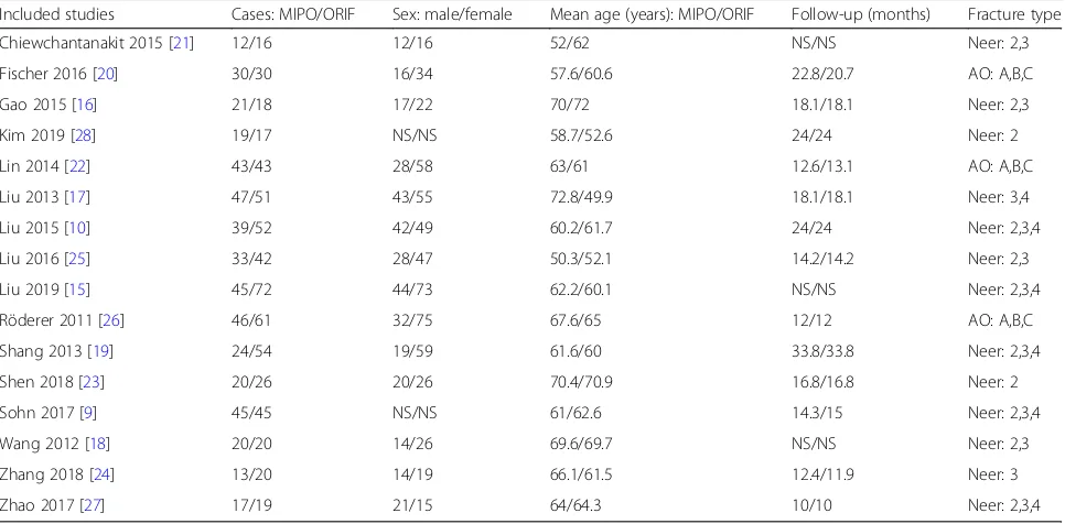
Table 1Characteristics of included studies
Included studies Cases: MIPO/ORIF Sex: male/female Mean age (years): MIPO/ORIF Follow-up (months) Fracture type
Chiewchantanakit 2015 [21] 12/16 12/16 52/62 NS/NS Neer: 2,3
Fischer 2016 [20] 30/30 16/34 57.6/60.6 22.8/20.7 AO: A,B,C
Gao 2015 [16] 21/18 17/22 70/72 18.1/18.1 Neer: 2,3
Kim 2019 [28] 19/17 NS/NS 58.7/52.6 24/24 Neer: 2
Lin 2014 [22] 43/43 28/58 63/61 12.6/13.1 AO: A,B,C
Liu 2013 [17] 47/51 43/55 72.8/49.9 18.1/18.1 Neer: 3,4
Liu 2015 [10] 39/52 42/49 60.2/61.7 24/24 Neer: 2,3,4
Liu 2016 [25] 33/42 28/47 50.3/52.1 14.2/14.2 Neer: 2,3
Liu 2019 [15] 45/72 44/73 62.2/60.1 NS/NS Neer: 2,3,4
Röderer 2011 [26] 46/61 32/75 67.6/65 12/12 AO: A,B,C
Shang 2013 [19] 24/54 19/59 61.6/60 33.8/33.8 Neer: 2,3,4
Shen 2018 [23] 20/26 20/26 70.4/70.9 16.8/16.8 Neer: 2
Sohn 2017 [9] 45/45 NS/NS 61/62.6 14.3/15 Neer: 2,3,4
Wang 2012 [18] 20/20 14/26 69.6/69.7 NS/NS Neer: 2,3
Zhang 2018 [24] 13/20 14/19 66.1/61.5 12.4/11.9 Neer: 3
Zhao 2017 [27] 17/19 21/15 64/64.3 10/10 Neer: 2,3,4
Results
Literature search
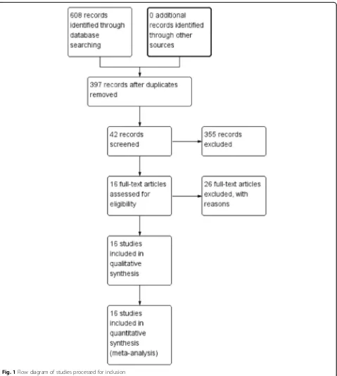
A total of 608 potentially relevant studies were identi-fied. The full search strategy for PubMed database is shown as Additional file 1. After removing 211 dupli-cates, we screened 397 papers. By reading the title and abstract, 355 papers were excluded according to the in-clusion and exin-clusion criteria. A total of 42 studies were assessed by reading the full text; eventually, 16 studies involving 1050 patients (464 patients in the MIPO group and 586 patients in the ORIF group) were included in the meta-analysis [9, 10, 15–28]. The flow diagram of the included studies is shown in Fig. 1. The characteris-tics of the included studies are listed in Table1.
Methodological quality
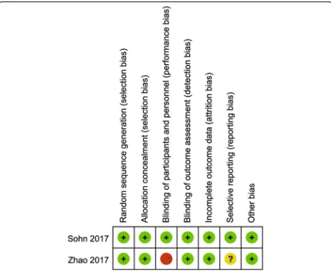
The methodological quality of the RCTs [9, 27] was assessed by theCochrane Handbook, the assessment results are summarized in Fig.2. The quality index scores of the
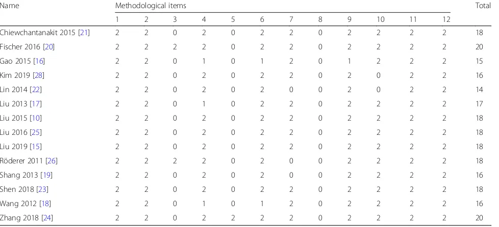
non-RCTs [10,15–26,28] were 14–20. The assessment re-sults are summarized in Table2.
Results of the meta-analysis Blood loss
Nine studies involving 610 patients reported blood loss [10, 15–18, 21, 22, 25, 27]. Heterogeneity tests indicated high heterogeneity (P< 0.00001; I2= 98%); a random-effects model was used. The result showed lesser blood loss in the MIPO group than that in the ORIF group (MD =−115.26; 95% CI:−167.48 to−63.03;P< 0.0001; Fig.3).
Operation time
Thirteen studies [9,10, 15–19, 21,22, 25–28], with 921 patients, mentioned operation time, and the heterogen-eity test indicated significant heterogenheterogen-eity (P< 0.00001; I2= 96%); thus, a random-effects model was adopted. The results showed shorter operation time in the MIPO
[image:4.595.58.537.321.715.2]group than that in the ORIF group (MD =−20.71; 95% CI:−30.21 to−11.22;P< 0.0001; Fig.4).
Radiation time
Three articles [23,26,28], with 189 patients, stated radi-ation time, but used different units of time; therefore, the SMD was adopted. A random-effects model was used, with obvious heterogeneity (P < 0.00001; I2= 98%). The duration of radiation exposure in the MIPO group was longer than that in the ORIF group (MD = 4.36; 95% CI: 1.21 to 7.51;P= 0.007; Fig.5).
Postoperative pain
The visual analogue scale (VAS) was used to evaluate post-operative pain, and seven studies [15,18, 19,24–27], with
486 patients, reported the VAS score. A random-effects model was used, with obvious heterogeneity (P < 0.0001; I2= 79%). The meta-analysis showed a significantly lower VAS score in the MIPO group than in the ORIF group (MD =−0.54; 95% CI:−1.04 to−0.04;P= 0.04; Fig.6).
Union time
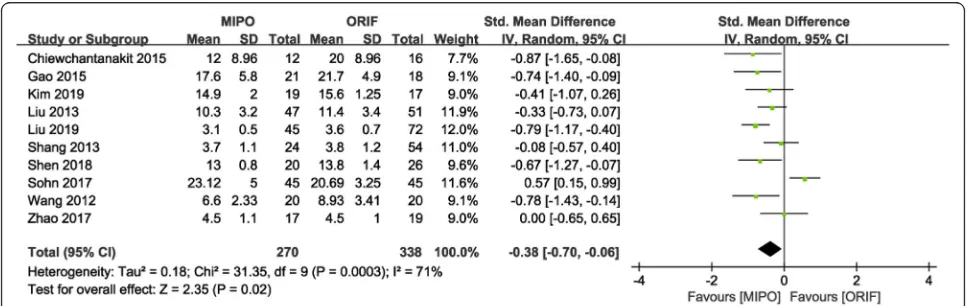
[image:5.595.57.546.100.323.2]Ten studies [9,15–19,21,23,27,28], with 608 patients, indicated postoperative union time, but used different units of time; therefore, the SMD was adopted. Hetero-geneity tests indicated that significant heteroHetero-geneity (P= 0.0003; I2= 71%); thus, a random-effects model was adopted. The meta-analysis showed a shorter time to union in the MIPO group than in the ORIF group (SMD =−0.38; 95% CI:−0.70 to−0.06;P= 0.02; Fig.7). Table 2MINORS appraisal scores for the included nonrandomized control trial
Name Methodological items Total
1 2 3 4 5 6 7 8 9 10 11 12
Chiewchantanakit 2015 [21] 2 2 0 2 0 2 2 0 2 2 2 2 18
Fischer 2016 [20] 2 2 2 2 0 2 2 0 2 2 2 2 20
Gao 2015 [16] 2 2 0 1 0 1 2 0 1 2 2 2 15
Kim 2019 [28] 2 2 0 2 0 2 2 0 2 0 2 2 16
Lin 2014 [22] 2 2 0 2 0 2 0 0 2 0 2 2 14
Liu 2013 [17] 2 2 0 1 0 2 2 0 2 2 2 2 17
Liu 2015 [10] 2 2 0 2 0 2 2 0 2 2 2 2 18
Liu 2016 [25] 2 2 0 2 0 2 2 0 2 2 2 2 18
Liu 2019 [15] 2 2 0 2 0 2 2 0 2 2 2 2 18
Röderer 2011 [26] 2 2 2 2 0 2 0 0 2 2 2 2 18
Shang 2013 [19] 2 2 0 2 0 2 0 0 2 2 2 2 16
Shen 2018 [23] 2 2 0 2 0 2 2 0 2 2 2 2 18
Wang 2012 [18] 2 2 0 1 0 1 2 0 2 2 2 2 16
Zhang 2018 [24] 2 2 0 2 2 2 2 0 2 2 2 2 20
MINORSMethodological index for nonrandomized studies
(1) A clearly stated aim; (2) inclusion of consecutive patients; (3) prospective collection of data; (4) endpoints appropriate to the aim of the study; (5) unbiased assessment of the study endpoint; (6) follow-up period appropriate to the aim of the study; (7) loss to follow-up that is < 5%; (8) prospective calculation of the study size; (9) an adequate control group; (10) contemporary groups; (11) baseline equivalence of groups; (12) adequate statistical analyses. The items were scored as“0”(not reported),“1”(reported but inadequate), or“2”(reported and adequate)
[image:5.595.61.540.569.703.2]Functional outcomes
The constant score of Neer type II fractures were provided in four studies [9, 15, 23, 27, 28]. A fixed-effects model was used (P= 0.16; I2= 39%), and ana-lysis showed significantly higher score in the MIPO group than in the ORIF group (MD = 2.24; 95% CI: 0.82 to 3.65; P = 0.02; Fig. 8).
The constant score of Neer type III fractures were stated in four studies [9, 15, 24, 27]. A fixed-effects model was used (P= 0.42; I2= 0%), and analysis showed significantly higher score in the MIPO group than in the ORIF group (MD = 1.95; 95% CI: 0.98 to 2.92; P< 0.001; Fig. 8).
Meta-analysis was not performed for the constant score of Neer type IV PHFs because it was reported by only one study.
Axillary nerve injury
Four studies [17,22,23,25], with 305 patients, reported axillary nerve injury. A fixed-effects model was used (P= 0.95; I2= 0%), and results showed a significantly higher rate of axillary nerve injury in the MIPO group than in the ORIF group (OR = 4.88; 95% CI: 1.03 to 23.25;P= 0.05).
Complications
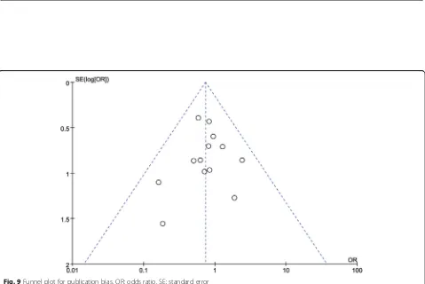
Thirteen studies reported complications. A fixed-effects model was used (P= 0.88; I2= 0%), and pooled results showed no significant difference in total complication rate between the two groups (OR = 0.74; 95% CI: 0.51 to 1.07; P= 0.11; Fig.9). However, the MIPO group had a significantly higher rate of axillary nerve injury that the ORIF group (OR = 4.88; 95% CI: 1.03 to 23.25; P = 0.05; I2= 0%). The pooled results of the following complica-tions showed no significant difference between the two groups (Table3): avascular necrosis, impingement, screw perforation, implant loosening, delayed union or non-union, limited abduction, and varus.
Publication bias
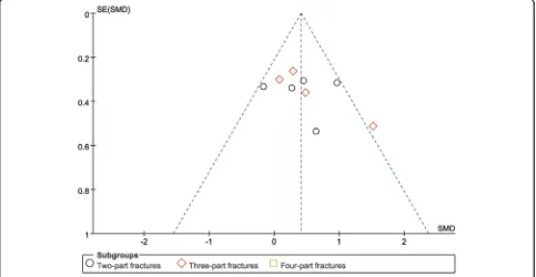
Funnel plots of the total complication rate (Fig. 9), and functional outcomes (Fig. 10) showed no substan-tial asymmetry, indicating no significant risk for pub-lication bias.
Sensitivity analysis
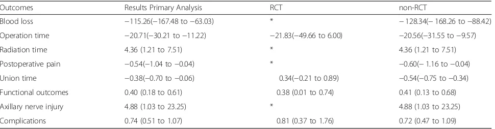
After sensitivity analysis, operation time and union time became insignificant for randomized trials. This change may be due to the inclusion of only 2 randomized Fig. 4Forest plot for operation time between the MIPO and ORIF groups. MIPO: minimally invasive plate osteosynthesis; ORIF: open reduction– internal fixation; OR: odds ratio; CI: confidence interval
[image:6.595.59.539.87.264.2] [image:6.595.66.539.612.702.2]controlled trials. Results for all other outcomes remained unchanged (Table4).
Discussion
We compared the clinical outcomes and complications of MIPO and ORIF in patients with PHFs in this analysis. In contrast to a previously published meta-analysis [12], our meta-meta-analysis involved a larger number of studies, but did not completely yield the same results. The findings of this study suggested that MIPO had ad-vantages in operation time, blood loss, postoperative pain, fracture union time, and constant score compared with ORIF. However, MIPO had a higher rate of axillary nerve injury and longer radiation time compared with ORIF. There was no significant difference in complica-tions between the two groups.
Over the past decade, the MIPO technique has be-come a more popular treatment for PHF [29]. Kim et al. [28] reported that the MIPO technique via the deltoid-splitting approach can provide sufficient field of vision of the plate location by minimal soft tissue dissection. Thus, it is easy to perform a reduction of a large greater
tuberosity fragment [22], significantly reducing operation time and blood loss.
The MIPO technique minimizes incision and avoids damage to the deltoid muscle, which will reduce postop-erative pain and facilitate early functional training [27]. Early functional training plays a positive role for recov-ery of shoulder joint function.
In this meta-analysis, the MIPO group had a sig-nificantly longer radiation duration because the
pa-tients underwent indirect reduction under
fluoroscopy [28]. MIPO’s longer radiation time, compared with that of ORIF, is a negative aspect of MIPO.
Restoration to normal shoulder function is an import-ant goal of the treatment of PHF. The present study showed that MIPO provides a better constant score of Neer type II or III PHFs. The result was similar to that of previous studies [23, 27]. Therefore, MIPO achieves better shoulder function in the treatment of PHFs.
Another important finding of this meta-analysis was the higher rate of axillary nerve injury in the MIPO group than in the ORIF group. Acklin et al. [30] reported that axillary nerve injury is the risk factor of the MIPO. However, Fig. 6Forest plot for postoperative pain between the MIPO and ORIF groups. MIPO: minimally invasive plate osteosynthesis; ORIF: open
reduction–internal fixation; OR: odds ratio; CI: confidence interval
[image:7.595.61.539.88.218.2] [image:7.595.56.540.550.703.2]Fig. 8Forest plot for constant score between the MIPO and ORIF groups. MIPO: minimally invasive plate osteosynthesis; ORIF: open reduction– internal fixation; OR: odds ratio; CI: confidence interval
[image:8.595.57.539.87.376.2] [image:8.595.59.538.405.725.2]Koljonen et al. [31] reported no axillary nerve injury in pa-tients treated with MIPO. Whether axillary nerve lesions are more frequent in the MIPO approach remains controversial. Axillary nerve injury in the MIPO group may be re-lated to the incisions in the deltoid-splitting approach extending more than 5 cm distal to the tip of the acromion [32]. To prevent injury to the axillary nerve with the MIPO technique, incisions should not extend more than 5 cm distal to the tip of the acromion [32]. In addition, the axillary nerve should be identi-fied and protected by positioning the index finger on the nerve during the insertion of the plate on the proximal humerus [33].
Meta-analysis results indicated that MIPO had shorter time to union compared with that ORIF in PHFs. Similar results were also reported by five of the included studies [15, 16, 18, 21, 23]. The MIPO
technique is commonly believed to provide advantages of fracture union process, as it maintains the perios-teum and soft tissue around the fracture site [28].
The meta-analysis results showed no significant differ-ence in impingement, screw perforation, implant loosen-ing, avascular necrosis, delayed union or nonunion, limited abduction, and varus collapse between the MIPO and ORIF groups.
[image:9.595.58.544.99.225.2]Our study has some limitations. First, the outcomes, except for the constant score, were not analyzed separ-ately according to Neer classification. The main reason was that most studies did not show the data of interest in a separate form. Second, only two RCTs were in-cluded. Finally, the follow-up duration was short; longer follow-up may identify more complications. Therefore, RCTs with longer follow-up duration and larger number of samples are needed to confirm our results.
Table 3Meta-analysis of reported complications
Outcomes No. of trials No. of patients: MIPO/ORIF OR (95% CI) Pvalue I2(%) Pvalue for heterogeneity
Nerve injury 4 143/162 4.88 (1.03, 23.2) 0.05 0 0.95
Impingement 4 154/179 0.96 (0.36,2.54) 0.94 0 0.94
Screw perforation 3 134/149 0.97 (0.42,2.23) 0.94 0 0.65
Implant loosening 6 222/281 0.70 (0.28,1.75) 0.44 24 0.25
Avascular necrosis 7 251/299 0.41 (0.16,1.05) 0.06 0 0.78
Delayed union or nonunion 6 207/307 0.37 (0.12,1.13) 0.08 0 0.97
Limited abduction 3 108/178 0.73 (0.17,3,26) 0.69 0 0.58
Varus 5 140/155 1.35 (0.47,3.90) 0.58 0 0.62
MIPOMinimally invasive plate osteosynthesis,ORIFOpen reduction–internal fixation,OROdds ratio,CIConfidence interval
[image:9.595.57.540.466.716.2]Conclusion
The meta-analysis results showed that in comparison with ORIF, MIPO had advantages in operation time, blood loss, postoperative pain, and fracture union time for the treat-ment of PHFs. The MIPO technique was associated with better shoulder function in Neer type II or III PHFs. How-ever, the MIPO technique had a higher rate of axillary nerve injury and longer radiation time compared to ORIF. There was no significant difference in complication rates between MIPO and ORIF. Recently, a network meta-analysis demon-strated that non-surgical treatment (NST) was associated with lower adverse event rates compared to ORIF for 3- and 4-part PHFs [34]. We recommend that future studies should not only compare MIPO to ORIF but also to NST to obtain thorough evidence-based treatment guidelines.
Supplementary information
Supplementary informationaccompanies this paper athttps://doi.org/10. 1186/s12891-019-2936-y.
Additional file 1.Full search strategy for Pubmed database.
Abbreviations
CI:Confidence intervals; MD: Mean difference; MINORS: Methodological Index for Nonrandomized Studies; MIPO: Minimally invasive plate osteosynthesis; OR: Odds ratio; ORIF: Open reduction–internal fixation; PHF: Proximal humeral fracture; PRISMA: Preferred Reporting Items for Systematic Reviews and Meta-Analyses; RCT: Randomized control trial; SMD: Standard mean difference; VAS: Visual analogue scale
Acknowledgements
We greatly appreciate the assistance of the company Editage in Shanghai, which provided English language editing.
Authors’contributions
FLL and ZPG conducted literature search and determined studies for exclusion and inclusion. FLL, FQW extracted data from the included studies, performed the meta-analysis, and drafted the manuscript. FLL, TN, XQL con-ceived the idea of the study, designed the study. QYT, XWL, CY, XQL, and TN reviewed and Edited the manuscript. TN performed major revision and minor revision. All authors read and approved the manuscript.
Funding
This study was supported by the Foundation of Natural Science Foundation of Jiangxi Province (20161BAB205235), the National Natural Science Foundation of China (81460341). The funding body had no role in the
design of the study and collection, analysis, and interpretation of data and in writing the manuscript.
Availability of data and materials
All data generated or analysed during this study are included in this published article and its supplementary information files.
Ethics approval and consent to participate
Not applicable.
Consent for publication
Not applicable.
Competing interests
The authors declare that they have no competing interests.
Received: 9 July 2019 Accepted: 8 November 2019
References
1. Calvo E, Morcillo D, Foruria AM, Redondo-Santamaría E, Osorio-Picorne F, Caeiro JR, et al. Nondisplaced proximal humeral fractures: high incidence among outpatient-treated osteoporotic fractures and severe impact on upper extremity function and patient subjective health perception. J Shoulder Elbow Surg. 2011;20:795–801.
2. Horak J, Nilsson BE. Epidemiology of fracture of the upper end of the humerus. Clin Orthop Relat Res. 1975;112:250–3.
3. Kannus P, Palvanen M, Niemi S, Parkkari J, Järvinen M, Vuori I. Osteoporotic fractures of the proximal humerus in elderly Finnish persons: sharp increase in 1970-1998 and alarming projections for the new millennium. Acta Orthop Scand. 2000;71:465–70.
4. Gaebler C, McQueen MM, Court-Brown CM. Minimally displaced proximal humeral fractures: epidemiology and outcome in 507 cases. Acta Orthop Scand. 2003;74:580–5.
5. Pinkas D, Wanich TS, DePalma AA, Gruson KI. Management of malunion of the proximal humerus: current concepts. J Am Acad Orthop Surg. 2014;22: 491–502.
6. Maier D, Jaeger M, Izadpanah K, Strohm PC, Suedkamp NP. Proximal humeral fracture treatment in adults. J Bone Joint Surg Am. 2014;96:251–61. 7. Hirschmann MT, Fallegger B, Amsler F, Regazzoni P, Gross T. Clinical
longer-term results after internal fixation of proximal humerus fractures with a locking compression plate (PHILOS). J Orthop Trauma. 2011;25:286–93. 8. Brunner F, Sommer C, Bahrs C, Heuwinkel R, Hafner C, Rillmann P, et al.
Open reduction and internal fixation of proximal humerus fractures using a proximal humeral locked plate: a prospective multicenter analysis. J Orthop Trauma. 2009;23:163–72.
[image:10.595.57.542.98.225.2]9. Sohn HS, Jeon YS, Lee J, Shin SJ. Clinical comparison between open plating and minimally invasive plate osteosynthesis for displaced proximal humeral fractures: a prospective randomized controlled trial. Injury. 2017;48:1175–82. 10. Liu K, Liu P-c, Liu R, Wu X. Advantage of minimally invasive lateral approach relative to conventional deltopectoral approach for treatment of proximal humerus fractures. Med Sci Monit. 2015;21:496–504.
Table 4Sensitivity analyses
Outcomes Results Primary Analysis RCT non-RCT
Blood loss −115.26(−167.48 to−63.03) * −128.34(−168.26 to−88.42)
Operation time −20.71(−30.21 to−11.22) −21.83(−49.66 to 6.00) −20.56(−31.55 to−9.57)
Radiation time 4.36 (1.21 to 7.51) * 4.36 (1.21 to 7.51)
Postoperative pain −0.54(−1.04 to−0.04) * −0.60(−1.16 to−0.04)
Union time −0.38(−0.70 to−0.06) 0.34(−0.21 to 0.89) −0.54(−0.75 to−0.34)
Functional outcomes 0.40 (0.18 to 0.61) 0.38 (0.01 to 0.74) 0.41 (0.13 to 0.68)
Axillary nerve injury 4.88 (1.03 to 23.25) * 4.88 (1.03 to 23.25)
Complications 0.74 (0.51 to 1.07) 0.81 (0.37 to 1.76) 0.72 (0.47 to 1.09)
11. Acklin YP, Sommer C. Plate fixation of proximal humerus fractures using the minimally invasive anterolateral delta split approach. Oper Orthop Traumatol. 2012;24:61–73.
12. Zang JC, Du JJ, Li C, Wang JB, Ma XL. Comparison between minimally invasive plate osteosynthesis and open plating for proximal humeral fractures: a meta-analysis. J Comp Eff Res. 2018;7:1001–8.
13. Moher D, Liberati A, Tetzlaff J, Altman DG. Preferred reporting items for systematic reviews and meta-analyses: the PRISMA statement. BMJ. 2009; 339:b2535.
14. Slim K, Nini E, Forestier D, Kwiatkowski F, Panis Y, Chipponi J.
Methodological index for non-randomized studies (minors): development and validation of a new instrument. ANZ J Surg. 2003;73:712–6. 15. Liu BC, Yang ZW, Zhou F, Ji HQ, Zhang ZS, Guo Y, et al. Application of the
modified internal fixation method of minimally invasive percutaneous plate osteosynthesis in treatment of proximal humeral fracture. Beijing Da Xue Xue Bao. 2019;51:277–82.
16. Gao YB, Tong SL, Yu JH, Lu WJ. Case control study on open reduction internal fixation (ORIF) and minimally invasive percutaneous plate osteosynthesis (MIPPO) for the treatment of proximal humerus fractures in aged. Zhongguo Gu Shang. 2015;28:335–9.
17. Liu J, Li SH, Li ZH, Wang JG, Yang CX, Zhang L, et al. Case-control study on minimally invasive percutaneous new plate osteosynthesis applied in proximal humerus fractures in elder patients. Zhongguo Gu Shang. 2013;26:4–8.
18. Wang JF, Song HB, Gu HJ, Ling ZD, Ma HH. Case-control study on minimally invasive plate osteosynthesis for the treatment of proximal humeurs fractures in elderly patients. Zhongguo Gu Shang. 2012;25:487–9. 19. Shang LP, Zhou F, Ji HQ, Zhang ZS, Liu XG, Tian Y. Comparison of curative
effects between minimally invasive locking plate internal fixation and open reduction with internal fixation for the treatment of proximal humerus fractures. Beijing Da Xue Xue Bao. 2013;45:711–6.
20. Fischer C, Frank M, Kunz P, Tanner M, Weber MA, Moghaddam A. Dynamic contrast-enhanced ultrasound (CEUS) after open and minimally invasive locked plating of proximal humerus fractures. Injury. 2016;47:1725–31. 21. Chiewchantanakit S, Tangsripong P. Locking plate fixation of proximal
humeral fracture: minimally invasive vs. standard delto-pectoral approach. J Med Assoc Thai. 2015;98:196–200.
22. Lin T, Xiao B, Ma X, Fu D, Yang S. Minimally invasive plate osteosynthesis with a locking compression plate is superior to open reduction and internal fixation in the management of the proximal humerus fractures. BMC Musculoskelet Disord. 2014;15:206.
23. Shen QF, Wen X, Yang SW, Chen X, Fan WX, Xu GZ, et al. MIPPO and ORIF for the treatment of elderly proximal humerus fractures of type Neer II:a case control study. Zhongguo Gu Shang. 2018;31:160–4.
24. Zhang Z, Zhang G, Peng Y, Wang X, Guo H, Zhang W, et al. Modified minimally invasive approach and intra-osseous portal for three-part proximal humeral fractures: a comparative study. J Orthop Surg Res. 2018;13:24.
25. Liu YW, Wei XE, Kuang Y, Zheng YX, Gu XF, Zhan HS, et al. Open vs. closed reduction combined with minimally invasive plate osteosynthesis in humeral fractures. Minim Invasive Ther Allied Technol. 2016;25:215–21. 26. Röderer G, Erhardt J, Kuster M, Vegt P, Bahrs C, Kinz L, et al. Second
generation locked plating of proximal humerus fractures--a prospective multicentre observational study. Int Orthop. 2011;35:425–32. 27. Zhao L, Yang P, Zhu L, Chen AM. Minimal invasive percutaneous plate
osteosynthesis (MIPPO) through deltoid-pectoralis approach for the treatment of elderly proximal humeral fractures. BMC Musculoskelet Disord. 2017;18:187.
28. Kim YG, Park KH, Kim JW, Oh JK, Yoon JP, Kim HJ, et al. Is minimally invasive plate osteosynthesis superior to open plating for fixation of two-part fracture of the proximal humerus? J Orthop Surg (Hong Kong). 2019;27: 2309499019836156.
29. Gonç U, Atabek M, Teker K, Tanrıöver A. Minimally invasive plate osteosynthesis with PHILOS plate for proximal humerus fractures. Acta Orthop Traumatol Turc. 2017;51:17–22.
30. Acklin YP, Jenni R, Walliser M, Sommer C. Minimal invasive PHILOS®- plate osteosynthesis in proximal humeral fractures. Eur J Trauma Emerg S. 2009; 35:35–9.
31. Koljonen PA, Fang C, Lau TW, Leung F, Cheung NWK. Minimally invasive plate osteosynthesis for proximal humeral fractures. J Orthop Surg (Hong Kong). 2015;23:160–3.
32. Ruchholtz S, Hauk C, Lewan U, Franz D, Kühne C, Zettl R. Minimally invasive polyaxial locking plate fixation of proximal humeral fractures: a prospective study. J Trauma. 2011;71:1737–44.
33. Laflamme GY, Rouleau DM, Berry GK, Beaumont PH, Reindl R, Harvey EJ. Percutaneous humeral plating of fractures of the proximal humerus: results of a prospective multicenter clinical trial. J Orthop Trauma. 2008;22:153–8. 34. Orman S, Mohamadi A, Serino J, Murphy J, Hanna P, Weaver MJ, et al.
Comparison of surgical and non-surgical treatments for 3- and 4-part proximal humerus fractures: a network meta-analysis. Shoulder Elbow. 2019. https://doi.org/10.1177/1758573219831506.
Publisher’s Note