R E S E A R C H A R T I C L E
Open Access
Biomechanical and functional indicators in male
semiprofessional soccer players with increased
hip alpha angles vs. amateur soccer players
Matthias Lahner
1*, Christoph von Schulze Pellengahr
1, Philipp Alexander Walter
1, Carsten Lukas
2, Andreas Falarzik
3,
Kiriakos Daniilidis
4, Lars Victor von Engelhardt
5, Christoph Abraham
6, Ewald M Hennig
6and Marco Hagen
6Abstract
Background:Femoroacetabular impingement (FAI) is predominant in young male athletes, but not much is known
about gait differences in cases of increased hip alpha angles. In our study, the hip alpha angle of Nötzli of soccer players was quantified on the basis of magnetic resonance imaging (MRI) with axial oblique sequences. The aim of the current study was to compare the rearfoot motion and plantar pressure in male semiprofessional soccer players with increased alpha angles to age-matched amateur soccer players.
Methods:In a prospective analysis, male semiprofessional and amateur soccer players had an MRI of the right hip to measure the alpha angle of Nötzli. In a biomechanical laboratory setting, 14 of these participants in each group ran in two shoe conditions. Simultaneously in-shoe pressure distribution, tibial acceleration, and rearfoot motion measurements of the right foot were performed.
Results:In the semiprofessional soccer group, the mean value of the alpha angle of group was 55.1 ± 6.58° (range 43.2-76.6°) and 51.6 ± 4.43° (range 41.9-58.8°) in the amateur group. In both shoe conditions, we found a significant difference between the two groups concerning the ground reaction forces, tibial acceleration, rearfoot motion and plantar pressure parameters (P< 0.01,P< 0.05,P= 0.04). Maximum rearfoot motion is about 22% lower in the semiprofessional group compared to the amateur group in both shoe conditions.
Conclusions:This study confirmed that semiprofessional soccer players with increased alpha angles showed
differences in gait kinematics compared to the amateur group. These findings support the need for a screening program for competitive soccer players. In cases of a conspicuous gait analysis and symptomatic hip pain, FAI must be ruled out by further diagnostic tests.
Keywords:Rearfoot motion, Tibial acceleration, Alpha angle of Nötzli, Femoroacetabular impingement, Magnetic
resonance imaging, Soccer players
Background
Femoroacetabular impingement (FAI) is a morphological hip disorder which shows a novel approximation to mechanical etiology of hip osteoarthritis [1,2]. A large share of idiopathic hip arthritis can be attributed to FAI, which is why early diagnosis is very important [3,4]. In the pathogenesis of FAI, there are two anatomical deformities either at the acetabulum or the proximal
end of the femur or in both structures. A femoral type (cam impingement) is anatomically differentiated from the acetabular type of FAI (pincer impingement) [5]. The cam impingement is caused from a prominence at the anterolateral femoral head-neck junction [6]. The alpha angle of Nötzli is described to quantify the asphericity of the femoral head in axial oblique sequences of magnetic resonance images (MRI). Causes for the cam impingement are aspheric deformity of the femoral head, slipped capital femoral epiphysis, late closure of the femoral epiphysis and Legg-Calve-Perthes disease [7-9]. The pincer impingement is caused by an immoderate
* Correspondence:[email protected] 1
Department of Orthopaedic Surgery, Ruhr-University Bochum, St. Josef-Hospital, Gudrunstr. 56, 44791 Bochum, Germany
Full list of author information is available at the end of the article
acetabular cover of the head of femur and is linked with acetabular retroversion, protrusio acetabuli or coxa profunda [10,11]. Symptomatic FAI can be treated by arthroscopic procedures or open surgery [12].
FAI is assumed to be predominant in young male ath-letes with sport activities with high impact for the hip joints like soccer [13]. Agricola et al. demonstrated that FAI was more prevalent in 89 elite soccer players than in 92 nonathletic controls [14]. Cam-type deformity develops during adolescence and is probably to be af-fected by high-impact sports practice [14]. The soccer game is primarily characterized by running-related ac-tions [15]. Therefore, the shock attenuation capacity of soccer players with FAI during running is of special biomechanical interest.
Clinically, FAI must be differentiated from insertional tendinopathy of the adductor muscles. The tendinopathy of the adductor can be associated with arthropathy of the symphysis and insertional pubic area [16]. The major clinical symptoms of the tendinopathy of the adductor muscles are groin or lower abdomen pain [16].
Only few biomechanical studies exist on the gait ana-lyses in patients with FAI [17-20]. In these studies, symptomatic patients were compared to healthy control probands. However, to our best knowledge, this is the first study which analyzed a strongly selective risk group like male semiprofessional soccer players who are dis-proportionately affected by FAI, but usually do not show clinical symptoms, yet.
Therefore, the aim of our biomechanical study was to compare the foot rollover process during running between male semiprofessional soccer players with increased alpha angles and age-matched amateur soccer players. It was postulated that an increased alpha angle would lead to different rearfoot motion, tibial acceleration and plantar pressure parameters.
Methods Study participants
This study follows the Declaration of Helsinki. All pro-bands have voluntarily agreed to the study and gave their informed consent. All study persons have received and signed patient education for MRI. The study protocol was approved by the local ethical committee of the Ruhr-University Bochum (registration number 4370-12). Between January 2012 and July 2013, 14 male semipro-fessional soccer players and 14 male amateur soccer players underwent a clinical examination of the mechan-ical leg axis, an MRI and a gait analysis. The MRI data was published in a previous study [21]. The soccer players were semiprofessional athletes who played 4 training units per week for 2 hours with a seasonal duration of 10 months. The control group consisted of age-matched male amateur soccer players with a physical activity less
than 5 hours a week. In all cases, the right leg was the kicking leg. For both groups, exclusion criteria were any kind of previous hip surgery in either hip joint, inflam-matory or metabolic rheumatic disease or a history of haemophilia. Height and weight measurements were performed. Dorsal or knee pain was excluded by the clinical examination.
MRI protocol
Each study volunteer underwent a nonarthrogram 1.5-T MRI (Magnetom, Siemens, Erlangen, Germany) of the right hip. All patients were examined in the supine pos-ition with a neutral pospos-ition of the hip joint. The MRI sequences were obtained on each study test person by one specially-trained radiology technician. The MRI se-quences parameters of the turbo spin-echo sequence were as follows: repetition times (TR) 637 ms, echo time (TE) 14 ms, a field of view (FOV) of 350 × 350 mm, a matrix of 512 × 256, a slice thickness of 6 mm and flip angle 150°. In addition, we used a coronal T1-weighted sequence (TR 530 ms, TE 14 ms, FOV 400 × 400 mm, slice thickness 5 mm, flip angle 150°), axial oblique T1-weighted sequence (TR 530 ms, TE 14 ms, FO 350 × 265 mm, slice thickness 5 mm, flip angle 150°) ori-ented along the axis of the femoral neck and fat-suppressed T1-weighted fast low angle shot (FLASH) sequences (TR 795 ms, TE 11 ms, FOV 400 × 400 mm, slice thickness 3 mm, flip angle 60°). For ethical reasons, neither intraarti-cular nor intravenous contrast was injected. The alpha angle was analyzed using the technique described by Nötzli [6]. We considered an alpha angle >55° as cut-off value because an alpha angle >55° is associated with FAI [22]. The alpha angle was subsequently measured by a radi-ologist (C.L.) and an orthopaedic surgeon (M.L.) both experienced in musculoskeletal imaging. The radio-logist was blinded to the level of activity of the subjects.
Biomechanical measurements
were fastened under the foot with adhesive tape. The physical properties of the piezoceramic transducers were described by Hennig et al. [23]. To measure tibial accel-eration, an Entran EGAX-F-25 miniature accelerometer was glued to the skin above the medial aspect of the tibia at a location midway between medial malleolus and tibial plateau. The accelerometer was fastened by an elastic strapt to improve the mechanical coupling to the underlying bone. Rearfoot motion was measured by using an electrogoniometer (Megatron MP 10) that was at-tached to the heel counter of the shoe. Rearfoot angle was defined as the angle between rearfoot bisection and the direction of the achilles tendon. The detailed biomechan-ical setup was presented in the study of Milani et al. [24].
Data collection and processing
The ground reaction force, axial tibial acceleration, pres-sure distribution, and rearfoot motion were collected simultaneously by a computer in a pretrigger mode. The data were sampled at a rate of 1 kHz per channel with a resolution of 12 bits. A threshold of 5 N of the vertical ground reaction force was chosen to determine the time onset of foot strike. The force and acceleration values were determined as multiples of body weight (bw) and
gravitational acceleration (g), respectively. The maximum force rate was calculated as the highest differential quo-tient of adjoining vertical ground reaction force divided by the time resolution of 1 ms. The median power fre-quency of the vertical force signal was calculated from a 1,024-point FFT power spectrum analysis. A low fre-quency cut-off value of 10 Hz was chosen, since only the initial impact force was of interest. Under the seven anatomical locations, peak pressures were determined for all participants. Maximum range of rearfoot motion was chosen as a descriptor value for the pronation be-haviour of the foot.
Statistical analysis
The selected patient cohort was grouped in an Excel file (version 2003, Microsoft Corporation, Seattle, USA). Distribution of data was assessed by the D’ Agostino-Pearson test. The arithmetic mean value, SD and 95% confidence intervals were calculated for the variables above and measured with Microsoft Excel. The values were recorded in IBM SPSS Statistics 14 (PASW 14, SPSS Inc., Chicago, IL, USA). The measurements on the alpha angles was compared using the Student’st-test. Inter-rater reliability was measured of the MRI readings. Statistical significance was defined as a P value <0.05.
Results MRI
[image:3.595.58.291.101.193.2]The demographic data is presented in Table 1. There was no statistically significant difference concerning the Table 1 Demographic information
Group (n) Age (years)
Height (cm)
Weight (kg)
Body mass index (kg/cm2)
Semiprofessional group (14)
22.21 ± 2.28 180 ± 0.07 76.0 ± 9.18 23.2 ± 1.25
Amateur group (14)
22.71 ± 2.88 181 ± 0.07 81.28 ± 11.11 23.80 ± 3.03
[image:3.595.304.540.111.168.2]Pvalue 0.496 0.722 0.182 0.500
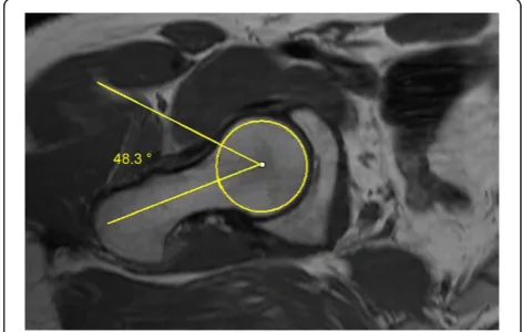
[image:3.595.58.291.515.665.2]Figure 1Axial MRI scan of the right hip of a participant of the soccer group.The alpha angle is formed by the connecting lines (yellow lines) between the longitudinal mid-axis of the femoral neck and the axis which marked the point first exceeded the radius of the cartilage-covered femoral head. The alpha angle was 76.8° in this example.
Table 2 Comparison of the mean of the alpha angles of the two groups
Group division (n) Mean alpha angle ± SD
Semiprofessional group (14) 55.16 ± 6.58°
Amateur group (14) 51.65 ± 4.43°
Pvalue 0.11
[image:3.595.303.539.555.705.2]mean age of both groups. There was no statistically sig-nificance difference in height, weight and body-mass index. 7 (50%) subjects of the semiprofessional group and 5 (35.7%) subjects of the amateur group clinically showed a varus malalignement of the mechanical axis. In both groups, no one had a history of hip dysplasia or disease in the childhood. In the soccer group, the mean value of the alpha angle was 55.16 ± 6.58° (Figure 1). The mean value of the alpha angle was lower in the amateur group, but there was no significant difference in the amateur group (51.65 ± 4.43°, Table 2, Figure 2). Con-cerning the inter-rater reliability, the values ranged from 0.87 to 0.99 (Table 3).
Biomechanical measurements
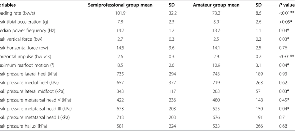
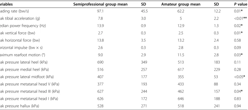
The results (mean values and standard deviations) of the ground reaction forces, peak tibial acceleration, rearfoot motion and plantar pressure parameters are presented in Tables 4 and 5. In both shoe conditions, we found similar statistically significant differences between the groups. In-creases in median power frequency (NW: +7%, p < 0.05, VW: +8%, p < 0.05) and the rate of the vertical ground reaction force (NW: +39%, p < 0.01, VW: +56%, p < 0.05)
reveal higher loading of the lower extremities and shock transmission to the upper body in the semiprofessional subjects.
The reduced shock absorption for the semiprofessional subjects is also demonstrated by an increased peak tibial acceleration (NW: +32%, p < 0.05, VW: +56%, p < 0.01) when compared to the amateur soccer players. Maximum rearfoot motion is about 22% lower in the semiprofes-sional soccer players compared to the amateur group in both shoe conditions. In the second part of the stance phase, peak vertical force is increased (NW/VW: +8%, p < 0.05) while the horizontal impulse is reduced (NW: -10%, p < 0.05; VW: -7%, p = 0.08) in the semiprofessional subjects. Additionally, we found increased peak plantar pressures under the lateral midfoot (NW: + 30%, p < 0.05, VW: +15%, p < 0.05) and the third metatarsal head (NW: + 30%, p < 0.05, VW: +36%) in the semiprofessional group for running at the same speed (3.3 m/s).
Discussion
[image:4.595.56.291.112.203.2]Purpose of the present study was to investigate biomech-anical manifestations of soccer players with increased alpha angles during running, especially the rearfoot mo-tion, which was not been analyzed before. To our best knowledge, this was the first gait analysis of predomantly asymptomatic athletes which had radiologically in-creased alpha angles. Despite no apparent differences in foot structure, the semiprofessional group experience higher impact forces and load transfer at initial ground contact as compared to the subjects of the amateur group. Increases in the median power frequency, maximum verti-cal ground reaction force rate, and peak tibial acceleration indicate the semiprofessional group’s lower limb to be a more rigid structure during ground contact in running Table 3 Inter-rater reliability of the alpha angles between
the two observers
Variables Inter-rater reliability
95%-confidence interval
Oberserver 1 vs. observer 2
Minimum level Maximum level
Alpha angle,
semiprofessional group
0.966 0.87 0.99
Alpha angle, amateur group
[image:4.595.63.539.514.725.2]0.967 0.899 0.989
Table 4 NW shod running without inserted valgus wedges
Variables Semiprofessional group mean SD Amateur group mean SD Pvalue
Loading rate (bw/s) 101.9 32.2 73.2 8.6 <0.01**
Peak tibial acceleration (g) 7.8 2.3 5.9 2.6 <0.05*
Median power frequency (Hz) 14.7 1.2 13.7 1.1 0.04*
Peak vertical force (bw) 2.7 0.3 2.5 0.3 0.03*
Peak horizontal force (bw) 14.5 3.6 14.1 2.5 0.76
Horizontal impulse (bw × s) 2.6 0.3 2.9 0.2 <0.01**
Maximum rearfoot motion (°) 8.5 2.6 10.9 3.1 0.04*
Peak pressure lateral heel (kPa) 735 294 743 189 0.93
Peak pressure medial heel (kPa) 657 377 719 263 0.62
Peak pressure lateral midfoot (kPa) 343 117 263 57 0.03*
Peak pressure metatarsal head V (kPa) 422 236 480 148 0.45*
Peak pressure metatarsal head III (kPa) 673 203 525 150 0.04*
Peak pressure metatarsal head I (kPa) 713 203 676 191 0.71
Peak pressure hallux (kPa) 581 224 533 266 0.68
when compared to the amateur subjects. The lower rear-foot motion in the semiprofessional group also contributes to increased shocks of the body during ground contact. A more lateral placement of the foot, causing higher peak pressures under the lateral heel and third metatarsal head, may explain the more rigid foot structure with less subta-lar angusubta-lar joint motion.
Although hip motion during running was not mea-sured, the present data allow us to assume that the os-cillation of the center of mass is different between the investigated groups. In the semiprofessional group, in-creased vertical forces are accompanied by reduced horizontal forces which both are related to a more accen-tuated upward-downward movement of the center of mass. Compared to non-symptomatic subjects, FAI-patients are restricted in hip flexion during squatting which may be caused by hip pain [25]. The half of the semiprofessional subjects showed clinically a varus deviation of the lower limb. In a previous study, our study group found a correl-ation between increased alpha angles and devicorrel-ation of the mechanical leg axis [26]. The mechanical axis deviation can have effects on the kinematics on gait. To compensate limitations in hip flexion which was found in FAI patients during walking, increasing vertical oscillation of the center of mass could be used to counteract the restrictions in sagittal plane hip motion [17]. By applying this strategy, soccer players with increased alpha angles may achieve the same step length but with higher lower extremity loading as well as, obviously, higher energy consumption due to increases in mechanical work. Obviously, all com-pensatory mechanisms become manifest as a more rigid lower limb structure during the entire stance phase.
It can be concluded that limitations in range of motion (ROM) which were observed in the studies of Kennedy
et al. [17] and Lamontagne et al. [25] seem to be compensated by readjustments of the locomotor system. However, restrictions in ROM combined with reduced shock absorption capacity constitute interlocking risk factors for joint disease. Especially in running-related activities, such as in many team sports, the need to be competitive has to be discussed critically according to the aftereffects, especially the pathomechanism of osteoarthrosis.
Consistent with previous findings, our study suggests that specific characteristics of FAI, for instance functional constraints in ROM, can be diagnosed by biomechanical screening. Limitations in hip ROM, reduced shock at-tenuation capacity and accentuated vertical movement of the center of mass should be considered as functional indicators of FAI.
Conclusions
Future research is needed to identify characteristic features which display the different stages of FAI process. A chal-lenge for future research is to develop physical training programs for preventing FAI or decelerating the FAI process. These findings support the need for a screening program for competitive soccer players. In cases of a con-spicuous gait analysis, FAI must be ruled out by further diagnostic tests.
Abbreviations
FAI:Femoroacetabular impingement; FOV: Field of view; MRI: Magnetic resonance imaging; NW: No wedges; TE: Echo time; TR: Repetition time; ROM: Range of motion; VW: Valgus wedges.
Competing interests
[image:5.595.64.540.99.310.2]The authors declare that they have no competing interests.
Table 5 VW shod running with inserted valgus wedges
Variables Semiprofessional group mean SD Amateur group mean SD Pvalue
Loading rate (bw/s) 97.1 45.5 62.2 12.2 0.01*
Peak tibial acceleration (g) 7.8 3.0 5 2.2 <0.01**
Median power frequency (Hz) 13.9 0.9 12.9 1.3 0.02*
Peak vertical force (bw) 2.7 0.3 2.5 0.3 0.01*
Peak horizontal force (bw) 13.8 3.5 13.2 2.4 0.58
Horizontal impulse (bw × s) 2.6 0.3 2.8 0.3 0.09
Maximum rearfoot motion (°) 9.0 2.9 11.5 2.8 0.03*
Peak pressure lateral heel (kPa) 690 349 513 183 0.11
Peak pressure medial heel (kPa) 516 257 617 229 0.28
Peak pressure lateral midfoot (kPa) 407 177 355 53 <0.05*
Peak pressure metatarsal head V (kPa) 377 193 433 88 0.34
Peak pressure metatarsal head III (kPa) 627 244 462 157 0.04*
Peak pressure metatarsal head I (kPa) 626 172 646 188 0.83
Peak pressure hallux (kPa) 528 271 518 241 0.94
Authors’contributions
ML, AF, CA, EMH and MH conceived and designed the study. ML, PAW, CL and LVvE performed the MRI measurements. ML, CvSP, PAW, KD, CA, EMH and MH are involved in the execution of the study and the writing of this manuscript. All authors read and approved the final manuscript.
Acknowledgments
The authors thank Coach André Pawlak and the soccer players of the Sportgemeinschaft Wattenscheid 09. The authors thank Dr. Nina Lahner for correcting the manuscript.
Author details
1Department of Orthopaedic Surgery, Ruhr-University Bochum, St. Josef-Hospital,
Gudrunstr. 56, 44791 Bochum, Germany.2Department of Diagnostic and Interventional Radiology and Nuclear Medicine, Ruhr-University Bochum, St. Josef-Hospital, Gudrunstr. 56, 44791 Bochum, Germany.3Olympic Training Center Westfalen/Bochum, Hollandstr. 95, 44866 Bochum, Germany. 4Department of Orthopaedic Surgery, Annastift Hannover, (Medical School Hannover, MHH), Anna-von-Borries-Str. 1-7, 30625 Hannover, Germany.5Faculty of Health Sciences, University of Witten/Herdecke, Alfred-Herrhausen-Str. 50, 58448 Witten, Germany.6Biomechanics Laboratory, Department of Sport and Movement Sciences, University of Duisburg-Essen, Gladbecker Str. 182, 45141 Essen, Germany.
Received: 20 November 2013 Accepted: 12 March 2014 Published: 16 March 2014
References
1. Beck M, Kalhor M, Leunig M, Ganz R:Hip morphology influences the pattern of damage to the acetabular cartilage: femoroacetabular impingement as a cause of early osteoarthritis of the hip.J Bone Joint Surg (Br)2005,87:1012–1018.
2. Ganz R, Leunig M, Leunig-Ganz K, Harris WH:The etiology of osteoarthritis of the hip: an integrated mechanical concept.Clin Orthop Relat Res2008,
466:264–272.
3. Imam S, Khanduja V:Current concepts in diagnosis and management of femoroacetabular impingement.Int Orthop2011,35:1427–1435. 4. Leunig M, Ganz R:Femoroacetabular impingement. A common cause of
hip complaints leading to arthrosis.Unfallchirurg2005,108:9–10. 12-17. 5. Ganz R, Parvizi J, Beck M, Leunig M, Nötzli H, Siebenrock KA:Femoroacetabular
impingement: a cause for osteoarthritis of the hip.Clin Orthop Relat Res2003,
417:112–120.
6. Nötzli HP, Wyss TF, Stoecklin CH, Schmid MR, Treiber K, Hodler J:The contour of the femoral head-neck junction as a predictor for the risk of anterior impingement.J Bone Joint Surg (Br)2002,84:556–560.
7. Siebenrock KA, Wahab KH, Werlen S, Kalhor M, Leunig M, Ganz R:Abnormal extension of the femoral head epiphysis as a cause of cam
impingement.Clin Orthop Relat Res2004,418:54–60.
8. Leunig M, Casillas MM, Hamlet M, Hersche O, Nötzli H, Slongo T, Ganz R:
Slipped capital femoral epiphysis: early mechanical damage to the acetabular cartilage by a prominent femoral metaphysic.Acta Orthop Scand2000,413:370–375.
9. Snow SW, Keret D, Scarangella S, Bowen JR:Anterior impingement of the femoral head: a late phenomenon of Legg-Calve-Perthes’disease. J Pediatr Orthop1993,13:286–289.
10. Kim WY, Hutchinson CE, Andrew JG, Allen PD:The relationship between acetabular retroversion and osteoarthritis of the hip.J Bone Joint Surg (Br)
2006,88:727–729.
11. Beck M, Leunig M, Parvizi J, Boutier V, Wyss D, Ganz R:Anterior femoroacetabular impingement: part II. Midterm results of surgical treatment.Clin Orthop Relat Res2004,418:67–73.
12. Papalia R, Del Buono A, Franceschi F, Marinozzi A, Maffulli N, Denaro V:
Femoroacetabular impingement syndrome management: arthroscopy or open surgery?Int Orthop2012,36:903–915.
13. Johnson AC, Shaman MA, Ryan TG:Femoroacetabular impingement in former high-level youth soccer players.Am J Sport Med2012,40:1342–1346. 14. Agricola R, Bessems JH, Ginai AZ, Heijboer MP, van der Heijden RA, Verhaar JA,
Weinans H, Waarsing JH:The development of Cam-type deformity in adolescent and young male soccer players.Am J Sports Med2012,
40:1099–1106.
15. Clemente FM, Couceiro MS, Martins FM, Ivanova MO, Mendes R:Activity profiles of soccer players during the 2010 world cup.J Hum Kinet2013,
38:201–211.
16. Valent A, Frizzerio A, Bressan S, Zanella E, Giannotti E, Masiero S:Insertional tendinopathy of the adductors and rectus abdominis in athletes: a review.Muscles Ligaments Tendons J2012,2:142–148.
17. Kennedy MJ, Lamontagne M, Beaulé PE:Femoracetabular impingement alters hip and pelvic biomechanics during gait Walking biomechanics of FAI.Gait Posture2009,30:41–44.
18. Rylander JH, Shu B, Andriacchi TP, Safran MR:Preoperative and postoperative sagittal plane hip kinematics in patients with
femoracetabular during level walking.Am J Sports Med2011,39:S36–S42. 19. Hunt MA, Gunether GR, Gilbart MK:Kinematic and kinetic differences
during walking in patients with and without symptomatic femoroacetabular impingement.Clin Biomech (Bristol, Avon)2013,28:519–523.
20. Brisson N, Lamontagne M, Kennedy MJ, Beaulé PE:The effects of cam femoroacetabular impingement corrective surgery on lower-extremity gait biomechanics.Gait Posture2013,37:258–263.
21. Lahner M, Walter PA, von Schulze Pellengahr C, Hagen M, von Engelhardt LV, Lukas C:Comparative study of the femoroacetabular impingement (FAI) prevalence in male semiprofessional and amateur soccer players. Arch Orthop Trauma Surg2014. Minor Revision.
22. Kassarjian A, Yoon LS, Belzile E, Connolly SA, Millis MB, Palmer WE:Triad of MR arthrographic findings in patients with cam-type femoroacetabular impingement.Radiology2005,236:588–592.
23. Hennig EM, Cavanagh PR, Albert H, Macmillan NH:A piezoelectric method of measuring the vertical contact stress beneath the human foot. J Biomed Eng1982,4:213–222.
24. Milani TL, Hennig EM:Measurements of rearfoot motion during running. Sportverletz Sportschaden2000,14:115–120.
25. Lamontagne M, Kennedy MJ, Beaulé PE:The effect of cam FAI on hip and pelvic motion during maximum squat.Clin Orthop Relat2009,
467:645–650.
26. Lahner M, Jahnke NL, Zirke S, Teske W, Vetter G, Von Schulze Pellengahr C, Daniilidis K, Hagen M, Von Engelhardt LV:The deviation of the mechanical leg axis correlates with an increased hip alpha angle and could be a predictor of femoroacetabular impingement.Int Orthop2014,38:19–25.
doi:10.1186/1471-2474-15-88
Cite this article as:Lahneret al.:Biomechanical and functional indicators in male semiprofessional soccer players with increased hip alpha angles vs. amateur soccer players.BMC Musculoskeletal Disorders201415:88.
Submit your next manuscript to BioMed Central and take full advantage of:
• Convenient online submission
• Thorough peer review
• No space constraints or color figure charges
• Immediate publication on acceptance
• Inclusion in PubMed, CAS, Scopus and Google Scholar
• Research which is freely available for redistribution