(AND PATIENT) SHOULD KNOW
This volume includes the proceedings of the
Forty-Fourth Annual Moyers Symposium and the Forty-Second
Annual International Conference on Craniofacial Research
March 4-5, 2017
Ann Arbor, Michigan
Editors
James A. McNamara, Jr.
Anita Valanju Shelgikar
Associate Editor
Kristin Y. De Koster
Volume 54
Craniofacial Growth Series
Department of Orthodontics and Pediatric Dentistry
School of Dentistry; and
Center for Human Growth and Development
The University of Michigan
Center for Human Growth and Development The University of Michigan, Ann Arbor, MI 48109
Publisher’s Cataloguing in Publication Data
Department of Orthodontics and Pediatric Dentistry and Center for Human Growth and Development
Craniofacial Growth Series
Sleep Apnea: What Every Clinician (and Patient) Should Know Volume 54
ISSN 0162 7279 ISBN 0-929921-00-3 ISBN 0-929921-50-x
BHOOMIKA AHUJA, Assistant Professor in Orthodontics, School of Den-tistry, Department of Developmental Sciences, Marquette University, Marquette, MI.
SHARON ARONOVICH, Clinical Assistant Professor, Department of Oral and Maxillofacial Surgery, The University of Michigan, Ann Arbor, MI. SUELEN W. BORGES, Assistant Professor, School of Dentistry, University Positivo, Curitiba, Paraná, Brazil.
MARY E. BURNS, private practice, New Hope, PA.
SEAN K. CARLSON, Associate Professor, School of Dentistry, Department of Orthodontics, University of the Pacific, San Francisco, CA; private prac-tice, Mill Valley, CA.
JOHN C. CARTER, Assistant Professor, Division of Pulmonary, Critical Care and Sleep Medicine, Case Western Reserve University School of Medi-cine, Cleveland, OH; Assistant Professor, Divisions of Pulmonary and Sleep Medicine, Pediatric Neurology, Seattle Children’s Hospital, Seattle, WA. PAOLO M. CATTANEO, Associate Professor, Section of Orthodontics, De-partment of Dentistry and Oral Health, Faculty of Health, Aarhus Univer-sity, Aarhus, Denmark.
LUCIA H.S. CEVIDANES, Associate Professor of Orthodontics, School of Dentistry, Department of Orthodontics and Pediatric Dentistry, The Uni-versity of Michigan, Ann Arbor, MI.
CAUBY M. CHAVEZ, JR., Associate Professor of Orthodontics, School of Dentistry, Federal University of Ceará, Fortaleza/Ceará, Brazil.
LOUIS CHMURA, private practice, Marshall, MI.
R. SCOTT CONLEY, LB Badgero Endowed Associate Professor and Chair, School of Dental Medicine, University of Buffalo, Buffalo, NY.
GABRIELE DI CARLO, PhD Fellow, Department of Oral and Maxillofacial Science, Sapienza University of Rome, Rome, Italy; Section of Orthodon-tics, Department of Dentistry and Oral Health, Faculty of Health Science, Aarhus University, Aarhus, Denmark.
CARLOS FLORES-MIR, Professor and Division Head, School of Dentistry, Division of Orthodontics, University of Alberta, Edmonton, Alberta, Can-ada.
JAMES K. HARTSFIELD, JR., E. Preston Hicks Endowed Professor of Or-thodontics and Oral Health Research, College of Dentistry, University of Kentucky, Lexington, KY.
JOSEPH I. HELMAN, C.J. Lyons Endowed Professor, Department of Oral and Maxillofacial Surgery, The University of Michigan, Ann Arbor, MI. TIMOTHY F. HOBAN, Director of Pediatric Sleep Medicine and Clinical Neurophysiology, Professor of Pediatrics and Communicable Diseases, Professor of Neurology, Department of Neurology, The University of Michigan, Ann Arbor, MI.
MARCOS IOSHIDA, Research Fellow, School of Dentistry, Department of Orthodontics and Pediatric Dentistry, The University of Michigan, Ann Arbor, MI.
GEORGE JERYN JACOB, Craniofacial Genetics Extern, College of Dentist-ry, University of Kentucky, Lexington, KY.
VISHESH K. KAPUR, Professor, Division of Pulmonary, Critical Care and Sleep Medicine, University of Washington, Seattle, WA.
LAEHYUN KIM, Principal Researcher, Center for Bionics, Korea Institute of Science and Technology, Seoul, South Korea.
YOON-JI KIM, Associate Professor, Department of Orthodontics, Korea University Anam Hospital, Seoul, South Korea.
YOUNGJUN KIM, Senior Researcher, Center for Bionics, Korea Institute of Science and Technology, Seoul, South Korea.
DOUGLAS KIRSCH, Medical Director, CHS Sleep Medicine Associate Pro-fessor, UNC School of Medicine, Carolinas HealthCare System, Charlotte, NC.
G. THOMAS KLUEMPER, Professor and Division Chief of Orthodontics, College of Dentistry, University of Kentucky, Lexington, KY.
ry, University of Kentucky, Lexington, KY.
ALEXANDRE MORO, Associate Professor, Department of Orthodontics, Federal University of Paraná and Positivo University, Curitiba/Paraná, Brazil.
ALEXANDRE T. MOTTA, Associate Professor, School of Dentistry, Depart-ment of Orthodontics, Fluminense Federal University, Rio de Janeiro, Brazil.
TUNG NGUYEN, Associate Professor, School of Dentistry, Department of Orthodontics, University of North Carolina, Chapel Hill, NC.
EUNG-KWON PAE, Chair and Program Director, School of Dentistry, De-partment of Orthodontics and Pediatric Dentistry, University of Mary-land, Baltimore, MD.
JOSEANE PIZZATTO, Research Associate, Department of Orthodontics, University of Alberta, Vancouver, British Columbia, Canada.
BENJAMIN PLISKA, Assistant Professor, Department of Oral Health Sci-ences, Faculty of Dentistry, University of British Columbia, Vancouver, British Columbia, Canada.
SUNDARALINGAM PREM PREMARAJ, Associate Professor and Graduate Program Director, College of Dentistry, Orthodontic Section, University of Nebraska Medical Center, Lincoln, NE.
THYAGASEELY SHEELA PREMARAJ, Assistant Professor, College of Den-tistry, University of Nebraska Medical Center, Lincoln, NE.
ANTONIO C.O. RUELLAS, Associate Professor of Orthodontics, Federal University of Rio de Janeiro, Rio de Janeiro, Brazil.
JAE-JUN RYU, Professor, Department of Prosthodontics, Korea Univer-sity Anam Hospital, Seoul, South Korea.
JASON ROBERT SCOTT, private practice, Spokane, WA.
ANITA VALANJU SHELGIKAR, Director, Sleep Medicine Fellowship; As-sistant Professor, Department of Neurology, The University of Michigan, Ann Arbor, MI.
PAULA P. SPADA, Associate Professor, School of Dentistry, University Positivo, Curitiba/Paraná, Brazil.
Bauru/São Paolo, Brazil.
IVY KIEMLE TRINIDADE SUEDAM, Associate Professor, Bauru Dental School, Hospital for Rehabilitation of Craniofacial Anomolies, University of São Paulo, Bauru/São Paolo, Brazil.
Obstructive sleep apnea (OSA) is a relatively common disorder characterized by the repeated collapse of the upper airway, resulting in sleep fragmentation and episodic hypoxemia. The consequences of untreated sleep apnea can be significant and include increased risk of cardiovascular events (e.g., heart attack, stroke), decreased quality of life and motor vehicle accidents. It is estimated that over 80% of individuals who have sleep apnea do not know that they have this medical problem, emphasizing the need for greater awareness among both clinicians and the lay population.
OSA has a complex multi-factorial etiology and is more common in older adults who are overweight, but it can affect individuals of any age and body type. Even children, especially those with enlarged tonsillar or constricted nasopharyngeal tissues, may have OSA. It requires long-term management; lifestyle changes, positive airway pressure, oral appliances and/or surgery can be used treat sleep apnea successfully once the condition is diagnosed.
The Moyers Symposium has had a long history of dealing with interdisciplinary topics and the 44th Annual Moyers Symposium was no exception. We brought together ten healthcare providers in both medicine and dentistry who have expertise in sleep-disordered breathing to discuss the diagnosis and treatment of OSA patients, considering in detail the multiple treatment approaches that are available. Some of the attendees also may have discovered that this condition was relevant personally, as the signs, symptoms and history of OSA were presented during the meeting.
Founding Director of the Center for Human Growth and Development at The University of Michigan. This meeting was co-sponsored by the School of Dentistry and the Center for Human Growth and Development.
First, I would like to thank my co-editor of this volume, Anita V. Shelgikar, MD, for her tireless work in helping us plan and execute the 2017 Moyers Symposium. Anita was instrumental in identifying out-standing speakers in sleep medicine to participate, as well as delivering the keynote 21st Annual Robert E. Moyers Memorial Lecture entitled Sleep Apnea: What Is It and Why Should We Care? She then demon-strated excellent editing skills in critiquing chapters from both medical and dental disciplines, making many suggestions that have clarified and improved each chapter. Her participation was integral from beginning to end both for the Symposium and the subsequent volume.
We continue to recognize the enormous contribution of Kris De Koster, Associate Editor of the Craniofacial Growth Series, for her efforts on this book. For the past ten years, Kris has facilitated the publication of this annual volume through her interactions with the authors, editing, manipulating a variety of figure formats and formatting the layout of the book. It is always a challenge for us to produce such a volume in the time frame prior to the next Symposium; Kris has a stellar record of producing a high-quality book within this limited period. In addition, we recognize the work of Kathy Ribbens who provided assistance in the final format-ting the book for publication. We also thank the contributors for sending us their material in a timely fashion.
We acknowledge and thank Dr. Nan Hatch, Chair of the Depart-ment of Orthodontics and Pediatric Dentistry, for providing the financial resources to underwrite partially the publication of this book. We also must thank Dr. Brenda Volling, the Director of the Center for Human Growth and Development, for the continued financial and moral sup-port of the Moyers Symposium provided by the Center for the last 44 years.
both meetings in an exceptionally smooth and efficient fashion.
We all are very pleased that the 44th Moyers Symposium was so successful. The topic obviously was of heightened clinical relevance to clinicians and researchers in both medicine and dentistry. The chapters within this volume are written with both the clinician and patients in mind, so that we all can benefit from sharing our knowledge provided from a variety of perspectives.
James A. McNamara, Jr.
Editor-in-Chief, Craniofacial Growth Series The University of Michigan
Contributors iii
Preface vii
Obstructive Sleep Apnea: What Is It and Why Should We Care?
Anita Valanju Shelgikar The University of Michigan
1
What’s Your Number? Diagnosis of Obstructive Sleep Apnea Douglas Kirsch
UNC School of Medicine
15
CPAP in the Treatment of Sleep Apnea John C. Carter and Vishesh K. Kapur University of Washington
31
Pediatric Sleep-disordered Breathing: Basic Concepts Pertinent to Orthodontists
Joseane Pizzatto and Carlos Flores-Mir University of Alberta
57
Obstructive Sleep Apnea in Children: More Than Just Large Tonsils
Timothy F. Hoban
The University of Michigan
87
Pediatric Obstructive Sleep Apnea: The Orthodontic Perspective
Benjamin Pliska
University of British Columbia
99
Obstructive Sleep Apnea and Primary Snoring in the Pediatric Patient
Bhoomika Ahuja Marquette University
R. Scott Conley University of Buffalo
Surgical Management of Obstructive Sleep Apnea: From Childhood to Adulthood
Sharon Aronovich and Joseph I. Helman The University of Michigan
155
Obstructive Sleep Apnea: A Patient’s Perspective Louis Chmura
Private practice, Marshall, MI
175
Genetic Factors Affecting Facial Morphology Associated with Sleep Apnea
James K. Hartfield, Jr., Lorri Ann Morford, George Jeryn Jacob, G. Thomas Kluemper
University of Kentucky
213
CBCT: Its Role in Detecting Obstructive Airway Problems in Adults and Children
Sean K. Carlson
University of the Pacific, private practice
237
Critical Concepts in the Diagnosis of the Airway Using 3D Images
Lucia H.S. Cevidanes, Cauby M. Chaves, Jr., Tung Nguyen, Alexandre Moro, Paula P. Spada, Suelen W. Borges, Alexandre T. Motta, Yoon-Ji Kim, Marilia Yatabe, Marcos Ioshida, Antonio C.O. Ruellas
The University of Michigan
259
Characterization of the Upper Airway Starting from CBCT Data
Paolo M. Cattaneo, Gabriele Di Carlo, Sirwan Fernandez Gurani Aarhus University, Sapienza University of Rome,
University of Southern Denmark
Marilia Yatabe, Ivy Kiemle Trindade-Suedam, Inge Elly Kiemle Trindade
University of São Paulo
Cervical Spine Angles, Craniocervical Posture, Neck Length and Oropharyngeal Airway Analyses of Obstructive Sleep Apnea Patients in Both Supine and Upright Positions: A Retrospective Three-dimensional Imaging Study
Sundaralingam Prem Premaraj, Brian Luong, Kim Sung, Thyagaseely Sheela Premaraj
University of Nebraska Medical Center
319
The Effects of Tongue, Hyoid and Pharyngeal Airway Space on Craniofacial Growth
Yoon-Ji Kim, Youngjun Kim, Laehyun Kim, Jae-Jun Ryu Korea Institute of Science and Technology, Korea University Anam Hospital
349
Disordered Breathing in the Perinatal Period Induces Bone and Metabolic Injury
Eung-Kwon Pae University of Maryland
363
CBCT Evaluation of Volumetric Changes in the Upper Airway Following Repositioning the Mandible to Centric Relation
Thyagaseely Sheela Premaraj, Jason Robert Scott, Mary E. Burns, Sundaralingam Prem Premaraj
University of Nebraska Medical Center, private practice
WHAT IS IT AND WHY SHOULD WE CARE?
Anita Valanju Shelgikar
ABSTRACT
Obstructive sleep apnea (OSA) is a chronic medical condition caused by repeti-tive collapse of the upper airway during sleep. This condition, in turn, can lead to sleep fragmentation and poor sleep quality. Excessive daytime sleepiness, decreased alertness, impaired mood and diminished quality of life may result from untreated OSA. Other consequences of untreated OSA include increased risk of hypertension, cardiac arrhythmia, myocardial infarction and stroke. Mul-tiple other biological processes and organ systems are affected adversely by OSA as well. Untreated OSA can be dangerous, especially for those individuals who work in transportation. Treatment of OSA can yield improvement in quality of life, reduction in associated medical comorbidities and have a positive economic effect on a population level.
key words: obstructive sleep apnea (OSA), physiology, treatment
PHYSIOLOGY OF OBSTRUCTIVE SLEEP APNEA
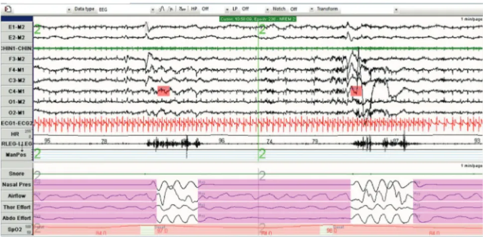
Obstructive sleep apnea (OSA) is a sleep-related breathing disor-der characterized by repetitive collapse of the upper airway during sleep, which causes complete or partial cessation in airflow (Eckert and Mal-hotra, 2008). Respiratory effort persists despite the reduction in airflow. Apnea refers to complete cessation of airflow; hypopnea indicates limited airflow. Figure 1 shows an example of an obstructive apnea, in which air-flow is absent, but respiratory effort continues with subsequent oxygen desaturation.
Figure 1. Image from a baseline polysomnogram showing oxygen desaturations following an obstructive apnea. NPRE = nasal pressure; N/O = nasal-oral therm-istor; THOR = thoracic effort belt; ABD = abdominal effort belt; SpO2 = blood oxygen saturation; Pleth = plethysmography.
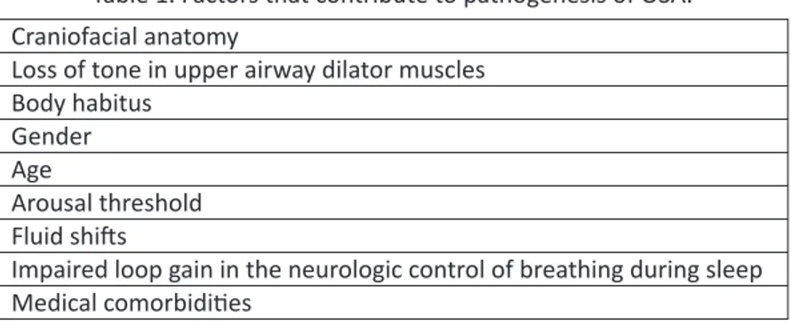
Table 1. Factors that contribute to pathogenesis of OSA. Craniofacial anatomy
Loss of tone in upper airway dilator muscles Body habitus
Gender Age
Arousal threshold Fluid shifts
Impaired loop gain in the neurologic control of breathing during sleep Medical comorbidities
varies from one person to the next, as does the combination of risk fac-tors in a given individual. This variation yields a heterogeneous patient population. This inherent diversity of disease adds to the scientific in-trigue surrounding OSA and also shapes the multitude of treatment op-tions currently available for OSA.
upper airway. This particular image does not indicate significant narrow-ing at one particular level along the upper airway. However, the upper airway cross-sectional area is known to be reduced in individuals with OSA, compared with those without OSA (Schwab et al., 1995).
In addition to anatomic considerations, upper airway muscle tone is another important consideration. The role of neuromuscular tone in the pathogenesis of OSA should not be forgotten; OSA is not simply a “structural problem.” As the brain transitions from wake to sleep, upper airway dilator muscle tone decreases and resistance within the upper airway increases. Understanding this physiology helps explain changes in upper airway caliber in different states (e.g., wake versus sleep).
As sleep is sustained, lung volumes decrease and chemoreceptors become less sensitive. Decreased chemoreceptor sensitivity affects the input provided for neural control of breathing. As a result of reduced lung volumes, relative hypoventilation ensues. As the upper airway dilator muscles become even less active, the upper airway caliber narrows further, either to partial or complete obstruction. This condition leads to an obstructive hypopnea (persistent work of breathing in the setting of a partially occluded airway) or an obstructive apnea (persistent work of breathing in the setting of a fully occluded airway).
When airflow is restricted, blood oxygen saturation may drop while carbon dioxide levels may rise. Respiratory effort increases as the respiratory apparatus continues to work against a partially or fully ob-structed airway. The increased work of breathing causes an arousal, in which the brain wakes from sleep. During this transient period of wake-fulness, upper airway muscle activity increases and chemosensitivity im-proves. This situation is followed quickly by a brisk opening of the upper airway. Once airflow is restored, the brain again transitions back from wake to sleep and the cycle resumes (Eckert and Malhotra, 2008).
Sleep-wake instability results from untreated OSA. Increased re-spiratory effort leads to frequent arousals from sleep, which last only a few seconds and then the brain changes from wake to sleep. In patients with frequent respiratory disturbances, sleep can become highly frag-mented as a function of endless transitions from wake to sleep and back again.
In addition to regulation of sleep-wake transitions, the brain also modulates ventilatory control. Neural control of breathing is an intricate process with a number of variable inputs. Loop gain refers to sensitiv-ity of a variable system (Naughton, 2010). In OSA, the variable system is a feedback cycle between peripheral inputs and central regulation of breathing. Components include:
1. The blood gas response to a change in ventilation; 2. The speed at which blood carbon dioxide level is
sig-naled to the central controller; and
3. The central ventilatory response to carbon dioxide.
In patients with untreated OSA, the loop gain is impaired; chemorecep-tors are less sensitive to small changes in blood carbon dioxide levels and ventilation becomes more unstable (Kapur, 2010).
and may allow for development of new therapeutic pathways for OSA patients.
COMORBIDITIES ASSOCIATED WITH OSA
The heart and brain are the two end organs most studied in un-treated OSA. The aforementioned fluctuations in blood levels of oxygen and carbon dioxide and sleep-wake instability can lead to increased sym-pathetic neural activity. The increased symsym-pathetic tone causes acute in-creases in heart rate, blood pressure and cardiac stress, particularly left ventricular stress. The accumulation of these sympathetic surges and re-lated consequences can lead to clinical manifestations of hypertension, cardiac arrhythmias, stroke and sudden cardiac death (Gami and Somers, 2007).
Untreated OSA is an independent risk factor for hypertension; the risk increases as the severity of OSA increases (Peppard et al., 2000). In a prospective study of men with OSA, odds of fatal and non-fatal car-diovascular increased with severe untreated OSA (Marin et al., 2005). The cumulative frequency of atrial fibrillation increases over time for individ-uals with OSA compared with those without (Gami et al., 2007). Just as the risk of cardiovascular events increases with untreated OSA, the risk of stroke or death rises as the severity of OSA increases (Yaggi et al., 2005). The risk of these adverse outcomes is mitigated with continuous positive airway pressure (CPAP) for treatment of OSA (Park et al., 2011).
Ongoing research indicates many other organs and homeostatic processes are affected adversely by untreated OSA. For example, blood glucose regulation can be impaired as patients with untreated OSA have increased insulin resistance. Frequent, repetitive sympathetic surges can lead to increased oxidative stress, endothelial dysfunction, hypercoagu-lability and systemic inflammation. These cellular-level changes not only increase risk for cardiovascular and cerebrovascular morbidities, but also may contribute to development of certain cancers.
Table 2. Examples of conditions that can be exacerbated by untreated OSA. Hypertension
Atrial fibrillation Depression
Myocardial infarction Stroke
Cancer
Adverse perioperative outcomes Epilepsy
Diabetes mellitus
For some patients, discussion of increased risk of myriad medi-cal comorbidities may seem abstract and depersonalized. Untreated OSA also can have a very personal, real-life impact for an affected individual, as exemplified in Figure 3. Excessive daytime sleepiness and decreased quality of life are two of the most commonly discussed consequences of untreated OSA (Knauert et al., 2015). For symptomatic individuals, exces-sive daytime sleepiness can have implications on employment options, social engagements and interpersonal relationships. Impaired academic and/or professional performance and restricted social interactions can have strong bearing on quality of life for those with untreated OSA. Effec-tive OSA treatment can help to reverse these negaEffec-tive symptoms and can yield improved quality of life.
CLINICAL EVALUATION OF OSA
The most critical component of the clinical evaluation for OSA is a detailed history. The examiner should inquire about sleep-related toms and consequences on daytime functioning. Sleep-related symp-toms suggestive of OSA include snoring, witnessed apneas, frequent noc-turnal awakenings and non-restorative sleep. Patients also may endorse frequent mouth breathing and waking with a dry mouth or sore throat. Other symptoms experienced by untreated OSA patients include acid re-flux, nocturnal urination and morning headaches. History of weight gain also may provide insight in the context of the patient’s other symptoms.
Clinical History: A 36-year-old gentleman presents to the Sleep Disorders Clinic for evaluation of daytime sleepiness. He has been experiencing excessive daytime ness for years. He often nods off during conversations and finds that his daytime sleepi-ness makes it difficult for him to interact with people for longer than an hour. He re-counts a few instances during which he dozed while waiting at a stoplight; he woke to the sound of another driver honking the horn. He denies any motor vehicle collisions due to sleepiness.
He sleeps from 10:30 p.m.-5:30 a.m. during the week and until 7:30 a.m. on weekends. He feels refreshed in the morning, but feels sleepy after a couple hours. He has three cups of coffee during the day and does not take naps. He snores when he sleeps on his back and rarely awakens gasping for air. He has been witnessed to have apneas during sleep.
He is a university faculty member and has been teaching for about ten years. He re-ports that he has difficulty keeping up with the demands of his job because his daytime sleepiness interferes with his ability to concentrate and work efficiently. He decided to pursue medical evaluation of his sleepiness because he is concerned that he may be placed on probationary status at work.
Diagnostic Evaluation: A baseline polysomnogram showed that he had 38 apneas + hypopneas/hour and oxygen desaturations to 78%. He was diagnosed with OSA and was prescribed continuous positive airway pressure (CPAP).
Treatment Response: He committed to using CPAP nightly. He had resolution of his excessive daytime sleepiness and no longer felt sleepy while driving. Due to improved attention and focus, he was able to improve the quality of his work and be considered for promotion.
Figure 3. Case example of a how untreated OSA can affect quality of life.
avoid high-risk situations (e.g., driving) when experiencing excessive day-time sleepiness. Occupational considerations also should be discussed to ensure that excessive daytime sleepiness does not impair the patient’s ability to perform his/her job safely.
The STOP-Bang questionnaire (Chung et al., 2013) is a quick, ef-fective screening tool that can help identify patients at risk for severe OSA. The screening items are:
Snoring Tiredness Observed apnea
high blood Pressure (STOP)
Body mass index (BMI) above 35 kg/m2
Age above 50 years
One point can be assigned for each category, for a maximum of 8 points. A validation study in obese and morbidly obese surgical patients (Chung et al., 2013) showed that a STOP-Bang score of 4 has 88% sensitiv-ity for severe OSA. A STOP-Bang score of 6 is more specific for severe OSA in obese and morbidly obese surgical patients.
Even the STOP questionnaire, which contains the first four ele-ments (Snoring, Tiredness, Observed apnea and high blood Pressure), is 79.5% sensitive in pre-surgical patients with severe OSA (Chung et al., 2008). For clinicians who are unable to obtain measurements of a pa-tient’s BMI and neck circumference, the STOP questionnaire may be a useful screening tool to incorporate into the clinical evaluation for OSA.
EPIDEMIOLOGY OF OSA
The estimated prevalence of OSA in the U.S. often is cited as 3-7% in men and 2-5% in women (Park et al., 2011). Other studied populations, including those in Australia, India, China and Korea also have a higher prevalence in men compared to women (Punjabi, 2008). The prevalence of OSA is higher among adults aged 65 years and older than it is for adults aged 30 to 64 (Bixler et al., 1998).
The OSA prevalence estimate is higher in other subgroups of the general population. In patients with obesity, OSA prevalence rises as BMI increases. A retrospective study of morbidly obese patients who pre-sented for weight loss surgery showed a total OSA prevalence of 78% across all BMI categories. In patients with a BMI above 60 kg/m2, the
prevalence measured 95% (Lopez et al., 2008). The prevalence of OSA can be expected to rise as the obesity epidemic continues in the U.S. and other countries. As a result, estimates from earlier landmark epidemio-logic studies on OSA may underrepresent the current prevalence of OSA in the general population.
PUBLIC HEALTH CONCERNS RELATED TO OSA
with individuals who do not have OSA (Karimi et al., 2015). This risk is reduced by 70% in OSA patients who use CPAP for at least four hours/ night.
The increased accident risk for untreated OSA patients is danger-ous, especially in those employed in transportation: truck drivers, train operators and pilots. News reports can be found to describe tragic ac-cidents in each of these settings, with the inac-cidents attributable at least partly to the operator’s/driver’s untreated OSA. These accidents occur around the globe. In the U.S., federal agencies are reviewing published data on OSA prevalence in transportation industries to inform policy-making about OSA screening, evaluation and management.
The prevalence of OSA in commercial drivers has been studied in multiple populations, with most studies showing a higher OSA preva-lence compared with the general population. For instance, a study of 388 commercial drivers showed a 78% OSA prevalence (Stoohs et al., 1995). In light of this high clinical risk, coupled with the risk of injury to self and others in the event of a crash, commercial drivers, pilots and train opera-tors may be subject to OSA screening, evaluation and treatment as indicated. The American Academy of Sleep Medicine has outlined detailed recommendations for transportation workers with safety-sensitive du-ties who are diagnosed with OSA (http://www.aasmnet.org/resources/ pdf/AASMResponseFMCSAFRA.pdf). These recommendations include considerations for “No Restrictions,” “Conditional Restrictions” and “Immediate Disqualification” from occupational responsibilities. The different designations are conferred based on clinical symptoms, sever-ity of OSA and adherence to OSA treatment. These individuals require longitudinal follow-up to ensure that OSA is treated fully and that the management plan is reassessed and adjusted if the patient’s clinical pic-ture changes.
ECONOMIC IMPACT OF OSA
of healthcare costs, undiagnosed OSA patients cost $1,950 to $3,899 per year more than patients without OSA. Cost is increased even when patients are diagnosed with OSA, but remain untreated. One study esti-mated that the annual cost of treating the medical consequences of OSA in the U.S. is $3.4 billion (Kapur et al., 1999).
The aforementioned review of 106 studies showed that in terms of healthcare utilization, OSA patients treated with CPAP cost $2,700-$5,200 less per year, compared with OSA patients who are not treated (Knauert et al., 2015). Incremental cost-effectiveness ratio (ICER)—the ra-tio of the incremental cost and incremental change in quality adjusted life years (QALY) that stems from use of a specified treatment—is a measure of cost effectiveness (Tan et al., 2008). An ICER/QALY value of $50,000 typically is considered to be acceptable or beneficial (Ubel et al., 2003). Economic analyses with ICER/QALY values have shown the cost effective-ness of CPAP therapy in OSA patients. Use of CPAP therapy compared with no treatment in moderate-to-severe OSA patients had an ICER/QALY value of $15,915, indicative of significant cost-effectiveness of CPAP ther-apy in these patients (Pietzsch et al., 2011).
CONCLUSIONS
OSA is an intriguing, multi-faceted disease process. Craniofacial anatomy and upper airway neuromuscular tone are just two of many fac-tors that contribute to development of OSA in an individual. The con-sequences of OSA affect not only the patient, but society at large. Un-treated OSA patients are at increased risk for a number of cardiovascular comorbidities; risk of stroke, endocrinologic disorders and cancer also in-crease when OSA is not treated. Patients with untreated OSA may experi-ence excessive daytime sleepiness and have a higher risk of motor vehicle accidents compared with those who do not have OSA.
REFERENCES
American Academy of Sleep Medicine. http://www.aasmnet.org/re-sources/pdf/AASMResponseFMCSAFRA.pdf. Accessed May 8, 2017. Bixler EO, Vgontzas AN, Ten Have T, Tyson K, Kales A. Effects of age on
sleep apnea in men: I. Prevalence and severity. Am J Respir Crit Care Med 1998;157(1):144-148.
Chihorek AM, Abou-Khalil B, Malow BA. Obstructive sleep apnea is asso-ciated with seizure occurrence in older adults with epilepsy. Neurology 2007;69(19):1823-1827.
Chiu KL, Ryan CM, Shiota S, Ruttanaumpawan P, Arzt M, Haight JS, Chan CT, Floras JS, Bradley TD. Fluid shift by lower body positive pressure increases pharyngeal resistance in healthy subjects. Am J Respir Crit Care Med 2006;174(12):1378-1383.
Chung F, Yang Y, Liao P. Predictive performance of the STOP-Bang score for identifying obstructive sleep apnea in obese patients. Obes Surg 2013;23(12):2050-2057.
Chung F, Yegneswaran B, Liao P, Chung SA, Vairavanathan S, Islam S, Kha-jehdehi A, Shapiro CM. STOP questionnaire: A tool to screen patients for obstructive sleep apnea. Anesthesiology 2008;108(5):812-821. Eckert DJ, Malhotra A. Pathophysiology of adult obstructive sleep apnea.
Proc Am Thorac Soc 2008;5(2):144-153.
Gami AS, Hodge DO, Herges RM, Olson EJ, Nykodym J, Kara T, Somers VK. Obstructive sleep apnea, obesity, and the risk of incident atrial fibrilla-tion. J Am Coll Cardiol 2007;49(5):565-571.
Gami AS, Somers VK. Sleep apnea and cardiovascular disease. In: Libby P, Bonow RO, Mann DL, Zipes DP, eds. Braunwald’s Heart Disease: A Textbook of Cardiovascular Medicine. 8th ed. Philadelphia: Saunders 2007.
Kapur V, Blough DK, Sandblom RE, Hert R, de Maine JB, Sullivan SD, Psaty BM. The medical cost of undiagnosed sleep apnea. Sleep 1999;22(6): 749-755.
Karimi M, Hedner J, Häbel H, Nerman O, Grote L. Sleep apnea-related risk of motor vehicle accidents is reduced by continuous positive airway pressure: Swedish Traffic Accident Registry data. Sleep 2015;38(3): 341-349.
Knauert M, Naik S, Gillespie MB, Kryger M. Clinical consequences and economic costs of untreated obstructive sleep apnea syndrome. World J Otorhinolaryngol Head Neck Surg 2015;1(1):17-27.
Lopez PP, Stefan B, Schulman CI, Byers PM. Prevalence of sleep apnea in morbidly obese patients who presented for weight loss surgery evalu-ation: More evidence for routine screening for obstructive sleep ap-nea before weight loss surgery. Am Surg 2008;74(9):834-838.
Marin JM, Carrizo SJ, Vicente E, Agusti AG. Long-term cardiovascular out-comes in men with obstructive sleep apnoea-hypopnoea with or with-out treatment with continuous positive airway pressure: An observa-tional study. Lancet 2005;365(9464):1046-1053.
Naughton MT. Loop gain in apnea: Gaining control or controlling the gain? Am J Respir Crit Care Med 2010;181(2):103-105.
Park JG, Ramar K, Olson EJ. Updates on definition, consequences, and management of obstructive sleep apnea. Mayo Clin Proc 2011;86(6): 549-555.
Peppard PE, Young T, Palta M, Skatrud J. Prospective study of the associa-tion between sleep-disordered breathing and hypertension. N Engl J Med 2000;342(19):1378-1384.
Pietzsch JB, Garner A, Cipriano LE, Linehan JH. An integrated health-eco-nomic analysis of diagnostic and therapeutic strategies in the treat-ment of moderate-to-severe obstructive sleep apnea. Sleep 2011; 34(6):695-709.
Punjabi NM. The epidemiology of adult obstructive sleep apnea. Proc Am Thorac Soc 2008;5(2):136-143.
Schwab RJ, Gupta KB, Gefter WB, Metzger LJ, Hoffman EA, Pack AI. Up-per airway and soft tissue anatomy in normal subjects and patients with sleep-disordered breathing: Significance of the lateral pharyngeal walls. Am J Respir Crit Care Med 1995;152(5 Pt 1):1673-1689.
Tan MC, Ayas NT, Mulgrew A, Cortes L, FitzGerald JM, Fleetham JA, Schul-zer M, Ryan CF, Ghaeli R, Cooper P, Marra CA. Cost-effectiveness of continuous positive airway pressure therapy for patients with obstruc-tive sleep apnea-hypopnea in British Columbia. Can Respir J 2008; 15(3):159-165.
Ubel PA, Hirth RA, Chernew ME, Fendrick AM. What is the price of life and why doesn’t it increase at the rate of inflation? Arch Intern Med 2003;163(14):1637-1641.
DIAGNOSIS OF OBSTRUCTIVE SLEEP APNEA
Douglas Kirsch
ABSTRACT
The understanding of obstructive sleep apnea (OSA) has advanced significantly over the last several decades, but there still is much to discover. The diagnostic testing for OSA initially was limited to specialized sleep laboratories, but now often is performed in the patient’s home with more limited equipment.
The primary severity marker for OSA is a work in progress, given dis-agreement about the definition of abnormal respiratory events. This chapter will review the recent evolution in testing and diagnostic criteria for OSA, while also forecasting what the near future might bring in terms of OSA testing.
key words: polysomnography, home sleep apnea test, obstructive sleep apnea, Apnea-Hypopnea Index, respiratory event index
INTRODUCTION
Obstructive sleep apnea (OSA) is a relatively modern diagnosis within the long history of the field of medicine. In past generations, the primary sign of OSA—snoring—was viewed as a sign of a deep and rest-ful sleep. Sleep medicine clinicians currently understand that the loud rumbles emanating from a person’s upper airway are not likely to be a sign of high-quality sleep, but a potential harbinger of a significant sleep disorder and a potential long-term health risk. This chapter will review the history of the diagnosis of OSA, the evolution in sleep apnea testing that has occurred in the last few decades and a glimpse of the possible future for evaluation of sleep-disordered breathing (SDB).
THE HISTORY OF POLYSOMNOGRAPHY
found himself being poked by needles to restart his breathing while sleeping (Kryger, 1983). A few thousand years later, Charles Dickens cre-ated the character Fat Joe in the Posthumous Papers of the Pickwick Club, noting that this corpulent character would fall asleep while chewing food and talking to others, suggestive of symptoms of severe SDB. Even more eloquently, H. V. Morton (1930) described the “Snorer of Kilkenny” thusly:
How that man made me suffer. His ghastly organ recital was as regular in its devilish rhythm as a sawmill. Once every half hour he was seized with a kind of convulsion. I hoped that he was dy-ing. The debasing sounds shuddered to a pianissimo and ceased, then he gave a violent gasp, a snort, appeared to be choking, grunted, gasped, and got into top gear again.
These examples, spread over millennia, are recognition of seri-ous snoring, apneic episodes and significant daytime sleepiness. The physiological evaluation of sleep, however, was stimulated in part by the discovery of Richard Caton, a Scottish scientist, who was the first to re-cord electrical rhythms originating from the brains of rabbits and mon-keys (Caton, 1875). It was not until decades later, however, that Hans Berger (1929), a German psychiatrist, was able to record human brain ac-tivity, including pattern changes with sleep, with his newly named “elec-troencephalogram.”
The study of sleep on American soil began with two groups, one at Harvard and another at the University of Chicago. Loomis and the Har-vard group began to describe different sleep stages (A-E) while study-ing sleepstudy-ing patients with electroencephalography (EEG). Loomis (1937), Aserinsky and Kleitman (1953) and other researchers at the University of Chicago took another step forward by studying eye movements during sleep, discovering the presence of rapid eye movements during sleep. Fi-nally, Dement and Kleitman (1957) recognized the patterns of sleep stag-es, including that of rapid eye movement (REM) sleep and its relationship to dreaming, through the use of entire night recordings with EEG, elec-tromyography (EMG) and electrooculography (EOG). Thus, the full-night sleep recording was born.
Bickelmann and colleagues (1956) described a patient with obesity, hy-poventilation and sleepiness, naming the symptom set “Pickwickian syn-drome” after the previously referenced Dickens story. Shortly thereafter, research groups in Europe evaluated patients with Pickwickian syndrome, discovering the presence of apneic spells with continued respiratory ef-fort (Gastaut et al., 1965). After postulating that this finding suggested ob-struction at the upper airway, physicians treated these patients with tra-cheostomy, eliminating the symptoms and the apneas (Kuhlo et al., 1969).
Further research on respiration during sleep led to differentiation between obstructive, mixed and central apneas. Moving beyond respira-tion, scientists in Europe and the United States published data leading to the addition of cardiac and leg movement leads for a sleep study. Finally, the components were in place for the modern polysomnogram (PSG) that we use clinically today. Figure 1 demonstrates a few epochs of a typical PSG, including sleep staging, respiratory assessment and, in this case, a diagnosis of OSA.
MEASURING OSA
Once OSA was recognized as a disorder of sleep, more clear guidelines for making a diagnosis were required. Early studies of the air-way with oral/nasal thermistor and effort bands identified the range of normal and abnormal with clear obstructive apneas. Over time, however, scientific study clarified that airway closure was not an all-or-none phe-nomenon, but was variable continuously. Partial obstruction of the air-way, known as a hypopnea, was demonstrated to be relevant equally as an apnea to sleep disruption.
Having multiple respiratory events definitions led to complica-tions in scientific research and clinical activities; an effort then was made to standardize these definitions. Multiple committee and task forces were convened over the course of several years to clarify the definitions of respiratory events. An American Academy of Sleep Medicine (AASM; 1999) consensus report recommended standardized scoring criteria for a range of respiratory events in clinical research (known as the Chicago criteria). This report described two types of hypopneas: 1) respiratory events with a > 50% decrease in a valid measure of airflow without a requirement for associated oxygen desaturation or arousal; and 2) those with a lesser airflow reduction in association with oxygen desaturation of > 3% or an arousal (Sleep, 1999).
Soon thereafter, a clinically-focused paper from the AASM Clini-cal Practices Review Committee used the hypopnea definition from the Sleep Heart Health Study as an abnormal respiratory event lasting ≥ 10 seconds in length with ≥ 30% reduction in airflow or chest wall movement and with ≥ 4% oxygen desaturation (Meoli et al., 2001). This particular
definition has been used by the Centers for Medicare and Medicaid Ser-vices (CMS) to determine treatment eligibility for patients with OSA diag-nosed according to this definition.
In order to better codify the variety of available scoring rules and improve clinical standardization, a scoring manual was created by the AASM (Iber et al., 2007). The first edition of the Manual for the Scoring of Sleep and Associated Events was published in 2007 with two defini-tions for hypopneas. The recommended definition in this edition was the same as the CMS definition: hypopnea scoring requires ≥ 30% reduction in nasal pressure signal excursions from baseline and associated ≥ 4% de-saturation from pre-event baseline.
The alternative hypopneas definition includes a ≥ 50% reduction in nasal pressure signal excursions and associated ≥ 3% desaturation or arousal. Later editions of the scoring manual have reversed the recom-mended and alternative definitions; the recomrecom-mended definition now is hypopneas associated with 3% oxygen desaturation and/or arousal.
Using the different AASM definitions for respiratory events can have dramatic differences on determining OSA severity. For instance, in Ruehland and associates’ study (2009), applying the Chicago criteria to patients led to a median Apnea-Hypopnea Index (AHI) of 25.1/hour, 3% oxygen desaturation and/or arousal criteria led to a median AHI of 14.9/hour and 4% oxygen desaturation criteria led to a median AHI of 8.3 events/hour. These variations would lead on average to the same patient being diagnosed with mild OSA using one definition and high-to-mod-erate OSA with another, thus leading to potentially different treatment algorithms for the same patient. Debate on a “true” definition for hypop-neas is ongoing and unlikely to be resolved in the near future.
THE AHI: IMPORTANT, BUT PROBLEMATIC
Severity Total AHI/hour of sleep
No OSA 0-4.99
Mild OSA 5-14.99
Moderate OSA 15-29.99
Severe OSA ≥ 30
total sleep hours. Another potential metric used to evaluate SDB is the oxygen desaturation index (ODI), which is the total number of oxygen desaturations divided by the hours of sleep.
The reason for the predominant use of the AHI in current clini-cal practice is that crucial research comprising the backbone of cliniclini-cal sleep medicine used the AHI as the primary determinant. In the 1993 Wisconsin Sleep Cohort epidemiological study, the AHI was the prima-ry metric used to define the prevalence of OSA; this large cohort study found in middle-aged adults that 4% of men and 2% of women had an AHI > 5 along with excessive daytime sleepiness. Sleepiness is one of the most frequent symptoms of OSA; for instance, patients with OSA have as much as a 5x higher risk of motor vehicle crash than control subjects (Tregear et al., 2009).
When evaluating the health risks to patients who suffer from OSA, the AHI typically has been used as the severity determinant. Multi-ple papers have demonstrated that AHI-determined OSA leads to hyper-tension (HTN) and that treatment of OSA will improve blood pressure. Epidemiological studies have linked OSA (based on AHI) with myocar-dial infarction, congestive heart failure and stroke (Gottlieb et al., 2010). However, a single definition for the respiratory events comprising the AHI may not elicit all the possible disease associations, as OSA may have different effects on different organ systems. For instance, 4% oxygen de-saturations better predict hypertension, while 2% oxygen dede-saturations better predict fasting hyperglycemia and the arousal frequency predicts impaired memory consolidation (Jordan et al., 2014).
Having a mostly formalized definition for the AHI has some ad-vantages in that it encapsulates a complex disorder into a single metric. However, as noted above, there are aspects of the AHI that raise concern; in particular, the AHI may not tell the entire story about OSA severity.
Questions about the metric are encapsulated in Table 2 and will be dis-cussed in further detail.
First, while research suggests that apneas and hypopneas are similar in physiological terms, it does not define them necessarily as bio-logically equivalent or even that all hypopneas have the same biological impact. This non-equivalence is true, particularly when a scored hypop-nea is associated with an arousal rather than an oxygen desaturation. While that arousal from sleep may have downstream impact on quality of life, wellbeing and medical conditions, it likely has a different impact than those events associated with oxygen desaturation, whether that de-saturation is 3%, 4% or other. Thus, the AHI—regardless of the definition used—is unlikely to encapsulate completely the variety of effects that OSA causes because of the variety of events that comprise the metric.
Second, respiratory events—whether apneas or hypopneas—are associated with different levels of oxygen desaturation. Thus, a hypopnea with a 4% desaturation is considered of equivalent severity to that asso-ciated with a 10% desaturation, though knowledge of the human body’s response to hypoxia would suggest that these hypopneas likely have a differential effect. While some studies suggest that increasing depth of oxygen desaturation during sleep has a negative impact on health and increases mortality (Muraja-Murro et al., 2013), there are limited data to inform whether depth or duration of oxygen desaturation may lead to an increased impact.
Third, by definition, all respiratory events must occur for at least ten seconds. However, does a ten-second hypopnea have a similar effect to a 60-second apnea? And could a shorter than ten seconds respiratory event have a health-related impact? In children, respiratory events of shorter length (two-breath minimum) are deemed to be important physi-ologically; however, currently only longer (more than ten seconds) respi-ratory events have been defined as relevant in adults.
1. Are apneas and hypopneas equivalent?
2. Does the depth of oxygen desaturation associated with each individual respiratory event matter?
3. Does the temporal length of a respiratory event matter?
Fourth, respiratory events typically may not distribute evenly over the course of a sleep study. In some cases, respiratory events may occur on a positional basis (e.g., supine versus non-supine) or may oc-cur on a sleep stage basis (e.g., REM sleep versus non-REM sleep versus during wake-sleep transitions). Thus, we can compare two patients who have the same overall AHI. Patient 1 has a high supine-position AHI, but only sleeps on his/her back for an hour during a PSG. Patient 2 has a high AHI only during REM sleep, but limited respiratory events during NREM sleep. Technically these patients have an equivalent severity of apnea based on their total AHI, though whether they truly are equivalent dis-ease severity or are likely to have dissimilar symptoms or disdis-ease-specific outcomes currently is unclear.
BEYOND THE LABORATORY-BASED PSG
For many years, patients questioned whether their sleep tested in a sleep laboratory was reflective of their typical sleep at home. With the advent of home sleep apnea testing (HSAT), patients now are able to be tested in their usual sleeping environment. The home testing de-vices are designed to be used by a patient at nighttime without significant technical assistance to evaluate them for OSA.
The unattended nature of the test is one of several differences between testing with HSAT devices and in-laboratory PSG. Other signifi-cant differences include: the absence of measurement of the patient’s total sleep time and EEG-related arousals from sleep from most HSAT de-vices; HSAT devices do not assess appropriately for disorders other than OSA; and there is a lower cost of testing with use of a HSAT device com-pared to in-laboratory PSG. Table 3 compares and contrasts HSAT devices and in-laboratory PSG.
Though HSATs have been available for decades, the common use of these devices was discouraged due to insufficient data regarding their use in clinical practice. As research study publications increased, national medical societies and the CMS re-evaluated and eventually approved use of HSATs for diagnosis of OSA in 2007-2008. The AASM released Clinical Guidelines for the Use of Unattended Portable Monitors in the Diagnosis of Obstructive Sleep Apnea in Adult Patients (Collop et al., 2007).
Differences HSAT device In-lab PSG
Location Home Sleep laboratory
Attended with a technologist No Yes
Measures sleep time Rarely Always
Typical measurement leads # 3-4 16-20
Measures EEG arousals No Yes
Measures EEG-based sleep time No Yes
Can diagnose occasionally CSAOSA, Many sleep disorders
Expense per test Low ($200-$400) High ($800 to multiple thousands)
Population Adults only Children and adults
Suggested restrictions
Patients without serious co-morbid
medical or sleep disorders
None
severe OSA. HSAT use in this population improves the specificity and sensitivity of the test. The guidelines also suggest that the devices not be used in patients with significant sleep co-morbidities (e.g., periodic limb movements of sleep, insomnia, parasomnias) and severe medical co-morbidities that may affect breathing (e.g., chronic obstructive pul-monary disease, congestive heart failure, neuromuscular conditions). On a practical level, however, local insurance regulations may affect the strict application of these guidelines to clinical care.
The use of HSATs now applies a potentially different structure to the assessment of respiratory event frequency. While laboratory testing includes both nasal pressure and oronasal thermistor leads, HSATs gener-ally include one of those two leads, typicgener-ally nasal pressure. EEG-staged sleep also is not measured on most HSAT devices, thus the breathing index is determined by the denominator of total recording time, rather than total sleep time. This change may lead to underestimation of OSA severity, given that most patients do not sleep for the entire recording of the home test.
The absence of EEG leads on the HSAT also means that arous-als from sleep cannot be scored. The fallout from this absence is that only the oxygen desaturation hypopnea criteria can be utilized. Thus, the AASM has opted to call a HSAT breathing index the “respiratory event index” or “REI.” Though similar, the REI truly is not synonymous with the AHI, thus further muddying the already cloudy waters of OSA severity. A sample image of a HSAT from a commonly used device can be seen in Figure 2.
SMARTPHONES AND BEYOND
Less data is available about Smartphone apps and other consum-er devices that evaluate SDB. FitbitsTM and other personal trackers
evalu-ate sleep via privevalu-ately designed algorithms that have mixed data when compared to PSG (Mantua et al., 2016). To this point, however, while the devices may detect movement-based arousals, they do not have a clear method for assessing respiratory-based sleep disorders.
Smartphone application is another method of assessment of sleep and SDB breathing at home. These applications typically use gy-roscopic movement and noises to determine sleep quality. Limited data exists comparing these applications to PSG, but what does exist suggests that the sensitivity and specificity of the applications may not be high (Bhat et al., 2015). A sample patient image from the Smart Alarm applica-tion can be seen in Figure 3, demonstrating reported depth of sleep and areas of user re-playable noise during the night (at the circles).
Figure 2. This two-minute epoch from a Philips Alice Night One HSAT demon-strates OSA (Philips Respironics, Pittsburgh, PA). From top to bottom, the leads are: pulse rate, snoring, nasal pressure (PFlow), thoracic effort (THO), oxygen saturation (SpO2) and body position (S=supine). The red shaded images in the nasal pressure signal demonstrate the absence of air flow in the presence of continued effort and are obstructive apneas. Oxygen desaturations associated with the respiratory events are seen in the tan shaded areas in the SpO2 lead.
via Smartphone appears to be moving forward quickly, using similar met-rics to those being used currently in the laboratory.
FUTURE THOUGHTS
Though the AHI has much data supporting its use, other metrics have been proposed. Some of the areas that are being explored actively are shown in Table 4. These metrics take into account some of the issues raised with the AHI above. At this point, these options are theoretical constructs; little clinical data are able to support the use of these metrics. In a paper evaluating interactions between event duration and oxygen desaturation, however, it is clear that two patients with simi-lar AHIs can have different findings when looking at these novel met-rics (Kulkas, 2013). These differences are just one of several possible explanations for why patients with different AHIs have different clinical symptoms and health outcomes. Another study evaluated patients with mild-to-moderate OSA retrospectively, using the integrated area of de-saturation curve (IAD) to re-evaluate them. Patients with high IADs were more likely to have had a cardiovascular event compared to those with lower IADs, when AHI alone did not distinguish these groups (Asano et al., 2009). Punjabi (2016) remarks about the breadth of available data on the sleep study in a recent article:
The field of sleep disorders medicine should not settle for taking a large series of time varying signals from the PSG that often ex-ceed one gigabyte of data and just use one metric to summarize all key features. Certainly, a “one size fits all” solution will never exist that can embody the physiologic diversity of apneas and hy-popneas.
Quantification of depth of desaturation Interaction of event length and desaturation Heart-rate variability
Sleep stage transitions
CONCLUSIONS
The young field of sleep medicine has come a long way in a short time, medically speaking. As sleep medicine continues to evolve in ways both predictable and not, practitioners should embrace the idea that while our current metrics have value based on our history, new metrics may evaluate OSA better severity by linking more strongly with patient symptoms and important clinical outcomes. This knowledge should aid in the shared decision-making processes around treatments and lead to better overall patient health outcomes.
REFERENCES
Asano K, Takata Y, Usui Y, Shiina K, Hashimura Y, Kato K, Saruhara H, Ya-mashina A. New index for analysis of polysomnography, ‘integrated area of desaturation,’ is associated with high cardiovascular risk in patients with mild to moderate obstructive sleep apnea. Respiration 2009;78(3):278-284.
Aserinsky E, Kleitman N. Regularly occurring periods of eye motility, and concomitant phenomena, during sleep. Science 1953;118(3062):273-274.
Berger H. Ueber das Elektroenkephalogramm des Menschen [On the hu-man electroencephalogram]. Archiv Psychiatr Nervenkr 1929;87:527-570. [In German.]
Bhat S, Ferraris A, Gupta D, Mozafarian M, DeBari VA, Gushway-Henry N, Gowda SP, Polos PG, Rubinstein M, Seidu H, Chokroverty S. Is there a clinical role for Smartphone sleep apps? Comparison of sleep cycle de-tection by a Smartphone application to polysomnography. J Clin Sleep Med 2015;11(7):709-715.
Bickelmann AG, Burwell CS, Robin ED, Whaley RD. Extreme obesity as-sociated with alveolar hypoventilation: A Pickwickian syndrome. Am J Med 1956;21(5):811-818.
Caton R. The electric currents of the brain. Br Med J 1875;2:278.
Collop NA, Anderson WM, Boehlecke B, Claman D, Goldberg R, Gottlieb DJ, Hudgel D, Sateia M, Schwab R; Portable Monitoring Task Force of the American Academy of Sleep Medicine. Clinical guidelines for the use of unattended portable monitors in the diagnosis of obstructive sleep apnea in adult patients. J Clin Sleep Med 2007;3(7):737-747. Dement W, Kleitman N. Cyclic variations in EEG during sleep and their
relation to eye movements, body motility, and dreaming. Electroen-cephalogr Clin Neurophysiol 1957;9(4):673-690.
Gastaut H, Tassinari CA, Duron B. [Polygraphic study of diurnal and noc-turnal (hypnic and respiratory) episodal manifestations of Pickwick syndrome]. Rev Neurol (Paris) 1965;112(6):568-579. [In French.] Gottlieb DJ, Yenokyan G, Newman AB, O’Connor GT, Punjabi NM, Quan
SF, Redline S, Resnick HE, Tong EK, Diener-West M, Shahar E. Prospec-tive study of obstrucProspec-tive sleep apnea and incident coronary heart disease and heart failure: The sleep heart health study. Circulation 2010;122(4):352-360.
Iber C, Ancoli-Israel S, Chesson AL Jr, Quan S; for the American Academy of Sleep Medicine. The AASM Manual for the Scoring of Sleep and As-sociated Events: Rules, Terminology and Technical Specifications. 1st ed. Westchester, IL: American Academy of Sleep Medicine 2007. Jordan AS, McSharry DG, Malhotra A. Adult obstructive sleep apnoea.
Lancet 2014;383(9918):736-747.
Kryger MH. Sleep apnea: From the needles of Dionysius to continuous positive airway pressure. Arch Intern Med 1983;143(12):2301-2303. Kuhlo W, Doll E, Franck MC. [Successful management of Pickwickian
syndrome using long-term tracheostomy]. Dtsch Med Wochenschr 1969;94(24):1286-1290. [In German.]
Kulkas A, Tiihonen P, Eskola K, Julkunen P, Mervaala E, Töyräs J. Novel pa-rameters for evaluating severity of sleep disordered breathing and for supporting diagnosis of sleep apnea-hypopnea syndrome. J Med Eng Technol 2013;37(2):135-143.
Mantua J, Gravel N, Spencer RM. Reliability of sleep measures from four personal health monitoring devices compared to research-based ac-tigraphy and polysomnography. Sensors (Basel) 2016;16(5).
Meoli AL, Casey KR, Clark RW, Coleman JA Jr, Fayle RW, Troell RJ, Iber C; Clinical Practice Review Committee. Hypopnea in sleep-disor dered breathing in adults. Sleep 2001;24(4):469-470.
Morton HV. The Search for Ireland. Methuen, London:1930;66-67. Muraja-Murro A, Kulkas A, Hiltunen M, Kupari S, Hukkanen T, Tiihonen P,
Mervaala E, Töyräs J. The severity of individual obstruction events is related to increased mortality rate in severe obstructive sleep apnea. J Sleep Res 2013;22(6):663-669.
Nakano H, Hirayama K, Sadamitsu Y, Toshimitsu A, Fujita H, Shin S, Taniga-wa T. Monitoring sound to quantify snoring and sleep apnea severity using a Smartphone: Proof of concept. J Clin Sleep Med 2014;10(1): 73-78.
Nandakumar R, Gollakota S, Watson NF. A contactless system for respira-tory event identification. Sleep 2015;38:A163 (abstract supplement). Punjabi NM. Counterpoint: Is the apnea-hypopnea index the best way to quantify the severity of sleep disordered breathing? No. Chest 2016; 149(1):16-19.
Ruehland WR, Rochford PD, O’Donoghue FJ, Pierce RJ, Singh P, Thornton AT. The new AASM criteria for scoring hypopneas: Impact on the ap-nea hypopap-nea index. Sleep 2009;32(2):150-157.
Sleep-related breathing disorders in adults: Recommendations for syn-drome definition and measurement techniques in clinical re search. The Report of an American Academy of Sleep Medicine Task Force. (No authors listed) Sleep 1999;22(5):667-689.
Stradling JR, Davies RJ. Sleep. 1: Obstructive sleep apnoea/hypopnoea syndrome: Definitions, epidemiology, and natural history. Thorax 2004;59(1):73-78.
John C. Carter
and Vishesh K. Kapur
ABSTRACT
Continuous positive airway pressure (CPAP) is a first-line therapy for adult pa-tients with obstructive sleep apnea (OSA), particularly those with daytime symp-toms. CPAP effectively resolves upper airway obstruction and is associated with improvements in daytime sleepiness and hypertension in patients with OSA. Ad-herence to CPAP is a key consideration in the effectiveness of this therapy and remains suboptimal for many patients, despite technological advancements in CPAP aimed at improving patient comfort. Educational and behavioral interven-tions have been associated with improvements in adherence.
key words: CPAP, sleep apnea, effectiveness, efficacy, adherence
INTRODUCTION
Continuous positive airway pressure (CPAP) is the most efficacious treatment for obstructive sleep apnea (OSA) and, therefore, the most fre-quently prescribed. Regardless of which of several potential pathogenic causes is at play, application of positive pressure inside the upper airway typically relieves the upper airway obstruction that defines the disorder. Since the discovery of CPAP, a number of technological innovations have improved its utility. Despite these advancements, suboptimal adherence remains a factor that limits effectiveness in a significant proportion of patients who are prescribed CPAP. The scientific literature regarding the effectiveness of CPAP reflects the interplay of the application of a highly efficacious therapy with suboptimal adherence. Research also reveals av-enues to realize the potential of CPAP more fully by improving adherence.
HISTORY OF CPAP
occurred in a patient with severe sleep apnea who had refused placement of a tracheostomy. This first CPAP unit consisted of little more than a commercially available blower fan with an attached pressure regulator that delivered positive air pressure to the patient via a jury-rigged nasal mask fashioned with rapid-setting silicone sealant. As the air pressure delivered was increased, the patient’s repetitive apneas quickly gave way to normal respiratory flow patterns. When pressure was decreased, apneas recurred. A polysomnographic recording indicated a fairly rapid entry into REM sleep with seven subsequent hours of stable sleep. The next morning, the patient reported feeling more awake and alert than he had in years (Sullivan et al., 1981).
MECHANISMS OF ACTION
The mechanism by which CPAP treats OSA is straightforward: positive pressure generated by an air pump is transmitted to the upper airway to maintain a constant intraluminal positive pressure sufficient to prevent airway collapse (Strohl and Redline, 1986). CPAP is highly effica-cious; if positive airway pressure (PAP) is applied at the level required to overcome the forces favoring airway collapse, OSA typically is resolved. This phenomenon is documented regularly in patients undergoing manu-al CPAP titration in the sleep laboratory (Sanders et manu-al., 1993; Kushida et al., 2006; Fig. 1). CPAP may not be efficacious fully in a small minority of OSA patients for whom instability of breathing control is a prominent fac-tor; these patients may develop central apneas with application of CPAP (Javaheri et al., 2009).
Figure 1. Split-night polysomnogram (PSG) showing response of OSA to CPAP. A: Sleep architecture and stages. B: Respiratory events including obstructive ap-neas and hypopap-neas as well as central apap-neas. C: Oxyhemoglobin saturations measured by oximetry. D: CPAP delivered to the patient. Note fragmentation in sleep architecture (A) prior to the initiation of CPAP, accompanied by frequent respiratory events and severe oxyhemoglobin desaturations that improve after CPAP is applied beginning at 5 cmH2O and titrating up to 10 cmH2O.
ADVANCEMENTS IN PAP TECHNOLOGY
PAP technology has advanced significantly over time in several respects. Modern devices are compact, quiet and typically incorporate humidification and monitoring capability that can be accessed remotely by care providers. In addition, devices have been developed that deliver variable pressure during individual respiratory cycles, as well as over suc-cessive respiratory cycles (Johnson and Johnson, 2015).
Figure 2. Cardiovascular consequences of positive pressure breathing in heart failure (HF). The application of CPAP increases intra-thoracic pressure, thereby reducing the pressure gradient across the cardiac wall. This increased pressure reduces cardiac pre- and after-load that may increase cardiac output in some HF patients. Ao = aorta; LA = left atrium; LV = left ventricle; RV = right ventricle; PA = pulmonary artery. Image courtesy of William DePaso; reproduced with permission.
bi-level PAP may relieve the sensation of difficulty with exhalation (Reeves-Hoche et al., 1995; Powell et al., 2012; Carlucci et al., 2015).
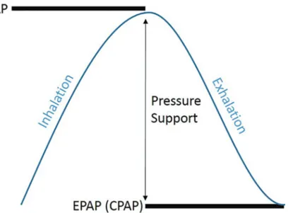
Second, in the case of hypoventilation, bi-level PAP provides venti-latory support by way of additional pressure (pressure support) delivered during inspiration relative to expiration. When used to treat hypoventila-tion, the EPAP is used to maintain patency of the airway, while the IPAP is set to maintain adequate ventilation. Bi-level devices also may include a backup rate, which prompts the device to provide inspiratory pressure au-tomatically if a sufficient inspiratory effort is not generated by the patient to trigger the device to do so.
Figure 3. Schematic representing the pressure cycle provided a bi-level PAP de-vice. EPAP maintains airway patency at the end of exhalation. IPAP provides addi-tional pressure during inhalation, allowing for greater lung expansion and ventila-tion. The pressure differential between IPAP and EPAP is called pressure support.
trigger bi-level PAP may not occur consistently (Loube et al., 1999; Ep-stein et al., 2009). More advanced bi-level PAP devices have been devel-oped that can adjust inspiratory and/or expiratory pressures during the night to maintain minimum levels of ventilation (for hypoventilation) or to limit variability of ventilation (for CSA).
Auto-titrating positive airway pressure (APAP) is designed to adjust CPAP settings continuously over the sleep period to provide the minimum pressure needed to relieve OSA (Roux and Hilbert, 2003). Pro-prietary algorithms adjust pressure on a continuous basis. The algorithms function based upon two basic mechanisms of action: 1) episodically monitor markers of airway patency; and 2) adjust pressure to achieve desired airway patency at the lowest pressure within pre-set pressure boundaries. The proprietary algorithms differ in terms of their aggres-siveness in changing pressures and the breathing-related parameters used to monitor adequacy of pressures.
minimization of PAP delivered to relieve OSA (Ayas et al., 2004; Aloia et al., 2005; Morgenthaler et al., 2008). By minimizing PAP pressures, pressure-related side effects of PAP (pressure intolerance, mask leak and aerophagia—excessive swallowing of air that goes to the stomach) may be ameliorated, leading to improved patient comfort. A potential disad-vantage of APAP is failure to provide appropriate pressures (either too high/low) to relieve OSA (Ip et al., 2012), due to algorithmic miscalcula-tion or mask/oral leak.
Expiratory pressure relief (EPR) is a technology that varies pres-sure delivered over the respiratory cycle (inspiration and expiration). Lower PAP is needed to maintain airway patency at the start of exhala-tion because lung volumes are larger at end-inhalaexhala-tion (which stiffens the upper airway via tracheal tug) and intra-luminal airway pressures are positive during exhalation. When pressure relief is activated, PAP is re-duced during the beginning of exhalation in a manner that ideally should not affect efficacy.
Reduction of pressure at the start of exhalation can improve com-fort for some patients. The risk associated with EPR use is that efficacy may be reduced unintentionally (Aloia et al., 2005; Bakker and Marshall, 2011). Most CPAP devices also incorporate a ramp function that can be set to start at a more comfortable sub-therapeutic pressure and increase over a variable period of time (10 to 45 minutes) to the optimum pres-sure. This is useful for patients who find the optimum pressure difficult to tolerate while falling asleep.
Modern CPAP devices provide the clinician and respiratory tech-nologist with data on usage, pressure delivery, leak and efficacy. Such data now are available via remote download, streamlining the process of following up on CPAP therapy. Device settings can be viewed and modi-fied remotely. This remote management represents a significant step for-ward in the convenience of managing patients on PAP therapy.