Cellular Aging Significantly Promotes CEMP1 Expression
in a STRO-1+ Enriched PDL Cell Population
By
Arthur F. Lamia
Dissertation submitted in partial fulfillment
Of the requirements for the degree of
Masters of Science Endodontics
In The University of Michigan
2013
Thesis Committee
[2]
Acknowledgments
Thesis Committee
Yvonne L. Kapila
Tatiana M. Botero
Graham R. Holland
Sunil D. Kapila
Special Thanks
Taocong Jin
Alireza Aminlari
Bibiana Matte
Higinio Arzate
Funding
CRSE Graduate Master’s Funding
Research Lab
J. Xu
T. Hayami
K. Pachiyappan
Y. Park
Miscellaneous
Neville J. McDonald
[3]
Table of Contents
CHAPTER 1-INTRODUCTION
5CHAPTER 2-METHODS AND MATERIALS
17PDL ISOLATION 18
MAGNETIC BEAD ISOLATION 19
IMMUNOFLUORESCENCE 20
CELLULAR AGING 22
OSTEOGENIC/CEMENTOGENIC DIFFERENTIATION 22
ALIZARIN RED S STAINING 24
RNA EXTRACTION 24
REVERSE TRANSCRIPTION 26
QUANTITATIVE REAL TIME PCR 26
BASELINE M
RNA
EXPRESSION 27TIME COURSE M
RNA
EXPRESSION 27WESTERN BLOT 28
CHAPTER 3- RESULTS
30PDL ISOLATION 30
MAGNETIC BEAD ISLOATION 30
IMMUNOFLUORESCENCE 31
WESTERN BLOT 31
OSTEOGENIC/CEMENTOGENIC DIFFERENTIATION 32
MESSENGER
RNA
EXPRESSION (BASELINE & TIMECOURSE) 34CHAPTER 4- DISCUSSION
38TABLES & FIGURES
49[4]
List of Tables and Figures:
Table 1:
mRNA absorbance ratios and concentrations; young cells
Table 2:
mRNA absorbance ratios and concentrations; aged cells
Table 3:
TaqMan gene expression assay and probe sequences
Table 4:
Fold increase/decrease and P value calculations for CEMP1
Table 5:
Fold increase/decrease and P value calculations for Collagen 1
Table 6:
Fold increase/decrease and P value calculations for Osteopontin
Figure 1:
Image of cells from the periodontal ligament
Figure 2:
Image of magnetic bead isolated cells
Figure 3:
Image of aged P 11 bead isolated STRO-1 positive cells
Figure 4:
Immunostaining of P 3 STRO-1(+), STRO-1(-) and TCN cells
Figure 5:
Merged Immunostaining of P 11 STRO-1/CEMP1 (+) cells
Figure 6:
Immunostaining of P 11 STRO-1 (-) or supernatant cells
Figure 7:
Western blot for CEMP1
Figure 8:
Osteogenically induced vs control cells (young)
Figure 9:
Osteogenically induced vs control cells (aged)
Figure 10:
Baseline CEMP1 mRNA expression young vs aged cells
Figure 11:
Baseline Collagen 1 mRNA expression young vs aged cells
Figure 12:
Baseline Osteopontin mRNA expression young vs aged cells
Figure 13:
Time Course experiment image
Figure 14:
Time Course Trial RT-qPCR for CEMP1expression
Figure 15:
Time Course Trial RT-qPCR for Collagen 1 expression
Figure 16:
Time Course Trial RT-qPCR for Osteopontin expression
Figure 17:
Alizarin Red S staining of young cells
[5]
Chapter 1-Introduction
The term ‘Periodontium,’ describes the tissues supporting and investing the tooth. It is comprised of the periodontal ligament (PDL), root cementum, alveolar bone and that part of the gingiva facing the tooth (Nanci et al., 2006). Each of the periodontal components has its very specialized structure and these structural characteristics directly define function. The periodontium protects, supports, and provides nourishment to the tooth. The PDL is a connective tissue embedded between the cementum and the inner wall of the alveolar bone socket. It is composed mainly of collagen fibers, ground substance and cells. The cells include fibroblasts, osteoblasts, osteoclasts, epithelial rest cells of Malassez, macrophages,
undifferentiated mesenchymal cells, axons with Schwann cells, endothelial cells and
cementoblasts (Marchesan et al., 2011). They all contribute to the development, repair and regeneration of the PDL and the surrounding structures. The main mechanical function of the PDL is to provide an elastic resistance to the matiscative forces. It also plays an important role in the regeneration of cementum, bone, as well as maintaining itself. Proper functioning of the periodontium is only achieved through structural integrity and interaction between its
[6]
role in this process as it invests and securely attaches the PDL fibers to the root surface and therefore, cementogenesis is a critical process for homeostasis and regeneration of the periodontium (Bosshardt et al., 1996).
The complex processes that regulate cementogenesis and normal cementum
metabolism remain unclear to date. Recent evidence indicates that cementum formation is critical for appropriate maturation of the periodontium (Villarreal-Ramirez et al., 2009).
Although cementum was first described in 1835 and its recognition probably dates back to 1667 (Zander et al., 1958), only until recently has it remained a poorly defined tissue at the cellular and molecular level. Classification of cementum via light and electron microscopy has defined several subtypes based on the presence (cellular) or absence (acellular) of cells and the source of collagen fibers (extrinsic vs intrinsic). Acellular extrinsic fiber or primary cementum is found on the cervical half to two thirds of the root, whereas cellular intrinsic fiber or secondary cementum is distributed along the apical third of the root and considered less well mineralized than acellular extrinsic fiber cementum. All of these subtypes are quite different from bone, in that they lack a direct blood supply, lymph drainage, and innervation as well as exhibit little to no remodeling (Saygin et al., 2000). Despite these differences, cementum is still very similar to bone. Unlike dentin and enamel in which there are clear differences in the proteins present in these tissues as well as factors present regulating their function compared with bone,
[7]
sialoprotein, osteopontin, vitronectin, fibronectin, osteocalcin, osteonectin, proteoglycans and growth factors such as transforming growth factor (TGF-β) and bone morphogenetic protein-3 (BMP-3) (Saygin et al., 2000). Osteopontin (OPN) and bone sialoprotein (BSP) have been implicated in mineral deposition as well as cell- and matrix-matrix interactions. OPN has a poly-Asp and BSP two poly-Glu domains, whose repetitive sequences are known to bind calcium to mineral surfaces. OPN is a molecule associated with early stages of mineralization and its expression by cementum tumor cells suggests that this molecule could be associated with the initial process of mineralization observed in cementum tumor cells (Arzate et al., 1998). Research through the years has demonstrated that cementum is a unique tissue
[8]
line isolated a tissue-specific-gene product, CP-23, which was expressed by periodontal ligament cell subpopulations and cementoblasts (Alvarez-Perez et al., 2006). With the lack of specific cementum markers and an inherent difficulty obtaining a pure cell population, viable cementoblasts have not yet been isolated. Attempts have been made to isolate cells that express the cementoblast phenotype (Arzate et al., 1992). Arzate et al (1992) proposed culturing cementum tumor cells for at least three reasons: first, they could be used to obtain sufficient quantities of biologically active cementum components due to the paucity of
[9]
specifically in precementum as well as cells paravascular to the blood vessels in the rest of the PDL, but cells in the PDL were only lightly stained; therefore, CP is periodontal ligament and cementum-specific. This protein was termed “cementum protein-23” (CP-23). The group revealed 98% of putative cementoblasts and 15% of periodontal ligament cells cultured in vitro expressed the CP-23 gene product (Alvarez-Perez et al., 2006).
Subsequently, Arzate and his group (Arzate et al., 2007) demonstrated that transfection of a human cementoblastoma-derived protein named Cementum Protein 1 (CEMP1) into human gingival fibroblasts induced mineralization and expression of bone and cementum-matrix proteins. This protein like CP-23 was expressed by cementoblasts and progenitor cells localized in the periodontal ligament. The transfected human gingival fibroblasts had higher alkaline phosphatase activity and expressed genes for bone sialoprotein, osteocalcin, osteopontin and also produced biological-type hydroxyapatite (Carmona-Rodriguez et al., 2007). The findings of this study indicated that CEMP1 may participate not only in
differentiation and mineralization of nonosteogenic cells, but function in cementum and bone formation as well. Since CEMP1 is synthesized by cementoblast cells and a restricted
periodontal ligament cell subpopulation (cementoblast precursors), it has been suggested that this protein is a cementum-specific biological marker. Utilizing this theory, Arzate’s group in 2009 produced a full length human recombinant CEMP1 protein in human gingival fibroblasts (Arzate et al., 2009). The identity of the human recombinant protein was determined by
[10]
The human body has the remarkable capacity for regeneration. The unique cells that give rise to specialized tissues are termed stem cells. These extraordinary cells have the capacity for self-renewal and have the ability to give rise to one or more different cell types. In mature tissue, adult stem cells play a major role in homeostasis and tissue repair while even more remarkable, is the developmental capacity of embryonic stem cells. These cells can, in theory, give rise to all tissue types and are often referred to as pluripotent cells. Due to regulatory and ethical issues associated with the use of embryonic stem cells, attention has been redirected to stem cells derived from adult tissues. The regenerative potential of adult stem cells has been recognized for several decades. Adult stem cells can be categorized into Hematopoietic and Mesenchymal stem cells depending on their origin. Hematopoietic stem cell literature has been well established however, within the diverse population of bone marrow stromal cells, there exists a subset of stem cells called mesenchymal stem cells capable of self-renewal and they can differentiate into several phenotypes including bone, cartilage, adipocytes and
hematopoiesis-supporting stroma (Krebsbach and Robey, 2002). Since the discovery of
[11]
postnatal stem cells from the periodontal ligament using one of the early mesenchymal stem cell surface markers STRO-1. These stem cells were designated PDLSCs and the results of this work revealed that most colony-forming cells were contained within the STRO-1 positive cell population, confirming STRO-1 as an early progenitor marker for PDLSCs (Seo et al., 2004). Xu et al went on to quantify the percentage of PDLSCs in the adult human periodontal ligament using cultured periodontal ligament cells from freshly extracted teeth. Magnetic bead sorting and flow cytometric data revealed that on average, 2.6% of periodontal ligament cells present were positive for the stem cells surface markers STRO-1 and CD146. This small population of cells was capable of multilineage differentiation toward a chondrogenic, adipogenic and osteoblastic phenotype thereby verifying the multipotent nature of these cells (Xu et al., 2009).
[12]
proposed that cells positive for STRO-1 may also be positive for CEMP1 (Paula-Silva et al., 2010). This concept was expanded upon recently (Aminlari et al., 2011, unpublished) as STRO-1
positive PDL cells that are also CEMP1 positive, were shown to have properties consistent with cementoblasts and can be directed toward cementogenesis. This group revealed the presence of STRO-1 and CEMP1 markers on postnatal stem cells from the PDL and went further through magnetic bead sorting, flow cytometric analysis, immunofluorescence analyses, dilution
cloning, mRNA isolation, and mineralized tissue analysis to test whether STRO-1/CEMP1 double positive clones can be both successfully differentiated into cementoblast-like cells and produce cementum-like tissue. The results of their study showed successful cloning of STRO-1/CEMP1 positive cells able to produce mineralized-like tissue with features similar to those of
[13]
whereby CEMP1 expression was examined at 3 and 7 days and it was found that CEMP1 expression was very low at both 3 and 7 days. The authors suggested that perhaps earlier and later time points would best serve to examine the optimal time to assay for CEMP1 expression during differentiation processes. (Aminlari 2011, unpublished).
Cellular Senescence and Cementum Markers
In addition to identifying optimal time points to assay for high expression of CEMP1, the concept of cellular senescence became important since cell experiments often required
multiple cell passages. Senescence or biological aging is defined as the endogenous and hereditary process of accumulative changes to molecular and cellular structure disrupting metabolism and resulting in death. Senescence occurs at both the level of the entire organism as well as at the level of its individual cells (Hayflick 1977). Once cells have entered senescence, they cease to divide and undergo a series of dramatic morphologic and metabolic changes. Aged cells accumulate age-related damage and become post-mitotic when they can no longer replicate. They also can no longer express the full gamut of markers they once could at an earlier age or passage. Viewed historically as a cell-intrinsic mechanism, senescence functions to restrict unlimited cell proliferation. This concept originated from classic experiments
[14]
over time, and in order to ‘weed-out’ potentially cancerous-like cells, the cells will no longer continue to divide and pass on these mutations. These cells undergo senescence and stop dividing. Most recently, Komaki et al investigated the change in CEMP1 expression during the differentiation of human periodontal ligament cells. Their objective was to investigate if CEMP1 is a regulator of cementoblasts. Immunocytochemical analyses and RT-qPCR revealed CEMP1 expression was reduced when periodontal ligament cells differentiated into osteoblasts in vitro; more specifically, they found that CEMP1 expression was reduced when periodontal ligament cells were cultured under osteogenic conditions, i.e., cultured with osteogenic induction
medium. The results went on to reveal CEMP1 expression declines when cells are committed to osteo-/chondro-blastic lineages in an in vivo diffusion chamber assay, which suggests that CEMP1 expression may be differentially regulated in osteoblasts and cementoblasts (Komaki et al., 2012).
It is believed the degree of periodontal tissue breakdown and tooth loss increase with age. The most important elements of the periodontal ligament are the principal fibers, which are collagenous, arranged in bundles, and the terminal portions insert into cementum and alveolar bone. Therefore the periodontal ligament not only has a primary function of tooth support, but also an important function in the tissue regeneration of the periodontal ligament itself, alveolar bone and cementum (Nanci et al., 2006). While some studies describing the changes of the periodontal ligament with age have been reported, they were limited to histological investigations. Therefore clarifying biological changes of the periodontal ligament with the process of aging would be useful to elucidate a relationship (if any) between
[15]
lifetime (Goseki et al., 1996). Healthy periodontal ligament from old subjects for biochemical assay however, is difficult to obtain because healthy teeth are rarely removed from the aged.
Hypothesis and Specific Aims
Reports from the literature conveying the notion that human fibroblast cultures derived from a variety of tissues types have a limited proliferative life-span thereby defining the
measurable parameter of a finite number of population doublings in tissue culture and has been termed ‘cellular aging’ (Hayflick et al., 1961; Hayflick, 1977); therefore the life-span of certain cultured cells including human fibroblasts, indeed has a correlation with that of each species and the concept of a direct relationship between cellular life span and species longevity is supported (Rohme, 1981). As mentioned earlier, Paula-Silva et al showed that calcium hydroxide promotes cementogenesis and induces cementoblastic differentiation of
[16]
To test our hypothesis, we used human PDL cells obtained from the teeth of 12-16 year-old patients and investigated the effects of in vitro cellular aging on cementum protein 1 (CEMP1), Collagen 1 (Col 1), and Osteopontin (OPN) all of which may play important roles in periodontal tissue function. To confirm that STRO-1 and CEMP1 markers are present on postnatal stem cells from the PDL, and that these markers can be used to successfully isolate PDL cells from freshly extracted teeth for further differentiation to cementoblasts, we used magnetic bead sorting, immunofluorescence analyses, Western blotting and a baseline RT-qPCR to detect and quantify these markers. To test whether STRO-1/CEMP1 double positive PDL cells could be differentiated into cementoblast-like cells after they age, we differentiated these aged cells as well as aged parental PDL, double negative control counterparts, cementoblastoma controls, and human mesenchymal stem cells in a time course fashion toward a mineralization phenotype. Evaluation of cementoblast-like characteristics was assessed via mRNA
[17]
Chapter 2- Methods and Materials
[18]
PDL Cell Isolation
Freshly extracted maxillary and mandibular premolars from human subjects ages 12-16 were obtained from the Oral Surgery Department under informed consent at the University of
Michigan, School of Dentistry. Teeth were quickly placed in media containing α-minimal
essential media (α-MEM) with L-Glutamine and without Ribonucleosides or
Deoxyribonucleotides. Under sterile conditions, the teeth were washed twice with PBS. The PDL tissues from the middle third of the roots were scraped using a sterile blade as previously described (Xu, Wang et al. '09). The tissues were minced and covered with sterile glass coverslips, held down by Vaseline at three points, in 35mm tissue culture dishes in α-MEM
supplemented with 10% fetal bovine serum (FBS), 1% penicillin–streptomycin, and Fungizone
(Invitrogen Corp., Carlsbad, CA, USA). The plates were labeled ART PDL-1, ART PDL-2, and so on.
The plates were incubated in a humidified atmosphere (37°C, 5% CO2), and the media was
replaced every two to three days for up to a month. PDL cells that grew out of the PDL tissue
were passaged into 10-cm plates by first washing with 5mL PBS, then incubating with 1 mL of
0.05% Trypsin-EDTA for 5 minutes until the cells detached from the plate and were floating in
the media. Finally the Trypsin was inactivated with 5 mL of α-MEM medium containing the 1%
[19]
10% fetal bovine serum plus 1% penicillin/streptomycin. Human mesenchymal stem cells (hMSCs) purchased from Lonza (Lonza, MD, USA), were kindly provided by Jin Ping Xu from the University of Michigan and cultured in mesenchymal stem cell media. Both TCN and hMSC cell strains were prepared to passage 2 and 3 and then used for subsequent experiments.
Magnetic Bead Isolation
In order to isolate STRO-1 positive cells for experimentation, the CELLectin Biotin Binder Kit (Life Technologies, Grand Island, NY) was utilized. Fresh buffers were made and adjusted to pH of 7.0-7.4. Passage 3 PDL cells were washed twice with 5 mL of PBS. Cells were then incubated with 5mL of Cell Dissociation buffer at 37°C, 5% CO2 for up to 15 min. Once the cells
detached from the plate, 10 mL of the PDL Growth Media was added and centrifuged at 1000 rpm for 5 min. The pellet was then resuspended in 400 μL of Buffer 2 (PBS, 1% BSA, 0.6% Na Citrate). The cells were incubated on ice for 60min in the presence of Mouse Anti-human STRO-1 IgM antibody at STRO-1:20 dilution.
[20]
The cells were washed twice with Buffer 2 by centrifugation at 1000 rpm for 5 min. They were resuspended in 1 mL of Buffer 1. The Dynabeads were washed three times as above and resuspended in the initial 25 μL of Buffer 1. The cells and the Dynabeads were mixed together and incubated for 20 min at 4°C with gentle mixing. The mixture was placed in the magnet for 2 min and the bead-unbound cells and supernatant removed (these were saved as “Sup” cells). The cell-bead complex was washed twice in Buffer 2 and resuspended in 200 μL of pre-warmed Buffer 3 (RPMI 1640, 1% FBS, 1% CaCl2, 1% MgCl2). Finally 6 μL of DNAse Releasing Buffer was added tothe mixture and allowed to incubate for 15 min at room temperature. This step would help release the cells from the Dynabeads. The test tube was placed in the magnet again and the unbound cells were removed after 2 minutes. These cells would theoretically be the STRO-1 positive cells. The last step was repeated two more times along with pipetting up and down using a 200-μL pipetter to fully unbind the cells. All other cells were also grown; DNAsed STRO-1+ cells (STRO-1), Bead-bound cells (Beads), Pre-DNAse cells in the supernatant (Sup), and parental PDL cells.
Immunofluorescence
[21]
to attach to the plate over night at 37°C. The next day, the cells were washed twice with 200 μL of PBS. To fix the cells, they were incubated in 200 μL of 4% paraformaldehyde for 15 minutes at room temperature. The wells were washed twice with 200 μL PBS and then blocked with 200 μL 5% BSA in PBS with 0.3% Triton X-100 for 1 hr at room temperature. This was to prevent the nonspecific binding of the primary antibody. The Triton X-100 allowed the permeability of the cells. The wells were washed twice with PBS and 200 μL of the primary antibody buffer (PBS, 0.3% Triton X-100, 1:200 dilution of Mouse anti-human STRO-1 monoclonal IgM antibody (BioFx Labs, MD), and 1:300 dilution of hCEMP1 antibody (Kindly provided by Dr. H Arzate, UNAM, Mexico City, Mexico) was added. Negative controls were incubated with either no antibody or Normal mouse IgG at 1:150 dilutions. The plates were wrapped in paraffin to prevent evaporation and incubated overnight in 4°C.
[22]
Cellular Aging of PDL, STRO-1 positive, STRO-1 negative, TCN and hMSC cells
All cell strains were cultured in α-MEM, hMSC medium, or osteogenic medium as previously described, on 10cm plates and allowed to reach 90% confluency (~2.5x10⁶ cells). Cellular aging was performed by subcultivation according to Goseki and Hayflick (Goseki et al., 1996), (Hayflick, 1977). Briefly, cells were incubated on 10cm plates as either: PDL isolated, STRO-1 positive or STRO-1 negative bead isolated, hMSC or TCN donated cells at 5% CO2 and
37° C while changing the medium every 3 days. Once cellular confluence was achieved, cells were passaged with 0.05% Trypsin as previously described. Cells plated as P2 or P3 were either used immediately or passaged again to appropriate plates (6cm or 6 well) for specific assays and considered ‘young’ cells (P 3-4) for each experiment thereafter. When cells had reached confluence at around one-week intervals, they were subcultivated in a 4:1 split ratio so that two population doublings per subcultivation would result. After the 10th to 11th passage, the growth rate of the cells decreased (certain cell strains more markedly than others), thereby increasing the time required to reach confluence (up to two weeks for hMSC and TCN cells). The resulting cells were then split into a ratio of 2:1 if necessary in order to achieve passage 12 or more, and at this late passage the cells were considered ‘aged’ (P 11-13).
Osteogenic/Cementogenic differentiation
[23]
cells, and cementoblastoma cells (TCN cells) were washed and detached using 0.05% trypsin for 5 min and the trypsin deactivated with MSC growth media. The cells were counted, centrifuged, and resuspended in an appropriate type and amount of MSC growth media. A total of 5X10⁵ cells were transferred to each 6cm plate or 3x10⁴ cells were transferred to each well of a 6-well plate. 90 total 6cm plates and 8 total 6-well plates were seeded as seen in figure 13 and figures 17 - 18 respectively. Each 6-well plate was a single cell strain performed in triplicate for Alizarin Red staining using osteogenic medium and control medium. RNA analysis involved nine 6cm plates for each cell strain in a time course fashion (0hr, 6hr, 24hr, 72hr and 1wk) for both young and aged cells also using osteogenic medium and control medium. 3 wells of each 6-well plate were osteogenically induced, and the other 3 wells served as controls. Forty 6cm plates were also osteogenically induced and fifty 6cm plates then served as controls after which, RNA isolation would be performed at each specific time point for treated and untreated cells (0hr, 6hr, 24hr, 72hr, and 1wk). To induce differentiation, the control media was replaced with the
osteogenic induction media containing L-glutamine, ascorbate, dexamethasone, and
-glycerophosphate according to the manufacturer’s instructions (Lonza, MD USA). The media
was replaced every 3-4 days. At the end of one week, the 6cm plates were designated for RNA
extraction, reverse transcription and subsequent RT-qPCR and at the end of two weeks, the 6
[24]
Alizarin Red S Staining
Upon the completion of the osteogenic induction at two weeks, eight 6-well plates were stained for mineral content using the Alizarin Red S staining protocol. The solution was freshly made adding 0.5 mg of Alizarin red dye to 100 mL sterile deionized water (dH2O) and
mixed well. The media was discarded from each well and the cells washed two times with PBS. The cells were then fixed with 2 mL of 70% ethanol for 10 min. The ethanol was then removed and wells allowed to air dry. The cells were then stained with 2 mL of the Alizarin red dye solution for 5 min. Finally, they were washed two times with sterile dH2O; air dried, and
scanned using an Epson double-sided radiograph-scanning machine and Apple Mac computer.
To quantify the amount of Alizarin Red S staining, the plates were incubated for 30 min in 1 mL of 100 mM cetylpyridinium chloride. At this point, 200 L of the now blue solution was placed in triplicate in a 96-well plate and read using a spectrophotometer at an OD reading of 570nm. Student’s t-test was used for analysis of the results comparing aged vs young cells. P < 0.05 was considered significant.
RNA Extraction
[25]
[26]
Reverse Transcription
In order to perform RT-qPCR, the RNA samples required reverse transcription (RT) to produce complementary DNA (cDNA). Calculations were performed to allow for reverse
transcription reactions to be performed for the time course experiment involving 45 samples of ‘young cell’ RNA and 45 samples of ‘aged cell’ RNA as well as 8 samples of “young-, aged cell” RNA for the baseline experiment.A 40 µl RT reaction with 0.5µg total RNA and random hexamers was performed using High Capacity cDNA Reverse Transcription Kit (Life Technologies, Carlsbad, CA USA). This kit included all the necessary components for the
transcription and was utilized after removing from the -20C freezer and slowly thawed on ice. The thermal condition for RT was: 25°C 10 min, 37°C 120 min and 85°C 5 min and kept at 4C until the samples were removed from the machine. Within 24 hours or sooner, the samples were stored in the -20C freezer.
Quantitative Real-Time Polymerase Chain Reaction
[27]
1.5µl gene specific probe and primers mixture from Life Technologies TaqMan® Gene Expression Assays. The sequences for the probes are listed in Table 3. The real-time PCR
thermal condition was: 50°C 2 min, 95°C 10 min followed by 40 cycles of 95°C 15 sec and 60°C
1 min. Relative quantification was performed using the ∆∆Ct method (Livak '01), data were
normalized to the expression of GAPDH and Student’s t-test was applied to compare
statistically significant differences between groups (young vs aged cells). P values < 0.05 were considered significant.
Baseline Messenger RNA Expression
Baseline expression of CEMP1, osteopontin, and collagen 1 in ‘young’ and ‘aged’ parental PDL cells, STRO-1/CEMP1 positive cells, STRO-1 negative cells, and cementoblastoma (TCN) cells, was evaluated and normalized to the housekeeping gene glyceraldehyde 3-phosphate dehydrogenase (GAPDH) using RT-qPCR with TaqMan primer/probes. The relative mRNA expression from this baseline experiment was then compared with the relative mRNA expression of the same genes in the same ‘young and ‘aged’ cells osteogenically induced in a time course fashion with the addition of human mesenchymal stem cells (hMSCs) in the time course experiment.
Time Course Messenger RNA Expression
[28]
human mesenchymal stem cells (hMSCs) was evaluated and normalized to the housekeeping gene glyceraldehyde 3-phosphate dehydrogenase (GAPDH) in a time course fashion using RT-qPCR with TaqMan primer/probes. Briefly, cells were plated young and aged as previously described on nine 6cm plates to ~70-80% confluency (1x10⁶ cells). The first plate was designated ‘zero hour’ (0hr) and remained untreated through the one week time frame. Two subsequent plates were then used for each time point thereafter at six hours (6h), twenty four hours (24h), seventy two hours (72h), and one week (1wk), remaining either untreated as a control or treated with osteogenic induction media ‘UT’ or ‘Tx’ (Fig 13).
Western blot
[29]
[30]
Chapter 3- Results
PDL Isolation
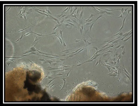
PDL cells grew out of the PDL tissues isolated from the freshly extracted teeth. On average PDL cells grew from 57% of the 30 teeth collected. Cell growth from the tissues was seen anywhere from five days to two weeks from the initial PDL extraction. Marked cell growth was noticed from extracted premolars compared to third molars. No PDL cells grew from the carious or periodontally involved teeth. Teeth not immediately placed in media, resulted in a very low success of PDL cell growth. The plated tissues were kept and their media replaced regularly for up to a month. If no growth was seen at that point, they were discarded. Under light microscopy at 10X magnification, the cells were spindle or stellate shaped with a single large nucleus, resembling the morphology of a typical fibroblast. Pictures were taken of the first cells to grow from the PDL tissue (Fig. 1).When confluent, the cells were arranged in parallel arrays. Once cells in the second to third passage looked approximately 90 percent confluent they were used for magnetic bead isolation.
Magnetic Bead Isolation
[31]
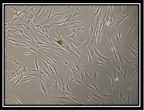
and grown as well. Figure 2 shows a phase contrast microscopic image of STRO-1 positive cells with bound beads creating a colony-forming unit. The cell is multipolar, stellate shaped, and able to form colonies. Figure 3 shows a phase contrast microscopic image of bead isolated aged STRO-1 positive cells (P 11) displaying discernible changes in morphology.
Immunofluorescence
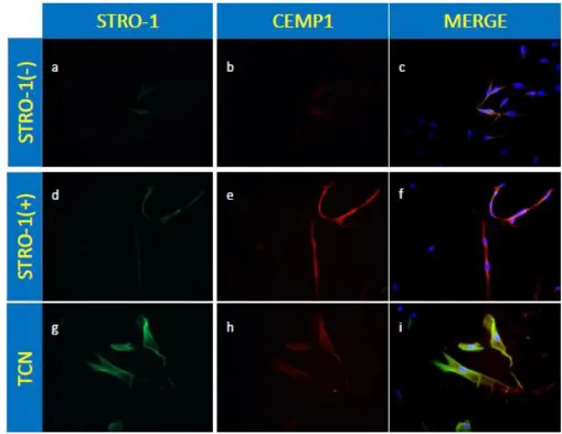
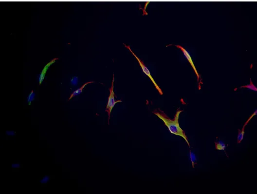
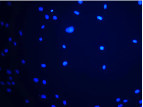
In addition to magnetic bead isolation, immunocytochemistry was performed to further show that the magnetic bead isolated cells were positive for the STRO-1 and CEMP1 marker. STRO-1 expression was identified with green fluorescence (FITC). CEMP1 expression was identified with red fluorescence (Texas Red). The isolated cell population expressed the cell surface molecule STRO-1 in both young (P 3) and aged (P 11) cells. A portion of the parental PDL cells stained green, while almost none of the Supernatant or STRO-1 negative cells stained green. Also note, the merged images w/ FITC and Texas Red as a yellowish green color indicating cells positive for STRO-1 are simultaneously positive for CEMP1 displaying expression for both markers in both young and aged cells (Fig. 4-6).
Western blot
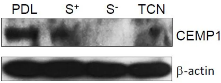
[32]
in parental PDL, STRO-1 positive and cementoblastoma (TCN) cells but not in STRO-1 negative cells. Bands are observed for CEMP1 at approximately 50 KDa as expected. All cell strain samples were normalized to Beta-actin with bands visible at approximately 42 KDa (Fig. 7).
Osteogenic/Cementogenic Induction
[33]
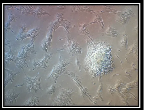
STRO-1 negative cells however maintained their confluency and parallel array. It was also noted that nodule formation appeared to increase in aged PDL, STRO-1/CEMP1 positive, STRO-1 negative and TCN cells. Images for young and aged parental PDL, STRO-1/CEMP1 positive, STRO-1 negative and cementoblastoma cells are shown in figures 8 and 9 respectively.
[34]
Messenger RNA Expression
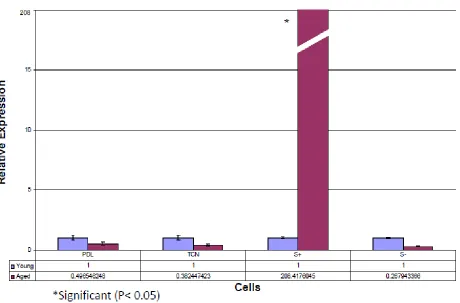
(Baseline Study and Time Course Study)A baseline expression of CEMP1, osteopontin, and collagen I in ‘young’ and ‘aged’ parental PDL, STRO-1/CEMP1 positive, STRO-1 negative, and cementoblastoma (TCN) cells was evaluated and normalized to the housekeeping gene glyceraldehyde 3-phosphate dehydrogenase (GAPDH). Comparing young versus aged cells, the results revealed a 208-fold upregulation of CEMP1 expression in aged STRO-1/CEMP1 positive cells (P< 0.05). Also revealed was a downregulation of CEMP1 expression in aged parental PDL, STRO-1 negative, and TCN cells which was not considered significant (P> 0.05). Relative expression for osteopontin showed a 10.2- and 12.9-fold upregulation in both aged PDL and STRO-1 positive cells respectively (P< 0.05), while a down regulation occurred in both TCN and STRO-1 negative cells which was not significant (P> 0.05). Relative expression for collagen 1 showed a 2.1-, 3.0- and 2.0-fold upregulation in aged PDL, STRO-1 positive and STRO-1 negative cells respectively (P< 0.05). A downregulation of expression for collagen 1 in aged TCN cells was noted as well however this was not considered significant (P> 0.05), (Fig. 10-12).
[35]
[36]
[37]
[38]
Chapter 4-Discussion
Despite the many methods proposed for periodontal regeneration ie; guided tissue
regeneration, guided bone regeneration, the use of membranes, bone morphogenetic proteins, and enamel matrix proteins, none have targeted cementogenesis specifically and sought to reliably initiate the regeneration of cementum and subsequent attachment of the
periodontium.Part of this is due to the lack of knowledge about the process of cementogenesis and about the potent capability of cementoblasts with particular interest to their precursors or origin. Unpublished work by Aminlari et al. 2011, has confirmed with the use of cementum protein 1 (CEMP1) as a cementum marker, that STRO-1/CEMP1 double positive cells from the PDL appear to be possible precursors to cementoblasts. Their study went on to demonstrate successful expression of both markers, STRO-1 and CEMP1 on these double positive cells via immunofluorescence as well as compositional data suggesting that the mineralized-like tissue formed by these osteogenically induced double positive cells displayed a Ca/P ratio of 1.89 which is very close to cellular cementum at 1.97 (Arzate et al. 2000). In light of this nonetheless, mRNA expression levels for CEMP1 however were highest in the osteogenically induced
[39]
protein 1, but possibly higher levels as well. Aminlari was able to conclude that STRO-1/CEMP1 double positive cells from the PDL exhibit characteristics consistent with precursors to
cementoblasts.
In this study, we investigated the effect of cellular aging on the temporal expression of CEMP1. This was organized as an initial baseline and then subsequent time course experiment including four different cell strains in the baseline study and an additional cell strain or a total of five different cell strains in the time course study. The cell strains included: parental periodontal ligament cells, cementoblastoma cells (TCN), STRO-1/CEMP1 double positive cells (CEMP1 positive), STRO-1 double negative cells and human mesenchymal stem cells (hMSC). We
[40]
The culturing, maintenance and aging of any cell strain can be challenging. Multiple variables coupled with operator error may wreak havoc with the simple plating of a specific cell strain or the ongoing maintenance of a particular time course experiment prior to its fruition. Hayflick defines and clarifies the terms “cell line” and “cell strain” and would argue the term “cell line” to be inapplicable to the type of cells described in this study. The term “cell line” is confined to only those cells grown in vitro for extended periods of time (years), which as a result, presumes potential “immortality” when serially cultivated in vitro. The cells described in this study are therefore assumed to lack this characteristic of potential immortality and exclude them from being referred to as “cell lines” rather, a more appropriate term such as “cell
strains.” A cell strain therefore, is a population of cells derived from animal tissue, subcultivated more than once in vitro, and lacking the property of indefinite serial passage while preserving the chromosomal karyotype characterizing the tissue of origin (Hayflick et al., 1961). Once decidedly operating in full force with a particular cell strain, the method of ‘aging’ this cell strain may be achieved in several ways. A cell may age through multiple passages or through a
substantial amount of time that it takes to achieve confluency, not to mention the stresses induced to the cell during the process.
[41]
[42]
difficult to maintain. It is believed that the cultured cementum tumor cells (TCN cells) in this study would be useful for at least three reasons: First, they could be used to obtain sufficient quantities of biologically active cementum components as this has been difficult so far due to the paucity of cementum in human teeth and labor intensive dissection procedures necessary. Second, the cells could be used to study the biosynthesis and processing of CAP and other cementum proteins. Third, early cementogenesis is believed to involve deposition of extracellular matrix produced by Hertwig’s epithelial root sheath (HERS) cells on dentin,
disruption of the HERS, migration and attachment of ectomesenchymal cells from dental follicle on to the matrix, their differentiation into cementoblasts and laying down of cementum (Arzate et al., 1992).
[43]
[44]
destruction, allows the investigator to admire the potential of these cells in an otherwise healthy environment as they age. Perhaps the STRO-1 negative cells which are theoretically CEMP1 negative because they express minimal to no CEMP1, and also express no upregulation of collagen in aged cells, require CEMP1 as a signaling agent or vehicle not only for mineral production, but regeneration and repair as well. We see the same influential shift for TCN cells as they continue to age through the time course experiment. Increased collagen 1 expression with the aged TCN cells is observed as time progresses, possibly due to the upregulation of CEMP1 occurring at the same time at the same shift point between 24 and 72 hours. It is at this time point and thereafter that we notice a significant upregulation of collagen 1 for induced aged cells (4 fold at 72hr and 5 fold at one week).
It might be suggested that Collagen 1 and CEMP1 are tied together sharing a common pathway with one requiring the other, and as the STRO-1/CEMP1 positive cell ages, this
pathway is either initiated or better yet, upregulated. Perhaps for the STRO-1 negative cell, this pathway no longer functions as the cell ages; due to possibly a manifestation of the Collagen 1 and CEMP1 expression no longer being tied together. Aminlari showed that when isolating STRO-1 positive PDL cells through magnetic bead isolation, only approximately 1.4% of the total cells along with a smaller percentage bound to the Dynabeads, were considered STRO-1
[45]
cementoblastomas; therefore a ‘negative feedback’ type of regulation must prevail. This would allow for cementum deposition with age, but not to the point of pathologic deposition. Also of interest to note, the untreated control aged human mesenchymal stem cells at one week revealed a 10 fold upregulation of collagen however, inducing the same aged cells by one week also revealed a 45 fold increase in collagen secretion. They also show a progressive
upregulation of CEMP1 (aged vs young) throughout the time course to nearly a 2 fold increase by one week as well which draws attention once again to both CEMP1 and Collagen 1 being tied together in a common pathway. The hMSC cells, which are less differentiated than a PDL or cementoblastoma cell, show tremendous potential to differentiate into collagen secreting and mineral producing cells especially in the aged state; perhaps in a regenerative or repair
capacity. This is indeed revealed after one week of induction with osteogenic media. Osteopontin (OPN) is a molecule associated with early stages of mineralization. Its
expression by cementum tumor cells (TCN cells) suggests that this molecule could be associated with the initial process of mineralization observed in these particular cells. The synthesis of this molecule may be regarded as the expression of a cell with an osteoblast-like phenotype and confirms its role as one of the molecules regulating the process of cementogenesis (Arzate et al., 1998). An upregulation of OPN is demonstrated for aged PDL, STRO-1/CEMP1 positive and STRO-1 negative cells however at one week a shift occurs in the ability to express OPN for STRO-1 negative cells with osteogenic media compared with STRO-1/CEMP1 positive cells. It is important to bear in mind that OPN is an early maker (osteogenic marker) in mineral
[46]
Untreated young and aged cells up to 72 hours revealed a progressive increase in OPN expression and then began to decrease thereafter. The cementoblastoma cells did in fact respond but at different times and to differential treatment. Perhaps a ‘generic’ type of osteogenic treatment as was used, may actually suppress OPN in these cells especially as they age. This might be necessary again as a possible type of negative feedback or rather, these terminally differentiated cells simply prefer laying down cementum rather than a bone-like mineralized material and the osteogenic media was quite possibly inhibiting the process. Lee et al evaluated the effects of aging on human dental pulp cells (HDPCs) with regard to
dentinogenesis and observed a decrease in OPN expression with aging. To better understand the mechanism of the aging process, stress-induced premature senescence (SIPS) was
performed by treating the HDP cells with H2O2. Odontogenic and osteogenic functions impaired
by senescence were assayed via several experiments including Western blotting, reverse transcriptase polymerase chain reaction and Alizarin Red S staining. Results of oxidative stress-mediated premature senescence with simple replicative senescence appeared to be similar manifesting a decrease of OPN expression (Lee et al., 2013). Environmental stresses can markedly differ from the pulp to the periodontium however, with one influencing the other as well. Inflammation, oxidative stress along with replicative senescence provided an environment in vitro that produced such results for Lee and his group; an environment that was not created in our study. To summarize for OPN expression, the STRO-1/CEMP1 positive cells were very responsive to mineralization treatment while the cementoblastoma cells were
[47]
The Arzate group not only showed CEMP1 regulates mineral deposition as demonstrated with anti-cementoblastoma protein antibodies thereby decreasing OPN and BSP levels, but they also showed using immunolocalization that this cementoblastoma protein was expressed
specifically in the precementum of acellular and cellular cementum (Arzate et al., 1998, 2006). The concept of CEMP1 regulating mineral deposition is therefore not new, nor is the concept of increased cementum deposition with aging. Kuttler studied the microscopic basis of the
anatomy of the dental apex using 402 extracted teeth from patients ranging in age with two groups: 18 - 25 and 55 – death. They concluded that the foramen of the root deviates more and more from the apical center with an increase of age caused by a thickening of the apical
cementum. Also the diameter of the foramen increased with age due to the apposition of new layers of cementum (Kuttler, 1955). Determinations of cementum thickness on 233 single-rooted teeth with healthy supporting tissues showed a straight-line relationship between age and cementum thickness. The thickness of cementum was approximately tripled between the ages of 11 and 76 years. This occurred more in the apical region and less around the
[48]
PDL, STRO-1/CEMP1 positive, and cementoblastoma cells, but not consistently or at all in hMSC, and STRO-1 negative cells. It was interesting to observe via spectrophotometry, significantly more nodule formation with Alizarin Red S staining for aged STRO-1 negative cells compared to young STRO-1 negative cells however not similarly with aged STRO-1/CEMP1 positive, PDL, and TCN cells despite the visibly increased staining. This may be due in part, to the differences in osteoinduction time of one week for the time course vs two weeks of induction for the Alizarin Red S staining. Confining the aged cells to a 6 well plate for two weeks could have dramatically altered the capabilities of these particular cell strains and perhaps warrants further
investigation.
[49]
[image:49.612.72.542.176.537.2]Tables and Figures
[50]
[51]
[52]
[53]
[54]
[55]
[56]
[57]
[58]
Figure 10: Baseline trial of real time PCR showing Cementum Protein 1 (CEMP1) mRNA relative expression comparing young (P 3-4) versus aged (P 11-13) cells in four different cell strains
[59]
[60]
[61]
[62]
[63]
[64]
[65]
[66]
[67]
Fig. 16: Time course trial of real time PCR showing Osteopontin expression for young and aged PDL, STRO-1/CEMP1 positive, TCN, STRO-1 negative and hMSC cells. Relative expression (Y-axis) evaluated from 0hr through one week (X-axis) comparing control cells (UT) and
[68]
[69]
Table 5: Fold increase/decrease and P value
[70]
[71]
[72]
[73]
References:
Alvarez Perez, M. A., S. Pitaru, et al. (2003). "Anti-cementoblastoma-derived protein antibody partially inhibits mineralization on a cementoblastic cell line." Journal of Structural Biology 143(1): 1-13.
Alvarez-Perez, M. A., S. Narayanan, et al. (2006). "Molecular cloning, expression and immunolocalization of a novel human cementum-derived protein (CP-23)." Bone 38(3): 409-19.
Arzate, H., M. A. Alvarez-Perez, et al. (1998). "Human cementum tumor cells have different features from human osteoblastic cells in vitro." Journal of Periodontal Research
33(5): 249-58.
Arzate, H., M. A. Alvarez-Perez, et al. (2000). "Electron microscopy, micro-analysis, and X-ray diffraction characterization of the mineral-like tissue deposited by human cementum tumor-derived cells." Journal of Dental Research 79(1): 28-34.
Arzate, H., L. F. Jimenez-Garcia, et al. (2002). "Immunolocalization of a human cementoblastoma-conditioned medium-derived protein." Journal of Dental Research 81(8): 541-6.
Arzate, H., S. W. Olson, et al. (1992). "Isolation of human tumor cells that produce cementum proteins in culture." Bone & Mineral 18(1): 15-30.
Ben-Porath, I., Weinberg, R. (2005). “The signals and pathways activating cellular senescence.” The International Journal of Biochemistry & Cell Biology 37: 961-976.
Bosshardt, D. D. and H. E. Schroeder (1996). "Cementogenesis reviewed: a comparison between human premolars and rodent molars." Anat Rec 245(2): 267-92.
Both, S. K., A. J. van der Muijsenberg, et al. (2007). "A rapid and efficient method for expansion of human mesenchymal stem cells." Tissue engineering 13(1): 3-9.
Carmona-Rodriguez, B., M. A. Alvarez-Perez, et al. (2007). "Human Cementum Protein 1 induces expression of bone and cementum proteins by human gingival fibroblasts." Biochemical & Biophysical Research Communications 358(3): 763-9.
Cuisinier, F. J., R. W. Glaisher, et al. (1991). "Compositional variations in apatites with respect to preferential ionic extraction." Ultramicroscopy 36(4): 297-305.
Engler, W. O., S. P. Ramfjord, et al. (1966). "Healing following simple gingivectomy. A tritiated thymidine radioautographic study. I. Epithelialization." Journal of periodontology
37(4): 298-308.
Fuji, S., Maeda, H., et al. (2008). “Investigating a clonal human periodontal ligament progenitor/stem cell line in vitro and in vivo.” J Cell Physiol 215: 743-749.
Gomez Flores, M., Hasegawa, M, et al. (2008). “Cementum-periodontal ligament complex regeneration using the cell sheet technique.” J Periodont Res 43: 364-371.
Goseki, T., Shimizu, N., et al. (1996). “Effects of in vitro cellular aging on alkaline phosphatase, cathepsin activities and collagen secretion of human periodontal ligament derived cells.” Mechanisms of Ageing and Development 91: 171-183
[74]
Hayami, T., Q. Zhang, et al. (2007). "Dexamethasone's enhancement of osteoblastic markers in human periodontal ligament cells is associated with inhibition of collagenase expression." Bone 40(1): 93-104.
Hayflick, L. (1977). “The cellular basis for biological aging.” Handbook of the biology of aging, Van Nostrand Reinhold, New York; 159-179
Hayflick, L., Moorhead, P. S. (1961). “The serial cultivation of human diploid cell strains.” Experimental Cell Research 25: 585-621.
Heijl, L., G. Heden, et al. (1997). "Enamel matrix derivative (EMDOGAIN) in the treatment of intrabony periodontal defects." Journal of clinical periodontology 24(9 Pt 2): 705-14.
Itaya, T., H. Kagami, et al. (2009). "Characteristic changes of periodontal ligament-derived cells during passage." Journal of periodontal research 44(4): 425-33.
Iwata, T., M. Yamato, et al. (2010). "Validation of human periodontal ligament-derived cells as a reliable source for cytotherapeutic use." Journal of Clinical Periodontology 37(12): 1088-1099.
Komaki, M., K. Iwasaki, et al. (2011). “Cementum Protein 1 (CEMP1) induces a cementoblastic phenotype and reduces osteoblastic differentiation in periodontal ligament cells.” Journal of Cellular Physiology 227: 649-657.
Krebsbach, P., Robey, P., (2002), “Dental and skeletal stem cells: Potential cellular therapeutics for craniofacial regeneration.” Journal of Dental Education 66 (6): 766-773.
Kuttler, Y. (1955). “Microscopic investigation of root apexes.” Journal of the American Dental Association 50(5): 544-552.
Lee, Y-H., G-E. Kim, et al. (2013). “Aging of in vitro pulp illustrates change of inflammation and dentinogenesis.” Journal of Endodontics 39: 340-345
Lenart, G., G. Bidlo, et al. (1968). "Use of x-ray diffraction method in investigations on mineral substances of bone and callus." Acta biochimica et biophysica; Academiae Scientiarum Hungaricae 3(3): 306-16.
Lin, N. H., D. Menicanin, et al. (2008). "Putative stem cells in regenerating human periodontium." Journal of Periodontal Research 43(5): 514-23.
Lindskog, S. (1982). "Formation of intermediate cementum. II: a scanning electron microscopic study of the epithelial root sheath of Hertwig in monkey." Journal of craniofacial genetics and developmental biology 2(2): 161-9.
Livak, K. (2001). "Analysis of Relative Gene Expression Data Using Real-Time Quantitative PCR and the 2−ΔΔCT Method." Methods 25(4): 402-408.
Marchesan, J. T., C. S. Scanlon, et al. (2011). "Implications of cultured periodontal ligament cells for the clinical and experimental setting: A review." Archives of oral biology.
McCulloch, C. A. (1985). "Progenitor cell populations in the periodontal ligament of mice." Anat Rec 211(3): 258-62.
Nanci, A. and D. D. Bosshardt (2006). "Structure of periodontal tissues in health and disease." Periodontol 2000 40: 11-28.
[75]
Potten, C. S. (1981). "Cell replacement in epidermis (keratopoiesis) via discrete units of proliferation." Int Rev Cytol 69: 271-318.
Rohme D. (1981). “Evidence for a relationship between longevity of mammalian species and life spans of normal fibroblasts in vitro and erythrocytes in vivo.” Proc Natl Acad Sci USA 78
(8): 5009-5013.
Ruparel, N., B., J. Affonso de Almeida. (2013). “Characterization of a stem cell of apical papilla cell line: Effect of passage on cellular phenotype.” Journal of Endodontics 39: 357-363.
Saito, E., A. Saito, et al. (2003). "Favorable healing following space creation in rhBMP-2-induced periodontal regeneration of horizontal circumferential defects in dogs with experimental periodontitis." Journal of periodontology 74(12): 1808-15.
Saito, M., M. Iwase, et al. (2001). "Expression of cementum-derived attachment protein in bovine tooth germ during cementogenesis." Bone 29(3): 242-8.
Saygin, N. E., W. V. Giannobile, et al. (2000). "Molecular and cell biology of cementum." Periodontol 2000 24: 73-98.
Seo, B. M., M. Miura, et al. (2004). "Investigation of multipotent postnatal stem cells from human periodontal ligament." Lancet 364(9429): 149-55.
Simmons, P. and B. Torok-Storb (1991). "Identification of stromal cell precursors in human bone marrow by a novel monoclonal antibody, STRO-1." Blood 78(1): 55-62.
Stein, Thomas John. (1990). “Anatomy of the root apex and its histologic changes with age.” Oral Surgery, Oral Medicine and Oral Pathology 69(2): 238-242.
Stewart, K., S. Walsh, et al. (1999). "Further characterization of cells expressing STRO-1 in cultures of adult human bone marrow stromal cells." Journal of Bone & Mineral Research
14(8): 1345-56.
Villarreal-Ramirez, E., A. Moreno, et al. (2009). "Characterization of recombinant human cementum protein 1 (hrCEMP1): primary role in biomineralization." Biochemical & Biophysical Research Communications 384(1): 49-54.
Wu, D., Ikezawa, K., et al. (1996). “Characterization of a collagenous cementum-derived attachment protein.” Journal of Bone and Mineral Research 11(5): 686-692.
Xu, J., W. Wang, et al. (2009). "Multiple differentiation capacity of STRO-1+/CD146+ PDL mesenchymal progenitor cells." Stem Cells & Development 18(3): 487-96.
Yoshikawa, G., Y. Murashima, et al. (2002). "Guided bone regeneration (GBR) using membranes and calcium sulphate after apicectomy: a comparative histomorphometrical study." International endodontic journal 35(3): 255-63.
Zander, H.A., and Beat Hurzeler. (1958). “Continuous Cementum Apposition.” Journal of Dental Research 37(5): 1035-1044.
Zeichner-David, M., K. Oishi, et al. (2003). "Role of Hertwig's epithelial root sheath cells in tooth root development." Developmental Dynamics 228(4): 651-63.