Mobilization and homing of peripheral blood
progenitors is related to reversible
downregulation of alpha4 beta1 integrin
expression and function.
F Prosper, … , J B McCarthy, C M Verfaillie
J Clin Invest.
1998;
101(11)
:2456-2467.
https://doi.org/10.1172/JCI188
.
Despite the wide use of mobilized peripheral blood (PB) progenitor cells (PBPC) for clinical
transplantation the mechanism(s) underlying their mobilization and subsequent engraftment
are still unknown. We compared the adhesive phenotype of CD34(+) colony-forming cells
(CFC) in bone marrow (BM) and PB of normal donors before and after administration of
granulocyte colony-stimulating factor (G-CSF) for 5 d. G-CSF-mobilized PB CFC cells
adhered significantly less to BM stroma, fibronectin, and to the alpha4 beta1 binding
fibronectin peptide, CS1, because of decreased expression of the alpha4 integrin. Since
incubation of BM CD34(+) cells for 4 d with CSF at concentrations found in serum of
G-CSF- treated individuals did not affect alpha4-dependent adhesion, G-CSF may not be
directly responsible for the decreased alpha4-mediated adhesion of PB CFC. Culture of
G-CSF-mobilized PB CD34(+) cells with cytokines at concentrations found in BM stromal
cultures upregulated alpha4 expression and restored adhesion of mobilized PB CFC to
stroma, fibronectin, and CS1. Adhesion of cultured, mobilized PB CFC to stroma and CS1
could not be further upregulated by the beta1 activating antibody, 8A2. This indicates
acquisition of a maximally activated alpha4 beta1 integrin once PB CFC have been
removed from the in vivo mobilizing milieu. Thus, decreased alpha4 expression on CD34(+)
CFC in PB may be responsible for the aberrant circulation of mobilized PB CD34(+) cells.
Reexpression of […]
Research Article
Find the latest version:
J. Clin. Invest.
© The American Society for Clinical Investigation, Inc. 0021-9738/98/06/2456/12 $2.00
Volume 101, Number 11, June 1998, 2456–2467 http://www.jci.org
Mobilization and Homing of Peripheral Blood Progenitors Is Related to Reversible
Downregulation of
a
4
b
1 Integrin Expression and Function
Felipe Prosper, David Stroncek,* James B. McCarthy,* and Catherine M. Verfaillie
Stem Cell Biology Program and Division of Hematology, Department of Medicine, and *Department of Laboratory Medicine and Pathology, University of Minnesota, Minneapolis, Minnesota 55455
Abstract
Despite the wide use of mobilized peripheral blood (PB) progenitor cells (PBPC) for clinical transplantation the mechanism(s) underlying their mobilization and subsequent engraftment are still unknown. We compared the adhesive phenotype of CD341 colony-forming cells (CFC) in bone marrow (BM) and PB of normal donors before and after ad-ministration of granulocyte colony-stimulating factor (G-CSF) for 5 d. G-CSF–mobilized PB CFC cells adhered significantly less to BM stroma, fibronectin, and to the a4b1 binding fi-bronectin peptide, CS1, because of decreased expression of the a4 integrin. Since incubation of BM CD341 cells for 4 d with G-CSF at concentrations found in serum of G-CSF– treated individuals did not affect a4-dependent adhesion, G-CSF may not be directly responsible for the decreased
a4-mediated adhesion of PB CFC. Culture of G-CSF–mobi-lized PB CD341 cells with cytokines at concentrations found in BM stromal cultures upregulated a4 expression and re-stored adhesion of mobilized PB CFC to stroma, fibronec-tin, and CS1. Adhesion of cultured, mobilized PB CFC to stroma and CS1 could not be further upregulated by the b1 activating antibody, 8A2. This indicates acquisition of a maximally activated a4b1 integrin once PB CFC have been removed from the in vivo mobilizing milieu. Thus, decreased
a4 expression on CD341 CFC in PB may be responsible for the aberrant circulation of mobilized PB CD341 cells. Reex-pression of a maximally activated a4b1 integrin on mobi-lized PB CFC removed from the mobilizing in vivo milieu may contribute to the early engraftment of mobilized PBPC. (J. Clin. Invest. 1998. 101:2456–2467.) Key words: mobiliza-tion • homing • bone marrow transplant • growth factors •
adhesion receptors
Introduction
Studies demonstrating the presence of hematopoietic progeni-tors with long-term lymphomyeloid repopulating ability in the peripheral blood (PB)1 have led to the use of PB progenitor
cells (PBPC) in transplantation (1–6). Several stimuli including
stress, exercise, endotoxin, dextran-sulfate, and chemotherapy can increase the number of circulating progenitors (7–11). Likewise, administration of cytokines such as granulocyte colony-stimulating factor (G-CSF), granulocyte-macrophage (GM)-CSF, or stem cell factor (SCF) greatly increases the frequency of CD341 cells, committed colony-forming cells (CFC), and more primitive progenitors in the blood (9, 12, 13). Since a small number of progenitors can be found in the PB under steady state conditions, mobilization could be seen as an exaggeration of an otherwise physiological process. Alterna-tively, mobilization may be caused by specific changes in the interaction between progenitors and the microenvironment or changes in the microenvironment itself (14–17).Currently, it is unknown if cytokines act by expanding the pool of available bone marrow (BM) progenitors and their subsequent release in the circulation, by expanding a specific subpopulation of marrow progenitors which interact less strongly with the mar-row microenvironment or by expanding and mobilizing pro-genitors present outside of the marrow (18).
Under steady state conditions, hematopoietic progenitors are mainly found in close contact with the marrow microenvi-ronment (19). Steady state BM CD341 cells express multiple adhesion receptors including the a4, a5, b1, CD11a/CD18 and CD11b/CD18 integrins, L-selectin, PECAM-1, and CD44 (16, 20–23). We and others have demonstrated in vitro that adhe-sion of BM progenitors to BM stroma is at least in part due to the interaction of the a4b1 and a5b1 integrins with stromal ex-tracellular matrix (fibronectin) and cell surface expressed ligands (VCAM) (24–27). In vivo studies have demonstrated a dominant role for the a4b1 integrin in the interaction between progenitors and the BM microenvironment. Repopulating stem cells fail to engraft when murine or sheep BM is incu-bated with anti-a4 antibodies before transplantation, and the number of progenitors found in the PB increases dramatically after infusion of anti-a4 antibodies into baboons (15, 28, 29). Several studies have examined cell adhesion receptor expres-sion on CD341 cells present in the PB after mobilization with cytokines or chemotherapy (14). Decreased levels of the a4 and CD11a/CD18 integrins and increased levels of the CD62L receptor have been described (16). In addition, several studies have demonstrated that incubation in vitro with GM-CSF, IL-3, and SCF may affect the adhesive behavior of progenitors by altering the affinity status of adhesion receptors (30–32). How-ever, whether changes in receptor expression and/or function are functionally relevant and are responsible for mobilization of progenitors is unknown. Since cytokines may also up- or downregulate expression of adhesive ligands in the
extracellu-Address correspondence to Catherine M. Verfaillie, M.D., Depart-ment of Medicine, Box 806, 420 Delaware St. SE, Minneapolis, MN 55455. Phone: 612-624-3921; FAX: 612-626-4074; E-mail: verfa001@ maroon.tc.umn.edu
Received for publication 20 March 1997 and accepted in revised form 31 March 1998.
lar matrix or on stromal cells, alterations in the microenviron-ment may promote premature release of progenitors in the PB (25, 33, 34).
It is even less clear how progenitors that were mobilized in the PB after administration of cytokines can result in the clini-cally observed early engraftment after transplant. Engraftment requires that progenitors home to the BM microenvironment. This depends at least in part on adhesions through the a4b1 in-tegrin (28, 35–37). Since alterations in the expression and/or function of the same a4b1 integrin may be involved in the mo-bilization of progenitors in the PB, one needs to assume that a fast reversal of the nonadhesive phenotype of mobilized he-matopoietic progenitors that have been removed from the mo-bilizing milieu must underlie their potential for fast engraft-ment.
In this study, we examined the effect of mobilization with G-CSF on progenitors that remain in the BM as well as on pro-genitors that are induced to circulate in the PB. We demon-strate that decreased expression, but not change in function, of the a4b1 integrin is responsible for decreased adhesion of PB progenitors to BM stroma, fibronectin, and CS1. This may ex-plain the premature circulation of progenitors in the PB after in vivo administration of G-CSF. We also demonstrate, in an in vitro system, that a4 expression and function is upregulated on PB CD341 cells, once removed from the in vivo milieu. This is associated with increased adhesion of PB CFC to BM stroma, fibronectin, and CS1 which in turn may be responsible for homing and engraftment of PBPC.
Methods
Samples
Donors. Normal healthy donors were selected using standard criteria of the American Association of Blood Banks for blood donors (38). Informed consent was obtained using guidelines approved by the Committee on the Use of Human Subjects for Research at the Uni-versity of Minnesota. All donors had a negative serologic test for HBsAg, anti-HCV, and anti-HIV and they had normal hemoglobin, white blood cell counts, white cell differentials, and platelet counts.
PBPC. Normal donors received a daily dose of 10 mg/kg per day of human recombinant G-CSF (Neupogen; Amgen, Inc., Thousand Oaks, CA) subcutaneously for 5 d (days 1–5). G-CSF was given as a single morning dose. 60 ml of blood was obtained by venopuncture from each donor before the first injection of G-CSF (day 0) and on day 16.
BM. 50 ml of heparinized BM was obtained from each normal donor before the first injection of G-CSF (day 0) and on day 16.
Processing of samples
Steady state PB and BM as well as G-CSF–mobilized PB and BM hematocrit white blood cell and differential count were determined using an automated cell counter (S1IV; Coulter Electronics Inc., Hialeah, FL). Flow cytometry, clonogenic assays, and long-term cul-tures were performed on steady state and G-CSF–mobilized PBPC, as well as BM obtained before and after treatment with G-CSF.
Cell selection. Steady state and G-CSF–mobilized PB and BM mononuclear cells (MNC) were separated by Ficoll Hypaque centrif-ugation (specific gravity, 1077) (Sigma Chemical Co., St. Louis, MO). CD34 enrichment was performed using the MACS® CD34 isolation
kit (Miltenyi Biotec Inc., Sunnyvale, CA) as described previously (12). FACS® analysis of cell surface receptors. The following antibod-ies coupled to FITC or phycoerythrin (PE) were used: mouse anti-bodies directed to CD29 (b1 integrin), CD34, CD44, CD49d (a4 in-tegrin), CD49e (a5 integrin), and CD62L (L selectin). Antibodies
purchased from Becton Dickinson (San Jose, CA) were CD44, CD62L, and CD34; CD29, CD49d, and CD49e were from Immuno-tech (Marseilles, France). Enumeration of CD341 cells in steady state or mobilized PB MNC and BM MNC was performed as described by Sutherland et al. (39).For dual-color analysis 100,000 column-selected CD341 cells or cells recovered from ex vivo culture initiated 1–4 d earlier with CD341 cells were resuspended in 100 ml of PBS plus 0.3% BSA labeled with 20 ng antiadhesion receptor antibodies in conjunction with 20 ng of anti-CD34 antibodies and incubated for 30 min at 48C, washed, and then analyzed with a FACStarPlus® flow
cy-tometer equipped with a CONSORT 32 computer. FITC- and PE-conjugated isotype-matched Igs were used as controls.
FACS® selection of CD341a4111 and CD341a41/2 cells. For FACS®
selection, CD341 cells were labeled with FITC-conjugated mouse anti-a4 and PE-conjugated mouse anti-CD34 (10 ng per 106 cells),
in-cubated for 30 min on ice, and then washed with cold PBS. Cells were selected on a FACStarPlus® laser flow cytometry system equipped
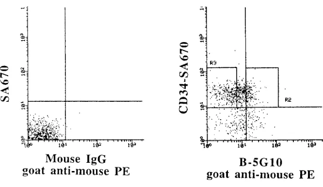
with a CONSORT 32 computer into a CD341a41/2 and CD341a4111 population based on a mouse IgG1-PE and IgG1-FITC control fluo-rescence profile. For experiments in which cells were subjected to subsequent adhesion assays, the anti-a4 antibody used for selection was a nonblocking antibody, B5G10 (40), kindly provided by Dr. M. Hemler (Harvard University, Boston, MA). Cells were stained with B5G10, washed, and stained with a goat anti–mouse IgG-PE (Tago Inc., Burlingame, CA) secondary antibody washed and stained se-quentially with anti-CD34–biotin (CellPro) and streptavidin-670 (SA-670; Life Technologies, Gaithersburg, MD). CD341a42 and CD341a4111cells were selected.
Progenitor cultures
Short-term methylcellulose progenitor culture. CD341 cells (2 3 103
cells/ml), MNC (2 3 105 cells/ml), or adherent and nonadherent
frac-tions from adhesion assays were cultured in methylcellulose as de-scribed (12) in the presence of SCF (Amgen, Inc.), GM-CSF (Immu-nex, Seattle, WA), and IL-3 (R&D Systems, Minneapolis, MN) each at a final concentration of 5 ng/ml and erythropoietin (Amgen, Inc.) 3 IU/ml.
Long-term culture. 2 3 106 PB or BM MNC (22 replicates:
61,224; 20,408; 6,802; and 2,267 cells/well) and 30,000 CD341 cells (22 replicates: 900, 300, 100, and 33 cells/well) were plated in limiting di-lutions onto previously irradiated M2-10B4 feeders in long-term BM culture (12, 24). After 8 wk, clonogenic culture medium was added and wells were scored for the presence or absence of secondary CFC 14 d later as described (12, 24). The absolute number of long-term culture-initiating cells (LTC-IC) present in the different cell popula-tions was calculated as described (41, 42).
Purification and synthesis of fibronectin and peptides from fibronectin
Human plasma fibronectin was purified as a by-product of Factor VIII production by sequential ion exchange and gelatin chromatogra-phy (43). Peptides from fibronectin were synthesized at the Micro-chemical Facility of the University of Minnesota (44–46). Peptide CS1 which has the sequence DELPQLVTLPHPNLHPGEILDVPST (47) was chemically conjugated to ovalbumin (44, 46).
Cell adhesion assays
MNC and CD341 cells from PB and BM were resuspended in serum-free IMDM and plated on preestablished, irradiated BM stromal lay-ers for 2 h. BM stroma was established from normal human marrow donors, as previously described. Once confluent, feeders were irradi-ated at 1,250 rad, and subcultured in wells of 24-well plates (24). Non-adherent cells were harvested by three gentle washes with warm IMDM and adherent cells were recovered after short-term trypsiniza-tion, as described (24).
CD341 cells from PB and BM or CD341a42 and CD341a4111 cells from Mob.PB were resuspended in serum-free IMDM and plated in contact with fibronectin or CS1 for 3 h in a humidified atmosphere at 378C. Nonadherent cells were removed by four standardized washes using warm IMDM and adherent cells by trypsinization (46).
To determine the percentage of adherent CFC, adherent and nonadherent fractions were replated in methylcellulose assays (24, 46). Percent adhesion was calculated as: (number of CFC in the ad-herent fraction)/(number of CFC in adherent 1 nonadherent frac-tions) 3 100.
Adhesion blocking experiments. In some experiments, PB and BM CD341 cells were incubated with 8A2 (1:100,000 dilution), P4C2 (1:400 dilution), P4C10 (1:400 dilution), P1D6 (1:400) control mouse IgG, or with media alone for 30 min before adhesion assays (48, 49). Monoclonal antibody, 8A2 (a kind gift from Dr. Nicholas Kovach, University of Washington, Seattle, WA), was used as a dilution of mouse ascites. The integrin-blocking monoclonal antibodies P4C2 (murine anti–human a4), P4C10 (murine anti–human b1), and P1D6 (murine anti–human a5) were purchased from GIBCO-BRL (Gai-thersburg, MD) and used as dilutions of mouse ascites (48). Control mouse IgG (Sigma Chemical Co.) was diluted in PBS and used at a concentration of 20 mg/ml.
Serum-free medium culture
Column-selected CD341 cells were plated in IMDM containing BSA (20 mg/ml) (GIBCO BRL), insulin (10 mg/ml), transferrin (200 mg/ml) (Sigma Chemical Co.), 1024 M 2-mercaptoethanol, penicillin 100 U/ml,
and streptomycin 100 U/ml (GIBCO BRL) (50) (serum-free media). The following cytokines were added to the medium: GM-CSF (200 pg/ml), G-CSF (0–100 ng/ml), SCF (200 pg/ml) (Amgen, Inc.), LIF (50 pg/ml) (R&D Systems), MIP-1a (200 pg/ml) (R&D Systems), and IL-6 (1 ng/ml) (Genetics Institute, Boston, MA). The concentration of the different cytokines corresponds to the physiological levels at which they are present in Dexter-type culture (51, 52). After 24 h to 7 d, cells were harvested, immunophenotyped, and used in adhesion assays.
Statistics
Results of experimental points obtained from multiple experiments were reported as the mean6SEM. Significance levels were deter-mined by two-sided Student’s t test analysis.
Results
G-CSF increases the number of progenitors present in PB but not in BM. As we and others have demonstrated (9, 12, 14,
53–56), G-CSF induced a 22-fold increase in the number of CD341 cells, a 26-fold increase in the number of CFC, and a 9-fold increase in week 8 LTC-IC per milliliter of PB in normal individuals (Fig. 1). Although a sevenfold increase in the num-ber of white blood cells was seen per milliliter of BM of nor-mal individuals after 5 d of G-CSF administration, the number of either CD341 cells, CFC, or week 8 LTC-IC per milliliter of BM did not change (Fig. 1).
Differences in adhesion receptor expression between steady state or mobilized PB or BM CD341 cells. To determine which
adhesion molecules may be involved in mobilization of he-matopoietic progenitors we examined the expression of adhe-sion receptors on CD341 cells obtained from PB or BM of nor-mal individuals before and after in vivo administration of G-CSF. No significant differences were seen in expression of
a2, a5, b1, CD11b, CD11c, or P-selectin. In vivo treatment with G-CSF increased L-selectin expression on CD341 cells present in PB or BM (percent CD341 cells positive: steady state PB: 26612% and mobilized PB: 6469%; steady state
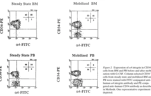
BM: 25614% and mobilized BM: 6168%; P , 0.001) and de-creased expression of the b2 integrin (CD11a/CD18) (percent CD341 cells positive: steady state PB: 5467% and mobilized PB: 4264%; steady state BM: 6265% and mobilized BM: 4464%; P , 0.05). In contrast, irrespective of G-CSF treat-ment, a4 integrin expression was lower on CD341 cells present in PB than in BM (percent CD341 cells positive: steady state PB: 3566% and mobilized PB: 2865%; steady state BM: 4764% and mobilized BM: 4564%). Aside from the fraction of CD341 cells that expressed a4 integrin, the density of a4 in-tegrin on CD341 cells (mean channel fluorescence of a4) was also lower on mobilized and steady state PB CD341 cells than on steady state or mobilized BM CD341 cells (Fig. 2).
[image:4.612.317.554.56.421.2]Because CFC represent only 10–20% of al CD341 cells, we FACS® selected CD341 cells from steady state BM and
Figure 1. Treatment of normal donors with G-CSF increases the number of progenitors in the PB but does not increase the number of progenitors in the BM. Normal donors (n 5 6) received 10 mg/kg of G-CSF daily for 5 d. BM and PB samples were obtained before treat-ment with G-CSF and 12–20 h after the last dose of G-CSF. The num-ber of CD341 cells, CFC, and LTC-IC per volume was calculated as: (number of CD341 cells, CFC, or LTC-IC per 105 MNC 3 number of MNC per ml)/105. The number of LTC-IC was evaluated after 8 wk in
G-CSF–mobilized PBPC based on their expression of a4 in-tegrin to determine if a4 integrin expression was different be-tween PB and BM CFC. In accordance with previous reports (27), . 80% of steady state BM CFC were recovered in the CD341/a4111 subpopulation. In contrast, . 80% of CFC were recovered in the CD341/a41/2 fraction of G-CSF–mobilized
[image:5.612.62.546.54.362.2]PB (Fig. 3). These studies indicate that BM CD341 cells and CFC express significantly more a4 integrin than mobilized PB CD341 cells and CFC irrespective of G-CSF treatment. This led to the hypothesis that the aberrant localization of steady
[image:5.612.315.556.444.590.2]Figure 2. Expression of a4 integrin in CD341 cells from BM and PB before and after mobili-zation with G-CSF. Column-selected CD341 cells from steady state and mobilized BM and PB were stained with FITC-conjugated anti– human a4 integrin antibody and PE-conju-gated anti–human CD34 antibody as described in Methods. One representative experiment is depicted.
Figure 3. Significantly less mobilized PB than steady state BM CFC are CD341a4111. Steady state BM and mobilized PB CD341 cells were selected by FACS® based on a4 integrin expression into
CD341a4111 and CD341a41/2 cells. Cells were then plated in meth-ylcellulose progenitor assay. The total number of CFC that express
a4 integrin was calculated as: (CFC in CD341a4111 cells) 3
(percent-age of CD341a4111 cells)/(total number of CFC in CD341 cells) 3 100. The number of CFC/2,000 plated cells was: BM 2766100 for CD341a4111 cells and 164660 for CD341a41/2 cells; mobilized PB 142644 for CD341a4111cells and 2706112 for CD341a41/2 cells. The percentage of CD341 cells present in the CD341a4111 window was 7168% for BM and 3766% for PB. *P , 0.01 comparison be-tween total CFC present in CD341a4111 and CD341a41/2 cells in steady state BM and mobilized PB (n 5 4).
[image:5.612.59.274.478.594.2]state and G-CSF–mobilized PB CD341 CFC may be related to the decrease in a4 integrin expression.
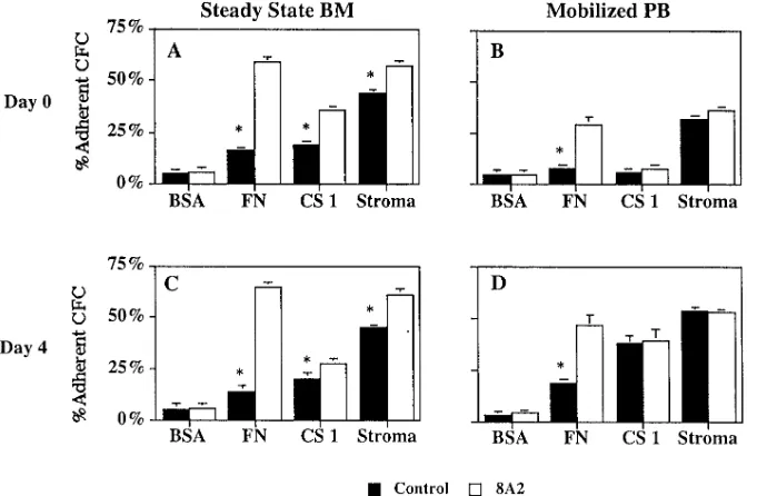
Decreased a4 integrin expression is responsible for de-creased PB CFC adhesion to stroma and fibronectin. We next
examined the capacity of CFC in PB and BM before and after treatment with G-CSF to adhere to BM stromal feeders (Fig. 4). 4764% of steady state BM CFC adhered to BM stroma, which is in accordance with our previous studies (24, 49). Like-wise, 4565% of CFC present in BM obtained after 5 d of in vivo treatment with G-CSF adhered to stromal feeders. In con-trast, only 2666 and 2965% of CFC present in PB before and after treatment with G-CSF adhered to stroma. No differences were seen in adhesion of CFC (Fig. 4) when assays were per-formed with MNC or CD341 enriched cells, suggesting that the selection with the QBEND-10 antibody did not alter the functional status of adhesion receptors.
Adhesion of steady state BM CD341 cells to BM stroma could be blocked in part by antibodies against the b1 integrin or a4 integrin but to a lesser extent by antibodies against the
a5 integrin (Fig. 5). The total number of CFC in the adherent plus nonadherent fraction was similar for cells exposed to me-dium only, mouse IgG, anti-a4, -a5, or -b1 antibodies indicat-ing that the short-term exposure to these antibodies does not affect progenitor growth in subsequent methylcellulose cul-tures. These studies confirm that adhesion of steady state BM CFC to BM stroma is mediated by a4–fibronectin or a4– VCAM interactions (25, 26). The low level adhesion of mobi-lized PB CFC to stroma was not affected by blocking antibodies against the a4, a5, or b1 integrin (Fig. 5), supporting the hy-pothesis that defects in a4b1-mediated adhesion may underlie the decreased adhesion to BM stroma and premature circula-tion of PB CFC.
[image:6.612.58.413.93.350.2]To further characterize the decreased adhesion of PB pro-genitors, we next compared the adhesion of steady state BM and mobilized PB CFC to purified fibronectin (adhesion through a4b1 and a5b1) and the fibronectin peptide CS1 cou-pled to ovalbumin (CS1-OVA) (adhesion through a4b1). Mo-bilized PB CFC adhered significantly less to fibronectin and to CS1-OVA than steady state BM CFC (P , 0.01) (Figs. 6 and
Figure 5. Role of the a4b1 integrin in steady state BM and mobilized PB CFC ad-hesion to BM stroma. Steady state BM (n 5 6) and mobilized PB (n 5 6) CD341 cells were tested either immediately after column selection (day 0 shown in A and B) or after culture ex vivo for 4 d in serum-free medium with cytokines at concentra-tions found in Dexter cultures (G-CSF at 250 pg/ml) (day 4 shown in C and D). 6,000 column-selected CD341 cells were incu-bated for 30 min with blocking anti a4 (P4C2), a5 (P1D6), or b1 (P4C10) anti-bodies, mouse IgG, or no antibody. Cells were then plated in the continued presence of the antibodies on irradiated stroma for 2 h. Adherent and nonadherent fractions were collected, washed, and replated in methylcellulose assay. The percentage of adherent CFC was calculated as described in Methods. The number of CFC per 1,000 CD341 cells on day 0 was: steady state BM 172654; mobilized PB 158632. The num-ber of CFC per 1,000 CD341 cells on day 4 was: steady state BM 214662; mobilized PB 195657. *P , 0.01 comparison between CFC adhesion in the presence of antibodies and no antibody control; #P , 0.01
compar-ison between adhesion of BM and PB CFC.
[image:6.612.314.533.494.614.2]7, A and B). In fact, adhesion of mobilized PB CFC to CS1-OVA was not significantly different than adhesion to BSA or ovalbumin (Fig. 6). In two experiments, we also showed that adhesion of mobilized BM CFC to fibronectin and OVA-CS1 is similar to that of steady state BM, but significantly higher than that of mobilized PB CFC (Table I). This confirms the notion that a4 integrin–mediated adhesion is defective in mo-bilized PB CFC but not in momo-bilized BM CFC.
Over the last 5 yr, it has become clear that the presence of an adhesion receptor on cells does not necessarily mean that
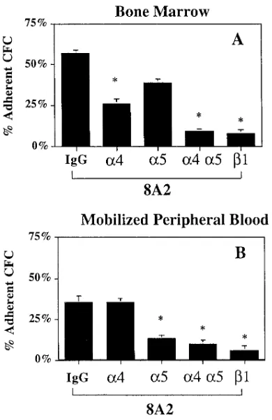
these receptors have functional relevance, i.e., they do not al-low binding and/or adhesion to ligands (31, 47, 49, 51, 56, 57). The function of integrins can be upregulated by the ligand it-self, activating antibodies, or through inside-out signals origi-nating from other adhesion receptors or cytokine signals (30, 31, 58–60). Thus, decreased integrin-mediated adhesion of mo-bilized PB CFC could be due not only to decreased a4 expres-sion but also decreased b1 integrin function. We used the b1 activating antibody 8A2 to determine if abnormal function rather than expression is responsible for the defect in PB CFC adhesion. Incubation of either steady state BM CD341 cells or mobilized BM CD341 cells with 8A2 increased adhesion of CFC to stroma, fibronectin, and CS1-OVA (Fig. 7 A and Table I). As we have reported previously, 8A2 did not increase or decrease colony growth in subsequent methylcellulose culture (not shown) (48). 8A2-induced adhesion of BM CFC to fi-bronectin could be significantly inhibited by anti-a4 or anti-b1 antibodies but not by anti-a5 antibodies. Combined addition of anti-a4 and anti-a5 antibodies resulted in almost complete abrogation of adhesion which was now similar to that seen with the anti-b1 antibody (Fig. 8). This indicates that, as for adhesion of BM CFC to stroma, the a4b1 integrin is the most important receptor for adhesion of steady state BM CFC to fi-bronectin.
Although incubation with 8A2 significantly increased ad-hesion of mobilized PB CFC to fibronectin, the percentage of mobilized PB CFC incubated with 8A2 adherent to fibronectin was still significantly lower than that of 8A2-treated BM CFC. 8A2 did not affect adhesion of PB CFC to BM stroma or CS1 (Fig. 7 B). Furthermore, unlike BM CFC, 8A2-induced adhe-sion of mobilized PB CFC to fibronectin could not be inhibited by anti-a4 blocking antibodies but was inhibited by anti-a5 or -b1 blocking antibodies (Fig. 8). These results strongly suggest that the decreased adhesion of mobilized PB CFC to stroma and
[image:7.612.58.403.105.328.2]fi-Figure 7. Restored adhesion of mobilized PB progenitors removed from the in vivo milieu as a result of reexpression and maxi-mal activation of the a4b1 integrin. Steady state BM (n 5 10) and mobilized PB (n 5 9) cells were tested either immediately af-ter column selection (day 0 shown in A and B) or after culture ex vivo for 4 d in serum-free medium with cytokines at concentra-tions found in Dexter cultures (G-CSF at 250 mg/ml) (day 4 shown in C and D). 6,000 column-selected CD341 cells were incu-bated for 30 min with the b1 activating an-tibody, 8A2, or mouse IgG before adhesion assays. Cells were then plated in the contin-ued presence of the antibodies onto stroma or on wells of plates previously adsorbed with fibronectin (50 mg/ml) or CS1 (20 mg/ ml) and incubated for 3 h in a humidified atmosphere. Adherent and nonadherent fractions were harvested as described in Methods and replated in methylcellulose assays. The percentage of adherent cells was calculated as described in Methods. FN,Fibronectin. *P , 0.01 comparison be-tween adhesion of CFC incubated with and without 8A2.
Table I. G-CSF Mobilized PB CFC, but Not G-CSF
Mobilized BM CFC, Do Not Adhere to the CS1 Binding Site of Fibronectin
Ligand
Mobilized BM CD341
Mobilized PB CD341
Steady state BM CD341
Stroma IgG 4665% 2865% 4765% 8A2 57611% 3662% 5763%
BSA IgG 3%, 7% 562% 562%
8A2 7%, 7% 562% 662%
FN IgG 30%, 34% 862% 1662%
8A2 65%, 62% 2964% 5963%
CS1 IgG 28%, 23% 662% 1962%
8A2 41%, 32% 862% 3262%
G-CSF–mobilized BM CD341 cells (n 5 6 for stroma and n 5 2 for
fi-bronectin and CS1), G-CSF–mobilized PB CD341 cells (n 5 6 for
stroma and n 5 10 for fibronectin and CS1), or steady state BM CD341
[image:7.612.56.297.536.664.2]bronectin is due to decreased expression but not function of the a4 integrin on mobilized PB progenitors. To demonstrate this further, we selected CD341a42 and CD341a4111 cells from mobilized PB collections using a nonblocking anti-a4 an-tibody, B-5G10 (40) (Fig. 9). Cells were then subjected to ad-hesion assays in the presence of mouse control IgG or the acti-vating anti-b1 antibody, 8A2. Mobilized PB CD341a4111 cells, like BM CD341 cells, adhered to both intact fibronectin and CS1, and this adhesive interaction could be increased with the activating antibody 8A2 (Table II). In contrast, mobilized PB CD341a42 cells failed to adhere to CS1 and adhesion could not be increased with 8A2, whereas their baseline adhe-sion to fibronectin was increased after treatment with 8A2. The observation that a4 integrin expressing mobilized PB CFC adhere to CS1 in a similar fashion as steady state BM CFC indicates further that decreased adhesion of the largely
a4-negative mobilized PB CFC is caused by lack of expression rather than decreased function of the a4 integrin.
Ex vivo exposure of BM CD341 cells to G-CSF does not decrease expression of a4 integrin nor decrease CFC adhesion to BM stroma. To determine if G-CSF per se induces the
de-crease in a4 receptor expression and lack of adhesion of PB CFC to BM stroma, we cultured steady state BM CD341 cells in serum-free medium and cytokines at concentrations found in supernatants of stromal feeders which may reflect cytokine concentrations found in the BM microenvironment. These cul-tures were performed with increasing concentrations of G-CSF (up to 100 ng/ml) for 1–4 d. Expression of a4 integrin on CD341 cultured cells was compared with a4 expression on fresh CD341 cells. We defined the mean channel fluorescence ratio (MCFR) as the ratio between MCF for a4, a5, or b1 ex-pression divided by MCF for their respective negative control. Ex vivo culture of steady state BM CD341 cells in defined me-dium with cytokines and 250 mg/ml G-CSF did not change the expression of a5 or b1 integrins but increased the a4 receptor density (a4 integrin MCF ratio: steady state BM 6.260.5 and after 4 d in culture 10.560.7) (P , 0.05). When the concentra-tion of G-CSF added to the ex vivo culture was increased from 250 pg/ml to 100 ng/ml, a similar increase in a4 expression was seen (MCFR after 4 d in culture with G-CSF at 100 ng/ml: 9.861.5). Thus, steady state BM CD341 cells did not acquire the a41/2 phenotype seen on mobilized PB CD341 cells.
[image:8.612.56.251.57.358.2]Adhesion of steady state BM CD341 cells cultured ex vivo with 250 pg/ml G-CSF remained unchanged when analyzed on days 1, 2, 3, 4, or 7 after culture (Fig. 10). As we had seen for uncultured BM CFC, adhesion of cultured BM CFC to stroma could be inhibited by anti-a4 and anti-b1 antibodies (Fig. 5) and could be enhanced by the activating antibody 8A2 (Fig. 7). These results indicate that the concentrations of cytokines used in the ex vivo culture system may reflect concentrations of cytokines present in the in vivo BM microenvironment mi-lieu. When G-CSF was added to this cytokine mixture in con-centrations up to 100 ng/ml, adhesion of BM CFC remained unchanged (percentage of CFC adherent to BM stroma after a 4-d culture with 100 ng/ml of G-CSF 4463%), and continued to be dependent on a4b1 integrins (adhesion in the presence of anti-b1 integrin antibody 2864%; adhesion in the presence
Figure 8. Role of the a4b1 and a5b1 integrin in steady state BM and mobilized PB CFC adhesion to fibronectin. 6,000 column-selected CD341 cells from steady state BM (n 5 3) or mobilized PB (n 5 3) were incubated for 30 min with the b1 activating antibody, 8A2, with either blocking anti a4 (P4C2), a5 (P1D6), and b1 (P4C10) or mouse IgG before adhesion assays. Cells were then plated in the continued presence of the antibodies on wells of 48-well plates previously ad-sorbed with fibronectin (50 mg/ml) and incubated for 3 h in a humidi-fied atmosphere. Adherent and nonadherent fractions were har-vested as described in Methods and replated in methylcellulose assays. The percentage of adherent cells was calculated as described in Methods. *P , 0.01 comparison between adhesion to fibronectin in the presence of blocking antibodies against a4 (P4C2), a5 (P1D6), or b1 (P4C10) and control (mouse IgG).
Table II. CD341a42–mobilized PB CFC, but Not
CD341a4111–mobilized PB CFC, Do Not Adhere to the CS1 Binding Site of Fibronectin
Ligand
Mob. PB CD341a42
Mob. PB CD341a4111
Mob. PB CD341
SS BM CD341
BSA IgG 2%, 2% 5%, 4% 5%62 5%62 8A2 2%, 7% 8%, 2% 5%62 6%62 FN IgG 2%, 11% 15%, 18% 8%62 16%62 8A2 40%, 46% 59%, 52% 29%64 59%63 CS1 IgG 1%, 6% 13%, 18% 6%62 19%62 8A2 3%, 4% 24%, 22% 8%62 32%62
[image:8.612.315.555.558.664.2]of anti-a4 integrin antibody 3465%). Thus, consistent with the observation that the adhesion of the majority of CFC present in the BM after treatment with G-CSF remains unchanged, these studies indicate that in vitro exposure to G-CSF per se does not induce the nonadherent phenotype, characteristic for PB CD341 cells and CFC, on the majority of BM CD341 cells.
Removal from the in vivo milieu reverses the nonadherent phenotype of PB CFC. Studies in animals have demonstrated
that the a4 integrin is involved in homing of hematopoietic progenitors to the BM (28, 29). Since mobilized PB progeni-tors can home and engraft, we hypothesized that removal from the in vivo milieu may reverse the nonadhesive phenotype of mobilized PB CFC. To evaluate this hypothesis we cultured G-CSF mobilized PB CD341 cells in defined medium and cy-tokines as described above including 250 pg/ml of G-CSF. Ex-pression of the a4 integrin on mobilized PB CD341 increased significantly after 48 h of culture and was maximal after 4 d in culture (a4 MCFR on day 0: 4.760.8; on day 4: 7.561.1; P , 0.001) (Fig. 11). Expression of the b1 integrin was also upregu-lated (b1 MCFR on day 0: 6.961.2; on day 4: 8.161.8) al-though to a lesser extent than a4 and there was no significant change in a5 integrin expression (a5 MCFR on day 0: 2.961.6; on day 4: 3.961.7) (Fig. 10).
We next examined adhesion of cultured PB CFC to stroma, fibronectin, and CS1. In contrast to steady state BM CFC, ex vivo culture of mobilized PB CD341 (Fig. 10) resulted in an in-crease in the fraction of CFC that adhered to BM stroma, no-ticeable after 24 h of ex vivo culture and maximal between days 3 and 4 after removal from the in vivo milieu. Adhesion of mobilized PB CFC that had been cultured for 4 d ex vivo to stroma could be blocked with antibodies against either the a4 or b1 integrin (Fig. 5), suggesting that increased adhesion of cultured mobilized PB depends, at least in part, on increased expression of a4 integrin. This was confirmed in experiments using purified ligands. Adhesion of mobilized PB CFC cul-tured in defined medium for 4 d to fibronectin (P , 0.01) and CS1 (P , 0.001) was significantly higher than that of freshly obtained mobilized PB CFC (day 0) (Fig. 7).
Interestingly, adhesion of ex vivo cultured mobilized PB CFC to BM stroma and CS1 surpassed that of steady state BM CFC before and after ex vivo culture (Figs. 7 and 10). Unlike what we observed for BM CD341 cells where 8A2 increased
integrin-mediated adhesion, adhesion of cultured PB CFC to stroma or CS1 could not be upregulated by 8A2 (Fig. 7). Even though adhesion of cultured mobilized PB CD341 cells to fi-bronectin could be increased by 8A2, this occurred to a lesser extent than observed for cultured BM CFC (Fig. 7). This sug-gests that aside from an increase in a4 expression, removal of mobilized PB CFC from the in vivo milieu also results in upregulation of the function of the a4b1 but not the a5b1 in-tegrin.
Discussion
That cytokines result in the temporary mobilization of com-mitted and primitive progenitors in the peripheral circulation has been well established (9, 12, 13). Although these cells are likely released from the BM, the origin and mechanism(s) through which these cells are released in the PB are not well understood. It is also unclear how these cells readhere to the BM microenvironment, a phenomenon known as homing. In this study we demonstrate that in vivo administration of G-CSF to normal individuals minimally affects the majority of BM progenitors since: (a) the absolute number of CD341, CFC, and LTC-IC present per milliliter of BM is unchanged after G-CSF; (b) in vivo exposure to G-CSF has no effect on adhesion of BM progenitors, as adhesion of BM CFC to stroma, fibronectin, and the a4b1 integrin binding domain of fibronectin, CS1, is the same before and after G-CSF treat-ment; and (c) ex vivo exposure of BM progenitors to high con-centrations of G-CSF does not induce the nonadherent pheno-type characteristic of PB progenitors. In contrast, progenitors found in the PB, both before and after in vivo treatment with G-CSF, fail to adhere to BM stroma, fibronectin, and the a4b1 binding peptide CS1 due to a decreased a4 integrin expression. However, removal of these progenitors from the in vivo milieu results in increased adhesion of PB progenitors to stroma, fi-bronectin, and CS1. The increase in adhesion of PB progeni-tors removed from the in vivo milieu is associated with a signif-icant increase in a4 integrin expression as well as a maximal activation of the a4b1 integrin.
The finding that the absolute number of CD341 cells, CFC, and LTC-IC in BM obtained after 5 d of in vivo G-CSF admin-istration is unaltered is consistent with recent studies in
[image:9.612.58.391.54.247.2]man primates (61). These studies demonstrated that treatment of baboons with G-CSF, SCF alone, or in combination did not change the number of CD341 cells present per milliliter of BM. This suggests that mobilization of progenitors in the PB is not likely the result of an expansion of progenitors in the BM and subsequent overflowing of progenitors in the PB. Since no depletion of progenitors in the BM is noted, G-CSF–induced mobilization may also not be the result of the displacement of a large fraction of BM progenitors into the PB. Using an in vitro assay in which BM CD341 cells are exposed to G-CSF at concentrations found in the circulation of individuals treated with G-CSF (62), we also provide evidence suggesting that G-CSF itself does not induce the nonadhesive phenotype of mobilized PB CFC. Culture of steady state BM CD341 cells in the presence of defined medium and 1–100 ng/ml of G-CSF did not decrease a4 expression nor adhesion to stroma of BM CD341 cells and CFC. These studies support the concept that mobilization of progenitors in the blood may be due to expan-sion of a small subpopulation of CD341a41/2 cells present in
the BM which, even under steady state conditions, is released in the blood due to lack of a4-mediated interactions. Studies are currently underway to determine if such a progenitor pool
is present in steady state BM which, unlike the majority of CD341a4111BM progenitors, is expanded in BM of individu-als treated with G-CSF. Our studies do not exclude the pos-sibility that the adhesive phenotype (a4 expression and a 4-mediated adhesion) of BM CD341 CFC is changed 1–2 d after G-CSF treatment has started and leads to the release of these cells into the blood on days 4–6 of G-CSF treatment. However, our in vitro culture studies failed to induce a decrease in a4 an-tigen expression and a decreased adhesion of steady state BM progenitors cultured for 1 or 2 d in the presence of up to 100 ng/ml of G-CSF.
CD341 cells present in PB both before and after treatment with G-CSF expressed significantly less a4 integrin than CD341 cells found in steady state or G-CSF–treated BM. Fur-ther, CFC circulating in the PB adhered significantly less to BM stroma and the a4b1 binding fibronectin peptide CS1. Thus, decreased expression of the a4 integrin and lack of a 4-dependent adhesion correlate with the abnormal circulation of CFC in the PB, and may therefore underlie mobilization. This is consistent with studies in baboons demonstrating that
anti-a4 antibodies mobilize progenitors in the blood (15). Further-more, this is consistent with observations in CML where the abnormal trafficking of progenitors is associated with de-creased a4b1- and a5b1-mediated adhesion to BM stroma (49, 56). We demonstrate that similar to CML, CFC in mobilized PB adhere poorly to BM stroma, fibronectin, and CS1. In con-trast to CML, where lack of adhesion is caused by a functional defect in the a4b1 integrin (48, 49, 56), our data indicate that lack of adhesion of PB progenitors is due mainly to decreased expression but not decreased function of a4 integrins since: (a) the majority of mobilized PB CFC, unlike steady state BM CFC, are present in the CD341a41/2 population; (b) in
com-parison with BM CFC, mobilized PB CFC fail to adhere to the
[image:10.612.61.298.56.392.2]a4b1 binding fibronectin peptide CS1; (c) unlike adhesion of BM CFC, adhesion of mobilized PB CFC to BM stroma could
Figure 10. Mobilized PB CD341 cells cultured ex vivo in defined me-dium upregulate expression of a4 and b1 integrin. Mobilized PB CD341 cells were labeled with PE-conjugated anti-CD34 antibody and FITC-conjugated antibodies against a4, a5, and b1 integrins or controls before and after culture for 4 d in serum-free media. One representative experiment is depicted.
Figure 11. Ex vivo culture increases adhesion of G-CSF–mobilized PB CFC but not steady state BM CFC to BM stromal feeders. Steady state BM and G-CSF–mobilized PB CD341 cells were cultured in se-rum-free medium in the presence of cytokines and adhesion experi-ments were performed after 1, 2, 3, 4, and 7 d in culture. For these ex-periments, the G-CSF concentration in ex vivo cultures was 250 pg/ml (n 5 6). *P , 0.01 comparison between adhesion of freshly selected mobilized PB CFC and ex vivo cultured mobilized PB CFC. #P , 0.05
[image:10.612.315.552.477.633.2]not be inhibited by anti-a4 antibodies; and (d) the b1 integrin activating antibody 8A2 did not increase adhesion of mobi-lized PB CFC to stroma or CS1 but could increase a5b 1-medi-ated adhesion to fibronectin. However, the small subpopula-tion of a4 expressing mobilized PB CFC adheres to CS1 and the activating antibody 8A2 enhances this adhesive interaction, similar to what is seen for steady state marrow-derived CFC.
We also present evidence that the nonadherent phenotype of progenitors found in PB can be reverted once progenitors are removed from the in vivo milieu, which may then allow homing and engraftment. The mechanism(s) involved in hom-ing of hematopoietic stem and progenitor cells is unclear. It is thought that homing of HSC is a two-step process where initial attachment to the marrow microvasculature may be mediated by lectins, while b1 integrins are responsible for securing he-matopoietic cells in the BM (28, 35–37). The importance of
a4–VCAM or a4–fibronectin interactions has been demon-strated in studies in which anti-a4 antibodies prevent HSC en-graftment in murine and ovine transplant models (28, 29). We demonstrate here that culture of mobilized PB progenitors with physiological concentrations of growth factors upregu-lates expression and function of the a4b1 integrin. Increased expression of the a4b1 integrin results in increased a 4-depen-dent adhesion of mobilized PB CFC. In this in vitro model, maximal upregulation of a4b1-mediated adhesion required 2 to 4 d. Adhesion through a4b1 integrin is thought to be re-sponsible for securing hematopoietic progenitors in the BM, while the initial steps of the homing process may depend on other receptors such as L-selectin, P-selectin, and PECAM-1 (22, 63, 64). L-selectin and PECAM-1 are more highly ex-pressed on PB or BM progenitors obtained after in vivo treat-ment with G-CSF and may allow the initial phase of the hom-ing process. We hypothesize that upregulation of the a4b1 integrin expression and function in vivo may occur within the BM microenvironment once the cells have migrated into the BM space (28). This hypothesis is currently being tested in a xenogeneic in vivo transplant model.
In vivo studies suggest that adhesion of progenitors to the BM microenvironment depends for a large part on the a4b1 integrin but not the a5b1 integrin, even though a5b1 integrins are present on CD341 cells (15, 28, 29). Likewise, our in vitro assays demonstrate that the a4b1 integrin, rather than the
a5b1 integrin, is responsible for CFC–stroma and CFC–fibro-nectin interactions. First, blocking antibodies against the a4 integrin decreased adhesion of BM CFC to BM stroma to a greater extent than antibodies against the a5 integrin. Second, adhesion of BM CFC activated with 8A2 to fibronectin could be inhibited significantly with antibodies against the a4 in-tegrin but not with antibodies against the a5 integrin. This sug-gests a dominant role for the a4b1 integrin in the adhesion of progenitors to stroma and fibronectin. However, a4 and a5 in-tegrins cooperate, since CFC adhesion to fibronectin was blocked to a much larger extent after addition of both anti-a4 and -a5 antibodies. Mobilized PB CFC, containing a majority of a41/2 cells, adhere to fibronectin solely through the a5
in-tegrin and likely explain the lower percent adhesion of mobi-lized PB CFC to fibronectin and the lesser increase in adhe-sion induced by the activating antibody 8A2. Of interest is the observation that the adhesion of ex vivo cultured mobilized PB CFC to stroma and CS1 was not increased in the presence of 8A2, but that 8A2 increased their adhesion to fibronectin, although to a lesser extent than that of uncultured or ex vivo
cultured BM CFC. This indicates that only the a5b1-mediated adhesion, but not the a4b1-mediated adhesion, could be in-creased by 8A2. As has been shown in other biological systems (65), our studies suggest that the ability to adhere through an
axb1 heterodimer is, at least in part, dictated by the a chain as-sociated with the b1 chain, a hypothesis that is currently being examined.
In summary, the data presented in this study support the concept that PB and BM progenitors may represent two dis-crete populations which differ in expression of a4 and in their ability to adhere to BM stromal elements, fibronectin, and peptides derived from fibronectin. Circulation of CD341 pro-genitors in the blood is associated with an a41/2 phenotype
and inability of progenitors to interact through the a4b1 in-tegrin with stromal feeders and purified ligands. After removal from the in vivo milieu, expression and function of the a4b1 integrin is upregulated on mobilized PB progenitors. This re-sults in the reestablishment of an adhesive phenotype that may allow engraftment. Future studies will address the origin of mobilized PB CD341 cells as well as the mechanisms underly-ing the decreased expression of the a4b1 integrin on circulat-ing CD341 cells and the upregulation of the a4b1 integrin ex-pression and function once PB CD341 cells have been removed from the in vivo milieu.
Acknowledgments
We thank Brad Anderson, Scott Wissink, Kelly Asselson, and Kirk Vanoberbeke for their excellent technical assistance, and the nurses of the donor center for collecting the samples for this study.
This study was supported by National Institutes of Health grants RO1-HL-49930, RO1-HL-48730, RO1-DK-53673, and PO1-CA 65493. We also acknowledge the support of the University of Minne-sota Hospital and Clinics and the Bone Marrow Transplant Research Fund. C.M. Verfaillie is a Scholar of the Leukemia Society of America.
References
1. Cavins, J.W., S.C. Scheer, E.D. Thomas, and J.W. Ferrebee. 1964. The re-covery of lethally irradiated dogs given infusions of autologous leukocytes pre-served at 2808C. Blood. 23:38–43.
2. Chervenick, P.A., and D.R. Boggs. 1971. In vitro growth of granulocytic and mononuclear cell colonies from blood of normal individuals. Blood. 37: 131–135.
3. Clarke, B.J., and D. Housman. 1977. Characterization of an erythroid precursor cell of high proliferative capacity in normal human peripheral blood.
Proc. Natl. Acad. Sci. USA. 74:1105–1109.
4. Goodman, J.W., and G.S. Hodgson. 1962. Evidence for stem cells in the peripheral blood of mice. Blood. 19:702–714.
5. McCredie, K.B., E.M. Hersh, and E.J. Freireich. 1970. Cells capable of colony formation in the peripheral blood of man. Science. 171:293–294.
6. Kessinger, A., J.O. Armitage, J.D. Landmark, D.M. Smith, and D.D. Weisenburger. 1986. Reconstitution of hematopoietic function with autologous cryopreserved circulating stem cells. Exp. Hematol. 14:192–196.
7. Barrett, A.J., P. Longhurst, P. Sneath, and J.M. Watson. 1978. Mobiliza-tion of CFU-c by exercise and ACTH induced stress in man. Exp. Hematol. 6: 570–594.
8. Cline, M.J., and D.W. Golde. 1977. Mobilization of hematopoietic stem cells (CFU-c) into the peripheral blood of man by endotoxin. Exp. Hematol. 5: 186–190.
9. Bensinger, W., J. Singer, F. Appelbaum, K. Lilleby, K. Longin, S. Row-ley, E. Clarke, R. Clift, J. Hansen, T. Shields, et al. 1993. Autologous transplan-tation with peripheral blood mononuclear cells collected after administration of recombinant granulocyte stimulating factor. Blood. 81:3158–3163.
10. Ma, E.P., S.H. Guo, and H.D. Wei. 1992. Experimental study and nor-mal individual trial of hematopoietic stem cell mobiliser DS. Int. J. Cell
Clon-ing. 10(Suppl.):41–44.
intra-venous recombinant human granulocyte-macrophage colony stimulating factor.
Blood. 74:1905–1914.
12. Prosper, F., D. Stroncek, and C.M. Verfaillie. 1996. Phenotypic and functional characterization of long-term culture initiating cells (LTC-IC) present in peripheral blood progenitor collections of normal donors treated with G-CSF. Blood. 88:2033–2042.
13. Sheridan, W.P., C.G. Begley, C. Juttner, J. Szer, L.B. To, D. Maher, K.M. McGrath, G. Morstyn, and R.M. Fox. 1992. Effect of peripheral-blood progenitor cells mobilized by filgrastin (G-CSF) on platelet recovery after high-dose chemotherapy. Lancet. 1:640–644.
14. Simmons, P.J., D.I. Leavesley, J.P. Levesque, B.W. Swart, D.N. Hay-lock, L.B. To, L.K. Ashman, and C.A. Juttner. 1994. The mobilization of primi-tive hematopoietic progenitors into the peripheral blood. Stem Cells. 12(Suppl. 1):187–202.
15. Papayannopoulou, T., and B. Nakamoto. 1993. Peripheralization of he-matopoietic progenitors in primates treated with anti-VLA 4 integrin. Proc.
Natl. Acad. Sci. USA. 90:9374–9378.
16. Mohle, R., S. Murea, M. Kirsch, and R. Haas. 1995. Differential
expres-sion of L-selectin, VLA-4, and LFA-1 on CD341 progenitor cells from bone
marrow and peripheral blood during G-CSF-enhanced recovery. Exp. Hematol. 23:1535–1542.
17. Turner, M.L., K. McIlwaine, R.S. Anthony, and A.C. Parker. 1995. Dif-ferential expression of cell adhesion molecules by human hematopoietic pro-genitor cells from bone marrow and mobilized adult peripheral blood. Stem
Cells. 13:311–316.
18. Tavassoli, M. 1993. Expansion of blood stem cell pool or mobilization of its marrow counterpart. Exp. Hematol. 21:1205–1206.
19. Coloumbel, L., A.C. Eaves, and C.J. Eaves. 1983. Enzymatic treatment of long-term cultures reveals the preferential location of primitive hematopoi-etic progenitors in the adherent layer. Blood. 62:291–299.
20. Kinashi, T., and T.A. Springer. 1994. Adhesion molecules in hematopoi-etic cells. Blood Cells. 20:25–44.
21. Dercksen, M.W., W.R. Gerritsen, S. Rodenhuis, M.K.A. Dirkson, I.C.M. Slaper-Cortenbach, W.P. Schaasberg, H.M. Pinedo, A.E.G. von dem Borne, and C.E. van der Schoot. 1995. Expression of adhesion molecules on CD341 cells: CD341 L-selectin1 cells predict a rapid platelet recovery after peripheral blood stem cell transplantation. Blood. 85:3313–3319.
22. Watt, S.M., J. Williamson, H. Genevier, J. Fawcett, D.L. Simmons, A. Hatzfeld, S.A. Nesbitt, and D.R. Coombe. 1993. The heparin binding PECAM-1
adhesion molecule is expressed by CD341 hematopoietic precursor cells with
early myeloid and B-lymphoid cell phenotypes. Blood. 82:2649–2663. 23. Laszik, Z., P.J. Jansen, R.D. Cummings, T.F. Tedder, R.P. McEver, and K.L. Moore. 1996. P-selectin glycoprotein ligand-1 is broadly expressed in cells of myeloid, lymphoid, and dendritic lineage and in some nonhematopoietic cells. Blood. 88:3010–3021.
24. Verfaillie, C.M., K. Blakolmer, and P.B. McGlave. 1990. Purified primi-tive human hematopoietic progenitor cells with long-term in vitro repopulating capacity adhere selectively to irradiated bone marrow stroma. J. Exp. Med. 172: 509–520.
25. Teixido, J., M.E. Hemler, J.S. Greenberger, and P. Anklesaria. 1992.
Role of b1 and b2 integrins in the adhesion of human CD34hi stem cells to
bone marrow stroma. J. Clin. Invest. 90:358–367.
26. Simmons, P.J., B. Masinovsky, B.M. Longenecker, R. Berenson, B. Torok-Storb, and W.M. Gallatin. 1992. Vascular cell adhesion molecule-1 ex-pressed by bone marrow stromal cells mediates the binding of hematopoietic progenitor cells. Blood. 80:388–395.
27. Martijn Kerst, J., J.B. Sanders, I.C.M. Slaper-Cortenbach, M.C. Doorakkers, B. Hooibrink, R.H.J. van Oers, A.E.G.K. von dem Borne, and C.E. van der Schoot. 1993. a4b1 and a5b1 are differentially expressed during
myelopoiesis and mediate the adherence of human CD341 cells to fibronectin
in an activation-dependent way. Blood. 81:344–351.
28. Papayannopoulou, T., C. Craddock, B. Nakamoto, G.V. Priestley, and N. Wolf. 1995. The VLA4/VCAM-1 adhesion pathway defines contrasting mechanisms of lodgment of transplanted murine hematopoietic progenitors be-tween bone marrow and spleen. Proc. Natl. Acad. Sci. USA. 92:9647–9651.
29. Williams, D.A., M. Rios, C. Stephans, and V.T. Patel. 1991. Fibronectin and VLA-4 in haematopoietic stem cell microenvironment interactions. Nature. 352:438–441.
30. Levesque, J.P., D.I. Leavesley, S. Niutta, M. Vadas, and P.J. Simmons. 1995. Cytokines increase human hematopoietic cell adhesiveness by activation of very late antigen (VLA)-4 and VLA-5 integrins. J. Exp. Med. 181:1805–1815. 31. Kovach, N.L., N. Lin, T. Yednock, J.M. Harlan, and V.C. Broudy. 1995. Stem cells factor modulates avidity of a4b1 and a5b1 integrins expressed on he-matopoietic cell lines. Blood. 85:159–167.
32. Kodama, H., M. Nose, S. Niida, S. Nishikawa, and S.I. Nishikawa. 1994. Involvement of the c-kit receptor in the adhesion of hematopoietic stem cells to stromal cells. Exp. Hematol. 22:979–984.
33. Rice, G.E., J.M. Munro, and M.P. Bevilacqua. 1990. Inducible cell adhe-sion molecule 110 (INCAM-110) is an endothelial receptor for lymphocytes. A CD11/CD18-independent adhesion mechanism. J. Exp. Med. 171:1369–1378.
34. Jalkanen, S., M. Jalkanen, R. Bargatze, M. Tammi, and E.D. Butcher. 1988. Biochemical properties of glycoproteins involved in lymphocyte
recogni-tion of high endothelial venules in man. J. Immunol. 141:1615–1623.
35. Papayannopoulou, T., and C. Craddock. 1997. Homing and trafficking of hemopoietic progenitor cells. Acta Haematol. 97:97–104.
36. Hardy, C.L. 1995. The homing of hematopoietic stem cells to the bone marrow. Am. J. Med. Sci. 309:260–266.
37. Simmons, P.J., A. Zannettino, S. Gronthos, and D. Leavesley. 1994. Po-tential adhesion mechanisms for localisation of haemopoietic progenitors to bone marrow stroma. Leuk. Lymphoma. 12:353–363.
38. Widman, F.K. 1993. Standards for Blood Bank and Transfusion Ser-vices. 15th Ed. Bethesda, MD.
39. Sutherland, D.R., A. Keating, R. Nayar, S. Anania, and K. Stewart.
1994. Sensitive selection and enumeration of CD341 cells in peripheral and
cord blood by flow cytometry. Exp. Hematol. 22:1003–1010.
40. Kassner, P.D., and M.E. Hemler. 1993. Interchangeable alpha chain cy-toplasmic domains play a positive role in control of cell adhesion mediated by VLA-4, a b1 integrin. J. Exp. Med. 178:649–660.
41. Taswell, C. 1981. Limiting dilution assays for the determination of im-munocompetent cell frequencies. I. Data analysis. J. Immunol. 126:1614–1619.
42. Verfaillie, C.M. 1992. Direct contact between human primitive hemato-poietic progenitors and bone marrow stroma is not required for long term he-matopoiesis. Blood. 79:2821–2826.
43. McCarthy, J.B., S.T. Haugen, and L.T. Furcht. 1986. Human fibronectin contains distinctive adhesion and motility promoting domains of metastatic melanoma cells. J. Cell Biol. 102:179–188.
44. McCarthy, J.B., A.P.N. Skubitz, Q. Zhao, X. Yi, D.J. Mickelson, D.J. Klein, and L.T. Furcht. 1990. RGD-independent cell adhesion to the carboxy-terminal heparin-binding domains of fibronectin involves heparin dependent and independent activities. J. Cell Biol. 110:777–787.
45. Stewart, J.M., and J.D. Young. 1984. Solid Phase Peptide Synthesis. 2nd Ed. Pierce Chemical Co., Rockford, IL. 1–135.
46. Verfaillie, C.M., J.B. McCarthy, and P.B. McGlave. 1991. Differentia-tion of primitive human multipotent hematopoietic progenitors into single lin-eage clonogenic progenitors is accompanied by alterations in their interaction with fibronectin. J. Exp. Med. 174:693–703.
47. Guan, J.L., and R.O. Hynes. 1990. Lymphoid cells recognize an alterna-tively spliced segment of fibronectin via the integrin receptor a4b1. Cell. 60:53–61. 48. Lundell, B.I., J.B. McCarthy, N.L. Kovach, and C. Verfaillie. 1996.
Acti-vation-dependent a5b1 integrin-mediated adhesion to fibronectin decreases
proliferation of chronic myelogenous leukemia progenitors and K562 cells.
Blood. 87:2450–2458.
49. Bhatia, R., E. Wayner, P.B. McGlave, and C. Verfaillie. 1994.
Inter-feron-a restores normal adhesion of chronic myelogenous leukemia
hemato-poietic progenitors to bone marrow stroma by correcting impaired b1 integrin receptor function. J. Clin. Invest. 94:384–391.
50. Petzer, A.L., D.E. Hogge, P.M. Lansdorp, D.S. Reid, and C.J. Eaves. 1996. Self-renewal of primitive human hematopoietic cells (long-term-culture-initiating cells) in vitro and their expansion in defined media. Proc. Natl. Acad.
Sci. USA. 93:1470–1474.
51. Gupta, P., J.B. McCarthy, and C.M. Verfaillie. 1996. Stromal fibroblast heparan sulfate is required for cytokine-mediated ex vivo maintenance of hu-man long-term culture-initiating cells. Blood. 87:3229–3236.
52. Verfaillie, C.M. 1994. Can human hematopoietic stem cells be cultured ex vivo? Stem Cells. 12:466–476.
53. Fujisaki, T., T. Otsuka, M. Harada, Y. Ohno, and Y. Niho. 1995. Granu-locyte colony-stimulating factor mobilizes primitive hematopoietic stem cells in normal individuals. Bone Marrow Transplant. 16:57–62.
54. Lane, T.A., P. Law, M. Maruyama, D. Young, J. Burgess, M. Mullen, M. Mealiffe, L.W. Terstappen, A. Hardwick, M. Moubayed, et al. 1995. Harvesting and enrichment of hematopoietic progenitor cells mobilized into the peripheral blood of normal donors by granulocyte-macrophage colony-stimulating factor (GM-CSF) or G-CSF: potential role in allogeneic marrow transplantation.
Blood. 85:275–282.
55. Teofili, L., M.S. Iovino, S. Sica, L. Pierelli, G. Menichella, V. De Ste-fano, C. Rumi, and G. Leone. 1994. Characterization of peripheral blood
CD341 progenitor cells mobilized with chemotherapy and granulocyte
colony-stimulating factor. Exp. Hematol. 22:990–995.
56. Bhatia, R., P. McGlave, and C. Verfaillie. 1995. Treatment of marrow stroma with interferon-a restores normal b1 integrin-dependent adhesion of
chronic myelogenous leukemia hematopoietic progenitors. Role of MIP-1a. J.
Clin. Invest. 96:931–939.
57. Kovach, N.L., T.M. Carlos, E. Yee, and J.M. Harlam. 1992. A
mono-clonal antibody to b1 integrin (CD29) stimulates VLA-dependent adhesion of
leukocytes to human umbilical vein endothelial cells and matrix components. J.
Cell Biol. 116:499–509.
58. Leavesley, D.I., J.M. Oliver, B.W. Swart, M.C. Berndt, D.N. Haylock, and P.J. Simmons. 1994. Signals from platelet/endothelial cell adhesion mole-cule enhance the adhesive activity of the very late antigen-4 integrin of human
CD341 hemopoietic progenitor cells. J. Immunol. 153:4673–4683.
60. Faull, R.J., N.L. Kovach, J.M. Harlan, and M.H. Ginsberg. 1994. Stimu-lation of integrin-mediated adhesion of T lymphocytes and monocytes: two mechanisms with divergent biological consequences. J. Exp. Med. 179:1307– 1316.
61. Donahue, R.E., M.R. Kirby, M.E. Metzger, B.A. Agricola, S.E. Sellers,
and H.M. Cullis. 1996. Peripheral blood CD341 cells differ from bone marrow
CD341 cells in Thy-1 expression and cell cycle status in non human primates
mobilized or not mobilized with granulocyte colony-stimulating factor and/or stem cell factor. Blood. 87:1644–1653.
62. Stuta, N., V.M. Santana, J.H. Rodman, M.J. Schell, J.N. Ihle, and W.E. Evans. 1992. Pharmacokinetics of subcutaneous recombinant human granulo-cyte colony-stimulating factor in children. Blood. 79:2849–2854.
63. Fuhlbrigge, R.C., R. Alon, K.D. Puri, J.B. Lowe, and T.A. Springer. 1996. Sialylated, fucosylated ligands for L-selectin expressed on leukocytes me-diated tethering and rolling adhesions in physiological flow conditions. J. Cell
Biol. 135:837–848.
64. Zannettino, A.C.W., M.C. Berndt, C. Butcher, E.D. Butcher, M.A. Va-das, and P.J. Simmons. 1995. Primitive human hematopoietic progenitors ad-here to P-selectin (CD62P). Blood. 1995:3466–3477.
65. Huhtala, P., M.J. Humphries, J.B. McCarthy, P.M. Tremble, Z. Werb, and C.H. Damsky. 1995. Cooperative signaling by a5b1 and a4b1 integrins reg-ulates metalloproteinase gene expression in fibroblasts adhering to fibronectin.