driver’s seat
Claudio Anasetti, James J. Mulé
J Clin Invest.
2007;
117(2)
:306-310.
https://doi.org/10.1172/JCI30973
.
The combination of the induction of lymphopenia and vaccination and/or T cell transfer is
garnering much attention for cancer treatment. Preclinical studies have shown that the
induction of lymphopenia by chemotherapy or radiation can enhance the antitumor efficacy
of several distinct, cell-based immunotherapeutic approaches. The mechanism(s) by which
such enhancement is achieved are being intensively studied, yet there is much opportunity
for improvement. The animal studies reported by Wrzesinski and colleagues in this issue of
the
JCI
are a promising and timely step in this direction (see the related article beginning on
page 492). The authors have evaluated both the effect of increasing the intensity of
lymphodepletion and the influence of HSC transfer on the in vivo function of adoptively
transferred CD8
+T cells. We discuss their results in light of the evolving field and their
implications for advancing cell-based immunotherapies for cancer.
Commentary
Find the latest version:
5. Okada, H., et al. 1998. Identification of a novel cis-acting element for fibroblast-specific transcription of the FSP1 gene. Am. J. Physiol. 275:F306–F314. 6. Venkov, C.D., et al. 2007. A proximal activator of
transcription in epithelial-mesenchymal transition. J. Clin. Invest. 117:482–491. doi:10.1172/JCI29544. 7. Kamada, S., and Miwa, T. 1992. A protein binding
to CArG box motifs and to single-stranded DNA functions as a transcriptional repressor. Gene.
119:229–236.
8. Bemark, M., Olsson, H., Heinegard, D., and Lean-derson, T. 1998. Purification and characterization of a protein binding to the SP6 kappa promoter. A potential role for CArG-box binding factor-A in kappa transcription. J. Biol. Chem. 273:18881–18890. 9. Mikheev, A.M., Mikheev, S.A., Zhang, Y., Aeber-sold, R., and Zarbl, H. 2000. CArG binding factor A (CBF-A) is involved in transcriptional regula-tion of the rat Ha-ras promoter. Nucleic Acids Res.
28:3762–3770.
10. Friedman, J.R., et al. 1996. KAP-1, a novel core-pressor for the highly conserved KRAB repression
domain. Genes Dev. 10:2067–2078.
11. Schultz, D.C., Friedman, J.R., and Rauscher, F.J., 3rd. 2001. Targeting histone deacetylase complexes via KRAB-zinc finger proteins: the PHD and bro-modomains of KAP-1 form a cooperative unit that recruits a novel isoform of the Mi-2alpha subunit of NuRD. Genes Dev. 15:428–443.
12. Zeisberg, M., et al. 2003. BMP-7 counteracts TGF- beta1-induced epithelial-to-mesenchymal transi-tion and reverses chronic renal injury. Nat. Med.
9:964–968.
To ablate or not to ablate?
HSCs in the T cell driver’s seat
Claudio Anasetti and James J. Mulé
H. Lee Moffitt Comprehensive Cancer Center, Tampa, Florida, USA.
The combination of the induction of lymphopenia and vaccination and/or
T cell transfer is garnering much attention for cancer treatment. Preclinical
studies have shown that the induction of lymphopenia by chemotherapy or
radiation can enhance the antitumor efficacy of several distinct, cell-based
immunotherapeutic approaches. The mechanism(s) by which such
enhance-ment is achieved are being intensively studied, yet there is much opportunity
for improvement. The animal studies reported by Wrzesinski and colleagues
in this issue of the
JCI
are a promising and timely step in this direction (see
the related article beginning on page 492). The authors have evaluated both
the effect of increasing the intensity of lymphodepletion and the influence
of HSC transfer on the in vivo function of adoptively transferred CD8
+T cells. We discuss their results in light of the evolving field and their
impli-cations for advancing cell-based immunotherapies for cancer.
Current immunization approaches attempt to activate and expand the tumor-reactive T cell population in hosts with an intact immune system. There is much evidence that within the immune system of can-cer patients, tumor-induced suppression and immune-based regulatory factors are present that may limit the effectiveness of vaccine-induced, tumor-specific T cells (1). An alternative approach is to induce lymphopenia (a reduction in lymphocyte number) in hosts, allowing residual host or transferred naive or antigen-specific donor T cells to undergo homeostasis-driven proliferation to restore the memory T cell compartment. Several potential advantages are offered by this strategy. For example, in
addition to eliminating inhibitory immune cells in the host such as Tregs, lymphomy- eloid reconstitution may overcome inher-ent defects in T cell signaling and may strengthen the costimulatory functions of APCs (2). Induction of lymphopenia can lead to an increased production and avail-ability of immune response–stimulating cytokines such as IL-7 and IL-15, result-ing in enhanced CD8+ T cell activity (3, 4).
Other studies have shown enhanced T cell trafficking into tumors after induction of lymphopenia (5, 6), as well as enhanced intratumoral proliferation of effector cells (7). It is postulated that vaccination during homeostasis-driven proliferation may serve to educate the developing T cell repertoire and lead to enhanced T cell memory against tumor-associated self antigens (8, 9).
Common methods to induce lymphope-nia include treatment with low-dose total body irradiation (TBI) that produces mild, reversible myelosuppression (hence nonmy- eloablative) or treatment with chemothera-peutic drugs such as cyclophosphamide
(Cy) alone or in combination with fludara- bine, which can induce short-term and lon-ger-term lymphopenia in mice and humans, respectively (10). Cy-induced lymphopenia can also enhance the induction of tumor-specific CD8+
T cells and can lead to protec-tive immunity against tumors (11, 12). In this issue of the JCI, Wrzesinski and colleagues (13) report on a series of animal studies undertaken to determine whether it is possible to augment adoptively trans-ferred T cell–mediated tumor destruction by increasing the intensity of lymphodeple-tion. The work moves the immunotherapy field forward by demonstrating, for the first time to our knowledge, a positive influence of HSC transfer on the in vivo function of adoptively transferred CD8+ T cells.
Immunotherapy in the setting of nonmyeloablative lymphodepletion
Adoptive transfer of naive or activated antigen-specific T cells immediately after induction of lymphopenia has been suc-cessful in inducing tumor regression in several murine models as well as in human clinical trials. In murine tumor models, induction of lymphopenia followed by adoptive transfer of peptide-specific T cells alone led to regression of established tumors (8). Active vaccination in combi- nation with lymphodepletion and adop-tive T cell transfer may further enhance immunity (14, 15). Studies have shown that active vaccination can skew the T cell repertoire toward self or tumor-associated antigens during homeostatic proliferation (16, 17).Hu et al. (18) have reported that tumor-specific T cells can preferentially expand in tumor vaccine–draining lymph nodes following adoptive transfer of naive
Nonstandard abbreviations used: BMT, bone mar-row transplantation; MART-1, melanoma-associated antigen recognized by T cells 1; TBI, total body irradia-tion; Tcm cell, central memory T cell; Tem cell, effector
memory T cell; TIL, tumor-infiltrating lymphocyte.
Conflict of interest: The authors have declared that no conflict of interest exists.
T cells and injection of a melanoma vaccine in RAG1-deficient mice. DC-based vaccines are particularly attractive as a treatment adjunct, as homeostasis-driven prolifera-tion has been shown to be dependent on interactions of T cells with self peptide MHC on DCs (19), and such vaccines can boost the antitumor activity of adoptively transferred, tumor antigen peptide–spe-cific T cells in vivo (20). Lymphodepleting chemotherapy in combination with adop-tive transfer of naive T cells and DC-based immunotherapycan lead to rejection of established melanoma (14). The most effective antitumor immunity is induced when vaccination and reconstitution are performed concomitantly, as delayed vac- cination may result in T cells with less anti-tumor potency (14).
In the mouse, lymphopenia drives not only the proliferation of CD44hiCD62
ligandlo (CD44hiCD62Llo) effector memory
T (Tem) cells, but also induces naive T cells
undergoing homeostasis-driven prolifera-tion to convert to a memory or activated state (21–23). Both Tem cells and “central”
memory T (Tcm) cells, expressing high levels
of CD62L and C-C motif chemokine recep- tor 7 (CCR7), have been shown to be impor-tant for long-lasting immune responses (24, 25). While T cells are phenotypically Tem
cells during homeostatic proliferation, DC- based vaccines can accelerate the develop-ment of Tcm cells (26). Tcm cells proliferate
better in response to antigenic stimulation, leading to enhanced protection against antigenic challenge (27). A recent study demonstrated in melanoma-bearing mice
that cytokine therapy in combination with adoptive T cell transfer and a DC-based vac-cine in the setting of lymphopenia can lead to a higher number of Tcm
cells, which cor-related with long-term survival(28). In cancer patients, nonmyeloablative induction of lymphopenia enhanced the efficacy of adoptively transferred, tumor antigen–specific T cells (29). Transfer of melanoma-associated antigen recognized by T cells 1–specific (MART-1–specific), autologous tumor-infiltrating lympho-cytes (TILs) and high-dose IL-2 therapy after chemotherapy-induced lymphope-nia resulted in the rapid expansion in vivo of a clonal population of T cells specific for the MART-1 antigen and resulted in the destruction of metastatic tumors and
induction of autoimmunity against nor-Figure 1
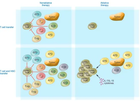
Schematic of the promotion of expansion and function of adoptively transferred antitumor CD8+ T cells following myeloablation and HSC rescue.
HSC transplant, given as part of the myeloablative regimen, can significantly augment the expansion and the antitumor impact of adoptively transferred self/tumor antigen–reactive T cells. Myeloablation effectively removes host inhibitory cells, opens up available space (the so-called Lebensraum effect), and destroys cells serving as a cytokine sink. The operative mechanism by which HSCs positively impact the transferred T cells is currently unknown but could include the production of APCs and T cell homeostatic cytokines (e.g., IL-7 and IL-12). GR1, suppressive monocytes; HSC, lin–c-kit+ HSC; pmel-1 TCR, gp100 melanoma–associated antigen-specific T cell; Treg, CD4+CD25+Foxp3+ Tregs; tumor,
[image:3.585.46.535.306.659.2]mal tissues expressing the MART-1 antigen (29–31). In some of these patients, regres-sion and resolution of large tumors and disseminated disease were observed.
Immunotherapy in the setting of myeloablative lymphodepletion
In mice recovering from lymphopenia fol-lowing myeloablative doses of TBI and transplantation of whole bone marrow (i.e., containing resident T cells) (BMT), DC- based vaccines can expedite T cell reconsti-tution and activation (32, 33). DC-based vaccination following myeloablative TBI and whole BMT is an effective means to induce regression or growth retardation of established tumors in murine syngeneic (34) and allogeneic models (35), respectively.
It is possible that resident T cells con-tained in the bone marrow from a naive donor may be more reactive to vaccination with self antigens than the endogenous T cells remaining after nonmyeloablative chemotherapy in a tumor-bearing host. Indeed, homeostasis-driven proliferation restores only the memory T cell compart-ment, whereas thymopoiesis is required to reconstitute the naive T cell compartment (36). After whole BMT, both homeostatic proliferation of resident T cells in the bone marrow and thymopoiesis together lead to reconstitution of the T cell compart-ment. In addition, bone marrow contains a high percentage of Tcm cells (37). Whereas
induction of Tem cells is important for a
peripheral immune response, Tcm cells are
superior in trafficking to peripheral lymph nodes and inducing strong systemic immu-nity (27). Transfer of purified T cells from the bone marrow of tumor lysate–pulsed DC–vaccinated mice could lead to regres-sion of breast cancer and melanoma (32). In another study, adoptive transfer of Tcm
cells was more effective than transfer of Tem cellsat inducing tumor regression in a
murine model of melanoma (24), indicat-ing the importance of CD8+ Tcm cells for
the induction of strong, systemic antitu-mor immunity.
In the setting of lymphopenia after HSC transplantation, adoptive T cell immuno-therapy as a prophylactic or strategy for treating CMV, EBV, and adenovirus infec- tions after transplantation has been con-ducted with promising outcomes (38–40). Strategies for employing T cells specific for minor histocompatibility antigens (mHA) to augment the graft-versus-leukemia effect that contributes to tumor eradi-cation are also being employed (41, 42).
In other clinical studies, strategies have included transfer of HSCs from a donor immunized with tumor antigen (idiotype proteins) to an allogeneic HSC recipient to avoid relapse in the setting of multiple myeloma (43, 44). Moreover, combina-tion immunotherapy consisting of a single early posttransplantation infusion of in vivo vaccine-primed and ex vivo costimu- lated autologous T cells followed by post-transplantation booster immunizations can improve the severe immunodeficiency associated with high-dose chemotherapy and can lead to the induction of clini-cally relevant immunity in adults within a month after transplantation (45). In cases where the functional quality of T cells for adoptive transfer against tumors or microbial/viral infections is compromised, rescue can be achieved by the use of IL-15 (46), anti-CD40 (47), or program death 1 (PD-1) engagement blockade (48), poten-tially allowing for a broader use of adoptive immunotherapy in these settings.
HSCs appear to be the key
The study by Wrzesinski et al. reported in this issue of the JCI shows that prior treatment with a myeloablative TBI dose requiring an HSC transplant can enhance both the expansion of the adoptively trans-ferred, tumor-reactive T cell population and the efficacy of tumor treatment in mice as compared with a nonmyeloablative TBI dose not requiring an HSC transplant (13). Importantly, the mere reduction of the levels of regulatory host elements and host cells competing for cytokines (so-called cytokine sinks) was not sufficient to increase T cell proliferation. Transplanta-tion of purified HSCs drove the expansion of both transferred effector T cells and cells that had survived lymphodepletion, which correlated with elevated serum lev- els of both IL-7 and IL-15 (Figure 1). HSC-driven T cell expansion was also observed in nonmyeloablated mice that also received an HSC transplant. However, it was only profound lymphodepletion, by radia-tion-induced myeloablation or by genetic means, that preferentially boosted the expansion of the transferred tumor-specific T cells, resulting in successful tumor treat-ment. Moreover, transferred T cells did not require in vitro preactivation to achieve successful treatment in the myeloablated setting. However, TCR engagement of naive T cells was a requirement, because HSC-driven expansion of naive T cells was
observed in WT mice and not in mice lack-ing β2-microglobulin (β2m–/– mice, which
are virtually devoid of functional MHC class I). This was not the case for effector cells, the HSC-driven expansion of which required neither MHC class I molecules on host cells nor additional vaccination. Last, surviving host cells negatively influenced the tumor treatment capacity of trans-ferred CD8+ T cells even when they were
present in small numbers after TBI-medi-ated lymphodepletion. It is noteworthy that these new findings could probably not have been uncovered without a very intri-cate mouse model. The authors employed their highly sensitive pmel-1 TCR-trans-genic mouse model, in which CD8+ T cells
recognize the gp100 tumor antigen, which is also relevant as a target in human mela-noma clinical trials (49).
The current study raises a few key points for further consideration. First, condition-ing with irradiation per se has at least two opposing activities on tumor immunity: (a) It depletes, in a dose-dependent manner, res-ident hematopoietic cells, including APCs (50), thus impairing antigen-specific and costimulation-driven expansion of tumor-specific T cells. The role of resident APCs in the stimulation of transferred, tumor-reac- tive T cells after allogeneic HSC transplan-tation has been shown by others (51, 52). (b) It depletes Tregs, NKT cells, and Gr1+
monocytes, which inhibit tumor-specific T cells. Multiple cell types have been shown to regulate tumor-specific responses.
Second, HSC transplants lead toat least two opposing activities that promote immu-nity and tolerance: activating naive CD8+ T
cells through MHC class I and all T cells through costimulation; and activating regulatory cells that dampen the immune response. Lin–/c-kit+ HSCs might have the
ability to directly affect T cell homeostasis, or, perhaps more likely, HSCs rapidly dif-ferentiate into APCs that present tumor antigen (53–55). Only when the potential effects of regulation are removed do the positive effects of HSC transplants on anti-tumor immunity become apparent. With respect to naive T cells, it can be surmised that HSC-derived APCs — rather than HSCs per se — amplify tumor immunity by prim-ing naive tumor-specific CD8+ T cells via
independent of MHC class I and indepen- dent of host T cells. Although in some mod-els,expansion of transferred, experienced T cells is APC independent and IL-7 and IL-15 dependent, Zaft et al. (56) have shown that it can be dependent on resident CD11c+ DCs.
Thus, HSC-derived CD11c+ DCs or other
APCs might affect homeostasis of tumor- specific effector T cells. Whether costimula-tion is provided through soluble factors, as Wrzesinski and colleagues speculate (13), or cell surface molecules such as CD40, CD80, CD86, or CD134 ligand also remains to be elucidated. With respect to regulatory cells, HSC-derived APCs provide survival signals to residual radioresistant regulatory cells, including Tregs and NKT cells. Third, the authors’ supplemental data suggest that nonablative irradiation plus vaccination appears to be as effective as ablative irradiation and HSC transplanta-tion: what then is the benefit of employing the more aggressive preparative regimen? On the face of it, the benefit could lie in cases where a suitable vaccine is not avail-able to stimulate/activate the transferred T cells. In this scenario, it is likely that mel-anoma-reactive CD8+ T cells can still be
generated from resected tumors (i.e., TILs), activated, and expanded ex vivo for adop- tive transfer into hosts following myeloab-lation and HSC rescue (57). Fourth, how generalizable are the find-ings? Although the transgenic mouse model in which the pmel-1 TCR is expressed— with the very high frequency (>90%) of murine gp100 melanoma–associated antigen-reac-tive CD8+ T cells — is attractive from the
standpoint of assay sensitivity/specificity as well its ability to tease out the opera-tive mechanisms involved, it is unknown whether the added benefit of myeloablation and HSC transplant can be replicated in a more standard model, e.g., in conventional C57BL/6 mice undergoing lethal TBI and syngeneic HSC transplantation followed by the adoptive transfer of TILs containing a polyclonal population of tumor-specific CD4+ and CD8+ T cells. The latter would
be more akin to the ongoing clinical trial in melanoma patients (30). Also of impor-tance, earlier clinical studies involving the adoptive transfer of cloned CD8+ T cells
did not result in any objective responses or any persistence of the transferred cells (including in the setting of nonmyeloabla-tive conditioning) (31). While it will be important to define the exact mechanism whereby HSCs can pro-
mote the expansion of adoptively trans-ferred antitumor CD8+ T cells, perhaps
more important will be to put these excit-ing findings to the test in the clinic. Given the initial promising clinical results to date of the adoptive transfer of TILs (30) and TCR gene–modified peripheral blood lym-phocytes in nonmyeloablated, lymphopenic melanoma patients(58), the stage is now set to carry the strategy further in the setting of myeloablation with HSC transplant rescue. Address correspondence to: James J. Mulé, H. Lee Moffitt Cancer Center and Research Institute, University of South Florida, 12902 Magnolia Drive, SRB-3, Tampa, Florida 33612, USA. Phone: (813) 745-1536; Fax: (813) 745-6188; E-mail: mulejj@ moffitt.usf.edu.
1. Lizee, G., Radvanyi, L.G., Overwijk, W.W., and Hwu, P. 2006. Improving antitumor immune responses by circumventing immunoregulatory cells and mechanisms. Clin. Cancer Res. 12:4794–4803. 2. Finke, J.H., et al. 1993. Loss of T-cell receptor zeta
chain and p56lck in T-cells infiltrating human renal cell carcinoma. Cancer Res. 53:5613–5616. 3. Wang, L.X., et al. 2005. Interleukin-7-dependent
expansion and persistence of melanoma-specific T cells in lymphodepleted mice lead to tumor regres-sion and editing. Cancer Res. 65:10569–10577. 4. Gattinoni, L., et al. 2005. Removal of homeostatic
cytokine sinks by lymphodepletion enhances the efficacy of adoptively transferred tumor-specific CD8+ T cells. J. Exp. Med. 202:907–912.
5. Mulé, J.J., Jones, F.R., Hellstrom, I., and Hellstrom, K.E. 1979. Selective localization of radiolabeled immune lymphocytes into syngeneic tumors. J. Immunol. 123:600–606.
6. Lugade, A.A., et al. 2005. Local radiation therapy of B16 melanoma tumors increases the generation of tumor antigen-specific effector cells that traffic to the tumor. J. Immunol. 174:7516–7523.
7. Wang, L.X., Shu, S., and Plautz, G.E. 2005. Host lymphodepletion augments T cell adoptive immu- notherapy through enhanced intratumoral prolif-eration of effector cells. Cancer Res. 65:9547–9554. 8. Dummer, W., et al. 2002. T cell homeostatic
proliferation elicits effective antitumor autoim-munity. J. Clin. Invest. 110:185–192. doi:10.1172/ JCI200215175.
9. Cho, B.K., Rao, V.P., Ge, Q., Eisen, H.N., and Chen, J. 2000. Homeostasis-stimulated proliferation drives naive T cells to differentiate directly into memory T cells. J. Exp. Med. 192:549–556. 10. Petrus, M.J., Williams, J.F., Eckhaus, M.A., Gress,
R.E., and Fowler, D.H. 2000. An immunoablative regimen of fludarabine and cyclophosphamide prevents fully MHC-mismatched murine marrow graft rejection independent of GVHD. Biol. Blood Marrow Transplant. 6:182–189.
11. Ercolini, A.M., et al. 2005. Recruitment of latent pools of high-avidity CD8(+
) T cells to the antitu-mor immune response. J. Exp. Med. 201:1591–1602. 12. Estin, C.D., Stevenson, U.S., Hellstrom, I., and Hell-strom, K.E. 1989. Cyclophosphamide potentiates the antitumor activity of v-p97NY. Cell. Immunol. 120:126–131. 13. Wrzesinski, C., et al. 2007. Hematopoietic stem cells promote the expansion and function of adoptively transferred antitumor CD8+ T cells. J. Clin. Invest.
117:492–501. doi:10.1172/JCI30414.
14. Nobusada, K., Kuhn, L., Pilon-Thomas, S.A., and
Mulé, J.J. 2005. Adoptive immunotherapy com-bined with a dendritic cell-based vaccine results in the rejection of established tumor in a murine melanoma model. J. Immunother. 28:614. 15. Ma, J., et al. 2003. Anti-tumor T cell response and
protective immunity in mice that received subleth-al irradiation and immune reconstitution. Eur. J. Immunol. 33:2123–2132.
16. La Gruta, N.L., Driel, I.R., and Gleeson, P.A. 2000. Peripheral T cell expansion in lymphopenic mice results in a restricted T cell repertoire. Eur. J. Immu-nol. 12:3380–3386.
17. Ge, Q., Rao, V.P., Cho, B.K., Eisen, H.N., and Chen, J. 2001. Dependence of lymphopenia-induced T cell proliferation on the abundance of peptide/ MHC epitopes and strength of their interaction with T cell receptors. Proc. Natl. Acad. Sci. U. S. A.
98:1728–1733. 18. Hu, H.M., Poehlein, C.H., Urba, W.J., and Fox, B.A. 2002. Development of antitumor immune respons-es in reconstituted lymphopenic hosts. Cancer Res. 62:3914–3919. 19. Ge, Q., Palliser, D., Eisen, H.N., and Chen, J. 2002. Homeostatic T cell proliferation in a T cell-den-dritic cell coculture system. Proc. Natl. Acad. Sci. U. S. A. 99:2983–2988.
20. Lou, Y., et al. 2004. Dendritic cells strongly boost the antitumor activity of adoptively transferred T cells in vivo. Cancer Res. 64:6783–6790.
21. Oehen, S., and Brduscha-Riem, K. 1999. Naive cytotoxic T lymphocytes spontaneously acquire effector function in lymphocytopenic recipients: a pitfall for T cell memory studies? Eur. J. Immunol.
29:608–614.
22. Kieper, W.C., and Jameson, S.C. 1999. Homeostatic expansion and phenotypic conversion of naive T cells in response to self peptide/MHC ligands. Proc. Natl. Acad. Sci. U. S. A. 96:13306–13311.
23. Goldrath, A.W., and Bevan, M.J. 1999. Low-affinity ligands for the TCR drive proliferation of mature CD8+ T cells in lymphopenic hosts. Immunity.
11:183–190.
24. Klebanoff, C.A., et al. 2005. Central memory self/ tumor-reactive CD8+
T cells confer superior anti-tumor immunity compared with effector memory T cells. Proc. Natl. Acad. Sci. U. S. A. 102:9571–9576. 25. Klebanoff, C.A., Gattinoni, L., and Restifo, N.P.
2006. CD8+ T-cell memory in tumor immunology
and immunotherapy. Immunol. Rev. 211:214–224. 26. Badovinac, V.P., Messingham, K.A., Jabbari, A.,
Haring, J.S., and Harty, J.T. 2005. Accelerated CD8+
T-cell memory and prime-boost response after den-dritic-cell vaccination. Nat. Med. 11:748–756. 27. Sallusto, F., Lenig, D., Forster, R., Lipp, M., and
Lanzavecchia, A. 1999. Two subsets of memory T lymphocytes with distinct homing potentials and effector functions. Nature. 401:708–712. 28. He, H., et al. 2006. Combined IL-21 and low-dose
IL-2 therapy induces anti-tumor immunity and long-term curative effects in a murine melanoma tumor model. J. Transl. Med. 4:24.
29. Dudley, M.E., and Rosenberg, S.A. 2003. Adoptive-cell-transfer therapy for the treatment of patients with cancer. Nat. Rev. Cancer. 3:666–675. 30. Dudley, M.E., et al. 2002. Cancer regression and
autoimmunity in patients after clonal repopulation with antitumor lymphocytes. Science. 298:850–854. 31. Dudley, M.E., et al. 2002. A phase I study of nonmy-eloablative chemotherapy and adoptive transfer of autologous tumor antigen–specific T lymphocytes in patients with metastatic melanoma. J. Immuno-ther. 25:243–251.
32. Asavaroengchai, W., Kotera, Y., Koike, N., Pilon-Thomas, S., and Mulé, J.J. 2004. Augmentation of antitumor immune responses after adoptive trans- fer of bone marrow derived from donors immu-nized with tumor lysate-pulsed dendritic cells. Biol. Blood Marrow Transplant. 10:524–533.
fungal RNA induce protective immunity to Can-dida albicans in hematopoietic transplantation. J. Immunol. 168:2904–2913.
34. Asavaroengchai, W., Kotera, Y., and Mulé, J.J. 2002. Tumor lysate-pulsed dendritic cells can elicit an effective antitumor immune response during early lymphoid recovery. Proc. Natl. Acad. Sci. U. S. A.
99:931–936.
35. Moyer, J.S., Maine, G., and Mulé, J.J. 2006. Early vaccination with tumor lysate-pulsed dendritic cells after allogeneic bone marrow transplantation has antitumor effects. Biol. Blood Marrow Transplant.
12:1010–1019.
36. Ge, Q., Hu, H., Eisen, H.N., and Chen, J. 2002. Dif- ferent contributions of thymopoiesis and homeo-stasis-driven proliferation to the reconstitution of naive and memory T cell compartments. Proc. Natl. Acad. Sci. U. S. A. 99:2989–2994.
37. Mazo, I.B., et al. 2005. Bone marrow is a major res-ervoir and site of recruitment for central memory CD8+ T cells. Immunity. 22:259–270.
38. Leen, A.M., et al. 2005. T-cell immunotherapy for adenoviral infections of stem-cell transplant recipi-ents. Ann. N. Y. Acad. Sci. 1062:104–115.
39. Bollard, C.M., et al. 2006. In vivo expansion of LMP 1- and 2-specific T-cells in a patient who received donor-derived EBV-specific T-cells after alloge-neic stem cell transplantation. Leuk. Lymphoma.
47:837–842.
40. Leen, A.M., et al. 2006. Monoculture-derived T lym-phocytes specific for multiple viruses expand and
produce clinically relevant effects in immunocom-promised individuals. Nat. Med. 12:1160–1166. 41. Riddell, S.R., Bleakley, M., Nishida, T., Berger, C.,
and Warren, E.H. 2006. Adoptive transfer of allo-geneic antigen-specific T cells. Biol. Blood Marrow Transplant. 12:9–12.
42. Riddell, S.R., and Greenberg, P.D. 1997. T cell ther- apy of human CMV and EBV infection in immuno-compromised hosts. Rev. Med. Virol. 7:181–192. 43. Kwak, L.W., Neelapu, S.S., and Bishop, M.R. 2004.
Adoptive immunotherapy with antigen-specific T cells in myeloma: a model of tumor-specific donor lymphocyte infusion. Semin. Oncol. 31:37–46.
44. Neelapu, S.S., et al. 2005. Tumor antigen immu-nization of sibling stem cell transplant donors in multiple myeloma. Bone Marrow Transplant.
36:315–323.
45. Rapoport, A.P., et al. 2005. Restoration of immu-nity in lymphopenic individuals with cancer by vaccination and adoptive T-cell transfer. Nat. Med.
11:1230–1237.
46. Teague, R.M., et al. 2006. Interleukin-15 rescues tol-erant CD8+
T cells for use in adoptive immunother-apy of established tumors. Nat. Med. 12:335–341. 47. Tuma, R.A., Giannino, R., Guirnalda, P., Leinr,
I., and Pamer, E.G. 2002. Rescue of CD8 T cell- mediated antimicrobial immunity with a non-specific inflammatory stimulus. J. Clin. Invest.
100:1493–1501.
48. Farrell, A. 2006. Defeating T-cell fatigue in HIV. Nat. Med. 12:1124–1125.
49. Overwijk, W.W., et al. 2003. Tumor regression and
autoimmunity after reversal of a functionally toler-ant state of self-reactive CD8+ T cells. J. Exp. Med.
198:569–580.
50. Collin, M.P., et al. 2006. The fate of human Langer- hans cells in hematopoietic stem cell transplanta-tion. J. Exp. Med. 203:27–33.
51. Matte, C.C., et al. 2004. Donor APCs are required for maximal GVHD but not for GVL. Nat. Med.
10:987–992.
52. Reddy, P., et al. 2005. A crucial role for antigen-pre- senting cells and alloantigen expression in graft-ver-sus-leukemia responses. Nat. Med. 11:1244–1249.
53. Feng, B., et al. 2000. Development of mouse den- dritic cells from lineage-negative c-kit(low) plu-ripotent hemopoietic stem cells in vitro. Stem Cells.
18:53–60.
54. Ginhoux, F., et al. 2006. Langerhans cells arise from monocytes in vivo. Nat. Immunol. 7:265–273.
55. Merad, M., et al. 2004. Depletion of host Langer- hans cells before transplantation of donor alloreac-tive T cells prevents skin graft-versus-host disease. Nat. Med. 10:510–517.
56. Zaft, T., Sapoznikov, A., Krauthgamer, R., Littman, D.R., and Jung, S. 2005. CD11c-high dendritic cell ablation impairs lymphopenia-driven prolifera-tion of naive and memory CD8+ T cells. J. Immunol.
175:6428–6435.
57. Gattinoni, L., Powell, D.J., Rosenberg, S.A., and Res-tifo, N.P. 2006. Adoptive immunotherapy of cancer: building on success. Nat. Rev. Immunol. 6:383–393. 58. Morgan, R.A., et al. 2006. Cancer regression in
patients after transfer of genetically engineered lymphocytes. Science. 314:126–129.
A link between protein translation and body weight
Liangyou Rui
Department of Molecular and Integrative Physiology, University of Michigan Medical School, Ann Arbor, Michigan, USA.
Nutrient overload induces obesity, a primary risk factor for type 2 diabetes.
Ribosomal biogenesis and protein synthesis, which are controlled by the
mammalian target of rapamycin (mTOR), are primary energy-consuming
processes in cells. mTOR phosphorylates and inactivates members of the
eukaryotic translation initiation factor 4E–binding (eIF4E-binding) protein
(4E-BP) family, which are translational repressors of 5
′
cap–dependent
pro-tein synthesis. In this issue of the
JCI
, Le Bacquer et al. report that
simultane-ous deletion of both 4E-BP1 and 4E-BP2 in mice results in insulin resistance,
decreased energy expenditure, and increased adipogenesis (see the related
article beginning on page 387). These findings link protein synthesis,
insu-lin sensitivity, and body weight.
Nonstandard abbreviations used: DKO, double knockout; 4E-BP, eIF4E-binding protein; eIF, eukary-otic translation initiation factor; IRS-1, insulin receptor substrate 1; mTOR, mammalian target of rapamycin; mTORC, mTOR complex; PDK1, phosphoinositide-dependent protein kinase 1;S6K, ribosomal protein S6 kinase; 5′ UTR, 5′ untranslated region.
Conflict of interest: The author has declared that no conflict of interest exists.
Citation for this article: J. Clin. Invest. 117:310–313 (2007). doi:10.1172/JCI31289.
Food (energy) shortage is a constant threat to the survival of a species. Individuals who can efficiently maintain their body weight via energy conservation have an increased
chance of survival and propagation during times when food supply is limited. Nutri- ents and hormones activate multiple evo-lutionarily conserved signaling pathways that govern the balance between energy intake and expenditure. Mammalian target of rapamycin (mTOR) is a well-conserved serine/threonine protein kinase that func-tions as an intracellular nutrient sensor to control protein synthesis, cell growth, and metabolism. In this issue of the JCI , Le Bac-quer et al. demonstrate that the eukaryotic translation initiation factor 4E–binding (eIF4E-binding) protein (4E-BP) family of
translational repressors, which are physi-ologic substrates of mTOR, play a key role in regulating body weight and glucose homeostasis in mice (1).
mTOR regulation of energy and glucose metabolism