Consequences of cellular cholesterol
accumulation: basic concepts and
physiological implications
Ira Tabas
J Clin Invest.
2002;
110(7)
:905-911.
https://doi.org/10.1172/JCI16452
.
The cells of most organs and tissues satisfy their requirements for membrane cholesterol via
endogenous cholesterol biosynthesis (1). Many cell types, however, have acquired
mechanisms to internalize exogenous sources of cholesterol, usually in the form of
plasma-derived lipoproteins (2). Examples include steroid-synthesizing cells, hepatocytes, and
macrophages and smooth muscle cells in atherosclerotic lesions, often referred to as foam
cells. In the case of steroidogenic cells, the internalization of lipoprotein-cholesterol
represents a physiological process that provides cells with precursor cholesterol stores, to
be used for “acute” steroid hormone production (3). Hepatocyte lipoprotein uptake mediates
the clearance of various classes of plasma lipoproteins (1), which can lead to whole-body
elimination of excess diet-derived cholesterol in the bile, a process known as reverse
cholesterol transport (Tall, this Perspective series, ref. 4). The uptake of arterial-wall
lipoproteins by macrophages and smooth muscle cells may be a type of physiological
scavenging response that initially helps rid the endothelium of potentially harmful
lipoprotein material (5). As will be discussed below, however, this cellular process
eventually contributes to the progression and complications of atherosclerotic vascular
disease. Cells that rely totally or mostly on endogenous cholesterol synthesis cannot
accumulate excess endogenous cholesterol because of homeostatic regulation at multiple
steps in the cholesterol biosynthetic pathway (6). Cells that internalize exogenous
cholesterol also repress endogenous cholesterol biosynthesis and LDL receptor […]
Perspective
Find the latest version:
The cells of most organs and tissues satisfy their require-ments for membrane cholesterol via endogenous cho-lesterol biosynthesis (1). Many cell types, however, have acquired mechanisms to internalize exogenous sources of cholesterol, usually in the form of plasma-derived lipoproteins (2). Examples include steroid-synthesizing cells, hepatocytes, and macrophages and smooth muscle cells in atherosclerotic lesions, often referred to as foam cells. In the case of steroidogenic cells, the internaliza-tion of lipoprotein-cholesterol represents a physiologi-cal process that provides cells with precursor cholesterol stores, to be used for “acute” steroid hormone produc-tion (3). Hepatocyte lipoprotein uptake mediates the clearance of various classes of plasma lipoproteins (1), which can lead to whole-body elimination of excess diet-derived cholesterol in the bile, a process known as reverse cholesterol transport (Tall, this Perspective series, ref. 4). The uptake of arterial-wall lipoproteins by macrophages and smooth muscle cells may be a type of physiological scavenging response that initially helps rid the endothe-lium of potentially harmful lipoprotein material (5). As will be discussed below, however, this cellular process eventually contributes to the progression and complica-tions of atherosclerotic vascular disease.
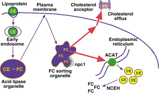
Cells that rely totally or mostly on endogenous cho-lesterol synthesis cannot accumulate excess endogenous cholesterol because of homeostatic regulation at multi-ple steps in the cholesterol biosynthetic pathway (6). Cells that internalize exogenous cholesterol also repress endogenous cholesterol biosynthesis and LDL receptor expression in response to cholesterol loading. Further-more, these cells have evolved other mechanisms to pre-vent the accumulation of excess unesterified, or “free,” cholesterol (FC). One mechanism is cholesterol esterifi-cation, which is mediated by the microsomal enzyme acyl-coenzyme A:cholesterol acyltransferase (ACAT) (7)
(Figure 1). The two forms, ACAT-1 and ACAT-2, differ in their sites of expression, with macrophages and most other cell types expressing ACAT-1. In humans, intes-tinal epithelial cells, but not hepatocytes, selectively express ACAT-2; mice, in contrast, express ACAT-2 in both of these cell types (7). Another important protec-tive mechanism against FC accumulation is cellular efflux of cholesterol and certain cholesterol-derived oxysterols (Tall, this Perspective series, ref. 4; Björkhem, this series, ref. 8) (Figure 1). In addition, some of the physiological pathways described above, such as steroid and bile acid biosynthesis, may help limit the accumu-lation of intracellular FC in steroidogenic cells and hepatocytes, respectively (2).
The evolution of multiple pathways to prevent intra-cellular FC accumulation reflects the toxic effects of excess cellular FC (9). In this article, I first discuss pos-sible mechanisms of FC-induced cytotoxicity. I then turn to the development of advanced atherosclerotic lesions, a critical pathophysiological scenario in which macrophages accumulate excess FC. This scenario pro-vides a valuable model to study mechanisms of FC accumulation, the adaptive processes that cells employ to help protect themselves from excess FC, and path-ways and consequences of FC-induced cell death. The conversion of the lesional macrophage from a choles-teryl ester–laden (CE-laden) foam cell into an FC-loaded cell may, I argue, represent a critical turning point in the progression of the atherosclerotic lesion.
Mechanisms of FC-induced cytotoxicity
A physiological FC/phospholipid ratio in cellular membranes is necessary to maintain proper membrane fluidity, or more precisely, a proper range of membrane fluidities (10). The degree of saturation of the fatty acyl moieties of membrane phospholipids is the major determinant of the fluidity of lateral membrane domains, which consist of well-packed, detergent-resistant liquid-ordered rafts and more fluid, deter-gent-soluble liquid-crystalline regions (Simons and Ehehalt, this Perspective series, ref. 11; Maxfield and Wüstner, this series, ref. 12). Nonetheless, the interac-tion of the hydrophobic rings of cholesterol with these fatty acyl chains has important effects (Tabas, Series Introduction, ref. 13). In particular, the ability of cholesterol to pack tightly with saturated fatty acyl
Consequences of cellular cholesterol accumulation:
basic concepts and physiological implications
Ira Tabas
Department of Medicine, and Department of Anatomy and Cell Biology, Columbia University, New York, New York, USA
J. Clin. Invest.110:905–911 (2002). doi:10.1172/JCI200216452.
PERSPECTIVE
Biology and biochemistry of cholesterol | Ira Tabas, Series Editor
Address correspondence to:Ira Tabas, Department of Medicine, Columbia University, 630 West 168th Street, New York, New York 10032, USA. Phone: (212) 305-9430; Fax: (212) 305-4834; E-mail: [email protected].
Conflict of interest:No conflict of interest has been declared.
groups of membrane phospholipids is critical for the formation of liquid-ordered rafts (10). Thus, choles-terol depletion causes marked disruption of these rafts. However, when the FC/phospholipid ratio rises above a physiological level, the liquid-ordered rafts may become too rigid, and the liquid-crystalline domains may begin to lose their fluidity. These events adversely affect certain integral membrane proteins that require conformational freedom for proper function and that can be inhibited by a high FC/phospholipid ratio (14). Such proteins include plasma membrane constituents like Na+-K+ATPase, adenylate cyclase, alkaline
phos-phatase, rhodopsin, and transporters for glucose, organic anions, and thymidine. Similar observations have been made with proteins residing in internal membranes, such as the Na+-Ca2+transporter in the
sarcoplasmic reticulum of cardiac muscle, the ATP-ADP transporter in the inner mitochondrial mem-brane, and UDP-glucuronyltransferase in liver micro-somes (14). Interestingly, inhibition of ACAT activity in Chinese hamster ovary cells transfected with amy-loid precursor protein blocks the generation of amyamy-loid
β-peptide (15). Although the mechanism of this effect is not yet known, one possibility is that the conforma-tion of the amyloid precursor or the β -peptide–gener-ating proteases is altered by an increase in the local FC/phospholipid ratio (Simons and Ehehalt, this series, ref. 11). High FC levels might therefore be pro-posed to kill cells in part by inhibiting one or more
integral membrane proteins whose func-tion is blocked or altered under condifunc-tions of high membrane rigidity. This and sever-al other models for FC-induced cell death, discussed below, are summarized in Table 1. Excess membrane cholesterol may also dis-rupt the function of signaling proteins that reside in membrane domains. For example, when human neutrophils with a normal plasma membrane cholesterol content are stimulated to migrate by exposure to chemokines, the actin-signaling protein Rac is recruited to detergent-sensitive non-raft membrane domains in leading lamella. However, when the plasma membrane of these cells are overloaded with cholesterol, Rac is recruited to the entire circumference of the plasma membrane, lamellar extension is nonvectorial, and neutrophil migration does not occur (L.M. Pierini and F.R. Max-field, personal communication). One inter-pretation of these data is that excess plasma membrane cholesterol disrupts the function of certain signaling molecules that normal-ly reside in non-raft domains. Experiments in vitro with model membranes suggest that, in membranes already enriched in sphin-golipids (like those that exist in several types of epithelial cells), increasing the FC con-centration even modestly above the physio-logical concentration can actually suppress the formation of membrane domains (16). Other mechanisms of cellular toxicity associated with FC accumulation include intracellular cholesterol crystallization, oxysterol formation (Björkhem, this series, ref. 8), and triggering of apoptotic signaling pathways (9). Needle-shaped cholesterol crystals form when the FC/phospholipid ratio reaches a very high level. Although typically seen in extracellular regions of advanced atherosclerotic lesions, intracellular cho-lesterol crystals have been observed both in cultured macrophages overloaded with cholesterol and in foam cells isolated directly from human coronary athero-sclerotic lesions (17, 18). Intracellular cholesterol crys-tals can probably damage cells by physically disrupt-ing the integrity of intracellular structures. Excess intracellular FC accumulation can also promote the oxidation of cholesterol to oxysterols, some of which may be cytotoxic (19). Finally, FC overloading of macrophages can trigger a series of apoptotic path-ways (20–22), as discussed below.
Accumulation of FC in macrophages during atherogenesis
[image:3.576.58.333.62.224.2]The hallmark of the early atherosclerotic lesion is the CE-laden macrophage foam cell (23). Remarkably, in the face of extremely high levels of CE, these early lesional cells maintain an FC content not markedly dif-ferent from non–foam cell macrophages. However, lipid assays of lesional material from various stages of human and animal atheromata reveal a steady increase
Figure 1
in FC content and a steady decrease in CE content as the lesions become more advanced (24). While a por-tion of this trend reflects extracellular events, analysis of foam cells isolated from advanced lesions clearly demonstrates an increase in macrophage FC content (9). As discussed below, cultured macrophage models of FC accumulation have revealed fascinating cellular responses to FC accumulation, as well as end-stage con-sequences of FC loading that are relevant to the pro-gression and complications of atherosclerosis.
Mechanisms of FC accumulation
Macrophages are normally protected from the accu-mulation of excess FC by ACAT-1–mediated esterifica-tion and by cholesterol efflux (2) (Figure 1). In addiesterifica-tion, the hydrolysis of stored CE by neutral CE hydrolase does not usually exceed a cell’s capacity to export or re-esterify this pool of cholesterol. Thus, the progressive accumulation of FC by lesional macrophages might be explained by failure of one or more of these protective mechanisms as the atherosclerotic lesion progresses.
Both cholesterol efflux and cholesterol esterification would be adversely affected by disruption of intracellu-lar cholesterol transport. Cholesterol that enters cells via internalization of lipoproteins is initially targeted to an organelle, probably a type of late endosome, that then distributes this cholesterol to the plasma mem-brane for efflux and the endoplasmic reticulum (ER) for esterification (2, 25, 26). Peripheral cholesterol trans-port is an energy-dependent vesicular process and involves molecules such as Niemann-Pick C protein 1 (npc1) and npc2 (HE1) and the lipids lysobisphospha-tidic acid and sphingomyelin (12, 25). Disturbance of cellular membrane vesiculation, depletion of cellular ATP stores, or disruption of the function of these mol-ecules could block FC efflux and esterification, thus favoring FC accumulation. Indeed, peripheral FC traf-ficking is inhibited in cultured macrophages incubated with oxidized LDL, a form of modified LDL found in atherosclerotic lesions. This response may be related to inhibition by oxysterols of lysosomal sphingomyelinase (27), leading to sphingomyelin accumulation. Excess intracellular sphingomyelin, in turn, disrupts normal peripheral cholesterol distribution by an unknown mechanism (28). Interestingly, experiments in cultured cells have revealed that lysosomal FC accumulation itself inhibits lysosomal sphingomyelinase activity (29). Thus, if cellular cholesterol influx begins to exceed the capacity of lysosomes to transport FC, an initially mod-est accumulation of lysosomal FC
may be amplified by subsequent inhibition of lysosomal sphin-gomyelinase. This mechanism may help explain the finding that cul-tured macrophages that phagocy-tose large quantities of CE droplets accumulate FC in lysosomes (30). Direct disruption of ACAT-1 or cellular efflux pathways would also promote FC accumulation. Efflux might be compromised by
inhibit-ing ABCA1, SR-BI, or other plasma membrane proteins that mediate cholesterol efflux. Indeed, recent data from our laboratory suggest that excess FC accumula-tion in macrophages compromises the funcaccumula-tion of ABCA1, which may then amplify further FC accumula-tion (31). Efflux may also be blocked if access is lost to plasma-derived cholesterol acceptors like HDL and apoA-1, as may occur for macrophages buried deep in the intima of atherosclerotic lesions. In addition, if cel-lular cholesterol influx exceeds the capacity of ACAT-1, FC may begin to accumulate in the ER membrane domains where this enzyme resides. Because these mem-branes are normally cholesterol-poor (32), an FC-induced change in the physical properties of these mem-branes might compromise ACAT-1 activity (33). According to this model, FC accumulation would be amplified by progressive cholesterol accumulation in ER membranes, leading to further ACAT-1 dysfunction. Finally, efflux pathways may also be inhibited by con-version of cholesterol into oxysterols. While certain oxysterols actually promote sterol removal (e.g., 27-hydroxycholesterol in macrophages and bile acids in hepatocytes), other oxysterols (e.g., 25-hydroxycholes-terol and 7-ketocholes25-hydroxycholes-terol) exacerbate choles25-hydroxycholes-terol accu-mulation by inhibiting FC efflux or ACAT-1–mediated cholesterol esterification (19).
Recent experiments in cultured macrophages suggest that neutral CE hydrolase activity can further complicate the problem of cellular FC homeostasis in advancing atherogenic lesions. When these cells are incubated for a short period with atherogenic lipoproteins, the CE-rich lipid droplets formed have a high CE content and essen-tially normal FC content, as expected. After subsequent culture in the absence of lipoproteins, however, CE con-tent drops and FC concon-tent rises correspondingly. Col-lections of FC can be seen in the cytoplasm of these cells by filipin staining, reflecting the ability of neutral CE hydrolase to act on preformed CE droplets and to gen-erate excess FC under at least some physiological condi-tions (34). Whether this enzyme is also active when cells are continuously exposed to atherogenic lipoproteins remains to be determined. Nonetheless, the action of this hydrolase is likely to contribute to FC accumulation in lesional macrophage foam cells whose FC efflux or re-esterification by ACAT-1 is compromised.
Adaptive processes
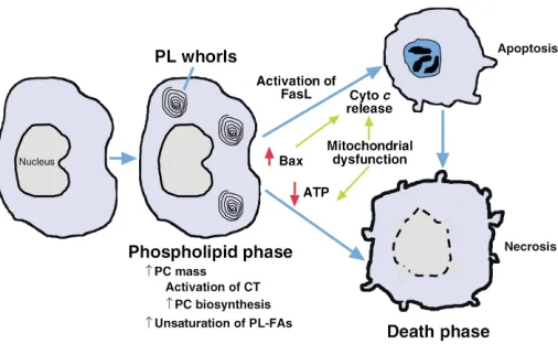
As shown in Figure 2, the responses of macrophages to FC loading can be divided into two phases — an initial
Table 1
Potential mechanisms of FC-induced cytotoxicity
Event Consequence
Loss of membrane fluidity Dysfunction of integral membrane proteins Disruption of membrane domains Disruption of signaling events
Induction of apoptosis Caspase-mediated cell death Intracellular cholesterol crystallization Organelle disruption Formation of toxic oxysterols ? Oxidative damage
adaptive stage in which phospholipid synthesis increas-es to offset the harmful effects of increasing FC, and a later stage when these defenses are overcome, leading to cell death. As investigators continue to explore the mechanisms of FC accumulation in vivo, a convenient cell-culture model has enabled the study of both of these stages (9, 35). In this model, macrophages are treated with one of several specific inhibitors of ACAT-1, either during or after incubation with athero-genic lipoproteins. Importantly, the perinuclear distri-bution of filipin-positive FC in the cultured cells mim-ics the distribution of FC observed in macrophages isolated from atherosclerotic lesions. Moreover, the var-ious effects observed in this model are not seen with ACAT inhibitors in the absence of lipoproteins and are thus dependent on FC loading per se.
One of the earliest responses of macrophages to FC loading is an increase in phospholipid biosynthesis (35). While this response is observed with sphin-gomyelin and several other classes of phospholipids, the major species produced is phosphatidylcholine (PC). The mechanism involves posttranslational acti-vation of the rate-limiting enzyme in PC biosynthesis, CTP:phosphocholine cytidylyltransferase (CT). Exactly how FC accumulation leads to CT activation is not known, but the process requires dephosphorylation of CT and possibly one or more CT regulatory proteins. The twofold increment in PC mass in FC-loaded macrophages is remarkable, and it is reflected by the appearance of phospholipid-containing whorl-like membrane structures in the cells. Importantly, such structures have also been seen in lesional macrophages (36). This observation, together with other reports showing an increase in phospholipid biosynthesis in
the cells of atherosclerotic lesions in vivo (37), suggests that the responses of FC-loaded cultured macrophages indeed mimic physiological events. The central issue to arise from these observa-tions concerned the functional role of this phospholipid response. The hypothesis that increasing PC represents an adaptive response, preventing FC-mediated cytotoxicity by keeping the FC/phospholipid ratio in check, received direct support from studies conducted with peri-toneal macrophages from CT-deficient mice (38). There are two genes that encode CT, CCTαand
CCTβ. Macrophages from mice with homozy-gous disruption of the CCTαgene have intact CTβactivity and thus retain approximately 20% of their total CT activity and PC biosynthetic capacity. Importantly, these cells appear healthy when grown under normal culture conditions and are essentially indistinguishable from wild-type macrophages. However, when subjected to an FC load, CTα-deficient macrophages cannot mount a substantial PC response, and they suc-cumb to the toxic effects of FC much earlier than do wild-type macrophages. Thus, the ability of FC-loaded macrophages to activate CT and increase PC biosynthesis helps protect them from FC-induced cytotoxicity.
Ikonen and coworkers (39) recently reported another phospholipid response to FC loading, namely, an increase in plasma membrane phospholipid species with polyunsaturated acyl chains. These studies were conducted with human fibroblasts that had been loaded with FC either by incubation with serum in the face of a mutation that blocks cholesterol esterification or, in wild-type fibroblasts, by acute plasma membrane loading. In both cases, there was a significant increase in the content of polyunsaturated fatty acids in several classes of phospholipids. Although the mechanism of these fatty acid alterations is not known, these changes may represent another adaptive effect to FC loading, because membranes rich in phospholipids with polyunsaturated acyl chains are more resistant to the stiffening effects of cholesterol (40).
FC-induced cell death
[image:5.576.60.313.58.214.2]Although cells are clearly capable of adaptive responses to FC loading, these mechanisms eventually fail with prolonged internalization of cholesterol, leading to cell death (9, 41) (Figure 2). The basis of adaptive failure is not known, although a decrease in CT activity has been observed before the onset of cellular toxicity (42). CT, like ACAT, is associated with normally cholesterol-poor cellular membranes and thus may become dysfunction-al when its lipid microenvironment becomes too rigid. By morphological criteria, cytotoxic FC-loaded macrophages show signs of both necrosis (e.g., dis-rupted cellular membranes) and apoptosis (e.g., con-densed nuclei) (9, 41). By biochemical criteria, apop-tosis-associated caspases and their signaling pathways are activated in a portion of the cells (below). In all likelihood, as depicted in Figure 3, some cells in a
Figure 2
population of FC-loaded macrophages become acute-ly necrotic due to direct and acute disruptive effects on membrane proteins, whereas others undergo a pro-grammed apoptotic response. Moreover, cells that ini-tially undergo an apoptotic program can subsequent-ly demonstrate morphological signs of necrosis (so-called aponecrosis), perhaps as a result of chronic ATP depletion or failure of neighboring cells to phagocytose the apoptotic bodies (43).
In a typical cell-culture model of FC-loaded macro-phages, approximately 25–30% of the cells show the apoptosis-associated hallmarks of phosphatidylserine externalization and DNA fragmentation, which can be completely prevented by inhibitors of effector caspases (21). Interestingly, partial inhibition of apoptosis is also observed when the Fas receptor signaling pathway is dis-rupted either by genetic mutations in Fas or its ligand or by use of a blocking anti–Fas ligand antibody. FC load-ing results in posttranslational activation of cell-surface Fas ligand, either by inducing a conformational change in the molecule or by stimulating its transport from intracellular stores to the plasma membrane (21).
Widespread mitochondrial dysfunction, indicated by a decrease in the mitochondrial transmembrane poten-tial, is also observed in FC-loaded macrophages (22). Furthermore, FC-loaded macrophages show evidence of mitochondrial cytochrome crelease and caspase-9 activation. Thus, in addition to involvement of the Fas pathway, a classic mitochondrial pathway of apoptosis is activated in FC-loaded macrophages. Importantly, these events are not mediated by oxysterols, because oxysterol-induced mitochondrial dysfunction and apoptosis are inhibited by the antioxidant glutathione, whereas the death pathways described above are resist-ant to this compound. The mechanisms by which FC overloading triggers these events is not known, but it appears to be independent of direct effects of FC on the plasma membrane (P.M. Yao and I. Tabas, unpublished data). Intracellular and mitochondrial levels of the proapoptotic protein Bax are increased in FC-loaded macrophages (22), but direct proof for the involvement of Bax is lacking. Other possibilities include direct toxic
effects of FC on mitochondrial membranes and activa-tion of a proapoptotic signaling pathway.
An important concept to arise from these cell-culture studies is the importance of intracellular cholesterol trafficking in FC-induced death. Rothblat and col-leagues (20) were the first to show that amphipathic amines, which markedly inhibit peripheral cholesterol trafficking, protect FC-loaded macrophages from death. These results suggest that, whether FC triggers death by direct membrane effects or by activation of death-promoting molecules or both, FC must be able to traffic to peripheral sites in the cell to effect cell killing. Our recent data indicate that, whereas FC traf-ficking to the plasma membrane may be responsible for necrotic-type death, excess FC accumulation in the plasma membrane cannot explain apoptotic death. Moreover, FC-induced apoptosis is blocked by very subtle disruptions of intracellular trafficking, such as occur in macrophages from heterozygous NPC mice or in macrophages treated with very-low-dose amphi-pathic amines (ref. 44; and P.M. Yao and I. Tabas, unpublished data). Thus, the peripheral apoptosis-sensing mechanisms appear to be quite sensitive to FC. Clearly, an important goal of further research in this area is to define the molecular mechanisms that under-lie the relationship between intracellular cholesterol trafficking and apoptosis in FC-loaded cells.
Relevance to atherosclerotic vascular disease
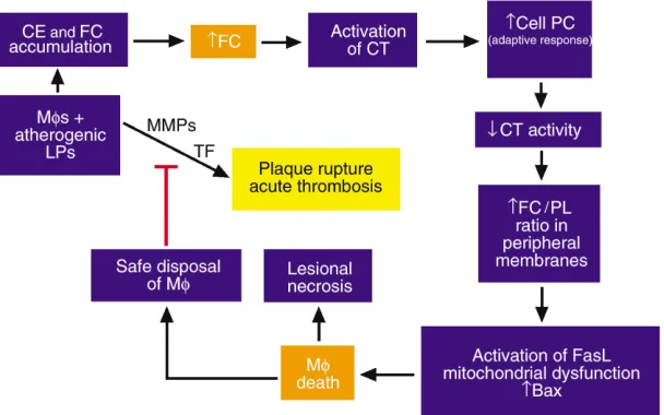
[image:6.576.237.541.540.730.2]The presence of apoptotic and necrotic macrophages in atherosclerotic lesions has been well documented in many human and animal studies (45, 46). Among the potential causes of lesional macrophage death, FC-induced toxicity needs serious consideration, because macrophages from advanced lesions are known to be loaded with FC, a potent inducer of macrophage death (9). The functional significance of the cell death path-ways depicted in Figure 3 remains uncertain. On the one hand, assuming harmless disposal of apoptotic bodies by neighboring phagocytes, macrophage apop-tosis may limit the number of intimal cells in a physio-logically “safe” manner that avoids inducing local
Figure 3
inflammation. On the other hand, death of macro-phages by either necrosis or apoptosis might lead to release of cellular proteases, inflammatory cytokines, and prothrombotic molecules, which could contribute to plaque instability, plaque rupture, and acute throm-botic vascular occlusion. Indeed, necrotic areas of advanced atherosclerotic lesions are known to be asso-ciated with death of macrophages, and ruptured plaques from human lesions have been shown to be enriched in apoptotic macrophages (46).
Two mouse models have begun to shed light on the in vivo consequences of FC-induced macrophage death. In the first model, LDL receptor knockout mice were recon-stituted with ACAT-1–deficient macrophages by bone marrow transplantation (47). Compared with control mice reconstituted with ACAT-1–positive macrophages, the atherosclerotic lesions of the experimental mice were larger and had increased FC content and more apoptot-ic macrophages. Thus, in this model, FC-induced macrophage death promotes lesion development.
The second model was designed to address the effect of preventing macrophage death in an otherwise ath-erosclerosis-prone mouse. By crossing a disrupted allele of Npc1, in heterozygous form, into an apoE knockout background, our group produced animals with ad-vanced atherosclerotic lesions whose macrophages were relatively resistant to FC-induced death. Control mice with normal npc1 function had large acellular areas filled with FC and macrophage debris, but not with CE. In contrast, the lesions of the mice with par-tial npc1 deficiency had a greater content of CE-rich macrophages (44). Because npc1 deficiency does not protect macrophages from other inducers of apoptosis, these data support the hypothesis that FC-induced death, requiring intact FC trafficking, is an important cause of lesional macrophage death in vivo. Moreover, the lesions in the death-resistant model appear more stable, but future studies with rupture-susceptible mice will be needed to substantiate this point.
In light of these recent data, the therapeutic value of ACAT inhibitors may require some further scrutiny. These agents, which are intended to block the accumu-lation of CE in vascular macrophages, have been pro-posed for the prevention or treatment of atherosclerot-ic vascular disease (48). Indeed, ACAT inhibitors have been shown to prevent atherosclerosis in several animal models, and one such inhibitor is currently undergoing trials in humans. The site of drug action may be key to explaining the beneficial effects of these drugs despite the apparent risk that they could further the progres-sion of atherosclerotic leprogres-sions. First, even for ACAT-1 inhibitors, which suppress macrophage-associated enzymatic activity in vitro, the drug’s ability to enter the lesion may be limited. Second, moderate suppression of ACAT activity in these cells can probably be offset by cholesterol efflux, as long as the cells have access to cho-lesterol acceptors such as HDL or apoA-1. ACAT-2 inhibitors, conversely, should have no direct effect on lesional macrophages and could be strongly beneficial because of their ability to suppress lipoprotein produc-tion by the intestine and — in mice — the liver (49). For
these reasons, ACAT inhibitors, whether directed at one or both of these isoforms, may be less likely to precipi-tate macrophage death and lesional necrosis than work with ACAT-1–deficient cells might suggest.
Concluding remarks and future directions
The study of cellular cholesterol excess provides an opportunity to address a variety of important topics ranging from biophysical chemistry to intracellular sig-naling pathways to mechanisms of clinical disease. To begin, a proper understanding of the FC-loaded cell requires an appreciation of membrane lipid phase behavior. One must then investigate the alterations of specific enzymes and other proteins to elucidate the mechanisms of specific consequences of FC loading on cellular physiology. These alterations may result either from a direct consequence of membrane alterations or from the activation of signaling reactions that them-selves are triggered by membrane alterations. Moreover, effects of FC or FC metabolites on gene expression must also be considered. Finally, the biology of the FC-loaded cell must be placed in the context of the whole tissue and organism, as demonstrated by the potential effects of the FC-loaded macrophage on atherosclerosis.
In each area, much remains to be done. While the effects of high levels of FC on the physical properties of model membranes have been studied widely in vitro, biophysical studies on excess FC in membranes of living cells have suffered from technological difficulties. New advances in fluorescence microscopy, including the use of domain-specific probes (Maxfield and Wüstner, this series, ref. 12), are beginning to close this critical gap. The consequences of cellular FC excess on specific molecules or signaling pathways are gradually entering the scien-tific literature, but insight into the mechanisms linking these consequences with changes in FC-induced alter-ations in membrane structure and possibly gene expres-sion is lacking. For example, much has been published on the effects of cellular cholesterol depletion on raft structure and function (Simons and Ehehalt, this series, ref. 11), but very little work has been published about the effects of cellular cholesterol excess on raft biology.
this concept will require much further work, particular-ly in vivo using geneticalparticular-ly altered mice. Nonetheless, given the current evidence and the importance of ather-osclerosis, the rationale for gaining a thorough under-standing of the biology of the FC-loaded cell is clear.
1. Dietschy, J.M., Turley, S.D., and Spady, D.K. 1993. Role of the liver in the maintenance of cholesterol and low density lipoprotein homeostasis in different animal species, including humans. J. Lipid Res.34:1637–1659. 2. Tabas, I., and Kreiger, M. 1999. Lipoprotein receptors and cellular cho-lesterol metabolism in health and disease. In Molecular basis of heart dis-ease. K.R. Chien, editor. W.B. Saunders Co. New York, New York, USA. 428–457.
3. Lin, D., et al. 1995. Role of steroidogenic acute regulatory protein in adrenal and gonadal steroidogenesis. Science.267:1828–1831. 4. Tall, A.R., Costet, P., and Wang, N. 2002. Regulation and mechanisms of
macrophage cholesterol efflux. J. Clin. Invest.110:899–904. doi:10.1172/ JCI200216391.
5. Glass, C.K., and Witztum, J.L. 2001. Atherosclerosis: the road ahead. Cell. 104:503–516.
6. Horton, J.D., Goldstein, J.L., and Brown, M.S. 2002. SREBPs: activators of the complete program of cholesterol and fatty acid synthesis in the liver. J. Clin. Invest.109:1125–1131. doi:10.1172/JCI200215593. 7. Chang, T.Y., et al. 2001. Roles of acyl-coenzyme A:cholesterol
acyltrans-ferase-1 and -2. Curr. Opin. Lipidol.12:289–296.
8. Björkhem, I. 2002. Do oxysterols control cholesterol homeostasis? J. Clin. Invest.110:725–730. doi:10.1172/JCI200216388.
9. Tabas, I. 1997. Free cholesterol-induced cytotoxicity. A possible con-tributing factor to macrophage foam cell necrosis in advanced athero-sclerotic lesions. Trends Cardiovasc. Med.7:256–263.
10. Simons, K., and Ikonen, E. 2000. How cells handle cholesterol. Science. 290:1721–1726.
11. Simons, K., and Ehehalt, R. 2002. Cholesterol, lipid rafts, and disease. J. Clin. Invest.110:597–603. doi:10.1172/JCI200216390.
12. Maxfield, F.R., and Wüstner, D. 2002. Intracellular cholesterol transport. J. Clin. Invest.110:891–898. doi:10.1172/JCI200216500.
13. Tabas, I. 2002. Cholesterol in health and disease. J. Clin. Invest. 110:583–590. doi:10.1172/JCI200216381.
14. Yeagle, P.L. 1991. Modulation of membrane function by cholesterol. Biochimie.73:1303–1310.
15. Puglielli, L., et al. 2001. Acyl-coenzyme A:cholesterol acyltransferase modulates the generation of the amyloid beta-peptide. Nat. Cell Biol. 3:905–912.
16. Milhiet, P.E., Giocondi, M.C., and Le Grimellec, C. 2002. Cholesterol is not crucial for the existence of microdomains in kidney brush-border membrane models. J. Biol. Chem.277:875–878.
17. Kellner-Weibel, G., et al. 1999. Crystallization of free cholesterol in model macrophage foam cells. Arterioscler. Thromb. Vasc. Biol. 19:1891–1898.
18. Lupu, F., Danaricu, I., and Simionescu, N. 1987. Development of intra-cellular lipid deposits in the lipid-laden cells of atherosclerotic lesions. A cytochemical and ultrastructural study. Atherosclerosis.67:127–142. 19. Brown, A.J., and Jessup, W. 1999. Oxysterols and atherosclerosis.
Athero-sclerosis.142:1–28.
20. Kellner-Weibel, G., et al. 1998. Effects of intracellular free cholesterol accumulation on macrophage viability: a model for foam cell death. Arte-rioscler. Thromb. Vasc. Biol.18:423–431.
21. Yao, P.M., and Tabas, I. 2000. Free cholesterol loading of macrophages induces apoptosis involving the Fas pathway. J. Biol. Chem. 275:23807–23813.
22. Yao, P.M., and Tabas, I. 2001. Free cholesterol loading of macrophages is associated with widespread mitochondrial dysfunction and activation of the mitochondrial apoptosis pathway. J. Biol. Chem.276:42468–42476. 23. Ross, R. 1995. Cell biology of atherosclerosis. Annu. Rev. Physiol.
57:791–804.
24. Katz, S.S., Shipley, G.G., and Small, D.M. 1976. Physical chemistry of the lipids of human atherosclerotic lesions. Demonstration of a lesion inter-mediate between fatty streaks and advanced plaques. J. Clin. Invest. 58:200–211.
25. Liscum, L., and Munn, N.J. 1999. Intracellular cholesterol transport. Biochim. Biophys. Acta.1438:19–37.
26. Lange, Y. 1998. Intracellular cholesterol movement and homeostasis. In Intracellular cholesterol trafficking. T.Y. Chang and D.A. Freeman, editors.
Kluwer Academic Publishers. Boston, Massachusetts, USA. 15–27. 27. Maor, I., Mandel, H., and Aviram, M. 1995. Macrophage uptake of
oxi-dized LDL inhibits lysosomal sphingomyelinase, thus causing the accu-mulation of unesterified cholesterol-sphingomyelin-rich particles in the lysosomes. A possible role for 7-ketocholesterol. Arterioscler. Thromb. Vasc. Biol.15:1378–1387.
28. Leventhal, A.R., Chen, W., Tall, A.R., and Tabas, I. 2001. Acid sphin-gomyelinase-deficient macrophages have defective cholesterol traffick-ing and efflux. J. Biol. Chem.276:44976–44983.
29. Reagan, J.W., Jr., Hubbert, M.L., and Shelness, G.S. 2000. Posttransla-tional regulation of acid sphingomyelinase in niemann-pick type C1 fibroblasts and free cholesterol-enriched chinese hamster ovary cells. J. Biol. Chem.275:38104–38110.
30. Tangirala, R.K., Mahlberg, F.H., Glick, J.M., Jerome, W.G., and Rothblat, G.H. 1993. Lysosomal accumulation of unesterified cholesterol in model macrophage foam cells. J. Biol. Chem.268:9653–9660.
31. Feng, B., and Tabas, I. 2002. ABCA1-mediated cholesterol efflux is defec-tive in free cholesterol-loaded macrophages. Mechanism involves enhanced ABCA1 degradation in a process requiring full npc1 activity. J. Biol. Chem. In press.
32. Chang, T.Y., Chang, C.C.Y., and Cheng, D. 1997. Acyl-coenzyme A:cho-lesterol acyltransferase. Annu. Rev. Biochem.66:613–638.
33. Moynault, A., Luciani, M.F., and Chimini, G. 1998. ABC1, the mam-malian homologue of the engulfment gene ced-7, is required during phagocytosis of both necrotic and apoptotic cells. Biochem. Soc. Trans. 26:629–635.
34. Mori, M., et al. 2001. Foam cell formation containing lipid droplets enriched with free cholesterol by hyperlipidemic serum. J. Lipid Res. 42:1771–1781.
35. Tabas, I. 2000. Cholesterol and phospholipid metabolism in macro-phages. Biochim. Biophys. Acta.1529:164–174.
36. Shio, H., Haley, N.J., and Fowler, S. 1979. Characterization of lipid-laden aortic cells from cholesterol-fed rabbits. III. Intracellular localization of cholesterol and cholesteryl ester. Lab. Invest.41:160–167.
37. Zilversmit, D.B., Shore, M.L., and Ackerman, R.F. 1954. The origin of aortic phospholipid in rabbit atheromatosis. Circulation.9:581–585. 38. Zhang, D., et al. 2000. Macrophages deficient in CTP:Phosphocholine
cytidylyltransferase-alpha are viable under normal culture conditions but are highly susceptible to free cholesterol-induced death. Molecular genetic evidence that the induction of phosphatidylcholine biosynthe-sis in free cholesterol-loaded macrophages is an adaptive response. J. Biol. Chem.275:35368–35376.
39. Blom, T.S., et al. 2001. Mass spectrometric analysis reveals an increase in plasma membrane polyunsaturated phospholipid species upon cellular cholesterol loading. Biochemistry.40:14635–14644.
40. Huster, D., Arnold, K., and Gawrisch, K. 1998. Influence of docosa-hexaenoic acid and cholesterol on lateral lipid organization in phos-pholipid mixtures. Biochemistry.37:17299–17308.
41. Warner, G.J., Stoudt, G., Bamberger, M., Johnson, W.J., and Rothblat, G.H. 1995. Cell toxicity induced by inhibition of acyl coenzyme A: cho-lesterol acyltransferase and accumulation of unesterified chocho-lesterol. J. Biol. Chem.270:5772–5778.
42. Tabas, I., Marathe, S., Keesler, G.A., Beatini, N., and Shiratori, Y. 1996. Evidence that the initial up-regulation of phosphatidylcholine biosyn-thesis in free cholesterol-loaded macrophages is an adaptive response that prevents cholesterol-induced cellular necrosis. Proposed role of an eventual failure of this response in foam cell necrosis in advanced ath-erosclerosis. J. Biol. Chem.271:22773–22781.
43. Formigli, L., et al. 2000. Aponecrosis: morphological and biochemical exploration of a syncretic process of cell death sharing apoptosis and necrosis. J. Cell. Physiol.182:41–49.
44. Zhang, D., et al. 2001. Niemann-Pick C heterozygosity confers marked protection from macrophage death and lesional necrosis. Circulation. 104:II-45. (Abstr.)
45. Kockx, M.M. 1998. Apoptosis in the atherosclerotic plaque: quantitative and qualitative aspects. Arterioscler. Thromb. Vasc. Biol.18:1519–1522. 46. Mitchinson, M.J., Hardwick, S.J., and Bennett, M.R. 1996. Cell death in
atherosclerotic plaques. Curr. Opin. Lipidol.7:324–329.
47. Fazio, S., et al. 2001. Increased atherosclerosis in LDL receptor-null mice lacking ACAT1 in macrophages. J. Clin. Invest.107:163–171.
48. Brown, W.V. 2001. Therapies on the horizon for cholesterol reduction. Clin. Cardiol.24:III24–III27.