Mature dendritic cells boost functionally
superior CD8
+
T-cell in humans without foreign
helper epitopes
Madhav V. Dhodapkar, … , Ralph M. Steinman, Nina
Bhardwaj
J Clin Invest.
2000;
105(6)
:R9-R14.
https://doi.org/10.1172/JCI9051
.
We have recently shown that a single injection of mature, antigen-pulsed, human dendritic
cells (DCs) rapidly elicits CD4
+and CD8
+T-cell immunity in vivo. The DCs were pulsed
with 2 foreign proteins, keyhole limpet hemocyanin (KLH) and tetanus toxoid (TT), as well
as an HLA A2.1-restricted influenza matrix peptide (MP). Responses to all 3 antigens
peaked at 30–90 days after immunization and declined thereafter. To determine if the
foreign helper proteins (TT and KLH) were essential for CD8
+T-cell responses to the viral
peptide, we reinjected 3 of the HLA-2.1 subjects with mature DCs pulsed with MP alone. All
3 volunteers showed a rapid boost in MP-specific immunity, and freshly sampled blood from
1 contained cytolytic T cells. In all 3 subjects, CD8
+T-cell responses to booster DCs were
faster and of greater magnitude than the responses to the first DC injection. Importantly, the
T cells that proliferated after booster DC treatment secreted interferon-
g
upon challenge with
much lower doses of viral peptide than those elicited after the first injection, indicating a
higher functional avidity for the ligand. These data begin to outline the kinetics of T-cell
immunity in response to DCs and demonstrate that booster injections of mature DCs
enhance both qualitative and quantitative aspects of CD8
+T-cell function in humans.
This article may have been published online in […]
Rapid Publication
We have recently shown that a single injection of mature, antigen-pulsed, human dendritic cells (DCs) rapidly elicits CD4+and CD8+T-cell immunity
in vivo. The DCs were pulsed with 2 foreign proteins, keyhole limpet hemo-cyanin (KLH) and tetanus toxoid (TT), as well as an HLA A2.1-restricted influenza matrix peptide (MP). Responses to all 3 antigens peaked at 30–90 days after immunization and declined thereafter. To determine if the foreign helper proteins (TT and KLH) were essential for CD8+T-cell responses to the
viral peptide, we reinjected 3 of the HLA-2.1 subjects with mature DCs pulsed with MP alone. All 3 volunteers showed a rapid boost in MP-specific immu-nity, and freshly sampled blood from 1 contained cytolytic T cells. In all 3 sub-jects, CD8+T-cell responses to booster DCs were faster and of greater
magni-tude than the responses to the first DC injection. Importantly, the T cells that proliferated after booster DC treatment secreted interferon-γupon challenge with much lower doses of viral peptide than those elicited after the first injec-tion, indicating a higher functional avidity for the ligand. These data begin to outline the kinetics of T-cell immunity in response to DCs and demonstrate that booster injections of mature DCs enhance both qualitative and quanti-tative aspects of CD8+T-cell function in humans.
This article may have been published online in advance of the print edition. The date of public-ation is available from the JCI website, http://www.jci.org. J. Clin. Invest.105:R9–R14 (2000).
Introduction
Although several approaches (such as DNA vaccines, viral vectors) generate protective T-cell immunity in mice, it has proven difficult to generate potent T-cell immunity in humans with current vac-cines (1). Dendritic cells (DCs), nature’s adjuvant, are antigen-presenting cells (APCs) specialized to initiate T-cell immunity (2). It is known that exposure to inflammatory stimuli leads to termi-nal differentiation or maturation of DCs, with enhanced capacity to stimu-late T-cell immunity (2). We have shown recently that a single injection of anti-gen-bearing, monocyte-derived, mature DCs leads to rapid enhancement of T-cell responses in humans (3). These data demonstrated that mature DCs are potent immune adjuvants in humans and provided the first controlled evi-dence of their immunogenicity.
More quantitative T-cell assays have now made it possible to study the kinetics and longevity of the T-cell immune response in humans after DC immunization. In previous work, serial quantitative measure-ments of immune parameters were not performed (4, 5). Here we serially characterize the kinetics and durabil-ity of CD4+and CD8+T-cell
respons-es after DC injection in humans using a panel of quantitative assays. We also show that a booster injection of mature DCs leads to greater and more rapid enhancement of the CD8+ T-cell response without the
need for “foreign helper epitopes.” Importantly, we demonstrate that T cells elicited after booster DCs are functionally superior and recognize lower doses of antigen than those elicited after the first injection.
Mature dendritic cells boost
functionally superior CD8
+T-cell in humans
without foreign helper epitopes
Madhav V. Dhodapkar, Joseph Krasovsky, Ralph M. Steinman, and Nina Bhardwaj
Laboratory of Cellular Physiology and Immunology, Rockefeller University, New York, New York 10021, USA
Address correspondence to: Madhav V. Dhodapkar, Laboratory of Immunology and Cellular Physiology, Rockefeller University, 1230 York Avenue, Box 176, New York, New York 10021, USA. Phone: (212) 327-7597; Fax: (212) 327-8875;
E-mail: [email protected].
Received for publication December 2, 1999, and accepted in revised form February 1, 2000.
Methods
Study subjects
Seven of 9 subjects who were injected with antigen-pulsed DCs in the initial study (3) were available for long-term monitoring and are subjects of the present analysis. Two subjects (P2, P3) had left the New York area and were unavailable for follow-up.
Schema for immune monitoring and booster injections
All subjects were monitored for immune responses to study antigens every 1–4 months after the first anti-gen-pulsed DC injection. In 3 of 4 HLA A2.1+subjects (P4, P5, P6),
boost-er DC injections wboost-ere pboost-erformed at 7–9 months after the first antigen-pulsed injection. A booster injection was not considered in the fourth HLA A2.1+subject (P1) because of
interven-ing pregnancy. All subjects who received a booster injection signed an updated informed consent. Subjects were monitored 2 days, 1 week, 1 month after the booster and every 1–4 months thereafter. The study was approved by the Rockefeller Universi-ty Institutional Review Board and the U.S. Food and Drug Administration.
Measurement of immune responses
Antigen-specific proliferation. Antigen-specific proliferation to keyhole limpet hemocyanin (KLH), tetanus toxoid (TT), and staphylococcal enterotoxin A (SEA) as a control, was performed as described earlier (3). All assays were performed on fresh PBMCs. When possible, cryopreserved samples from before and after immunization were also thawed and assayed together. In
Rapid
some cultures, the phenotype of the responding T cells was monitored at the end of culture by staining for CD4 and quantifying the percentage of CD4+T-cell blasts (high forward
scat-ter) using flow cytometry.
Enzyme-linked immunospot assay for IFN-γrelease from single antigen-specific T cells. Enzyme-linked immunospot (ELISPOT) assay for detection of influenza matrix protein peptide (MP) and influenza-specific T cells was performed as described earlier on freshly isolated PBMCs (3). To assess the peptide sensitivity of antigen-spe-cific T cells, pre- and postimmuniza-tion specimens were thawed together and cocultured overnight (16 hours) with freshly generated autologous mature DCs pulsed with graded doses (0.01–100 ng/mL) of MP (unpulsed DCs as control) at a DC/T cell ratio of 30:1. The number of antigen-specific IFN-γ–producing cells was quantified as noted above.
Bulk cytolytic effector assays. For detec-tion of MP-specific killers in bulk uncultured PBMCs, freshly isolated PBMCs were directly added to labeled T2 targets pulsed with 1 µM MP (unpulsed T2 as controls).
T-cell recall assays for bulk cytolytic effectors and IFN-γ–secreting cells. Recall T-cell memory was quantified using 2 methodologies, ELISPOT and cytolytic effector (CTL) assay. Recall CTL assay was performed as described earlier, using antigen-pulsed DCs pulsed with MP (1 µM) or infected with live influen-za virus (moi = 2) (3). For recall ELISPOT assay, pre- and postimmu-nization specimens were thawed togeth-er and cocultured with freshly gentogeth-erat- generat-ed mature DCs pulsgenerat-ed with MP (versus no peptide as control) as in the recall CTL assay. After 7 days, cells were trans-ferred to an ELISPOT plate at 105cells
per well and cultured for 14–18 hours with or without restimulation with MP.
Generation of dendritic cells
DCs were generated from blood monocyte precursors and maturation was induced by culture in monocyte-conditioned medium as described (3).
Booster DC injection
DCs were pulsed overnight 1 day before injection with 1 µM HLA
A*0201–restricted influenza MP (man-ufactured under Good Laboratory Practice [GLP] by Alan Houghton, Sloan Kettering Cancer Center, New York, New York, USA). On the day of injection, the DCs were resuspended in normal saline containing 5% autolo-gous plasma and injected as a superfi-cial subcutaneous. injection as described earlier (3). All injected DC preparations tested negative for bacte-rial and fungal contamination.
Detection of KLH-specific antibodies
KLH-specific antibodies were detected by an ELISA adapted from methods described by Holtl et al. (6). Quantita-tion was performed by subtracting OD (450 nm) in KLH wells from that in con-trol wells. Pre- and postimmunization sera from each volunteer were assayed together, and all experiments included a positive control serum known to con-tain high-titer KLH-specific antibodies.
Statistical analysis
For comparison of the immune response between first and booster DC injections, ratios of pre- and postim-munization values were compared by paired ttest after logarithmic transfor-mation of the data. Pvalues less than 0.05 were considered significant.
Results
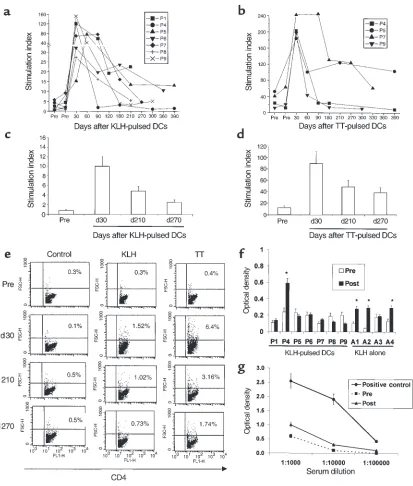
Longevity of KLH and TT-specific T-cell responses. All 9 subjects injected with KLH-pulsed DCs showed priming of KLH-specific immune response (3). T-cell proliferative responses to KLH peaked at a median of 30 days (range 30–90 days) after antigen-pulsed DC injection and declined thereafter (Figure 1a). However, at ≥6 months after the DC injection, proliferative responses to KLH were still detectable above baseline in 4 of 7 (P1, P5, P6, P9) subjects. Five of 6 subjects (P2, P4, P5, P7, P9) injected with TT-pulsed DCs had boosting of TT-specific immunity (3). TT-specif-ic T-cell response also peaked at a median of 30 days after injection and then declined, but remained detectable above baseline at ≥ 6 months in 2 (P5, P7) of 4 responding (P4, P5, P7, P9) subjects available for follow-up (Figure 1b). In the 3 sub-jects not injected with TT-pulsed
DCs, as well as those injected with antigen alone, there was no signifi-cant change in TT-specific responses during follow-up (data not shown). Injection of booster DCs (P4, P5, P6) that were pulsed with MP only also did not lead to a nonspecific increase in KLH- or TT-specific immunity.
To further confirm that the differ-ences between baseline measurements and ≥ 6-month postimmunization time points were not because of interassay variability, samples from these time points, when possible, were thawed and assayed together. Data from a representative volunteer (P5) are shown (Figure 1, c and d). The CD4 nature of the proliferative response in these cultures and the frequency of responding cells was also evident when we monitored T-cell phenotype and proportion of CD4 T-cell blasts (high forward scatter) using flow cytometry (Figure 1e). The maximum number of CD4 T blasts was detected at 30 days after immunization, consistent with the data from proliferation assays on both fresh and thawed cells. We showed previously that KLH and TT responses to antigen-pulsed DCs were CD4 dependent (3).
Development of KLH-specific humoral immunity. Sera from before nization and 3 months after immu-nization were examined for the pres-ence of KLH-specific antibodies in 7 volunteers injected with KLH-pulsed DCs (P1, P4–P9) and those injected with KLH alone (A1–A4). KLH-specif-ic antibody responses (at much lower titer than our positive control serum), were detected in 3 of 4 volunteers injected with KLH alone (Figure 1, f and g). In contrast, KLH-specific anti-bodies were detected in only 1 (P4) of 7 subjects tested who received KLH-pulsed DCs, and these were at simi-larly low titer.
Durability of MP-specific T-cell immu-nity after the first antigen-pulsed DC injec-tion. Circulating MP-specific
IFN-γ–producing CD8+T cells peaked at a
median of 60 days (range 7–90) after DC injection. In 3 of 4 HLA A 2.1+ volunteers (P4, P5, P6), the number of circulating MP-specific CD8+ T
Figure 1
TT- and KLH-specific immunity after DC injection. (a) Longevity of KLH-specific immune response after single DC injection.For each meas-urement shown, 105freshly isolated PBMCs were incubated in the presence or absence of 10 µg/mL KLH for 5 days, and proliferation was
measured as 3H-TdR incorporation. Results are expressed as stimulation index. SEM < 30%; cpm without antigen < 5 ×10³. (b) Longevity of
TT-specific immune response after single DC injection.For each measurement shown, 105freshly isolated PBMCs were incubated in the
pres-ence or abspres-ence of 3 µg/mL TT for 5 days, and proliferation was measured as 3H-TdR incorporation. Results are expressed as stimulation
index. SEM < 30%. (c) Evaluation of longevity of KLH-specific response using cryopreserved specimens. Pre- and postimmunization specimens were thawed together and cultured in the presence or absence of 10 µg/mL KLH for 5 days. Antigen-specific proliferation was measured as
3H-TdR incorporation. Results are shown as stimulation index. Data shown are for 1 subject (P5), representative of 3 subjects tested. (d)
Eval-uation of longevity of TT- specific response using cryopreserved specimens. Pre- and postimmunization specimens were thawed together and cultured in the presence or absence of 3 µg/mL TT for 5 days. Antigen-specific proliferation was measured as 3H-TdR incorporation. Results
are shown as stimulation index. Data shown are for 1 subject (P5), representative of 2 subjects tested. (e) Quantification of CD4 proliferative response as percent of CD4 blasts. T-cell cultures from the experiment in cand dwere stained for CD4 and analyzed by flow cytometry. Per-cent of CD4 blasts were quantified as perPer-cent of CD4+T cells with high forward scatter, noted in the upper-right quadrant. Data shown are
gated for CD4+cells. (fand g) Detection of KLH-specific antibodies. (f) Presence of KLH-specific antibodies was determined in sera before
and 3 months after immunization by ELISA as described in Methods. Data shown are at a serum dilution of 1:104. Because of variable
2d), the number of MP-specific T cells leveled off at a higher level.
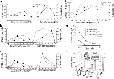
When T cells were boosted in culture with autologous DCs, there was an increase in MP-specific lytic effectors after immunization in 3 of 4 subjects (P1, P5, P6; Figure 2, a–d). In these 3 subjects, the response peaked at a mean of 70 days (range 30–90 days) after injection (Figure 2, b–d). Similar to the ELISPOT assay, MP-specific recall CTL responses had returned back to baseline by 6 months after the first injection in 3 of 4 subjects.
Booster DC injection: toxicity. All 3 HLA A2.1+subjects with declining
circulat-ing MP-specific T cells (P4, P5, P6) were boosted with mature DCs pulsed
with MP alone. The dose of booster DCs was 4.5–5 ×10⁶cells, and purity
ranged from 39–74%. KLH and TT were omitted from booster DCs to determine if these epitopes were required for eliciting MP-specific CD8+T-cell responses using mature
DCs. All booster injections were well tolerated with no greater than grade 1 toxicity or evidence of autoimmunity.
Local reaction to DC injection. Two of 3 subjects developed a local reaction at the injection site. In these 2 subjects these reactions were larger and devel-oped earlier (at 24 versus 48 hours), as compared with those after the first injection (data not shown).
T-cell response to booster DC injections:
assays on uncultured PBMCs. All 3 sub-jects who received a booster DC injec-tion had an increase in the number of circulating MP-specific IFN-γ –produc-ing T cells in freshly isolated PBMCs (Figure 2, a–c). This response was evi-dent as early as 2 days after the injec-tion in 2 subjects and by 7 days in all subjects. Immune response to booster injection was more rapid, with mean 18.4-fold increase in MP-specific T cells in the first week, compared with 4.8-fold increase after the first injection (P
= 0.01). In addition, the peak responses following booster DCs were higher than those after the first injection (19.7-fold versus 7-fold; P = 0.008). As with the first injection, increase in MP-Figure 2
Enhancement of antigen-specific CD8+T cells in vivo. (a–d) Kinetics of antigen-specific CD8+T cells following DC injection(s). MP-specific
interferon-γ–producing T cells in freshly isolated uncultured T cells were quantified using an ELISPOT assay (triangles). Results are shown as the number of spot-forming cells (SFC)/2 ×105PBMC. MP-specific CTLs were quantified after 7-day coculture with MP-pulsed mature
[image:5.612.78.516.54.358.2]specific IFN-γ–producing T cells after booster DCs in 2 subjects with longer follow-up (P5, P6) has not been sus-tained and has declined back to prein-jection baseline within 6 months.
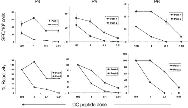
Increased peptide sensitivity of the expand-ed CD8+ T-cell response. We were unable to
detect cytolytic effectors in bulk uncul-tured T cells in any of the subjects after the first DC injection. However, bulk lytic effectors were detected after boost-er DCs in 1 of 3 subjects 7 days aftboost-er injection (P4; Figure 2e). The detection of bulk lytic effectors in this subject (and not in others) could not be explained simply by the frequency of MP-specific IFN-γ–producing T cells. Therefore, we next examined peptide sensitivity of elicited T-cell populations as a measure of functional maturation of the immune response. To control for vari-able thawing of resident APCs in these samples, freshly prepared autologous mature DCs pulsed with various doses of peptides were used as APCs in a 16-hour ELISPOT assay for IFN-γ–secreting cells with thawed uncultured PBMCs from 30 days after the first MP-pulsed DC immunization or booster DCs. T cells elicited after booster DCs demon-strated greater peptide sensitivity than those after the first injection in all 3 sub-jects. Remarkably, T cells in P4 (who developed circulating lytic effectors), showed the greatest peptide sensitivity, with half-maximal recognition at less than 0.01 ng/mL (Figure 3).
T-cell response to booster DCs: assays in cul-tured T cells. When T cells were boosted in culture with peptide-pulsed DCs, there was greater expansion of MP-spe-cific CTLs after booster DCs, relative to the initial DC injection in all 3 volun-teers (Figure 2, a–c). When T cells from before and after the second immuniza-tion were thawed together and cocul-tured with peptide-pulsed DCs, there was a greater expansion of MP-specific IFN-γ–producing T cells after booster DCs, which correlated with the CTL data (Figure 2f). As with the ELISPOT assays in uncultured cells, the increase in T-cell recall assays after the booster DCs was not sustained and was fol-lowed by a contraction phase similar to that after the first injection.
Discussion
In this study, we have followed several
aspects of the T-cell response to 2 DC immunizations in humans. The num-ber of circulating antigen-specific
IFN-γ–secreting CD8+ T cells is rapidly
enhanced following DC injection and peaks by 7–30 days after injection. The measured responses then decline over the next 3 months. This decline in T-cell reactivity may either be because of loss of measured function (e.g., the capacity to secrete cytokines or to pro-liferate in recall assays) or loss of T cells because of activation-induced cell death (AICD) and the rapid disappear-ance of antigen-bearing DC in vivo. Persistence of CD8+T cells may also
depend on the presence of influenza-specific CD4+T cells, which are not
induced by MP. These data about the kinetics of response have important implications for both the optimal schedule of immune monitoring and DC immunizations in clinical trials. Thus, peak immune responses may not be manifest until 1–3 months after the DC injection and frequent (e.g., weekly/bimonthly) injections of DCs may actually be detrimental by induc-ing AICD of recently activated T cells. CD4+T-cell responses to both a
prim-ing (KLH) and boostprim-ing (TT) antigen after a single DC injection also involves a similar rapid increase followed by a
decline. However, these responses remained detectable above baseline for
≥ 6 months in some subjects. The observed lack of KLH-specific antibody response after KLH-pulsed DCs can be attributed to a lack of free/soluble anti-gen when delivered with DCs.
In following the kinetics of T-cell activity using quantitative assays in the blood of humans after DC immuniza-tion, we find that the responses can be detected without the need to expand the T cells in vitro before the assay. The immune responses in humans last sev-eral months, in contrast to a recent study in mice wherein response was evi-dent only in terms of weeks (7).
Recent studies in mice have shown that CD4+T cells help the generation
of CD8+ T-cell responses through
CD40 ligand-mediated activation of DCs (8). Thus, KLH and TT may have provided help for the generation of CD8+T-cell responses after the first
DC injection. Alternatively, mature DCs may already be activated and therefore may not require CD4-medi-ated help, as has been observed in vitro (9). All 3 subjects rapidly responded to MP-pulsed mature DCs without KLH or TT, indicating that these epitopes are not required for eliciting CD8+
[image:6.612.229.539.453.631.2]T-cell responses using mature DCs.
Figure 3
However, exclusion of these foreign helper epitozpes does not stringently exclude CD4 help generated inadver-tently during DC culture (10). Ongo-ing studies will determine if the inclu-sion of these epitopes alters the generation of immune response.
Booster DCs led to significantly high-er and more rapid T-cell responses, detectable as early as 2 days after injec-tion. Thus, although all measured immune responses such as ELISPOT as well as recall assays using DCs had returned to baseline before booster DCs, these subjects had retained “memory” of the first DC injection at the level of the whole individual. Therefore, humans retain a memory for the anti-gen that was administered on the first dose of DCs that we are unable to meas-ure with current functional assays.
A more effective form of protective immunity may be provided by circulat-ing antigen-specific killers. Such responses have been detected in humans previously only following acute viral infections (e.g., acute HIV or Epstein-Barr virus infection) or certain live attenuated vaccines (e.g., measles), but not with subunit vaccines. The development of circulating lytic effec-tors in 1 subject after booster DCs in this study suggests that this may be feasible by using DCs in humans, par-ticularly when one optimizes variables such as DC dose, route of administra-tion, maturation status, helper epi-topes, and DC survival in vivo.
Another important and novel aspect of immune response to booster DCs observed here is the enhanced func-tional avidity of CD8+T-cell response,
manifest as greater peptide sensitivity of T cells elicited after booster DCs. Similar findings were made
independ-ently in mice; i.e., T cells elicited dur-ing secondary viral infection exhibited greater peptide sensitivity (11–13). To our knowledge, this is the first evi-dence of qualitative enhancement of T-cell function in humans after a vac-cination strategy. Higher-affinity T cells may be essential to achieve pro-tective immunity against viruses and tumors in vivo (14, 15). Additional research will be needed to pursue and therapeutically exploit the intriguing potential of DC immunization that is revealed in the current study. A first dose of DCs expands effector and memory T cells, whereas a second dose elicits more rapid, readily measurable responses that include lytic effectors and CD8+effector T cells with higher
functional avidity.
Acknowledgments
This work was supported in part by an Investigator Award from the Cancer Research Institute and a Clinical Research Career Development Award from the American Society of Clinical Oncology (both to M.V. Dhodapkar), grants from the National Institutes of Health (AI-40874 to R.M. Steinman, AI-39516 and AI-44628 to N. Bhard-waj), American Cancer Society (ROG-98-355-01 to R.M. Steinman), the SLE foundation (N. Bhardwaj), and a Gen-eral Clinical Research Center grant (M01-RR00102) from the National Center for Research Resources at the National Institutes of Health. We would like to thank all volunteers for their interest and participation in this study, Coraleen Fossella for help with clinical monitoring, Rockefeller Uni-versity nursing staff for their help with patient care, and Judy Adams for help with graphics.
1. Raychaudhuri, S., and Rock, K.L. 1998. Fully mobilizing host defense: building better vac-cines. Nat. Biotechnol. 16:1025–1031.
2. Banchereau, J., and Steinman, R.M. 1998. Den-dritic cells and the control of immunity. Nature.
392:245–252.
3. Dhodapkar, M.V., et al. 1999. Rapid generation of broad T-cell immunity in humans after a sin-gle injection of mature dendritic cells. J. Clin. Invest.104:173–180.
4. Murphy, G., Tjoa, B., Ragde, H., Kenny, G., and Boynton, A. 1996. Phase I clinical trial: T-cell therapy for prostate cancer using autologous dendritic cells pulsed with HLA-A020-specific peptides from prostate-specific peptides from prostate-specific membrane antigen. Prostate.
29:371–380.
5. Nestle, F.O., et al. 1998. Vaccination of melanoma patients with peptide- or tumor lysate-pulsed dendritic cells. Nat. Med.
4:328–332.
6. Holtl, L., et al. 1999. Cellular and humoral immune responses in patients with metastatic renal cell carcinoma after vaccination with anti-gen pulsed dendritic cells. J. Urol. 161:777–782. 7. Ludewig, B., et al. 1999. Protective antiviral cyto-toxic T cell memory is most efficiently main-tained by restimulation via dendritic cells. J. Immunol.163:1839–1844.
8. Ridge, J.P., Di Rosa, F., and Matzinger, P. 1998. A conditioned dendritic cell can be a temporal bridge between a CD4+T-helper and a T-killer
cell. Nature. 393:474–478.
9. Bhardwaj, N., et al. 1994. Influenza virus-infect-ed dendritic cells stimulate strong proliferative and cytolytic responses from human CD8+T
cells. J. Clin. Invest.94:797–807.
10. Livingstone, A.M., and Kuhn, M. 1999. Dendrit-ic cells need T cell help to prime cytotoxDendrit-ic T cell responses to strong antigens. Eur. J. Immunol.
29:2826–2834.
11. Bachmann, M.F., Speiser, D.E., and Ohashi, P.S. 1997. Functional maturation of antiviral T cell response. J. Virol.71:5764–5768.
12. Busch, D.H., and Pamer, E.G. 1999. T cell affin-ity maturation by selective expansion during infection. J. Exp. Med. 189:701–710.
13. Savage, P.A., Boniface, J.J., and Davis, M.M. 1999. A kinetic basis for T cell receptor reper-toire selection during an immune response.
Immunity. 10:485–492.
14. Alexander-Miller, M.A., Leggatt, G.R., and Berzofsky, J.A. 1996. Selective expansion of high- or low-avidity cytotoxic T lymphocytes and efficacy for adoptive immunotherapy. Proc. Natl. Acad. Sci. USA. 93:4102–4107.
15. Zeh, H.J., III, Perry-Lalley, D., Dudley, M.E., Rosenberg, S.A., and Yang, J.C. 1999. High avid-ity CTLs for two self-antigens demonstrate superior in vitro and in vivo antitumor efficacy.