von Willebrand factor.
Z M Ruggeri
J Clin Invest. 1997;
99(4)
:559-564.
https://doi.org/10.1172/JCI119195
.
Perspective
Find the latest version:
Perspectives Series:
Cell Adhesion in Vascular Biology
J. Clin. Invest.
© The American Society for Clinical Investigation, Inc. 0021-9738/97/02/0559/06 $2.00
Volume 99, Number 4, February 1997, 559–564
von Willebrand Factor
Zaverio M. Ruggeri
Roon Research Laboratory for Arteriosclerosis and Thrombosis, Division of Experimental Thrombosis and Hemostasis, Departments of Molecular and Experimental Medicine and of Vascular Biology, The Scripps Research Institute, La Jolla, California 92037
A perspective on von Willebrand factor (vWF)1 within a series
on cell adhesion in vascular biology offers the opportunity to review the current understanding of platelet function in hemo-stasis and thrombosis. Platelets contribute to maintaining the normal circulation of blood through the preservation of vascu-lar integrity and the control of hemorrhage after injury. Thus, the formation of platelet thrombi is a needed defense mecha-nism, but may precipitate diseases such as myocardial infarc-tion in the setting of atherosclerosis (1, 2). Acute thrombotic arterial occlusion is the leading cause of morbidity and mortal-ity in industrial societies, underscoring the relevance of studies aimed at unraveling how platelets respond to vascular injury. Particularly important in this regard is vWF, along with the subendothelial matrix components and membrane receptors that interact with it, owing to a key role in supporting unique aspects of platelet function. Indeed, vWF-dependent adhesion mechanisms can be viewed as the evolutionary adaptation to the need of establishing firm contact between a circulating ele-ment and the vessel wall meeting any mechanical challenge created by blood flow conditions.
Platelet function: A paradigm of adhesion mechanisms for circulating vascular cells
Platelets survey the inner lining of the vessel wall without in-teracting with it under normal circumstances, but respond rap-idly to alterations of endothelial cells by attaching firmly to the site of lesion, where exposure of subendothelial structures may have occurred. A first layer of platelets adheres to the reactive surface, subsequently growing by accrual of additional plate-lets through homotypic aggregation. Both processes depend on the binding of membrane receptors to immobilized or solu-ble ligands, and are modulated by stimulus-coupled biochemi-cal and cytoskeletal responses. Platelet thrombus formation is the paradigm of adhesion for circulating vascular cells, that can interact efficiently with the vessel wall only by withstanding opposing forces created by blood flow. In fact, the tendency of
cells to move with the layer of fluid adjacent to the reactive surface creates unique biomechanical requirements for the for-mation of adhesive bonds. In a vessel, the velocity of blood near the wall is lower than towards the center, a difference re-sulting in a shearing effect between contiguous layers of fluid moving at different speed. Thus, “shear” is the consequence of the relative parallel motion of fluid planes during flow; it is greatest near the wall and decreases progressively towards the center of the vessel (3). The local shear rate is expressed in cm/s per cm, or the equivalent inverse second (s21). Fluid shear
stress is force per unit area, the underlyingcause of the shear-ing motion of blood. Shear rate is directly proportional to shear stress andinversely proportional to fluid viscosity. As-suming for blood a viscosity of 4 centipoise, the numerical value of shear stress (in dyn/cm2) corresponds to shear rate
di-vided by 40.
The role of platelets requires that they become irreversibly attached at sites of vascular injury. The force opposing stable adhesion and aggregation is greater with increasing shear rate, i.e., in arteries more than in veins and, particularly, in arteri-oles. The highest wall shear rate in the normal circulation oc-curs in small arterioles of 10–50 mm diameter, where levels have been estimated to vary between 500 and 5,000 s21 (4).
Yet, these are the vessels where effective hemostasis is more strictly dependent on the ability of platelets to form thrombi. The relevance of adhesion mechanisms responsive to high flow conditions may be even greater in pathological conditions as-sociated with the occurrence of acute arterial occlusion. Wall shear rates of 3,000–10,000 s21 have been measured at the top
of plaques occluding the lumen of diseased coronary arteries by 50% (5), a degree of stenosis still considered of moderate clinical significance (1, 2), and considerably higher values may occur with more severe occlusion. These considerations high-light the importance of vWF as the essential adhesive substrate mediating platelet thrombus formation against high shear forces.
von Willebrand factor structure
The structural organization of the vWF molecule has been elu-cidated (reviewed in reference 6). The glycoprotein is synthe-sized in endothelial cells and megakaryocytes as a precursor polypeptide of 2813 amino acids (pre-pro-vWF), including the 12-residue signal peptide, the 751-residue propeptide and the 2050-residue mature subunit. After cleavage of the signal pep-tide, complex intracellular processing leads first to dimeriza-tion of pro-vWF, initiated by the noncovalent associadimeriza-tion of propeptide moieties, then to covalent polymerization with nu-merous interchain disulfide bonds both at the amino- and car-boxyl-terminal ends. Multimers are either stored in specific
or-Address correspondence to Zaverio M. Ruggeri, M.D., The Scripps Research Institute, SBR-8, 10550 No. Torrey Pines Road, La Jolla, CA 92037. Phone: 619-784-8950; FAX: 619-784-2026; E-mail: ruggeri@ scripps.edu
Received for publication 7 January 1997.
ganelles for regulated release—Weibel-Palade bodies in endothelial cells and a-granules in megakaryocytes (or plate-lets after thrombocytopoiesis)—or constitutively secreted. The latter pathway is operative only in endothelial cells, with bidi-rectional secretion both into the circulating blood and the sub-endothelial matrix. Megakaryocytes do not constitutively se-crete vWF, but platelets release it during thrombogenesis. The 741-residue propeptide is normally cleaved before secretion of the multimers, and is found in the circulation where it may act as an independent modulator of cell adhesion to collagen. The degree of vWF polymerization is to some extent directly cor-related to prothrombotic activity. The largest multimers are found in the subendothelium and in platelet a-granules, whereas in blood (where soluble vWF and platelet mem-brane receptors are exposed to one another) a specific pro-teolytic cleavage in the constitutive subunit reduces multimer size after secretion (7, 8). Yet, vWF remains one of the largest circulating proteins, with molecular mass that can exceed 10,000 kD. Preliminary information has become available on the enzyme responsible for cleaving circulating vWF (9), open-ing a potentially important field of research on thrombotic dis-orders linked to accumulation of large multimers in blood. As important as the polymeric nature of vWF is for establishing
multiple adhesive bonds with platelets, specific substrate and receptor specificities crucial for this function reside in distinct subunit domains.
[image:3.612.60.557.332.601.2]The binding sites for all molecules known to interact with vWF have been located with good approximation at the level of primary sequence. The amino-terminal domain within the first 272 residues of the mature subunit forms a complex with factor VIII, essential to maintain adequate circulating levels of this cofactor involved in thrombin generation. All other func-tional sites in the vWF molecule support platelet adhesion and aggregation by binding to extracellular matrix components or to membrane receptors. Two vWF domains, A1 and A3, have been shown to interact with collagen in a variety of different experimental models, but more recent evidence indicates that only the latter may be required for this function (10). How-ever,the A1 domain can also bind to heparin-like molecules (11)—thus, possibly, proteoglycans—and to sulfatides (12), playing in any case a potentially important role in the immobi-lization of soluble vWF onto complex extracellular matrices (13). Notwithstanding other possible functions, it is established that the A1 domain and flanking regions (residues 449–728 of the mature subunit) represent the binding site for GP Iba in the platelet membrane GP Ib-IX-V complex (14). The site of
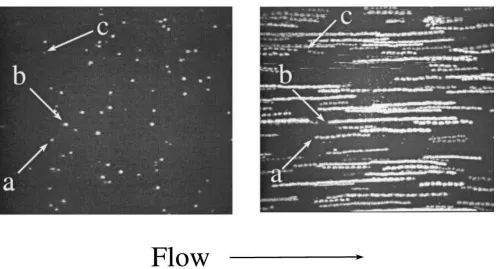
Figure 1. Low velocity surface translocation of platelets interacting with immobilized von Willebrand factor. These images are derived from a real time experiment recorded at the video rate of 30 frames per second. Whole blood containing as anticoagulant d-phenylalanyl-l -prolyl-l-arginine chloromethyl ketone dihydrochloride (PPACK), an inhibitor of a-thrombin, was perfused onto a surface coated with immobilized vWF at wall shear rate of 1,500 s21. The image on the left represents a single frame, corresponding to a time period of 1/30th of a second, recorded 1
min after initiating blood perfusion. Each fluorescent (white) particle is a single platelet tethered to the surface; three individual platelets are identified by arrows (a, b, and c). The image on the right is a composite created by the superimposition of 30 successive frames, taken at one sec-ond intervals beginning from the one shown on the left, thus covering a time period of 30 s. Each of the three platelets identified on the left ap-pears at the origin of a track extending for variable length in the direction of flow, demonstrating movement from the position originally occu-pied. The composite image shows that all platelets tethered to the surface translocate for variable distances, thus have different velocities. The speed of motion has been found to vary between 2 and 20 mm per second when the wall shear rate is 1,500 s21, corresponding to , 2% of the
cal-culated free flow velocity of platelets moving with the surrounding flowing blood in close proximity to the surface but not interacting with it (17). Freely moving platelets are not visible at the flow rate used for this experiment. Blocking aIIbb3 has no effect on the surface translocation of
interaction for aIIbb3, the second platelet receptor binding to
vWF, is the RGDS sequence located at residues 1744–1747 in the carboxyl-terminal C1 domain.
von Willebrand factor function in thrombogenesis
The function of vWF is to promote thrombus formation by mediating adhesion of platelets to the injured vessel wall and to one another. There is evidence that vWF in different ana-tomical locations participates in hemostatic processes. The cir-culating pool, however, is probably the most important in initi-ating platelet adhesion, since the distribution of subendothelial vWF is not homogeneous and the protein lacks in many vessels where platelet function is needed for hemostasis (15). More-over, platelet vWF is not released until after activation, and may not be immediately available at a site of injury to promote adhesion, although its contribution to later phases of thrombus formation has been convincingly demonstrated (16). Plasma vWF is well suited to mediate early adhesion because it binds rapidly and tightly to collagen (and, possibly, to other matrix structures such as proteoglycans) whenever blood is exposed to injured tissues. Differential reconstitution of vWF in body compartments of pigs with severe von Willebrand disease pro-vides good support for these concepts (16).
The unique mechanism supporting platelet adhesion to im-mobilized vWF under high flow conditions has been eluci-dated (17). The first step of this dynamic process is mediated
by the binding of platelet GP Iba to the vWF A1 domain, an interaction characterized by a fast association rate that can tether platelets to exposed thrombogenic surfaces even when the velocity of flowing blood relative to the vessel wallis ele-vated. This event is favored by the multimeric nature of vWF, providing a high local density of active A1 domain sites and re-sulting in formation of multiple bonds. However, the vWF-GP Iba interaction is also characterized by a fast dissociation rate and cannot provide bonds supporting irreversible adhesion, so that platelets tethered to the vessel wall in this manner move constantly in the direction of flow, albeit at a fraction of the free flow velocity (Fig. 1). During the slow translocation plate-lets become activated, and aIIbb3 can eventually mediate
irre-versible adhesion by binding to the RGDS sequence in the vWF C1 domain. The recognition specificity of aIIbb3 before
activation, relevant for initial adhesion, is limited to interac-tion with immobilized fibrinogen or fibrin, consequently bind-ing to vWF can only occur after platelet tetherbind-ing to the sur-face mediated by GP Iba. Moreover, the forward rate of binding interactions involving aIIbb3 appears to be relatively
[image:4.612.60.553.418.622.2]slow, explaining why this receptor is not sufficient to mediate initial platelet attachment to the vessel wall under high flow conditions, even though it is always required for firm adhesion (17). Thus, vWF is the indispensable adhesive substrate for thrombus formation in high shear environments because of the unique biomechanical properties of A1 domain binding to GP
Figure 2. Schematic representation of the dual step mechanism supporting platelet adhesion to immobilized von Willebrand factor. The first contact established between platelets and immobilized vWF is mediated by GP Iba. Nonactivated aIIbb3 cannot pair with the RGDS sequence in
vWF (38). The bond between the vWF A1 domain and GP Iba must form rapidly, since platelets can be tethered to a vWF-coated surface re-gardless of flow conditions (17), and must also have high resistance to tensile stress, since adherent platelets can oppose the drag force created by flow even when the resulting wall shear rate is greater than 30,000 s21, the highest tested (17). This bond, however, has an intrinsically high
disso-ciation rate, thus a limited half-life, resulting in detachment where tension is greatest, and forward rotational movement (rolling) due to the torque imposed by the flowing fluid. New bonds are formed as different regions of the membrane of rolling platelets come in closer contact with the surface. Translocation continues until aIIbb3 becomes activated and binds to the RGDS sequence in the vWF C1 carboxyl terminal domain.
Iba. Only this interaction can tether fast flowing platelets and markedly decrease their velocity relative to the vessel wall, al-lowing the occurrence of subsequent events (Fig. 2).
In experimental conditions where only purified vWF was exposed to platelets, and exogenous agonists were either not present or inhibited,demonstrable consequences of activation, i.e. thrombus formation, were seen only after several minutes and only at high shear rates (17).In the setting ofvascular le-sions, however, subendothelial components such as collagen, and soluble platelet agonists such as ADP and epinephrine, greatly enhance the efficiency of the hemostatic response by contributing synergistically to aIIbb3 activation. These
mecha-nisms critically accelerate the attainment of irreversible plate-let adhesion at sites of injury where plasma vWF becomes im-mobilized onto exposed collagen, and explain the functional relevance of a2b1 and other collagen receptors in hemostasis
(18, 19). Indeed, irreversible platelet adhesion onto matrices containing collagen type I or III is essentially instantaneous even at the highest levels of shear stress, and thrombus forma-tion ensues within seconds. This response is accelerated by the rapid activation of platelets, but it is still only possible owing to the unique function of GP Iba and the vWF A1 domain (Fig. 2). The relevant role of vWF in supporting initial platelet ad-hesion at sites of vascular injury is complemented by its func-tion in mediating platelet–platelet cohesion. In fact, aggrega-tion of platelets exposed to high shear stress is dependent on vWF binding to aIIbb3, as well as GP Iba, and fibrinogen cannot
substitute for this function (20, 21). Studies with whole blood perfused over subendothelial surfaces support these conclu-sions (22). The reasons why vWF is required for aggregation under high shear are not fully understood, but are likely to be explained by specific biomechanical and structural properties of the molecule. There is evidence that shear forces may in-duce vWF to take the shape of extendedfilaments (23). Thus, the repeating subunit structure of the large multimers may of-fer an array of interactionsites capable of binding in a multiva-lent manner to receptors on theplatelet membrane, increasing the strength of interaction and providing better linkage of platelets to one another. The divalent fibrinogen molecule, on the other hand, may provide sufficient adhesive strength to withstand opposing shear forces of lesser magnitude. More-over, high shear stress induces vWF binding to platelets in the absence of any other chemical or physical modulator, a process dependent on interaction with both GP Iba and aIIbb3 (24)
that may promote preferential vWF localization on the platelet membrane. Shear-induced vWF binding is associated with platelet activation independent of other agonists (3), an addi-tional factor favoring aggregation mediated by vWF in the ini-tial stages of thrombus development.
Perspective on future studies
In spite of considerable progress in our understanding of the mechanisms involved in platelet thrombus formation, we still lack comprehension of the specific contributions provided by individual adhesive interactions to the process as a whole. This is due, at least in part, to the fact that the experimental models of platelet function traditionally used to mimic events occur-ring in flowing blood provide only a limited representation of the complexity present at a site of vascular injury, both with re-spect to the cells and molecules involved as well as the relevant hemodynamic conditions. Some answers to this problem will come from the use of mouse models allowing targeted
manipu-lation of individual components of the hemostatic system, cou-pled with studies of platelet response to vascular damage in in-tact organisms. Moreover, ongoing developments of confocal videomicroscopy will result in more accurate volumetric and morphometric analysis of thrombus formation during blood flow in ex vivo experiments. These approaches should provide some of the information necessary to design and use rationally new inhibitors of platelet adhesion and aggregation as anti-thrombotic drugs.
Animal models with targeted gene obliteration or mu-tagenesis should also help in obtaining more definitive infor-mation on whether vWF is involved in the development of ath-erosclerotic lesions, the process that develops over a long period of time and precedes acute arterial occlusion. High plasma levels of the protein have been found to be an indepen-dent risk factor for recurrent myocardial infarction and death (25). Because endothelial cells are the origin of all circulating vWF, increased plasma levels may reflect the extent of vascu-lar damage. Experiments in animals have suggested a caus-ative link between vWF and atherosclerosis, demonstrating that pigs with severe von Willebrand disease, who have mark-edly reduced levels of vWF, are protected from developing aortic plaques even when fed a cholesterol-rich diet (26). Fur-thermore, there is good evidence that decreased concentra-tions of circulating vWF limit the extent of occlusive thrombo-sis in stenosed and injured coronary arteries (27). The latter effect is not surprising in view of the essential role played by vWF in platelet thrombus formation under high shear stress. Less clear are the mechanisms to explain the possible partici-pation of vWF in plaque development, although it seems rea-sonable to assume that influences on platelet adhesion and, consequently, generation of cytokines are involved. It also seems reasonable to anticipate that studies will be performed on genetic polymorphisms of vWF potentially linked to en-hanced function, in order to evaluate their possible role as a risk factor for occlusive cardiovascular disorders.
Studies on von Willebrand disease (28), the most common genetic disorder in humans, will continue to focus on the defi-nition of the molecular defects responsible for decreased vWF function, providing new information on the importance of sin-gle amino acid residues in supporting specific interactions. Progress has to be expected in elucidating the potential role of vWF in disorders such as thrombotic thrombocytopenic pur-pura and hemolytic uremic syndromes, that appear to be asso-ciated with the presence of unusually large vWF multimers in blood (29, 30). Preliminary evidence indicates that decreased plasma levels of a specific processing enzyme (9) are responsi-ble for the accumulation of hyperactive forms of vWF, poten-tially explaining the diffuse thrombotic occlusion seen in the arterial microvasculature of these patients. Clarification of such issues has the potential to lead to new therapeutic modal-ities for these serious and often life-threatening diseases.
With respect to vWF function, a number of unanswered questions must be addressed with additional experimental work. There is already sufficient evidence that any vWF-dependent platelet activity involves two membrane receptors, GP Iba and aIIbb3, and initial information has been obtained
by GP Ib-IX-V and its role in promoting subsequent vWF binding to aIIbb3. Moreover, the possibility that GP Iba-vWF
A1 domain bonds contribute directly to supporting platelet at-tachment to one another, in addition to any participation in ac-tivation, must still be evaluated, notwithstanding the current dogma that aIIbb3 is the only receptor mediating aggregation.
Equally needed is more definitive information on the mechanisms that regulate the initial recognition of vWF by GP Iba. It is generally assumed that conformational changes are induced in the vWF A1 domain by interaction with collagen or other matrix components at sites of vascular injury, and per-haps by shear forces in GP Iba as well as vWF, allowing ligand-receptor pairing normally prevented in the circulation. To date, there is only experimental evidence demonstrating that the overall shape of vWF multimers may be affected by shear forces (23), but nothing is known about more subtle con-formational changes in the relevant interactive sites. In vitro, soluble vWF binds to GP Iba only in the presence of exoge-nous modulators, but all those identified to date, such as risto-cetin and botroristo-cetin, are nonphysiologic substances. The mode of action of these modulators has been elucidated, at least in part. Ristocetin has been shown to dimerize in solution and the dimeric forms interact with both platelets and vWF, thus bridging GP Iba with its ligand (31). Botrocetin, on the other hand, forms a stoichiometric bimolecular complex with soluble vWF and this complex, in turn, interacts with GP Iba (32). In either case, irreversible vWF binding to platelets requires only GP Iba, an apparent contradiction with the results obtained in the absence of these exogenous substances indicating that the dissociation rate of the interaction may be too high to allow measurement of equilibrium binding. Thus, as an alternative hypothesis to be tested, the initiation of vWF-dependent plate-let responses may be controlled not by regulation of the A1 domain-GP Iba interaction (which may always occur with rapid on and off rates) but by modulation of the activation needed for irreversible vWF binding to platelets through en-gagement of aIIbb3. The observation that the largest plasma
vWF multimers are relatively decreased in patients with reac-tive thrombocythemia (33) agrees with the notion that vWF binding to GP Iba may be ongoing in the normal circulation. In these cases, the elevated platelet count, increasing receptor concentration per unit volume, would only contribute to mak-ing the event more easily detectable, while the preferential re-moval of larger multimers can be explained by their known higher binding affinity. The hypothesis that the vWF-GP Iba
interaction need not be regulated by an off/on switch is com-patible with the evidence that specific residues in the A1 do-main may be responsible for do-maintaining functional conforma-tions with different affinity for GP Iba, presumably allowing transitions from one to another. The occurrence in patients with type IIB von Willebrand disease of mutations causing measurable binding of soluble vWF to GP Iba in the absence of any exogenous modulator (28) further indicates that the function of the A1 domain can be regulated. To date, however, there is no information on the mechanisms that could induce such affinity changes during hemostasis in vivo. Detailed knowledge on the three-dimensional structure of important functional domains of vWF, including A1, is rapidly becoming available through x-ray crystallography, setting the stage for a rational approach to answering these questions.
Finally, more studies will undoubtedly be devoted to test the hypothesis that a drug preventing the binding of vWF to
GP Iba—or, presumably, the binding of vWF to collagen— should provide an efficacious antithrombotic intervention. Pigs with von Willebrand disease have a markedly decreased incidence of occlusive thrombosis in stenosed and injured cor-onary arteries (34), even in the setting of atherosclerosis (27). Experiments with a monoclonal antibody blocking vWF bind-ing to GP Iba have shown efficacy in preventing occlusive thrombosis in a pig coronary artery thrombosis model (35). At present, the only candidate GP Iba inhibitor for use in humans is a recombinant fragment representing essentially the Al do-main of vWF.Studies with this compound, VCL, have shown antithrombotic efficacy and enhancement of thrombolysis (36), as well as prevention of intimal thickening after balloon injury of vessels (37). Whether new drugs based on the con-cept of inhibiting the unique functions of vWF in thrombogen-esis will be developed remains conjectural at present.
References
1. Fuster, V., L. Badimon, J.J. Badimon, and J.H. Chesebro. 1992. The pathogenesis of coronary artery disease and the acute coronary syndromes (1). [Review.] N. Engl. J. Med. 326:242–250.
2. Fuster, V., L. Badimon, J.J. Badimon, and J. H. Chesebro. 1992. The pathogenesis of coronary artery disease and the acute coronary syndromes (2). [Review.] N. Engl. J. Med. 326:310–318.
3. Kroll, M.H., J.D. Hellums, L.V. McIntire, A.I. Schafer, and J.L. Moake. 1996. Platelets and shear stress. Blood. 88:1525–1541.
4. Tangelder, G.J., D.W. Slaaf, T. Arts, and R.S. Reneman. 1988. Wall shear rate in arterioles in vivo: least estimates from platelet velocity profiles. Am. J.
Physiol. 254:H1059–H1064.
5. Back, C.H., J.R. Radbill, and D.W. Crawford. 1977. Analysis of pulsatile viscous blood flow through diseased coronary arteries of man. J. Biomech. 10: 339–353.
6. Ruggeri, Z.M., and J. Ware. 1993. von Willebrand factor. FASEB J. 7: 308–316.
7. Dent, J.A., S.D. Berkowitz, J. Ware, C.K. Kasper, and Z.M. Ruggeri. 1990. Identification of a cleavage site directing the immunochemical detection of molecular abnormalities in type IIA von Willebrand factor. Proc. Natl. Acad. Sci. USA. 87:6306–6310.
8. Dent, J.A., M. Galbusera, and Z.M. Ruggeri. 1991. Heterogeneity of plasma von Willebrand factor multimers resulting from proteolysis of the con-stituent subunit. J. Clin. Invest. 88:774–782.
9. Furlan, M., R. Robles, and B. Lammle. 1996. Partial purification and characterization of a protease from human plasma cleaving von Willebrand fac-tor to fragments produced by in vivo proteolysis. Blood. 87:4223–4234.
10. Cruz, M.A., H. Yuan, J.R. Lee, R.J. Wise, and R.I. Handin. 1995. Inter-action of the von Willebrand factor (vWF) with collagen. J. Biol. Chem. 270: 10822–10827.
11. Fujimura, Y., K. Titani, L.Z. Holland, J.R. Roberts, P. Kostel, Z.M. Ruggeri, and T.S. Zimmerman. 1987. A heparin-binding domain of human von Willebrand factor. Characterization and localization to a tryptic fragment ex-tending from amino acid residue Val-449 to Lys-728. J. Biol. Chem. 262:1734– 1739.
12. Christophe, O., B. Obert, D. Meyer, and J. Girma. 1991. The binding domain of von Willebrand factor to sulfatides is distinct from those interacting with glycoprotein Ib, heparin, and collagen and resides between amino acid res-idues Leu 512 and Lys 673. Blood. 78:2310–2317.
13. Denis, C., D. Baruch, C.M. Kielty, N. Ajzenberg, O. Christophe, and D. Meyer. 1993. Localization on von Willebrand factor binding domains to endo-thelial extracellular matrix and to type VI collagen. Arterioscl. Thromb. 13:398– 406.
14. Fujimura, Y., K. Titani, L. Z. Holland, S.R. Russell, J.R. Roberts, J.H. Elder, Z.M. Ruggeri, and T.S. Zimmerman. 1986. von Willebrand factor.A re-duced and alkylated 52/48 kDa fragment beginning at amino acid residue 449 contains the domain interacting with platelet glycoprotein Ib. J. Biol. Chem. 261:381–385.
15. Bahnak, B.R., Q. Wu, L. Coulombel, Z. Assouline, D. Kerbiriou-Nabias, G. Pietu, L. Drouet, J.P. Caen, and D. Meyer. 1989. Expression of von Wille-brand factor in porcine vessels: Heterogeneity at the level of von WilleWille-brand factor mRNA. J. Cell. Physiol. 138:305–310.
16. Nichols, T., C. Samama, D. Bellinger, J. Roussi, R. Reddick, and M. Bonneau. 1995. Function of von Willebrand factor after crossed bone marrow transplantation between normal and von Willebrand disease pigs: effect on ar-terial thrombosis in chimeras. Proc. Natl. Acad. Sci. USA. 92:2455–2459.
Cell. 84:289–297.
18. Nieuwenhuis, H.K., J.W.N. Akkerman, W.P.M. Houdijk, and J.J. Sixma. 1985. Human blood platelets showing no response to collagen fail to express surface glycoprotein Ia. Nature (Lond.). 318:470–472.
19. Moroi, M., S.M. Jung, M. Okuma, and K. Shinmyozu. 1989. A patient with platelets deficient in glycoprotein VI that lack both collagen-induced ag-gregation and adhesion. J. Clin. Invest. 84:1440–1445.
20. Peterson, D.M., N.A. Stathopoulos, T.D. Giorgio, J.D. Hellums, and J.L. Moake. 1987. Shear-induced platelet aggregation requires von Willebrand factor and platelet membrane glycoproteins Ib and IIb-IIIa. Blood. 69:625–628. 21. Ikeda, Y., M. Handa, K. Kawano, T. Kamata, M. Murata, Y. Araki, H. Anbo, Y. Kawai, K. Watanabe, I. Itagaki, K. Sakai, and Z.M. Ruggeri. 1991. The role of von Willebrand Factor and fibrinogen in platelet aggregation under varying shear stress. J. Clin. Invest. 87:1234–1240.
22. Weiss, H.J., J. Hawiger, Z.M. Ruggeri, V.T. Turitto, P. Thiagarajan, and T. Hoffmann. 1989. Fibrinogen-independent platelet adhesion and thrombus formation on subendothelium mediated by glycoprotein IIb-IIIa complex at high shear rate. J. Clin. Invest. 83:288–297.
23. Siedlecki, C.A., B.J. Lestini, K.Kottke-Marchant, S.J. Eppell, D.L. Wil-son, and R.E. Marchant. 1996. Shear-dependent changes in the three-dimen-sional structure of human von Willebrand factor. Blood.88:2939–2950.
24. Goto, S., D.R. Salomon, Y. Ikeda, and Z.M. Ruggeri. 1995. Character-ization of the unique mechanism mediating the shear-dependent binding of sol-uble von Willebrand factor to platelets. J.Biol.Chem. 270:23352–23361.
25. Jansson, J.H., T.K. Nilsson, and O. Johnson. 1991. von Willebrand fac-tor in plasma: a novel risk facfac-tor for recurrent myocardial infarction and death. Br. Heart J. 66:351–355.
26. Fuster, W., E.J. Bowie, J.C. Lewis, D.N. Fass, C.A.J. Owen, and A.L. Brown. 1978. Resistance to arteriosclerosis in pigs with von Willebrand’s dis-ease. Spontaneous and high cholesterol diet-induced arteriosclerosis. J. Clin. Invest. 61:722–730.
27. Nichols, T.C., D.A. Bellinger, R.L. Reddick, M.S. Read, G.G. Koch, K.M. Brinkhous, and T.R. Griggs. 1991. Role of von Willebrand factor in arte-rial thrombosis. Studies in normal and von Willebrand disease pigs. Circulation. 83:56–64.
28. Cooney, K.A., D. Ginsburg, and Z.M. Ruggeri. 1994. von Willebrand disease. In Thrombosis and Hemorrhage. J. Loscalzo and A. Schafer, editors. Blackwell Scientific Publications, Boston. 657–682.
29. Moake, J.L., C.K. Rudy, J.H. Troll, M.J. Weinstein, N.M. Colannino, J. Azacar, R.H. Seder, S.L. Hong, and D. Deykin. 1982. Unusually large plasma factor VIII: von Willebrand factor multimers in chronic relapsing thrombotic thrombocytopenic purpura. N. Engl. J. Med. 307:1432–1435.
30. Moake, J.L., J.J. Byrnes, J.H. Troll, C.K. Rudy, M.J. Weinstein, N.M. Colannino, and S.L. Hong. 1984. Abnormal VIII: von Willebrand factor pat-terns in the plasma of patients with hemolytic-uremic syndrome. Blood. 64:592– 598.
31. Scott, J.P., R.R. Montgomery, and G.S. Retzinger. 1991. Dimeric risto-cetin flocculates proteins, binds to platelets, and mediates von Willebrand fac-tor-dependent agglutination of platelets. J. Biol. Chem. 266:8149–8155.
32. Read, M.S., S.V. Smith, M.A. Lamb, and K.M. Brinkhous. 1989. Role of botrocetin in platelet agglutination: formation of an activated complex of botrocetin and von Willebrand factor. Blood.74:1031–1035.
33. Budde, U., R.E. Scharf, P. Franke, K. Hartmann-Budde, J. Dent, and Z.M. Ruggeri. 1993. Elevated platelet count as a cause of abnormal von Wille-brand factor multimer distribution in plasma. Blood.82:1749–1757.
34. Nichols, T.C., D.A. Bellinger, T.A. Johnson, M.A. Lamb, and T.R. Griggs. 1986. von Willebrand's disease prevents occlusive thrombosis in stenosed and injured porcine coronary arteries. Circ. Res. 59:15–26.
35. Bellinger, D.A., T.C. Nichols, M.S. Read, R.L. Reddick, M.A. Lamb, K.M. Brinkhous, B.L. Evatt, and T.R. Griggs. 1987. Prevention of occlusive cor-onary artery thrombosis by a murine monoclonal antibody to porcine von Wille-brand factor. Proc. Natl. Acad. Sci. USA. 84:8100–8104.
36. Yao, S.K., J.C. Ober, L.I. Garfinkel, Y. Hagay, N. Ezov, J.J. Ferguson, H.V. Anderson, A. Panet, M. Gorecki, L.M. Buja, and J.T. Willerson. 1994. Blockade of platelet membrane glycoprotein Ib receptors delays intracoronary thrombogenesis, enhances thrombolysis, and delays coronary artery reocclu-sion in dogs. Circulation. 89:2822–2828.
37. Zahger, D., M.C. Fishbein, L.I. Garfinkel, P.K. Shah, J.S. Forrester, J. Regnstrom, J. Yano, and B. Cercek. 1995. VCL, an antogonist of the platelet GP1b receptor, markedly inhibits platelet adhesion and intimal thickening after balloon injury in the rat. Circulation. 92:1269–1273.
38. Savage, B., S.J. Shattil, and Z.M. Ruggeri. 1992. Modulation of platelet function through adhesion receptors: A dual role for glycoprotein IIb-IIIa (in-tegrinIIb 3) mediated by fibrinogen and glycoprotein Ib-von Willebrand fac-tor. J. Biol. Chem. 267:11300–11306.
“Cell Adhesion In Vascular Biology”
Series Editors, Mark H. Ginsberg, Zaverio M. Ruggeri, and Ajit P. Varki
October 15, 1996 Adhesion and signaling in vascular cell–cell interactions ... Guy Zimmerman, Tom McIntyre, and
... Stephen Prescott
November 1, 1996 Endothelial adherens junctions: implications in the control of vascular
permeability and angiogenesis ... Elisabetta Dejana
November 15, 1996 Genetic manipulation of vascular adhesion molecules in mice...Richard O. Hynes and Denisa D. Wagner
December 1, 1996 The extracellular matrix as a cell cycle control element in
atherosclerosis and restenosis ... Richard K. Assoian and ... Eugene E. Marcantonio
December 15, 1996 Effects of fluid dynamic forces on vascular cell adhesion... Konstantinos Konstantopoulos and
... Larry V. McIntire January 1, 1997 The biology of PECAM-1 ... Peter J. Newman January 15, 1997 Selectin ligands: Will the real ones please stand up?... Ajit Varki February 1, 1997 Cell adhesion and angiogenesis ... Joyce Bischoff February 15, 1997 von Willebrand Factor ... Zaverio Ruggeri
March 1, 1997 Therapeutic inhibition of carbohydrate-protein interactions in vivo ... John Lowe and Peter Ward
March 15, 1997 Integrins and vascular matrix assembly... Erkki Ruoslahti
April 1, 1997 Platelet GPIIb/IIIa antagonists: The first anti-integrin receptor therapeutics... Barry Coller
April 15, 1997 Importance of shear stress in endothelial adhesion molecule expression... Michael Gimbrone
May 1, 1997 Proteoglycans and proteoglycan-binding proteins in vascular biology ... Robert Rosenberg
May 15, 1997 New insights into integrin-ligand interaction... Robert Liddington and Joseph Loftus
June 1, 1997 Adhesive interactions of Sickle erythrocytes with endothelium... Robert Hebbel
June 15, 1997 Cell migration in vascular biology ... Stephen Schwartz
July 1, 1997 Integrin signaling in vascular biology ... Sanford Shattil and Mark Ginsberg
July 15, 1997 Multi-step mechanisms of leukocyte homing ... Eugene Butcher