Characterization of SR 121463A, a highly potent
and selective, orally active vasopressin V2
receptor antagonist.
C Serradeil-Le Gal, … , J P Maffrand, G Le Fur
J Clin Invest.
1996;
98(12)
:2729-2738.
https://doi.org/10.1172/JCI119098
.
SR 121463A, a potent and selective, orally active, nonpeptide vasopressin V2 receptor
antagonist, has been characterized in several in vitro and in vivo models. This compound
displayed highly competitive and selective affinity for V2 receptors in rat, bovine and human
kidney (0.6 < or = Ki [nM] < or = 4.1). In this latter preparation, SR 121463A potently
antagonized arginine vasopressin (AVP)-stimulated adenylyl cyclase activity (Ki =
0.26+/-0.04 nM) without any intrinsic agonistic effect. In autoradiographic experiments performed in
rat kidney sections, SR 121463A displaced [3H]AVP labeling especially in the
medullo-papillary region and confirmed that it is a suitable tool for mapping V2 receptors. In
comparison, the nonpeptide V2 antagonist, OPC-31260, showed much lower affinity for
animal and human renal V2 receptors and lower efficacy to inhibit vasopressin-stimulated
adenylyl cyclase (Ki in the 10 nanomolar range). Moreover, OPC-31260 exhibited a poor V2
selectivity profile and can be considered as a V2/V1a ligand. In normally hydrated
conscious rats, SR 121463A induced powerful aquaresis after intravenous (0.003-0.3
mg/kg) or oral (0.03-10 mg/kg) administration. The effect was dose-dependent and lasted
about 6 hours at the dose of 3 mg/kg p.o. OPC-31260 had a similar aquaretic profile but with
markedly lower oral efficacy. The action of SR 121463A was purely aquaretic with no
changes in urine Na+ and K+ excretions unlike that of known […]
Research Article
Find the latest version:
J. Clin. Invest.
© The American Society for Clinical Investigation, Inc. 0021-9738/96/12/2729/10 $2.00
Volume 98, Number 12, December 1996, 2729–2738
Characterization of SR 121463A, a Highly Potent and Selective, Orally Active
Vasopressin V
2Receptor Antagonist
Claudine Serradeil-Le Gal,* Colette Lacour,‡ Gérard Valette,* Georges Garcia,‡ Loïc Foulon,* Gérard Galindo,‡ Lise Bankir,§
Brigitte Pouzet,§ Gilles Guillon,iClaude Barberis,iDanielle Chicot,i Serge Jard,iPol Vilain,* Corine Garcia,* Eléonore Marty,*
Danielle Raufaste,* Gabrielle Brossard,* Dino Nisato,‡ Jean Pierre Maffrand,* and Gérard Le Fur*
*Sanofi Recherche, Exploratory Research Department, 31036 Toulouse Cedex, France; ‡Sanofi Recherche, Cardiovascular Department,
34184 Montpellier Cedex, France; §INSERM U 90, Hôpital Necker, 75743 Paris Cedex, France; and iINSERM U 401, Rue de la
Cardonille, 34094 Montpellier Cedex, France
Abstract
SR 121463A, a potent and selective, orally active, nonpep-tide vasopressin V2 receptor antagonist, has been
character-ized in several in vitro and in vivo models. This compound displayed highly competitive and selective affinity for V2
re-ceptors in rat, bovine and human kidney (0.6 # Ki [nM]
# 4.1). In this latter preparation, SR 121463A potently an-tagonized arginine vasopressin (AVP)-stimulated adenylyl cyclase activity (Ki5 0.2660.04 nM) without any intrinsic agonistic effect. In autoradiographic experiments performed in rat kidney sections, SR 121463A displaced [3H]AVP
la-beling especially in the medullo-papillary region and con-firmed that it is a suitable tool for mapping V2 receptors. In
comparison, the nonpeptide V2 antagonist, OPC-31260,
showed much lower affinity for animal and human renal V2
receptors and lower efficacy to inhibit vasopressin-stimu-lated adenylyl cyclase (Ki in the 10 nanomolar range).
More-over, OPC-31260 exhibited a poor V2 selectivity profile and
can be considered as a V2/V1a ligand. In normally hydrated
conscious rats, SR 121463A induced powerful aquaresis af-ter intravenous (0.003–0.3 mg/kg) or oral (0.03–10 mg/kg) administration. The effect was dose-dependent and lasted about 6 hours at the dose of 3 mg/kg p.o. OPC-31260 had a similar aquaretic profile but with markedly lower oral effi-cacy. The action of SR 121463A was purely aquaretic with no changes in urine Na1 and K1 excretions unlike that of known diuretic agents such as furosemide or hydrochloro-thiazide. In addition, no antidiuretic properties have been detected with SR 121463A in vasopressin-deficient Brattle-boro rats. Thus, SR 121463A is the most potent and selec-tive, orally active V2 antagonist yet described and could be a
powerful tool for exploring V2 receptors and the
therapeuti-cal usefulness of V2 blocker aquaretic agents in
water-retain-ing diseases. (J. Clin. Invest. 1996. 98:2729–2738.) Key
words: SR 121463A • vasopressin • nonpeptide antagonist • V2 receptor • aquaretic
Introduction
The importance of arginine vasopressin (AVP)1 in the regula-tion of blood pressure and volume and in the control of fluid and electrolyte balance is well established. AVP plays a major role as an antidiuretic hormone regulating water and solute ex-cretion by the kidney through specific interaction with the re-nal V2 receptors present all along the collecting duct from cor-tex to papilla in the mammalian nephron (1, 2).
So far, three AVP receptors subtypes, V1a, V1b, and V2 have been identified based upon their primary structure (3–10), their coupling mechanisms, their tissular distributions and their pharmacological properties (for review see 11, 12). The V2 receptor belongs to the seven transmembrane G protein-coupled receptor superfamily and is positively protein-coupled to a Gs/ adenylyl cyclase system. This V2 receptor has been cloned in different species including rat, pig, bovine and human (7–10). Moreover, several constitutive AVP V2 receptor gene muta-tions have now been identified as the molecular basis for the lack of urine concentration in Nephrogenic Diabetes Insipidus (13, 14).
Receptor-specific AVP V2 antagonists, so-called “aquaretic agents,” able to block the action of AVP in the collecting duct cells and thus to promote specifically water excretion, could be of high therapeutical value for the treatment of several water-retaining disorders such as SIADH (Syndrome of Inappropri-ate Antidiuretic Hormone secretion), liver cirrhosis, certain stages of congestive heart failure and hypertension, nephrotic syndrome (15–18). In most of these diseases an abnormal in-crease of circulating AVP plasma level, activating renal V2 re-ceptors, seems to be the key event in water retention and sub-sequent hypotonic hyponatremia (18–20). Thus, for these pathologies, there is great clinical interest in the development of potent V2 receptor antagonists to provide specific water di-uretic/aquaretic compounds devoid of the well-known side ef-fects of classical diuretic or saliuretic agents on the solute ex-cretion (urine Na1 and/or K1 loss).
Although several potent peptide vasopressin V2 receptor antagonists have been synthesized, the evaluation of their therapeutic utility has been severely hampered by their lack of oral bioavailability, species differences and especially by their agonist antidiuretic effects when tested in man (21). Recent years have marked a turning point with the design of the first nonpeptide, orally effective AVP V1a and V2 receptor
antago-Address correspondence to C. Serradeil-Le Gal, Sanofi Recherche, 195 route d’Espagne, 31036 Toulouse Cedex, France. Phone: 33 5 61 16 23 84; FAX: 33 5 61 16 25 86.
Received for publication 10 June 1996 and accepted in revised form 10 October 1996.
nists (22–24). In this field, Yamamura et al. reported an orally effective V2 compound, OPC-31260, exerting aquaresis in sev-eral animal models and in man without agonistic activity (25, 26).
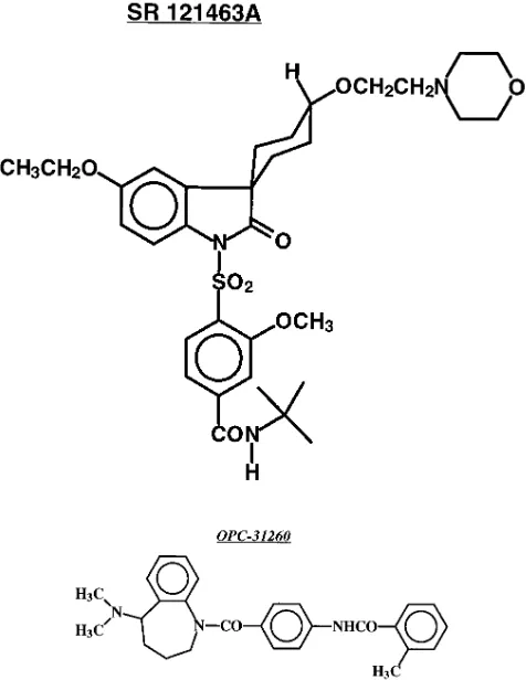
In this study, we describe the structure and the pharmaco-logical properties of SR 121463A, (1-[4-(N-tert-butylcarba-moyl)-2-methoxybenzene sulfonyl]-5-ethoxy-3-spiro-[4-(2-mor-pho-linoethoxy)cyclohexane]indol-2-one, fumarate), a novel, highly potent and V2-selective nonpeptide AVP receptor an-tagonist (see Fig. 1). In all the biological tests used, a close comparison was performed between the effects of the two nonpeptide molecules, SR 121463A and OPC-31260, belong-ing to different chemical series.
In addition, despite the close structural similarities between AVP V2 receptors from different species (7–10), marked dif-ferences between their pharmacological properties have been described (21). For these reasons, special attention was paid to the evaluation of SR 121463A in several human tissues and in predictive pharmacological models in order to design a com-pound devoid of partial agonist effects and with the expected aquaretic activity in man.
Methods
Materials
The newly described nonpeptide molecule, SR 121463A, (1-[4-(N-tert-butylcarbamoyl)-2-methoxybenzene sulfonyl]-5-ethoxy-3-spiro-[4-(2-morpholinoethoxy)cyclohexane]indol-2-one, fumarate; equato-rial isomer), (Fig. 1), was synthesized in Sanofi Recherche, Toulouse, (France) and belongs to an original chemical series of potent and se-lective V2 receptor antagonists. The only other published nonpeptide
V2 receptor antagonist, OPC-31260, (6
)(5-dimethylamino-1-{4-(2-methylbenzoylamino)benzoyl}-2,3,4,5-tetrahydro-1H-benzazepine) (24), was synthesized in Sanofi Recherche, (Montpellier, France) and used as a reference in the different biological tests (Fig. 1). The structures of SR 121463A and OPC-31260 were determined by 1H and 13C NMR
and infrared spectroscopy. The molecular weights, determined by mass spectrometry, are 736.6 and 427.5 for SR 121463A and OPC-31260, respectively. Melting points of 172 and 207.88C, respectively, were obtained. The purity, measured by high pressure liquid chroma-tography, thin layer chromatography and elemental analysis, was . 98%. The analytical parameters reported above for OPC-31260 are identical to those initially described for this molecule (27).
For in vitro experiments, SR 121463A and OPC-31260 were dis-solved in DMSO (1022 M) and then diluted in the appropriate test
solvent. SR 121463A was dissolved in saline and DMF for intrave-nous and intraperitoneal administration in rats, respectively. For oral treatment, all compounds used were administered in 0.6% methylcel-lulose solution.
AVP, polybrene, PMSF, OT, and bacitracin were from Sigma Chemical Co. (L’Isle d’Abeau, France). BSA type V was obtained from IBF (Villeneuve La Garenne, Paris, France). DME and PBS were from Boehringer Mannheim (Meylan, France). All other cell culture reagents were from GIBCO (Life Technologies, GIBCO BRL, France). EDTA, Tris and DMSO were purchased from Merck-Clevenot (Nogent sur Marne, France). All other chemicals were from Prolabo (Paris, France). SK&F 101926 (desGly-d(CH2)5
D-Tyr(Et)VAVP) (21), used as a reference in the Brattleboro rat exper-iments, was a generous gift from Dr. L. Kinter. The radioligands, [3H]AVP (80 Ci/mmole), [125I]OT antagonist, (d(CH
2)5Tyr(Me)2,
Thr4, Orn8 [125I]Tyr9-NH
2) (2,000 Ci/mmole), [3H]cAMP (40 Ci/
mmole) and [a-32P]-ATP (20 Ci/mmole) were obtained from New
England Nuclear, Les Ulis, France. Linear AVP antagonist (Phaa-D-Tyr(Me)-Phe-Gln-Asn-Arg-Pro-Arg-Tyr-NH2) was iodinated as
pre-viously described in (28).
Biological material
Human tissue samples from adrenals, kidneys, and pituitaries were collected in conformity with the French national ethical rules. Healthy human adrenals were obtained from human donors after brain death. Adrenals were chilled in cold saline and dissected. Crude plasma membranes were prepared within 3–5 h and stored in liquid nitrogen. Human kidneys were obtained from nephrectromy for renal carcinoma. The tissue was chilled into cold saline and dissected 2–8 h after excision. Only the non pathological part of the kidney was used to prepare crude plasma membranes which were stored in liquid ni-trogen. Human pituitary glands were collected from deceased per-sons within 6 h after death and immediately stored in liquid nitrogen. Crude plasma membranes were prepared from the frozen tissue be-fore each experiment. Bovine kidneys were obtained from a local slaughterhouse. Mammary tissue was taken from 19-d-old Sprague-Dawley pregnant rats and stored in liquid nitrogen until used. Male Sprague-Dawley rats, 250-–350 g, (Iffa-Credo, Lyon, France) were used for in vitro binding studies and for in vivo activity measure-ments. Two series of male homozygous Brattleboro rats with central Diabetes Insipidus (DI) weighing 300–350 g, bred in house (INSERM Unité 90, Hôpital Necker, Paris, France) or bought from Harlan Sprague-Dawley (Indianapolis, IN), were used for evaluation of po-tential agonist antidiuretic activities. All protocols performed in Sanofi Recherche have been approved by the Animal Care and Use Committee of Sanofi Recherche.
In vitro experiments
Membrane preparations.Human hypophyseal membranes were pre-pared as previously described in (23). Membrane preparations from human kidneys or adrenal cortex were obtained according to Guillon et al. (29) and (30), respectively. Membranes from Ltk2 cells,
[image:3.612.316.554.402.710.2]fected with the DNA encoding for the human oxytocin receptor, were prepared as in (31). Briefly, 72 h after transfection, cells were har-vested, washed twice in PBS without Ca21 and Mg21, polytron-homog-enized in lysis buffer (15 mM Tris-HCl pH 7.5; 2 mM MgCl2; 0.3 mM
EDTA), and centrifuged at 100 g for 5 min at 48C. Pellets were washed in a buffer A consisting of 50 mM Tris-HCl, pH 7.4; 5 mM MgCl2 and centrifuged at 44,000 g for 20 min at 48C. Membranes were
suspended in a small volume of buffer A and protein contents were determined. Aliquots of membranes were used immediately or stored at 2808C.
Membranes from rat and bovine kidney (papilla and inner me-dulla), rat liver and rat mammary glands, were prepared according to Stassen et al. (32), Prpic et al. (33) and Serradeil-Le Gal et al. (23), re-spectively.
Protein concentration was determined by the method of Bradford using bovine serum albumin as a standard (34).
Binding assays
Binding affinity constants of SR 121463A and OPC-31260 for the dif-ferent AVP/OT receptors investigated were deduced from compe-tition experiments using the appropriate radiolabeled ligands: [3H]AVP (bovine and rat kidney, rat liver and human pituitary);
[125I]linear AVP antagonist (adrenals); [125I]OT antagonist (rat
mam-mary glands and Ltk2 cells expressing the uterine oxytocin receptor). Experiments were performed as described earlier (23). Binding as-says of [3H]AVP to rat kidney medullary membranes were conducted
according to the method of Yamamura et al. (24).
For competition experiments, increasing concentrations of SR 121463A or OPC-31260 were incubated with membranes and the cor-responding ligand under the above-specified conditions. Saturation binding experiments using [3H]AVP (from 0.08 to 20 nM) as a ligand
were performed in bovine kidney in the absence (control) or pres-ence of SR 121463A (0.25, 0.50, 1, 2, and 4 nM).
Binding data analysis
The IC50 value was defined as the concentration of inhibitor required
to obtain 50% inhibition of the specific binding. Inhibition constant (Ki) values were calculated from the IC50 values using the Cheng and
Prusoff equation (34). Data for equilibrium binding (Kd, Bmax),
com-petition experiments (IC50, nHill), and kinetic constants (Kobs, K21)
were analyzed using an iterative non-linear regression program (35). IC50 values of SR 121463A and OPC-31260 were compared using
Stu-dent’s t test. Values of P , 0.05 were taken as significant. Autoradiography
Male Sprague-Dawley rats (250–300 g) were killed by decapitation and the kidneys were rapidly removed and frozen in isopentane at 2458C. Serial sections (15 mm) were cut in a cryostat microtome and thaw-mounted onto chrome alum gelatin-coated glass slides (gelatin 1%, chrome-alum 0.05%). Sections were stored at 2808C until use. Slide-mounted sections (3–4 sections/slide), brought to room temper-ature, were preincubated for 15 min in the binding buffer (50 mM Tris-HCl, pH 8.1, 2 mM MgCl2, 1 mM EDTA, 0.1% bovine serum
al-bumin and bacitracin). Incubation was carried out for 1 h at room temperature in the incubating medium in the presence of 2 nM [3H]AVP. Nonspecific binding was determined by incubating
addi-tional slides under the same conditions and in the presence of 1 mM unlabeled AVP. After incubation, the sections were washed three times for 10 min each in ice-cold buffer, dipped briefly in distilled wa-ter, and dried under a stream of cold air. Labeled sections were placed on a phosphor-imaging plate (Fuji) for 4 d and further ana-lyzed with a Bio-Image Analyser (BAS 2000, Fuji) as described in (37).
Adenylyl cyclase assay. Adenylyl cyclase activity was measured as previously described (29) by the rate of conversion of [a-32P]ATP
into labeled cyclic AMP. The incubation medium contained: 10 mM sodium azide; 0.1 mM ouabain; 1 mM cAMP; 0.25 mM ATP; 0.17 mCi/ml [3H]cAMP; 20 mM creatine phosphate; 1 mg/ml bovine serum
albumin and various amounts of the nonpeptide analogues. Mem-brane proteins, 10–30 mg per assay, were preincubated for 15 min at 308C and the reaction was initiated by adding ATP 0.25 mM plus 1 mCi of [a-32P]ATP with or without 10 nM AVP. The membranes
were incubated for an additional 10 min period and the reaction stopped by adding 500 ml of a solution containing: 100 mM Tris-HCl, pH 8.0; 80 mM sodium dodecylsulfate; 1 mM ATP and 1 mM cAMP. Labeled cAMP was separated and counted by liquid scintillation spectrometry as previously described in (29). All values were cor-rected for cAMP recovery estimated from the recovery of the [3H]cAMP added to the incubation medium and for a blank value
de-termined in the absence of membranes. Adenylyl cyclase activities were expressed as pmol cAMP accumulated/10 min per mg protein. All determinations were performed in duplicate.
In vivo experiments
Properties of SR 121463A in normally hydrated conscious rats. Male Sprague Dawley rats weighing 290–320 g were kept in an air-condi-tioned room at 22628C and fed with a standard rat diet (AO4, UAR Epinay sur Orge, France) with water provided ad libitum. In a first set of experiments, SR 121463A was administered intravenously at doses varying from 0.003 to 0.3 mg/kg (i.e. 0.004 to 0.4 mmol/kg) body weight (n 5 6) to normally hydrated conscious rats. SR 121463A was injected (1 ml/kg) in 0.9% NaCl through a catheter placed in a jugular vein, 48 h before the experiment. The animals were then housed indi-vidually in metabolic cages with water and food ad libitum. Urine was collected for 4 h. In a second set of experiments, SR 121463A, 0.03–10 mg/kg (0.04–14 mmol/kg, n 5 7), OPC-31260, 10 mg/kg (23 mmol/kg, n 5 8), furosemide, 30 mg/kg (91 mmol/kg, n 5 8), hydrochlorothia-zide, 30 mg/kg (101 mmol/kg, n 5 8) or vehicle (methylcellulose 0.6%, n 5 20) were administered orally to rats by gavage (3 ml/kg). After treatment, the rats were placed individually in metabolic cages with food and water ad libitum. Urine was collected throughout the 24-h period after treatment. In the time-course study, the aquaretic effect was measured by collecting urine at 2 h intervals for 6 h and then from 6 to 24 h in control (methylcellulose 0.6%, n 5 20) and treated groups (SR 121463A, 0.03–10 mg/kg p.o., and OPC-31260, 10 mg/kg). The effects of the drugs tested on urine osmolality, and urine Na1 and K1 excretions were tested on a 24-h urine collection period after drug administration. Urine osmolality was measured with a freezing point depression osmometer (model Fisk OS 110, Elvetec, Montpellier, France) and urinary sodium and potassium concentrations with a flame photometer (IL 943, Instrument Laboratories, Marseille, France).
Statistical significance of the results was analyzed by one-way analysis of variance on independent measurements followed by Dun-nett or Kruskal-Wallis’ test. A two-way analysis of variance followed by Dunnett’s test was also used when appropriate.
Activity of SR 121463A in vasopressin-deficient Brattleboro rats in comparison with the V2 peptide antagonist SK&F 101926 The activity of SR 121463A was compared with that of the reference AVP V2 peptide, SK&F 101926, in an experimental
Results
Interaction of SR 121463A with animal and human AVP V2
re-ceptors, and in vitro selectivity profile.As shown in Table I,
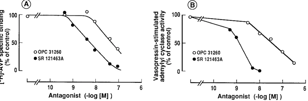
SR 121463A displayed a high affinity for AVP V2 receptors from several species including man. This nonpeptide com-pound dose-dependently inhibited [3H]AVP specific binding to kidney medullo-papillary membranes from rat, bovine and human origin with a Hill coefficient close to the unity, compat-ible with a single site competitive model as illustrated in hu-man kidney membranes (Fig. 2 A).
Moreover, saturation binding experiments performed in a bovine kidney preparation in the absence or presence of SR 121463A (0.25, 0.50, 1, 2, and 4 nM) confirmed that this com-pound interacted competitively with renal AVP V2 receptors. Indeed, in the presence of this molecule, the apparent dissocia-tion constant (Kd) was dose-dependently decreased, whereas the maximal binding capacity (Bmax) was not significantly mod-ified (Fig. 3). The Ki value calculated from Scatchard plots
(0.7860.15 nM) was consistent with that obtained according to the Cheng and Prusoff equation in competition experiments performed in bovine kidney preparations (Ki5 0.87 nM).
In terms of selectivity, the highly V2-specific profile of SR 121463A was firstly evidenced for other AVP receptor sub-types (V1a, V1b) and for the closely related oxytocin (OT) re-ceptor from both animal and human origin. As shown in Table I, SR 121463A exhibited only weak affinities for other AVP/ OT receptors and interacted with at least a 100 fold lower po-tency with V1a, V1b , and OT receptors than with V2. Second, the high degree of specificity of SR 121463A for the V2 recep-tor was further demonstrated in several additional binding as-says (n 5 50) showing that SR 121463A (1 mM) was unable to bind to a variety of receptors of nonpeptide (histamine, adren-ergic, dopamine, serotonin, adenosine, L-type calcium chan-nel, benzodiazepine) or peptide ligands (angiotensin II, endo-thelin, neuropeptide Y, cholecystokinin, CRF, neurotensin) (not shown).
The comparison with the nonpeptide compound,
OPC-Table I. Comparative Affinities of SR 121463A and OPC-31260 for Vasopression and Oxytocin Receptors in Animal and Human Species
Ki (nM)
V2 V1a V1b OT
Rat Bovine Human Rat Human Human Rat Human
Mammary glands
Uterus (Ltk2 cells) Kidney Kidney Kidney Liver Adrenal Pituitary
SR 121463A 1.42*60.98 0.64*60.14 4.1*60.8 10,60066,000 4606120 . 10,000 3,22061,200 1,2136383
OPC-31260 21.769.1 10.960.8 25.468.6 7486254 260630 . 10,000 2,0966618 1,0776319
Binding assays were performed as described in Methods. Inhibition constants (Ki) were determined from competition experiments and calculated
ac-cording to the equation of Cheng and Prusoff (35). Values are the mean6SD of at least three determinations. V2 IC50 values of SR 121463A and
[image:5.612.63.558.83.196.2]OPC-31260 were compared using Student’s t test. Values of P, 0.05 were taken as significant.
Figure 2. Effect of SR 121463A on binding to AVP V2 receptors (A) and on AVP-stimulated adenylyl cyclase activity (B) in human kidney
membranes. Comparison with OPC-31260. Binding assays were performed for 60 min at 378C in the presence of 1 nM [3H]AVP, 10–30 mg of
membranes and increasing concentrations of SR 121463A or OPC-31260. Nonspecific binding was determined by adding 1 mM unlabeled AVP in the incubating medium. Specific binding is the difference between total and nonspecific binding, and is expressed as percentage of specific binding determined without drug (100% 5 1364 fentomoles of [3H]AVP specifically bound/mg protein). For adenylyl cyclase activity, AVP 10
nM or vehicule (basal) and [a-32P]-ATP were added in the assay for 10 min at 308C. [32P]cAMP generated was then determined as described
[image:5.612.63.557.488.651.2]31260, tested under similar experimental conditions, showed that this molecule displayed significantly lower affinity (6 to 17-fold) than SR 121463A for AVP V2 receptors in the different species tested (Table I, Fig. 2 A). The affinity found here for OPC-31260 both in rat kidney and liver membranes is in agreement with the original published values (24). In addition, OPC-31260 showed only a relatively weak specificity for V2 re-ceptors since this molecule also exhibited significant affinity for AVP V1a receptors especially in human tissues (Table I). The selectivity index of SR 121463A and OPC-31260 for the human V2 receptor versus the human V1a, V1b, and OT recep-tors clearly evidenced a better specificity for SR 121463A
(. 100) (Table II) showing that OPC-31260 should rather be considered as a V2/V1a compound, whereas SR 121463A is the first potent and highly selective nonpeptide V2 ligand yet de-scribed.
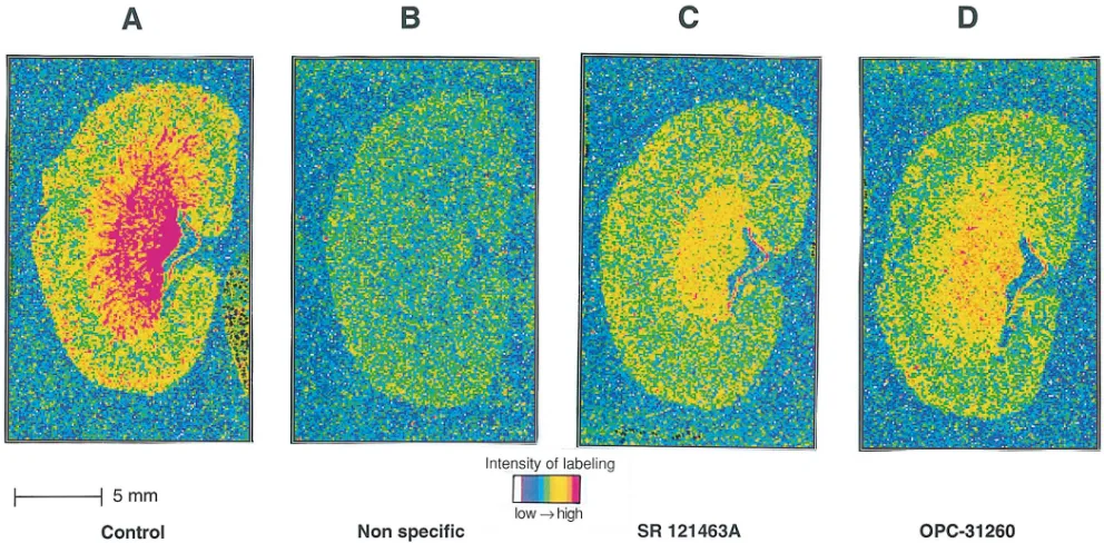
Autoradiographic localization of AVP V2 receptors in the
rat kidney using SR 121463A. Autoradiographic experiments,
using [3H]AVP as a ligand, provided the mapping and localiza-tion of AVP receptors in the rat kidney. As illustrated in Fig. 4
A, [3H]AVP intensively labeled the medullo-papillary region of the kidney and, to a lesser extent, the cortex, as previously described (39, 40). SR 121463A (100 nM) displaced most of [3H]AVP labeling in the medullo-papillary area (Fig. 4 C). The remaining amounts of labeling observed, particularly in the re-gion of the medulla, in the pelvic wall and in the cortex could be due to [3H]AVP binding to V
[image:6.612.55.298.55.234.2]1a receptors also present in this organ (37, 40). At the same 100 nM concentration, OPC-31260 appears to be less potent than SR 121463A in displacing [3H]AVP labeling in rat kidney sections (Fig. 4 D), as expected according to the affinity in rat kidney membranes observed for OPC-31260, almost 20-fold lower than for SR 121463A (Table
Table II. Selectivity Profile of the Two Nonpeptide AVP V2
Receptor Antagonists, SR 121463A and OPC-31260, for Human Vasopressin and Oxytocin Receptors
Selectivity index
KiV1a/KiV2 KiV1b/KiV2 KiOT/KiV2
SR 121463A 112 . 2400 296
OPC-31260 10 . 390 42
Inhibition constants (Ki) used in the calculation of these ratios are given
[image:6.612.313.555.92.165.2]in Table I.
Figure 3. Scatchard plots of [3H]AVP binding to bovine kidney
mem-branes without (d) or with 0.25 (,), 0.5 (.), 1 (s), 2 (j), and 4 (h) nM SR 121463A. Results represent data from a typical experiment. All experiments were performed in duplicate.
Figure 4. Autoradiograms of [3H]AVP binding to rat kidney sections in the absence (A) or presence of 1 mM AVP (B), 0.1 mM SR 121463A (C),
[image:6.612.57.553.456.700.2]I). Thus, SR 121463A represents a selective V2 probe for studying the in situ localization of V2 receptors.
Effect of SR 121463A on adenylyl cyclase activity in human
kidney membranes. To determine the agonist or antagonist
properties of SR 121463A, we examined the activity of this compound on the AVP-induced adenylyl cyclase activity in human kidney membranes. In these preparations, AVP maxi-mally stimulated cAMP production by 3.660.3-fold with a Kact of 2.5 nM (data not shown) in good agreement with previous results (29). Data shown in Fig. 2 B demonstrate that both SR 121463A and OPC-31260 dose-dependently inhibited AVP (10 nM)-induced adenylyl cyclase stimulation yielding respective
Ki values of 0.2660.04 and 17.668.3 nM (n5 3). These results clearly evidenced a higher potency of SR 121463A versus OPC-31260 in inhibiting AVP-stimulated adenylyl cyclase, as expected from the binding affinities found for these two com-pounds for the human kidney V2 receptors (Fig. 2 A). More-over, neither SR 121463A nor OPC-31260 were able to stimu-late the basal adenylyl cyclase activity in human kidney membranes in concentrations up to 100 mM, showing the total absence of agonistic properties (not shown).
In vivo activity of SR 121463A in normal conscious rats.
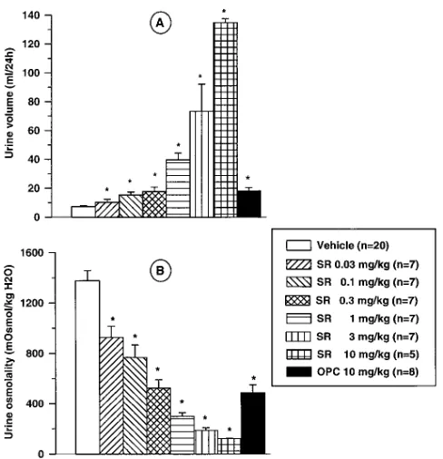
In normally hydrated conscious rats, oral administration of SR 121463A, 0.03–10 mg/kg (0.04–14 mmol/kg), increased urine excretion and decreased urine osmolality dose-dependently (Fig. 5). This effect was significant from 0.03 mg/kg on urine osmolality and from 0.1 mg/kg and upwards for urine volume (P, 0.05). The time-course showed a rapid effect on urine
flow rate. The maximal effect was reached during the first 2 hour-period after the administration of the different doses of SR 121463A (Fig. 6). The higher doses (3 and 10 mg/kg) had effects lasting into the 6–24 h sampling period as shown by a urine flow rate higher than that occurring in the vehicle-treated group. Under identical experimental conditions, OPC-31260, 10 mg/kg (23 mmol/kg) p.o., was equipotent to SR 121463A, 0.3–1 mg/kg (0.4–1.4 mmol/kg) p.o., on the drop in urine osmolality and on the increase in 24 h collected urine, in agreement with the lower V2 receptor affinity found for OPC-31260 in rat kidney membranes (Table I).
Intravenous administration of SR 121463A from 0.003 to 0.3 mg/kg, (0.004 –0.4 mmol/kg), induced a dose-dependent in-crease in urine flow rate which was exactly parallel to the oral dose-response curve (Fig. 7). From these plots, a ratio of 5 be-tween intravenous and oral efficacies could be deduced (0.1 mg/kg [0.14 mmol/kg] i.v. induced similar effect to 0.5 mg/kg [6.8 mmol/kg] p.o.) demonstrating good oral bioavailability for SR 121463A.
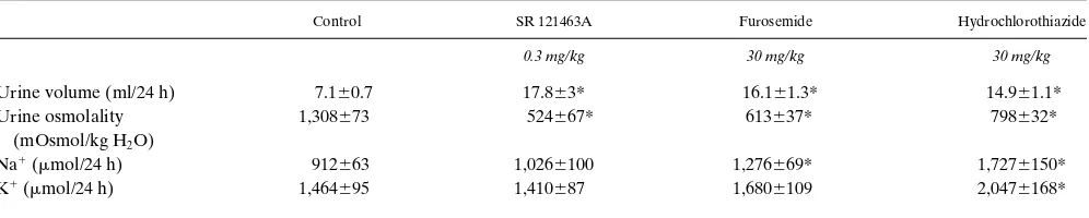
Table III further summarizes the effects of SR 121463A on urine flow rate, osmolality and Na1/K1 excretion in compari-son with those of traditional diuretic compounds such as furo-semide and hydrochlorothiazide. These agents were used at oral doses eliciting similar effects on urine volume excretion (0.3 mg/kg i.e. 0.4mmol/kg for SR 121463A, 30 mg/kg for furo-Figure 5.Effects of SR 121463A on cumulative urine volume (A) and
[image:7.612.56.300.56.309.2]urine osmolality (B) after oral administration (0.03–10 mg/kg, i.e. 0.04–14 mmol/kg) in conscious rats. Comparison with OPC-31260, 10 mg/kg (23 mmol/kg) p.o. Urine was collected for 24 h after SR 121463A (SR) or OPC-31260 (OPC) administration in a 0.6% meth-ylcellulose solution. Values are means6SEM of 5–20 determinations per group. Statistical analysis was performed using a Kruskall-Wallis’ test and the level of significance was taken as P , 0.05 for comparison with the control.
[image:7.612.316.555.57.294.2]semide (91 mmol/kg) and hydrochlorothiazide (101 mmol/kg). At this oral dose, SR 121463A caused an increase in urine vol-ume and a drop in urine osmolality with no significant change (P . 0.05) in Na1 and K1excretion over a 24-h period (Table III). Furosemide and hydrochlorothiazide, 30 mg/kg p.o., in-duced roughly similar effects to SR 121463A, 0.3 mg/kg, on urine flow rate and osmolality but, as well-known for classical diuretic/saliuretic agents, the urine Na1 excretion was mark-edly increased (Table III). It is important to note that hydro-chlorothiazide, studied during a 24 hour period, induced a strong effect on both urine Na1 and K1 excretion.
Thus, SR 121463A is the most potent orally-effective V2 an-tagonist i.e. aquaretic agent yet described. It is devoid of the effects of traditional diuretic agents on solute excretion (Na1 and/or K1 loss).
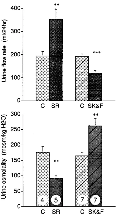
Activity of SR 121463A in vasopressin-deficient Brattleboro rats in comparison with SK&F 101926.SK&F 101926, a prom-ising peptide V2 receptor antagonist with a powerful aquaretic effect in several animal species and models, turned out to be a partial V2 agonist with potent antidiuretic properties when tested in man. In fact, the partial agonistic properties of this
compound could also be detected in the vasopressin-deficient Brattleboro rat strain because these rats have higher sensitivity to antidiuretic agonists than AVP-repleted rats (21, 41, 42). These findings suggest that this experimental model allows the assessment of the partial agonistic activities of putative aqua-retic agents. We studied the influence of SR 121463A in com-parison with SK&F 101926 on urine excretion and osmolality in these rats (Fig. 8). As expected, control Brattleboro rats, ex-hibiting hereditary central DI, were characterized by a high urine flow rate (z 200 ml/24 h) and low urine osmolality (z 170 mOsm/kg H2O). As shown in Fig. 8, SR 121463A (10 mg/kg i.p.) induced significant aggravation of these parameters by further enhancing urine flow rate by 77% (354642 ml/24 h) and lowering urinary osmolality by 54% (9268 mOsm/kg H2O) (P , 0.01). In contrast, SK&F 101926 induced antidi-uretic effects in this model by lowering urine volume and in-creasing urine osmolality by 60% each, as previously de-scribed. It is important to note that further experiments, performed with increasing concentrations of SR 121463A, have shown that the effects observed on urine volume and os-molality in DI rats are dose-dependent and are detectable with doses as low as 0.01 mg kg i.p. (data not shown). In conclusion, SR 121463A appears to be devoid of any antidiuretic proper-ties even when administered at a high dose in Brattleboro rats. Moreover, this molecule is able to enhance diuresis in this ani-mal model.
Discussion
Specific blockade of renal AVP V2 receptors seems to be a rel-evant approach for generating selective aquaretic agents for the treatment of water-retaining disorders (15–20). Therefore, intensive work and active investigations have been performed to design selective ligands for AVP receptors (22–24, 43). The present study describes the biochemical and pharmacological profile of SR 121463A, a newly potent and selective nonpep-tide AVP V2 receptor antagonist with powerful oral aquaretic properties in the rat.
[image:8.612.57.298.58.234.2]This new molecule (Fig. 1), belonging to an original chemi-cal series, shows high affinity and marked selectivity for AVP V2 receptors from animal and human origin (Table I). In bind-ing experiments, SR 121463A inhibits [3H]AVP labeling to rat, bovine and human kidney membranes with nanomolar and even subnanomolar potency (Ki values of 1.42, 0.64, and 4.1 nM, respectively). As demonstrated in bovine kidney prepara-tions, SR 121463A is a full competitive inhibitor (Fig. 3). Since Figure 7. Comparison of intravenous and oral administration of SR
121463A on urine flow rate in normally-hydrated conscious rats. Dose-response curves were obtained after i.v. injection of SR 121463A in isotonic saline and after p.o. administration in a 0.6% methylcellulose solution. Data are expressed as means6SEM of 5 to 7 determinations per group.
Table III. Effects of SR 121463A, Furosemide and Hydrochlorothiazide p.o. on Urine Volume and Osmolality, and on Na1 and K1
Urinary Excretion in Normally Hydrated Rats
Control SR 121463A Furosemide Hydrochlorothiazide
0.3 mg/kg 30 mg/kg 30 mg/kg
Urine volume (ml/24 h) 7.160.7 17.863* 16.161.3* 14.961.1*
Urine osmolality 1,308673 524667* 613637* 798632*
(mOsmol/kg H2O)
Na1 (mmol/24 h) 912663 1,0266100 1,276669* 1,7276150*
K1 (mmol/24 h) 1,464695 1,410687 1,6806109 2,0476168*
[image:8.612.59.557.602.702.2]we (23, 44), and others (29, 45, 46), have previously demon-strated marked species differences in the in vitro affinity and potency of several nonpeptide AVP/OT receptor antagonists, especially when tested in human tissues, it is of importance to underline that SR 121463A also potently interacts with human renal V2 receptors. Moreover, the high affinity of SR 121463A found in this latter preparation is consistent with the powerful antagonist effect of this compound in inhibiting AVP-stimu-lated adenylyl cyclase (Ki value of 0.26 nM) without any ago-nistic response (Fig. 2).
Another striking finding is the high degree of selectivity of SR 121463A for AVP V2 receptors as evidenced in several binding tests in vitro. Firstly, this compound has very low affin-ity for AVP V1a, V1b and for the related oxytocin receptors (se-lectivity index at least . 100; Table II) and secondly, SR 121463A does not interact with more than 50 receptors of other nonpeptide or peptide ligands. It is noteworthy that
de-spite the low affinity of SR 121463A for V1a receptors, this molecule is able to discriminate rat liver and human adrenal V1a receptors (Table I).
When the properties of OPC-31260, the only other pub-lished nonpeptide V2 receptor antagonist, and SR 121463A are compared under similar experimental conditions, OPC-31260 is significantly less potent (Ki. 10 nM versus Ki in the nano-molar range for SR 121463) in inhibiting [3H]AVP binding to rat, bovine and human kidney membranes and in antagonizing AVP-stimulated adenylyl cyclase in human kidney plasma membranes in vitro (Table I and Fig. 2). Moreover, SR 121463A is much more selective towards the V2 AVP receptor subtype than OPC-31260 (Table II). Indeed, despite a well-conserved affinity of OPC-31260 for human V2 receptors, this compound exhibits a poor V2 selectivity profile especially no-ticeable on human AVP and OT receptors (Tables I and II). These data are consistent with previous results showing a sig-nificant interaction of OPC-31260 with human V1a receptors, both in binding (44) and functional studies. In vitro, OPC-31260 effectively antagonizes AVP-induced contractions in human isolated internal mammary and coronary arteries (47, 48), known to express the AVP V1a receptor subtype. Thus, this molecule could be considered as a V2/V1a antagonist com-pound whereas SR 121463A is a pure V2 ligand.
In addition, autoradiographic experiments performed in rat kidney sections, using [3H]AVP as the ligand and SR 121463A, show an intense localization of V2 binding sites specifically in the medullo-papillary region of the kidney in agreement with previous reports using mRNA V2 receptor in situ hybridiza-tion and autoradiographic techniques (39, 40). Indeed, SR 121463A is the most V2 selective ligand so far described. Thanks to its high stability, it may represent a good pharmaco-logical tool for mapping V2 receptors in the organism and for the search of extrarenal V2 sites (49) and/or potential V2 sub-types. In that respect, a V2 receptor subtype has been sug-gested in rat limbic brain areas (50) and V2 receptor mRNA has also been detected in rat hippocampus (51).
[image:9.612.57.264.55.434.2]The pharmacological profile of SR 121463A described herein deserves to be underlined and demonstrates that this molecule is a potent fully aquaretic drug in several rat experi-mental models in vivo. After intravenous and oral administra-tion in normally hydrated rats, SR 121463A enhanced the urine flow rate and decreased urine osmolality in a dose-depen-dent manner showing that SR 121463A is able to counteract the antidiuretic effect of endogenous vasopressin. The com-parison between intravenous and oral responses shows that SR 121463A is well absorbed (oral route only 5 times less potent than i.v. route) and has good bioavailibility. The diuretic ef-fect, and the subsequent drop in urine osmolality, at 0.3 mg/kg (0.4 mmol/kg) p.o., are almost equipotent to those of oral OPC-31260, 10 mg/kg (23 mmol/kg), and furosemide 30 mg/kg (91 mmol/kg) or hydrochlorothiazide 30 mg/kg (91 mmol/kg). Moreover, the comparison of SR 121463A with these two clas-sical diuretics on urine parameters clearly evidences different pharmacological profiles due to the different intrinsic mecha-nisms of action of these drugs (Table III). Furosemide and hy-drochlorothiazide elicit a well-known effect on Na1 and/or K1 excretion, associated with water excretion, whereas SR 121463A has a pure aquaretic effect. In fact, the interaction of AVP V2 antagonists with the renal V2 receptors has been shown to pre-vent AVP-induced insertion of specific water channels (re-cently cloned aquaporin 2, AQP2) into the luminal membrane Figure 8. Effect of acute i.p. administration of SR 121463A and
and thus, to specifically block water reabsorption in collecting duct cells (52, 53). This original mechanism of action highlights the interest of such molecules in several diseases in which water retention is closely associated with hypotonic hyponatremia.
Another important question raised with AVP V2 receptor antagonists is partial agonist antidiuretic activity, in some of the species investigated. The problem encountered by SK&F researchers with peptide V2 antagonists illustrates the diffi-culty of designing effective aquaretic agents in man. One of these peptides, SK&F 101926, was identified as a potent aqua-retic agent in rats, dogs and squirrel monkeys and was the most potent AVP V2 analogue in inhibiting AVP-stimulated adeny-lyl cyclase in vitro in all these species. But this promising mole-cule turned out to be a potent antidiuretic agent in man during clinical trials. Interestingly, SK&F 101926 disclosed significant agonist effects in Brattleboro rats when injected at a dose which induced marked diuresis in normal rats (21, 41, 42). Therefore, despite the potent inhibitory effect of SR 121463A on AVP-stimulated adenylyl cyclase and the lack of an agonistic effect (at concentrations up to 100 mM) in this preparation, we fur-ther investigated SR 121463A in the vasopressin-deficient Brattleboro rat strain which seems to be a predictive and sensi-tive pharmacological model for detecting antidiuretic activity. Experiments performed in Brattleboro rats reveal that SR 121463A is devoid of any V2 agonistic effect, even when ad-ministered at a high dose (10 mg/kg i.p.). Moreover, in this ex-perimental model, this selective V2 antagonist compound was able to induce a further increase in urine flow rate and a de-cline in urine osmolality, suggesting that some V2-mediated antidiuretic action is present in the kidney of Brattleboro rats. This antidiuretic influence could be due to the possible secre-tion of small amounts of vasopressin by peripheral organs such as the adrenals and testes where immunoreactive AVP has been detected (54–56). Alternatively, oxytocin, which is present at enhanced plasmatic concentrations in Brattleboro rats, may be responsible for the residual antidiuretic activity in those an-imals (57). Oxytocin has indeed been shown to enable Brattle-boro rats to raise their urine osmolality, an effect which can be explained by the fact that oxytocin interacts with V2 receptors and increases water permeability in the rat collecting duct (58). In conclusion, the nonpeptide AVP V2 receptor antagonist, SR 121463A, can be considered as the most potent and selec-tive V2 ligand in both animal and human preparations de-scribed so far. SR 121463A is also the most powerful aquaretic agent in rats yet described, with long-lasting oral activity and lack of agonistic properties in vitro and in vivo. Therefore, this molecule is a suitable tool for exploring the pathophysiological role of V2 receptors and the therapeutical usefulness of V2 blocker aquaretic agents in water-retaining diseases. Thus, these results highlight the promise of obtaining with SR 121463A a full aquaretic drug devoid of agonist effects and suitable for clinical use.
Acknowledgments
We are grateful to Dr. M. Manning for kindly providing the Linear AVP antagonist (Phaa-D-Tyr(Me)-Phe-Gln-Asn-Arg-Pro-Arg-Tyr-NH2), and to Dr. L. Kinter for the generous gift of SK&F 101926. We
thank Dr. J. Simiand and her team for the use of the Bio-Image Anal-yser (BAS 2000, Fuji) and their technical assistance. We also thank A.J. Patacchini for comments on the manuscript and to M. Laborde for her excellent secretarial assistance.
References
1. Morel, F., M. Imbert-Teboul, and D. Charbardes. 1987. Receptors to
va-sopressin and other hormones in the mammalian kidney. Kidney Int. 31:512–
520.
2. Rouffignac, C. de, B. Corman, and N. Roinel. 1983. Stimulation by anti-diuretic hormone of electrolyte tubular reabsorption in rat kidney. Am. J. Phys-iol. 244(Renal Fluid Electrolyte Physiol. 13):F156–F164.
3. Morel, A., A. O’Carroll, M.J. Brownstein, and S.J. Lolait. 1992. Molecu-lar cloning and expression of a rat V1a arginine vasopressin receptor. Nature (Lond.). 356:523–526.
4. Thibonnier, M., C. Auzan, Z. Madhun, P. Wilkins, L. Berti-Mattera, and E. Clauser. 1994. Molecular cloning, sequencing, and functional expression of a
cDNA encoding the human V1a vasopressin receptor. J. Biol. Chem. 269(5):
3304–3310.
5. Sugimoto, T., M. Saito, S. Mochizuki , Y. Watanabe, S. Hashimoto, and H. Kawashima. 1994. Molecular cloning and functional expression of a cDNA
encoding the human V1b vasopressin receptor. J. Biol. Chem. 269(53):27088–
27092.
6. De Keyzer, Y., C. Auzan, F. Lenne, C. Beldjord, M. Thibonnier, X. Bertagna, and E. Clauser. 1994. Cloning and characterization of the human V3 pituitary vasopressin receptor. FEBS (Fed. Eur. Biochem. Soc.) Lett. 356:215– 220.
7. Lolait, S.J., A. M. O’Carroll, O.W. McBride, M. Konig, A. Morel, and M.J. Brownstein. 1992. Cloning and characterization of a vasopressin in V2 re-ceptor and possible link to nephrogenic diabetes insipidus. Nature (Lond.). 357: 336–339.
8. Birnbaumer, M., A. Seibold, S. Gilbert, M. Ishido, C. Barberis, A. Anta-ramian, P. Bradet, and W. Rosenthal. 1992. Molecular cloning of the receptor for human antidiuretic hormone. Nature (Lond.). 357:333–335.
9. Gorbulev, V., H. Büchner, A. Akhundova, and F. Fahrenholz. 1993. Mo-lecular cloning and functional characterization of V2 [8-lysine] vasopressin and oxytocin receptors from a pig kidney cell line. Eur. J. Biochem. 215:1–7.
10. Ufer, E., R. Postina, V. Gorbulev, F. Fahrenholz. 1995. An extracellular
residue determines the agonist specificity of V2 vasopressin receptors. FEBS
(Fed. Eur. Biochem. Soc.) Lett. 362:19–23.
11. Jard, S., J. Elands, A. Schmidt, and C. Barberis. 1988. Vasopressin and
oxytocin receptors: an overview. In Progress in Endocrinology. H. Imura and
K. Shizurne, editors. Excerpta Medica, Amsterdam. 1183–1188.
12. Burbach, J.P.H., A.H.A. Roger, S.J. Lolait, F.W. Van Leeuwen, E. Mezey, M. Palkovits, and C. Barberis. 1995. Molecular neurobiology and
phar-macology of the vasopressin/oxytocin receptor family. Cell. Mol. Neurobiol.
15(5):573–595.
13. Rosenthal, X., A. Seibold, A. Antaramian, M. Lonergan, M.F. Arthus, G.N. Hendy, M. Birnbaumer, and D.G. Bichet. 1992. Molecular identification of the gene responsible for congenital nephrogenic diabetes insipidus. Nature (Lond.). 359:233–235.
14. Bichet, D.G., M. Lonergan, M.F. Arthus, T.M. Fujiwara, and K. Mor-gan. 1995. Nephrogenic diabetes insipidus due to mutations in AVPR2 and
AQP2. In Neurohypophysis: Recent Progress of Vasopressin and Oxytocin
Re-search. T. Saito, K. Kurokawa, and S. Yoshida, editors. 605–613.
15. Laszlo, F.A., F. Laszlo, and D. De Wied. 1991. Pharmacology and clini-cal perspectives of vasopressin antagonists. Pharmacol. Rev. 43:73–108.
16. Mah, S.C., and K.G. Hofbauer. 1987. Antagonists of arginine-vaso-pressin: experimental and clinical applications. Drugs Future. 12:1055–1070.
17. Manning, M., and W.H. Sawyer. 1989. Discovery, development, and some uses of vasopressin and oxytocin antagonists. J. Lab. Clin.Med. 114:617– 632.
18. Sorensen, J.B., M.K. Andersen, and H.H. Hansen. 1995. Syndrome of inappropriate secretion of antidiuretic hormone (SIADH) in malignant disease.
J. Int. Med. 238:97–110.
19. Goldsmith, S.R., D.L. Dodge-Brown, and A. Katz. 1989. a2-adrenergic
stimulation and vasopressin in congestive heart failure. J. Cardiovasc. Pharma-col. 14:425–429.
20. Gavras, H. 1991. Role of vasopressin in clinical hypertension and con-gestive cardiac failure: interaction with the sympathetic nervous system. Clin.
Chem. 37/10(B):1828–1830.
21. Allison, N.L., C.R. Albrightson-Winslow, D.P. Brooks, F.L. Stassen, W.F. Huffman, R.M. Stote, and L.B. Kinter. 1988. Species heterogeneity and antidiuretic hormone antagonists: what are the predictors? In Vasopressin: Cel-lular and Integrative Functions, Allen W. Cowley, Jr., Jean-François Liard, and Dennis A. Ausiello, editors. Raven Press. Ltd., New York. 207–214.
22. Yamamura, Y., H. Ogawa, T. Chihara, K. Kondo, T. Onogawa, S. Naka-mura, T. Mori, M. Tominaga, and Y. Yabuuchi. 1991. OPC-21268, an orally
ef-fective, nonpeptide vasopressin V1 receptor antagonist. Science (Wash. DC).
252:572–574.
23. Serradeil-Le Gal, C., J. Wagnon, C. Garcia, C. Lacour, P. Guiraudou, B. Christophe, G. Villanova, D. Nisato, J.P. Maffrand, G. Le Fur et al. 1993. Bio-chemical and pharmacological properties of SR 49059, a new, potent, nonpep-tide antagonist of rat and human vasopressin V1a receptors. J. Clin. Invest. 92: 224–231.
Nakamura, T. Onogawa, T. Yamashita, T. Hosokawa, T. Mori, M. Tominaga, and Y. Yabuuchi. 1992. Characterization of a novel aquaretic agent, OPC-31260, as an orally effective, nonpeptide vasopressin V2 receptor antagonist.
Br. J. Pharmacol. 105:787–791.
25. Ohnishi, A., Y. Orita, R. Okahara, H. Fujihara, T. Inoue, Y. Yamamura, Y. Yabuuchi, and T. Tanaka. 1993. Potent aquaretic agent. A novel nonpeptide selective vasopressin 2 antagonist (OPC-31260) in men. J. Clin. Invest. 92:2653– 2659.
26. Ohnishi, A., Y. Orita, N. Takagi, T. Fujita, T. Toyoki, Y. Ihara, Y. Yamamura, T. Inoue, and T. Tanaka. 1995. Aquaretic effect of a potent, orally active, nonpeptide V2 antagonist in men. J. Pharmacol. Exp. Ther. 272:546–551. 27. Prous, J., N. Mealy, and J. Castañer. 1993. OPC-31260. Drugs Future. 18: 802–804.
28. Schmidt, A., S. Audigier, C. Barberis, S. Jard, M. Manning, A.S. Kolodziejezyk, and W.H. Sawyer. 1991. A radioiodinated linear vasopressin an-tagonist: a ligand with high affinity and specificity for V1A receptors. FEBS (Fed. Eur. Biochem. Soc.) Lett. 282:77–81.
29. Guillon, G., D. Butlen, B. Cantau, T. Barth, and S. Jard. 1982. Kinetic and pharmacological characterization of vasopressin membrane receptors from human kidney medulla: relation of adenylate cyclase activation. Eur. J. Phar-macol. 85:291–304.
30. Guillon, G., M. Trueba, D. Joubert, E. Grazzini, L. Chouinard, M. Coté, M. D. Payet, O. Manzoni, C. Barberis, M. Robert, and N. Gallo-Payet. 1995. Vasopressin stimulates steroid secretion in human adrenal glands:comparison with angiotensin-II effect. Endocrinology. 136:1285-1293.
31. Mouillac, B., B. Chini, M.N Balestre, J. Elands S. Trummp-Kallmeyer, J. Hoflack, M. Hibert, S. Jard, and C. Barberis. 1995. The binding site of neu-ropeptide vasopressin V1a receptor. Evidence for a major localization within transmembrane regions. J. Biol. Chem. 270(43):25771–25777.
32. Stassen, F.L., R.W. Erickson, W.F. Huffman, J. Stefankiewicz, L. Sulat, and V.D. Wiebelhaus. 1982. Molecular mechanisms of novel antidiuretic antag-onist: analysis of the effects on vasopressin binding and adenylate cyclase acti-vation in animal and human kidney. J. Pharmacol. Exp. Ther. 223:50–54.
33. Prpic, V., K.C. Green, P.F. Blackmore, and J.H. Exton, 1983. Vaso-pressin, angiotensin II-, and a1-adrenergic-induced inhibition of Ca21 transport
by rat liver plasma membranes vesicles. J. Biol. Chem. 259:1382–1385. 34. Bradford, M.M. 1976. A rapid and sensitive method for the quantitation of microgram quantities of protein utilizing the principle of protein-dye bind-ing. Anal. Biochem. 72:248–254.
35. Cheng, Y., and W. Prusoff. 1973. Relationship between the inhibition constant (Ki ) and the concentration of inhibition which cause 50 per cent inhi-bition (IC50) of an enzymatic reaction. Biochem. Pharmacol. 22:3099–3108.
36. Munson, P.V. and D. Rodbard. 1980. Ligand: a versatile computerized approach for characterization of ligand-binding systems. Anal. Biochem. 107: 220–239.
37. Serradeil-Le Gal, C., D. Raufaste, E. Marty, C. Garcia, J.P. Maffrand, and G. Le Fur. 1996. Autoradiographic localization of vasopressin V1a recep-tors in the rat kidney using [3H]-SR 49059. Kidney Int. 50:499–505.
38. Valtin, H. 1992. Genetic models of diabetes insipidus. In Handbook of Physiology, section 8: Renal Physiology. E.E. Windhager, editor. Oxford Uni-versity Press, New York. 1281–1315.
39. Tribollet, E., C. Barberis, J.J. Dreifuss, and S. Jard. 1988. Autoradio-graphic localization of vasopressin and oxytocin binding sites in rat kidney. Kid-ney Int. 33:959–965.
40. Ostrowski, N., S. III Young, M. Knepper, and S. Lolait. 1993. Expres-sion of vasopressin V1a and V2 receptor messenger ribonucleic acid in the liver
and kidney of embryonic, developing and adult rats. Endocrinology. 133(3):
1849–1859.
41. Kinter, L.B., W.F. Huffman, and F.L. Stassen. 1988. Antagonists of the
antidiuretic activity of vasopressin. Am. J. Physiol. 254(Renal Fluid Electrolyte Physiol. 23):F165–F177.
42. Kinter, L.B., B.E. Ilson, S. Caltabianol, D.K. Jorkasky, D.J. Murphy, H.A. Solleveld, G.R. Rhodes, D.P. Brooks, C.R. Algrightson-Winslow, R.M. Stote, and W.F. Huffman. 1991. Antidiuretic hormone antagonism in humans: are there predictors ? In Vasopressin. S. Jard and R. Jamison, editors. Colloque INSERM/John Libbey Eurotext Ltd. 208:321–329.
43. Manning, M., W.H. Sawyer, and H. Design. 1993. Synthesis and some uses of receptor-specific agonists and antagonists of vasopressin and oxytocin.
J. Recept. Res. 13:195–214.
44. Serradeil-Le Gal, C., D. Raufaste, E. Marty, C. Garcia, J.P. Maffrand,
and G. Le Fur. 1994. Binding of [3H]-SR 49059, a potent nonpeptide
vaso-pressin V1a antagonist, to rat and human liver membranes. Biochem. Biophys.
Res. Commun. 199:353–360.
45. Pettibone, D.J., T.M. Kishel, C.J. Woyden, B.V. Clineschmidt, M.G. Bock, R.M. Freidenger, D.F. Veber, and P.D. Williams. 1992. Radioligand binding studies reveal marked species differences in the vasopressin V1 recep-tor of rat, rhesus and human tissues. Life Sci. 50:1953–1958.
46. Williams, P.D., B.V. Clineschmidt, J.M. Erb, R.M. Freidinger, M.T. Guidotti, E.V. Lis, J.M. Pawluczyk, D.J. Pettibone, D.R. Reiss, D.F. Veber, and
C.J. Woyden. 1995. 1-{1-[4-[(N
-Acetyl-4-piperidinyl)oxy]-2-methoxybenzoyl]-piperidin-4-yl}-4H-3,1-benzoxazin-2(1H)-one (L-371,257): anew, orally bioavail-able, nonpeptide oxytocin antagonist. J. Med. Chem. 38:4634–4636.
47. Liu, J.J., J.R. Chen, B.B. Buxton, C.I. Johnston, and L.M. Burrell. 1995. Potent inhibitory effect of SR 49059, an orally active nonpeptide vasopressin V1a receptor antagonist, on human arterial coronary bypass graft. Clin. Sci. 89: 481–485.
48. Bax, W.A., D. Nisato, E. Bos, and P.R. Saxena. 1994. The effect of novel nonpeptide receptor antagonists on arginine vasopressin-induced contractions of the human isolated coronary artery. Br. J. Pharmacol. 114(Prod. Suppl.):164. 49. Liard, J.F. 1992. cAMP and extrarenal vasopressin V2 receptors in dogs.
Am. J. Physiol. 263 (Heart Circ. Physiol. 32):H1888–H1891.
50. Brinton, R.D., and E.A. Brownson. 1993. Vasopressin-induction of cy-clic AMP in cultured hippocampal neurons. Dev. Brain Res. 71:101–105.
51. Hirasawa, A., Y. Nakayama, N. Ishiharada, K Honda, R. Saito, G. Tsujimoto, Y. Takamo, and H. Kamiya. 1994. Evidence for the existence of
va-sopressin V2 receptor mRNA in rat hippocampus. Biochem. Biophys. Res.
Commun. 205:1702–1706.
52. Fushimi, K., S. Uchida, Y. Hara, Y. Hirata, F. Marumo, and S. Sasaki. 1993. Cloning and expression of apical membrane water channel of rat kidney collecting tubule. Nature (Lond.). 361:549–552.
53. Nielsen, S., and M. Knepper. 1995. Vasopressin regulation of aquaporin
2 water channels in kidney collecting duct. In Neurohypophysis: Recent
Progress in Vasopressin and Oxytocin Research. T. Saito, K. Urokawa, and S. Yoshida, editors. Elsevier Science B.V., Amsterdam, The Netherlands. 473–483.
54. Wathes, D.C., R.W. Swann, S.D. Birkett, D.G. Porter, and B.T. Picker-ing. 1983. Characterization of oxytocin, vasopressin, and neurophysin from the bovine corpus luteum. Endocrinology. 113:693–698.
55. Nicholson, H.D., 1983. More steroidogenic tissues containing oxytocin and vasopressin : human testis and fetal adrenal. Acta endocr. 103(suppl. 256): 243.
56. Ang, V.T., and J.S. Jenkins. 1984. Neurohypophysial hormones in the adrenal medulla. J. Clin. Endocrinol. Metab. 58:688–691.
57. Balment, R.J., M.J. Brimble, and M.L. Forsling. 1982. Oxytocin release and renal actions in normal and Brattleboro rats. Ann.NY Acad. Sci. USA. 394: 241–253.
58. Chou, C.L., S.R. DiGiovanni, A. Luther, S.J. Lolait, and M.A. Knepper. 1995. Oxytocin as an antidiuretic hormone II. Role of V2 vasopressin receptor.