Inhibitory effect of glucagon-like peptide-1 on
small bowel motility. Fasting but not fed motility
inhibited via nitric oxide independently of
insulin and somatostatin.
T Tolessa, … , S Efendic, P M Hellström
J Clin Invest.
1998;
102(4)
:764-774.
https://doi.org/10.1172/JCI942
.
Effects of glucagon-like peptide-1 (GLP-1)(7-36)amide on fasted and fed motility in the rat
small intestine were investigated in relation to its dependence on nitric oxide (NO), insulin,
and somatostatin. Small bowel electromyography was performed using bipolar electrodes
implanted 15, 25, and 35 cm distal to pylorus, and transit was studied with a radioactive
marker. In the fasted state, GLP-1 (5-20 pmol kg-1min-1), reaching physiological plasma
levels, prolonged the migrating myoelectric complex (MMC) cycle length along with slowed
transit. This effect was antagonized by exendin(9-39)amide. The NO synthase inhibitor
Nomega-nitro- L-arginine (L-NNA) also blocked the response to GLP-1, whereas L-arginine
restored the response. Insulin (80-200 pmol kg-1min-1) induced irregular spiking, whereas
somatostatin (100-500 pmol kg-1min-1) increased the MMC cycle length, independently of
NO. In the fed state, GLP-1 (20-40 pmol kg-1min-1) reduced motility, an inhibition unaffected
by L-NNA, whereas motility was stimulated by exendin(9-39)amide. Infusion of GLP-1
(20-100 pmol kg-1min-1) did not affect plasma insulin, but somatostatin was increased. In
conclusion, GLP-1 seems to inhibit small bowel motility directly via the GLP-1 receptor.
Inhibition of fasting motility is dependent of NO and not mediated via insulin or somatostatin,
whereas inhibition of fed motility is independent of NO.
Research Article
Find the latest version:
http://jci.me/942/pdf
J. Clin. Invest.
© The American Society for Clinical Investigation, Inc.
0021-9738/98/08/0764/11 $2.00
Volume 102, Number 4, August 1998, 764–774 http://www.jci.org
Inhibitory Effect of Glucagon-like Peptide-1 on Small Bowel Motility
Fasting but not Fed Motility Inhibited via Nitric Oxide Independently of Insulin and Somatostatin
Tesfaye Tolessa,* Mark Gutniak,‡ Jens Juul Holst,§ Suad Efendic,‡ and Per M. Hellström*
*Department of Medicine, ‡Department of Endocrinology and Metabolism, Karolinska Hospital, Karolinska Institute, SE-171 76
Stockholm, Sweden; and §Department of Medical Physiology, Panum Institute, DK-2100 Copenhagen, Denmark
Abstract
Effects of glucagon-like peptide-1 (GLP-1)(7-36)amide on fasted and fed motility in the rat small intestine were inves-tigated in relation to its dependence on nitric oxide (NO), insulin, and somatostatin. Small bowel electromyography was performed using bipolar electrodes implanted 15, 25, and 35 cm distal to pylorus, and transit was studied with a radioactive marker. In the fasted state, GLP-1 (5–20 pmol kg21min21), reaching physiological plasma levels, prolonged
the migrating myoelectric complex (MMC) cycle length along with slowed transit. This effect was antagonized by exendin(9-39)amide. The NO synthase inhibitor Nv
-nitro-L-arginine (L-NNA) also blocked the response to GLP-1, whereas L-arginine restored the response. Insulin (80–200 pmol kg21min21) induced irregular spiking, whereas
soma-tostatin (100–500 pmol kg21min21) increased the MMC
cy-cle length, independently of NO. In the fed state, GLP-1 (20–40 pmol kg21min21) reduced motility, an inhibition
unaffected by L-NNA, whereas motility was stimulated by exendin(9-39)amide. Infusion of GLP-1 (20–100 pmol kg21min21) did not affect plasma insulin, but somatostatin
was increased.
In conclusion, GLP-1 seems to inhibit small bowel motil-ity directly via the GLP-1 receptor. Inhibition of fasting mo-tility is dependent of NO and not mediated via insulin or somatostatin, whereas inhibition of fed motility is indepen-dent of NO. (J. Clin. Invest. 1998. 102:764–774.) Key words: gastrointestinal motility • glucagon-like peptide–1 •
migrat-ing myoelectric complex • rat • small intestine
Introduction
The gene-encoding glucagon is expressed both in the intestine and in the pancreas (1, 2). The 160–amino acid glucagon pre-cursor is processed differently in the intestine than in the pan-creas (1, 3). In pancreatic alpha cells the main processing prod-ucts are glicentin-related pancreatic polypeptide, glucagon, and a major proglucagon fragment (1, 3–5). In L-cells of the small intestine, the main products are glicentin, which may be cleaved further to glicentin-related pancreatic polypeptide and
oxyntomodulin plus the two glucagon-related peptides, gluca-gon-like peptide-1(7-36)amide (GLP-1)1 and glucagon-like
peptide-2 (1, 3–6).
Accumulating evidence show that one of the intestinal products, GLP-1, stimulates insulin secretion, inhibits gluca-gon secretion, and enhances insulin sensitivity with great im-pact on glucose metabolism (6–9). In other studies on isolated rat pancreatic cells, GLP-1 has also been found to stimulate so-matostatin secretion (10). Recent findings in man show that GLP-1 at physiological plasma concentrations is able to inhibit gastric emptying rate (11, 12), a mechanism that may further influence postprandial glucose metabolism. It is likely that ef-fects on both hormonal responses and gastric emptying partici-pate in the marked antidiabetogenic effect of GLP-1.
So far, the effect of the GLP-1 on motility of the small in-testine has not been studied. The purpose of this study was to investigate the effects of GLP-1 on the small intestinal motor pattern in the fasted, as well as the fed state, using the GLP-1 receptor antagonist exendin(9-39)amide. Because the motor responses to the peptide was of an inhibitory kind, the possible involvement of nitric oxide (NO) in its action on motility was investigated. Furthermore, the distinct possibility that the marked inhibitory effect of GLP-1 on motility is mediated by released insulin and somatostatin was studied.
Methods
107 male Sprague-Dawley rats (B&K, Sollentuna, Sweden) weighing 300–350 g were used for the study. The experimental protocol was ap-proved by the local Ethics committee for animal experimentation in northern Stockholm, Sweden.
Preparation of rats for electromyography. 53 rats were
anaesthe-tized with pentobarbital (50 mg kg21 intraperitoneally;
Apoteksbo-laget, Umeå, Sweden) and, through a midline incision, three bipolar stainless steel electrodes (SS-5T, Clark Electromedical Instruments, Reading, UK) were implanted into the muscular wall of the small in-testine 15 (J1), 25 (J2), and 35 (J3) cm distal to the pylorus. All ani-mals were supplied with one or two jugular vein catheters for admin-istration of drugs. The electrodes and catheters were tunneled subcutaneously to exit at the back of the animal’s neck. After surgery the animals were housed singly and allowed to recover for at least 7 d before experiments were undertaken. During recovery the rats were trained to accept experimental conditions. Experiments were then carried out in conscious animals after an 8-h fasting period in wire-bottomed cages with free access to water. During the experiments, the rats were placed in Bollman cages. The electrodes were con-nected to an EEG preamplifier (7P5B) operating a Grass Polygraph 7B (Grass Instruments, Quincy, MA). The time constant was set at 0.015 s and the low and high cut-off frequencies were set at 10 and 35 Hz, respectively.
Address correspondence to Per M. Hellström, Section of Gastroen-terology, Department of Medicine, Karolinska Hospital, SE-171 76 Stockholm, Sweden. Phone: 46 8 5177-3877; FAX: 46 8 5177-2058; E-mail: Per.Hellstrom@medks.ki.se
Received for publication 16 June 1997 and accepted in revised form 11 June 1998.
1. Abbreviations used in this paper: GLP-1, glucagon-like
peptide-1(7-36)amide; L-NNA, Nv-nitro-L-arginine; MMC, migrating myoelectric
Design of electromyography studies. All experiments started with a control recording of basal myoelectric activity with four activity fronts propagated over all three recording sites during a period of
z 60 min.
In experiments with fasted motility pattern, the infusion of GLP-1, insulin, or somatostatin was started immediately after the fifth activ-ity front had passed the first electrode site using a microinjection pump (CMA 100, Carnegie Medicine, Stockholm, Sweden). Simi-larly, in experiments with exendin(9-39)amide to antagonize the ef-fect of GLP-1, the antagonist was administered after the fifth activity front followed later by a parallel infusion of GLP-1.
In a first series of experiments, GLP-1 at doses of 1–20 pmol kg21min21 was administered intravenously for 60 min (n5 7).
A second series of experiments was carried out with an intrave-nous infusion of exendin(9-39)amide at a dose of 1,000 pmol kg21min21 starting 15 min before commencement of infusion of GLP-1
at a dose of 10 pmol kg21min21 for 60 min (n5 6).
A third series of experiments was performed with intravenous infusions of insulin at doses of 40–200 pmol kg21min21 during 60 min
(n5 7). In a fourth series, somatostatin at 50–500 pmol kg21min21
was administered intravenously for an equal period (n5 7). In a fifth series of experiments, the effect of the nitric oxide
syn-thase inhibitor Nv-nitro-L-arginine (L-NNA) was studied on the
re-sponse to GLP-1 (n5 6). After a 1-h control period, GLP-1 alone
was given intravenously at a dose of 10 pmol kg21min21. L-NNA, at a
dose of 1 mg kg21, was then administered intravenously 10 min before
the administration of GLP-1 was repeated. Subsequently, further treatment with L-arginine 300 mg kg21 was given before another
infu-sion of GLP-1.
In a sixth series, the effect of L-NNA on the response to soma-tostatin was evaluated (n5 8). Similarly, after a 1-h control period,
somatostatin was administered intravenously at 500 pmol kg21min21
for 60 min. Then, L-NNA at a dose of 1 mg kg21 was given
intrave-nously and the administration of somatostatin was repeated within 10 min.
In experiments with fed motility pattern, experiments were per-formed by feeding the rats 4–5 g of a cookie (McVitie’s Hob-nobs, United Biscuits, Göteborg, Sweden) after the fifth activity front had disappeared at the first electrode site. This feeding procedure dis-rupted the migrating myoelectric complex (MMC) and induced irreg-ular spiking as typically seen after food intake.
In a seventh series of experiments, GLP-1 at a dose of 5–40 pmol kg21min21 was administered intravenously 15 min before food intake.
The motility response to food after pretreatment with GLP-1 was compared to that without the peptide in the same animals (n5 8).
In an eighth series exendin(9-39)amide at a dose of 1,000 pmol kg21min21 was administered intravenously 15 min before food intake.
The motility response to food after pretreatment with exendin(9-39)amide was compared to the response without the compound in the same animals (n5 6).
In a ninth series of experiments, the effect of L-NNA was studied on the response to GLP-1 in the fed state. L-NNA at a dose of 1 mg kg21 was first administered intravenously, followed 10 min later by
GLP-1 at 20 pmol kg21min21. After 15 min, GLP-1 food was given
and the postprandial motility response to GLP-1 after pretreatment with L-NNA was compared to that without L-NNA in the same ani-mals (n5 6).
Preparation of rats for small bowel transit studies. 23 rats, previ-ously provided with three bipolar electrodes and a jugular vein cathe-ter for administration of drugs, were again anesthetized with
pento-barbital (50 mg kg21 intraperitoneally). Through an abdominal
midline incision, an indwelling polyethylene catheter (PE 50, Clay Adams, Becton Dickinson and Co., Parsippany, NJ) was implanted into the duodenum 1 cm distal to the first electrode site for adminis-tration of a radioactive marker solution. The catheter was tunneled subcutaneously to exit at the back of the animal’s neck. After surgery, the animals were allowed to recover for at least 3 d before experi-ments were undertaken. Experiexperi-ments were carried out with fasted
motility after a fasting period of 8 h and performed on conscious ani-mals in Bollman cages.
Design of studies. Immediately after an activity front of MMC had vanished from the first electrode site, an infusion of saline or GLP-1 was started and 0.4 ml of a radioactive transit marker solution
consisting of polyethylene glycol 4000 containing 1.48 MBq ml21 of
Na251CrO4 (pH 7.2, 300 mOsm kg21) was administered slowly over a
period of 30 s into the duodenum. After 60 min of infusion, the rats were then killed with an overdose of pentobarbital. The abdomen was opened, the gastrointestinal tract ligated at multiple points, and carefully removed in toto.
In a first series of experiments, saline was administered intrave-nously as control (n5 9). In a second and third series, GLP-1 was
ad-ministered intravenously at doses 10 (n5 8) or 20 (n5 6) pmol
kg21min21.
Preparation of rats for hormone release studies. 44 rats were kept under anesthesia using pentobarbital (50 mg kg21 intraperitoneally)
and supplied with a jugular vein catheter for administration of saline or peptides. After surgery, the rats were allowed to recover.
Design of hormone release studies. In conscious animals, continu-ous infusions of different peptides were carried out for 30 min. There-after, blood samples of 5 ml were obtained by heart puncture under pentobarbital sedation. Blood samples were collected using
heparin-ized Vacutainer® tubes (Becton Dickinson, Meylan Cedex, France)
and centrifuged at 3,000 rpm for 10 min. The plasma was pipetted off and stored in 2708C.
In a first series of experiments, saline (n5 6) was given, whereas in a second and third series, GLP-1 at doses of 20 (n 5 6) or 100 (n5
6) pmol kg21min21 were infused intravenously to analyze the plasma
concentrations of GLP-1, insulin, and somatostatin attained with these treatments, as well as circulating blood glucose levels.
In a fourth series of experiments, insulin 80 pmol kg21min21 (n5 6),
and in a fifth series, somatostatin 100 pmol kg21min21 (n5 6), were
given intravenously to measure the plasma concentrations of insulin and somatostatin attained at these infusion rates. The doses of insulin and somatostatin were chosen, as they were the lowest ones affecting motility. In a sixth series, the circulating levels of GLP-1 in response to feeding were analyzed. The conscious animals were fed 4–5 g of a cookie during a 30-min period. The rats were then anesthetized with pentobarbital sodium through the intravenous catheter and blood samples were obtained by heart puncture using heparinized tubes, as described above (n5 6).
In a seventh series, fasting plasma values of GLP-1 were obtained from a comparable group of rats that were deprived of food, studied for an equal time period, and killed in a similar manner (n5 8).
Assays. Immunoreactive insulin was measured by radioimmu-noassay using antibodies against porcine insulin, raised in guinea pig and 125I-labeled porcine insulin as tracer. Antibodies were incubated
with samples before adding the labeled hormone. Rat insulin (Novo Nordisk, Bagsværd, Denmark) was used as a standard. The sensitivity of the assay was z 1 pmol l21, and the sample volume 50 ml.
Dextran-coated charcoal was used to separate bound from free insulin. Intra-and interassay coefficients of variation were 5 Intra-and 10%, respectively.
The cross-reactivity with proinsulin was z 100%. There was no
cross-reactivity between C-peptide and insulin in the assay (13).
Immunoreactive somatostatin was analyzed with radioimmunoas-say as described previously. The detection limit was 2 pmol l21, and
the intra- and interassay coefficients of variation were 7 and 11%, re-spectively (14).
The plasma concentrations of immunoreactive GLP-1 were mea-sured as previously described (15) against standards of GLP-1 using antiserum 89390 raised against synthetic proglucagon 78-107 amide. This antiserum has an absolute requirement for the intact amidated
C-terminal of GLP-1 and cross-reacts , 0.01% with C-terminally
truncated or extended forms. The detection limit of the assay is 1 pmol l21 and the intra-assay coefficient of variation 5% at 20 pmol
l21. Before radioimmunoassay of GLP-1, plasma was extracted with
Blood glucose concentrations were measured with a glucose oxi-dase method (17).
Drugs and chemicals. GLP-1 was purchased from Saxon
Bio-chemicals GmbH (Hannover, Germany). Exendin(9-39)amide and somatostatin were purchased from Peninsula Laboratories Ltd.
(Mer-seyside, UK), whereas L-NNA and L-arginine was obtained from
Sigma Chemical Co. (St. Louis, MO). Insulin (Actrapid™) was ob-tained from Novo Nordisk (Bagsvaerd, Denmark). The radioactive
marker, Na251CrO4, was purchased from Amersham
(Buckingham-shire, UK). Saline solution (Natriumklorid 154 mmol l21, Baxter
Medical AB, Kista, Sweden) was used for control infusions and for dilution of peptides and drugs.
Data processing and statistical analysis. The main characteristic
fea-ture of myoelectric activity of the small intestine in the fasted state, the activity front, or phase III of MMC, was identified as a period of clearly distinguishable intense spiking activity with an amplitude at least twice that of the preceding baseline, propagating aborally through the whole recording segment and followed by a period of quiescence, phase I of MMC. Phase II of MMC was characterized as a period of irregular
spiking preceding the activity front. Prolonged periods of . 30 min
withscatteredspike potentials, but no discernible cyclic activity, were considered as periods of disrupted MMC activity. Periods with no de-tectable spike potentials were considered as quiescence. Calculations of characteristics of MMC, such as the MMC cycle length, and the du-ration, propagation velocity, and calculated length of phase III were ac-complished using a recently developed computer program (18, 19), which permits a detailed descriptive analysis of the MMC.
Small bowel transit was computed as the geometric center for the
distribution of the radioactive marker along the gut. The small intes-tine was divided into 10 segments of equal size and their content of radioactive marker was measured in a gamma counter (Compucount, Beckman, Fullerton, CA). The geometric center for the distribution of the radioactive marker was calculated as the relative amount of the radioactive marker in each segment multiplied by the individual num-ber of each segment (starting from the pylorus) and added to obtain the geometric center for the distribution of the marker in the small in-testine (20, 21).
Experiments in the fed state were carried out with computerized recordings of the spiking activity with a sampling frequency of 200 Hz and a cut-off level of 5% as described previously (19). In brief, intesti-nal spiking activity at all electrode sites was recorded for 10 min, be-ginning 30 min after start of food intake. The spiking activity was computed and expressed as number of spikes per 10 s.
Data are expressed as means and 95% confidence interval within parenthesis of n experiments. Data were statistically compared as re-peated measures using ANOVA followed by Bonferroni after test or paired observations using Student’s t test where appropriate. P , 0.05 in two-tailed tests was considered statistically significant.
Results
Studies of the fasted motor pattern
[image:4.612.61.555.61.176.2]Under fasting conditions, all rats exhibited a fasted motor pat-tern with recurring MMCs that were propagated through the intestinal segment under study.
Figure 1. Electromyographic recording from the rat jejunum at 15 (J1), 25 (J2), and 35 (J3) cm distal to the pylorus showing the effect of GLP-1
on the MMC.
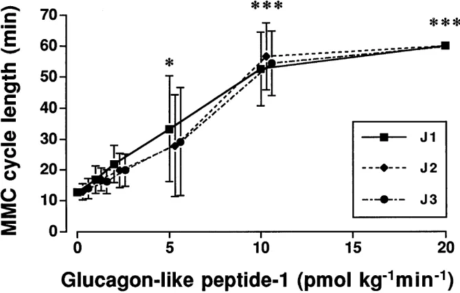
Figure 2. Dose-response curve showing the
[image:4.612.57.392.523.737.2]Effects of GLP-1 on MMC. Infusion of GLP-1 at doses of 10 and 20 pmol kg21min21 had an inhibitory effect on the
MMC. At 10 pmol kg21min21, the MMC was completely
dis-rupted during the infusion period (P , 0.05; Fig. 1). GLP-1 in-creased the MMC cycle length in a dose-dependent manner (Fig. 2). The characteristics of phase III in controls with a du-ration of 3.561.0, 3.660.8, and 3.560.5 min at the J1, J2, and J3 sites of the jejunum, a propagation velocity of 1.761.1 and
1.560.7 cm min21, as well as a calculated length of 6.264.2
and 5.462.3 min between the J1 and J2, as well as J2 and J3 sites, respectively, were not changed until motor quiescence was induced. During the infusions of GLP-1 the general condi-tion of the animals was not affected.
Pretreatment of the rats with exendin(9-39)amide intrave-nously at a dose of 1,000 pmol kg21min21 completely
[image:5.612.61.556.62.184.2]antago-nized the inhibitory effect of GLP-1 at 10 pmol kg21min21
Figure 3. Electromyographic recordings of the MMC propagated through the rat jejunum at 15 (J1), 25 (J2), and 35 (J3) cm distal to the pylorus.
Effect of exendin(9-39)amide on the basal MMC pattern and inhibition of response to GLP-1 (see Fig. 1).
Figure 4. Continuous electromyographic recording from a rat, starting at the upper row (A), passing the middle (B), and terminating at the
lower (C), showing the MMC propagated through the jejunum at 15 (J1), 25 (J2), and 35 (J3) cm distal to the pylorus. A: Effect of GLP-1 alone on the MMC. B: Effect of GLP-1 on the MMC after pretreatment with L-NNA given before administration of GLP-1. C: Effect of GLP-1 on the
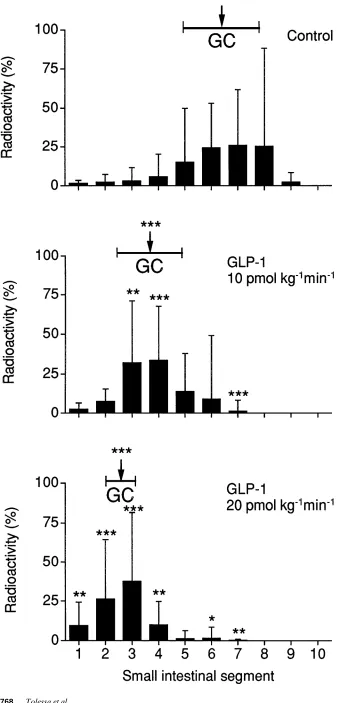
[image:5.612.59.561.357.692.2]Figure 5. Distribution of the radioactive
marker and calculated geometric center (GC) for the marker in the small intestine after 60 min in control rats receiving saline (n 5 9), and in rats receiving GLP-1 intra-venously at doses of 10 pmol kg21min21
(n 5 8) and 20 pmol kg21min21 (n 5 6).
without affecting the basal MMC pattern (P , 0.05). The con-trol MMC cycle length of 15.469.3, 19.7611.5, and 20.6610.2 min at J1, J2, and J3, respectively, was not significantly changed after infusion of exendin(9-39)amide alone or in com-bination with GLP-1. Similarly, the duration, propagation ve-locity, and calculated length of phase III during the infusions were not significantly changed compared to the controls above (Fig. 3).
In addition, pretreatment of the animals with L-NNA in-travenously at a dose of 1 mg kg21 effectively blocked the
ac-tion of GLP 10 pmol kg21min21 on MMC. Thus, the recycling
MMC pattern was preserved during the whole infusion period (P , 0.05), though with a lower amplitude. After L-NNA, the GLP-1–induced inhibition of MMC was reinstated by addi-tional administration of L-arginine at a dose of 300 mg kg21
be-fore administration of the peptide (P , 0.05; Fig. 4).
Infusions of GLP-1 at a dose of 20 pmol kg21min21
in-creased the plasma levels of GLP-1 from 14.7619.7 in controls to 77.8654.5 pmol l21 (P , 0.01).
Effects of GLP-1 on transit of contents in small bowel. GLP-1 at infusions of 10 and 20 pmol kg21min21 slowed the
propulsion of contents through the small bowel in a dose-dependent manner. This effect was verified by a progressive decrease of the geometric center for the distribution of the marker in the small bowel with increasing doses of GLP-1 (P , 0.001) (Fig. 5).
Effects of insulin and somatostatin on MMC. Insulin at a dose of 40 pmol kg21min21 did not change the MMC pattern
with an interval of phase III in controls of 17.9611.4 min at J1, 18.3612.6 min at J2, and 18.8614.5 min at J3. In addition, the characteristics of phase III in controls with a duration of 3.861.1, 3.860.7, and 3.361.0 min at the J1, J2, and J3 sites of the jejunum, a propagation velocity of 2.562.2 and 1.861.9 cm
min21, as well as a calculated length of 7.668.5 and 6.765.4
min between the J1 and J2, and J2 and J3 sites, respectively, were not affected. At a dose of 80 pmol kg21min21, the
recy-cling MMC pattern was abolished and replaced by continuous irregular spiking during the remaining infusion period (P , 0.05; Fig. 6). This myoelectric pattern was clearly different from that induced by GLP-1. Furthermore, at high infusion rates of 200 pmol kg21min21, the animals acutely appeared
more nervous and aggressive.
Insulin at a dose of 80 pmol kg21min21 increased the
plasma level of insulin from control levels of 279.06242.0 to 6600.066219.0 pmol l21 (P , 0.01), whereas the plasma levels
of GLP21 were unchanged (14.7619.7 versus 7.266.1 pmol l21,
respectively).
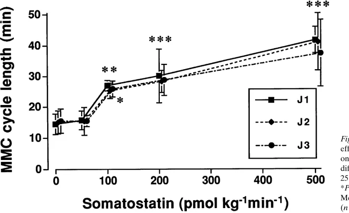
Somatostatin at doses of 100–500 pmol kg21min21 had
in-hibitory effects on the MMC. At 100 pmol kg21min21 the
MMC cycle length was increased (P , 0.05) (Fig. 7). With creasing doses of somatostatin, the MMC cycle length in-creased in a dose-dependent manner until quiescence was induced during the whole infusion period (Fig. 8). However, the inhibitory effect of somatostatin on MMC was different from that induced by GLP-1 and appeared more irregularly. The characteristics of phase III during infusion of somatosta-tin were not different from those observed during the control period, except for a slightly shorter duration of phase III seen at J1 with the highest dose of somatostatin (2.261.4 min; P , 0.05). Under control conditions, the duration of phase III of MMC was 3.760.9, 3.761.2, and 3.561.4 min at the J1, J2, and J3 sites of jejunum, the propagation velocity 1.561.0 and 1.960.7 cm min21, and the calculated length 5.262.8 and
[image:7.612.60.557.68.169.2]6.962.5 min between the J1 and J2, as well as the J2 and J3 sites, respectively. The general condition of the rats was not af-fected by somatostatin.
Figure 6. Electromyographic recording from the rat jejunum at 15 (J1), 25 (J2), and 35 (J3) cm distal to the pylorus showing the effect of insulin
on the MMC.
Figure 7. Electromyographic recording from the rat jejunum at 15 (J1), 25 (J2), and 35 (J3) cm distal to the pylorus showing the effect of
[image:7.612.61.554.609.711.2]Pretreatment of the rats with L-NNA at a dose of 1 mg kg21 intravenously failed to block the inhibitory action of
so-matostatin 500 pmol kg21min21 on the recycling MMC pattern
(P , 0.01). This effect was different compared to that obtained with GLP-1 in combination with L-NNA and L-arginine (Fig. 9).
Infusion of somatostatin 100 pmol kg21min21 increased
basal plasma somatostatin levels from 17.569.1 pmol l21 to
5213.562770.0 pmol l21 (P , 0.01), whereas the plasma levels
of GLP-1 were not significantly changed (14.7619.7 versus 3.762.0 pmol l21, respectively).
Effect of GLP-1 on circulating levels of insulin, somatosta-tin, and glucose. During infusions of GLP-1 at doses of 20 or 100 pmol kg21min21, the plasma levels of insulin did not
change, and the blood glucose levels were unaffected. How-ever, during the infusions of GLP-1, the plasma levels of soma-tostatin increased significantly in a dose-dependent manner (P , 0.01) (Table I).
Studies of the fed motor pattern
After food intake the fasted motor pattern was disrupted and replaced by a fed motor pattern with irregular spiking at all re-cording sites in the intestinal segment under study.
Effects of GLP-1 and exendin(9-39)amide on fed myoelec-tric activity. Intravenous infusion of GLP-1 at doses of 20 and 40 pmol kg21min21 inhibited the irregular spiking seen after
food intake. At a dose of 20 pmol kg21min21 the irregular
spik-ing was attenuated from 14.066.5 to 3.263.0 spikes 10 s21 at
J1, from 11.865.7 to 3.861.9 at J2, and from 12.166.8 to 4.162.9 at J3 (each P , 0.01; Fig. 10). At 40 pmol kg21min21
the irregular spiking was almost completely abolished with
only some scattered spikes during the infusion period, calcu-lated to 1.560.6, 1.961.0, and 2.461.4 spikes 10 s21 at J1, J2,
and J3, respectively (each P , 0.01).
The inhibitory action of GLP-1 at 20 pmol kg21min21 on
fed myoelectric activity was not affected by pretreatment with L-NNA at a dose of 1 mg kg21. Compared to control
experi-ments with GLP-1 alone at the same dose, the spiking activity remained constant at the J1, J2, and J3 levels with computed values of 9.464.3, 8.463.2, and 8.163.9 spikes 10 s21, respectively.
In contrast, pretreatment with exendin(9-39)amide intrave-nously at a dose of 1,000 pmol kg21min21 stimulated the
myo-electric response after food intake. Compared to food alone, the spiking activity at the J1 level increased from 13.564.3 to 17.964.5 spikes 10 s21 and at J2 from 12.964.4 to 18.865.9
spikes 10 s21 (both P , 0.05). At the J3 level only a modest
in-crease of spiking activity from 12.066.8 to 14.665.5 spikes 10 s21 was noted (Fig. 11).
Effect of food intake on circulating levels of GLP-1. After food intake, the circulating levels of GLP-1 increased. 30 min after feeding, the plasma level of GLP-1 was found to be 33.5616.2 pmol l21, with maximal values amounting to 54
pmol l21. In control rats the plasma level of the peptide was
11.869.3 pmol l21 (P , 0.01).
Discussion
The naturally occurring gut peptide GLP-1 is secreted from L-cells in the distal ileum and has prominent effects on the en-docrine pancreas and glucose metabolism. GLP-1 is an impor-tant incretin, i.e., a humoral factor from the intestinal tract, which at physiological concentrations potentiates the glucose-induced insulin release (22).
Motility of the small intestine is easiest to investigate under fasting conditions because of the presence of a motor pattern that can be predicted to recur within certain intervals, i.e., the MMC with phase I, phase II, and the prominent phase III, which exhibits a maximal contractile activity for that part of the intestine. After food intake, however, the motility pattern changes to a hitherto mathematically undefined pattern of ir-regular spiking activity all over the small intestine.
[image:8.612.58.413.60.277.2]Intravenous infusions of GLP-1 at doses as low as 10 pmol
Figure 8. Dose-response curve showing the
effect of increasing doses of somatostatin on the cycle length of the MMC at three different levels in the rat jejunum, 15 (J1), 25 (J2), and 35 (J3) cm distal to the pylorus. *P , 0.05, **P , 0.01 and ***P , 0.001. Mean values and 95% confidence interval (n 5 7).
Table I. Effect of Glucagon-like Peptide-1 on Circulating Levels of Insulin, Somatostatin, and Glucose
Infusion
P-Insulin (pmol l21)
P-Somatostatin (pmol l21)
B-Glucose (mmol l21)
Saline 279.06242.0 4.663.5 7.861.7
GLP-1 (20 pmol kg21min21) 370.06318.2 9.267.6 6.761.3
GLP-1 (100 pmol kg21min21) 239.86281.0 24.8625.8** 8.662.6
[image:8.612.56.298.651.721.2]kg21min21 disrupted the MMC and induced myoelectric
quies-cence in a dose-dependent manner. Concomitantly, the transit of contents in the small intestine was inhibited in a similar fashion. This effect is well in agreement with our earlier stud-ies in the rat showing that replacing the MMC by myoelectric quiescence slows propulsion through the gut (21, 23), and should also be valid for the replacement of fed myoelectric by quiescence, as irregular spiking has been considered to pro-mote a substantial propulsion through the gut (24, 25), whereas quiescence does not (21). The effect of GLP-1 on MMC and transit was substantiated by calculation of the geo-metric center for the distribution of the intestinal marker, which provides a robust measure of the transit rate (20).
At infusion rates of GLP-1 up to 20 pmol kg21min21, the
plasma concentrations reached were within or close to the physiological range as shown in our present experiments in the rat and in previous work in man (26). The relatively higher dosage required in rats to achieve similar plasma concentra-tions as in humans can be explained by the fact that the clear-ance rate of GLP-1 is 10–20 times higher in rats than in man (27). It is reasonable to use a higher infusion rate of the pep-tide in rats to achieve comparable plasma concentrations as in humans. In addition, the inhibitory motor response to GLP-1 was abolished by pretreatment with the GLP-1 receptor antag-onist exendin(9-39)amide (28). Commensurate with this, exen-din(9-39)amide used as a GLP-1 receptor antagonist has been shown to reduce the incretin effect in rats challenged with an intraduodenal glucose load (29), and hence,
exendin(9-Figure 9. Composite diagram of the effect
of the nitric oxide/L-arginine pathway on the response to somatostatin and GLP-1 on the cycle length of the MMC. A: Effect so-matostatin (500 pmol kg21min21) alone and
after pretreatment with L-NNA (1 mg kg21) (n 5 8). B: Effect of GLP-1 (10 pmol
kg21min21 i.v.) alone, and after
pretreat-ment with L-NNA (1 mg kg21 i.v.), as well
as L-arginine (L-Arg) (300 mg kg21 i.v.) in
combination with L-NNA (1 mg kg21 i.v.)
(n 5 6). Mean values and 95% confidence
[image:9.612.60.422.60.561.2]39)amide was considered a useful GLP-1 receptor antagonist for in vivo studies. Thus, our findings point in favor of a recep-tor-mediated mechanism for a physiological inhibition of GLP-1 on the MMC.
The effect of GLP-1 on MMC was clearly distinguishable from that of insulin, which induced irregular spiking similar to that seen after food intake (19, 30). Also, somatostatin
[image:10.612.58.555.61.352.2]disrupted the MMC and induced quiescence in a different manner to that of GLP-1, provided that the peptide was ad-ministered at high doses causing supraphysiological plasma concentrations. Therefore, it seems most likely that the inhibi-tory effect of GLP-1 on the MMC pattern is directly mediated by GLP-1 on the motility-regulating systems of the gut, and not via a hormonal release of insulin or somatostatin.
Figure 10. Electromyographic recording from the rat jejunum at 15 (J1), 25 (J2), and 35 (J3) cm distal to the pylorus showing the effect of food
intake alone (top) and after pretreatment with GLP-1 (bottom) in the same animal.
Figure 11. Electromyographic recording from the rat jejunum at 15 (J1), 25 (J2), and 35 (J3) cm distal to the pylorus showing the effect of food
[image:10.612.56.558.505.712.2]Because the inhibitory response of GLP-1 on MMC was blocked by treatment with the NO synthase inhibitor L-NNA, and as this effect was possible to revert by additional treatment with L-arginine at a high dose, it seems likely that the inhibitory effect of GLP-1 on MMC is dependent on the elaboration and release of NO in the enteric nervous system or smooth muscle. Alternatively, in support of this, earlier studies indicate a close relationship between relaxatory responses of the gut and NO (31, 32), especially the MMC (33, 34), where also the action of vasoactive intestinal peptide has been shown to be involved (35). The possibility of an unspecific inhibition of the effect of GLP-1 secondary to a blood pressure increase after L-NNA seems less likely, as L-NNA does not affect the inhibition of motility caused by other peptides, such as vasoactive intestinal peptide (35).
In addition to its inhibitory effects on the MMC, GLP-1 was shown to block the fed myoelectric pattern in the rat. This effect of the peptide seemed to require somewhat higher doses than the inhibition of the fasting myoelectric pattern and could not be shown to be dependent on NO. However, such a finding is not very surprising, as a host of different gastrointestinal peptides are considered to cooperate in the stimulation of mo-tility after food intake. In line with an inhibitory action of GLP-1 on fed motility, we found that the GLP-1 receptor an-tagonist exendin(9-39)amide modestly increased the motility response to food intake. Because the array of gastrointestinal peptides released after food intake all work through distinct receptor mechanisms mainly to stimulate postprandial motor activity, it is not very likely that one single peptide with an in-hibitory receptor, such as GLP-1, should be of vital importance for the regulation of postprandial motility.
The results from other studies indicate that GLP-1 exerts weak insulin-releasing properties at euglycemia (6, 12). In our present study, however, GLP-1 did not stimulate insulin re-lease even at doses as high as 100 pmol kg21min21. Thus,
dur-ing postabsorptive conditions, motility of the gastrointestinal tract seems to be considerably more sensitive to GLP-1 as compared to responses of pancreatic b cells.
GLP-1 has also been found to stimulate somatostatin re-lease in rat pancreatic cell cultures (10), and in both the per-fused pig and dog pancreas (36). In contrast to insulin release, GLP-1 markedly stimulates somatostatin release also at low glucose levels (9). Similarly, in our experiments GLP-1 at 100 pmol kg21min21 markedly enhanced somatostatin, but not
in-sulin, release. However, the release of somatostatin to periph-eral blood was obtained at doses of GLP-1 higher than those required to inhibit the MMC. In addition, in other species such as dog (37) and man (38), somatostatin has been shown to stimulate premature MMC, which is different from the rat. This supports our conclusion that the inhibitory effect of GLP-1 on motility under physiological circumstances is not mediated by a release of somatostatin. In this context, it is of interest that the insulin-stimulating effect of GLP-1, both in the ami-dated and nonamiami-dated form, has been shown to be glucose-dependent (8, 39–41). Therefore, our results using fasted rats for hormone release experiments may not be representative for postprandial conditions when GLP-1 may simultaneously influence both gastrointestinal motility and hormonal responses. Furthermore, the finding that the inhibitory action of soma-tostatin on the MMC was not affected by L-NNA strengthens our conclusion that the effect of GLP-1 on motility is not medi-ated by somatostatin, neither as a circulating hormone nor as a local mediator at the intestinal level.
Taken together, it seems that GLP-1 acts directly on gas-trointestinal motor functions to cause an inhibition of motility. Such an effect may be of great importance for the preabsorp-tive delivery of nutrients to the intestine via a slowed propul-sion through the gut. A decreased absorption rate of nutrients is considered beneficial to decrease the requirements of insulin to maintain normoglycemia. In support of this view, Gutniak and co-workers (26) have found that meal-related insulin re-quirements and plasma insulin levels after GLP-1 infusion in both insulin-dependent and non-insulin–dependent diabetic patients are greatly reduced. Hence, speculations have been made that GLP-1 may act as a novel treatment for diabetes mellitus, especially in non-insulin–dependent diabetes mellitus (42, 43). We believe that these mechanisms, together with the inhibition of gastrointestinal motility, involving also gastric emptying, play an important role for the marked antidiabeto-genic effect of GLP-1 demonstrated in patients with NIDDM and IDDM. In support of such an antidiabetogenic mecha-nism, Nauck and collaborators have recently found the inhibi-tory action of GLP-1 on gastric emptying to outweigh its insuli-notropic effect in healthy humans (44).
Interestingly enough, a number of peptide hormones from the proximal small intestine that have been considered as in-cretin candidates not only release insulin from the pancreas, but also inhibit gastrointestinal motility, mainly gastric empty-ing. Such an effect should diminish insulin requirements in re-lation to food intake. This double effect is obvious for gastric inhibitory peptide, which, however, has a lower insulin-releas-ing capacity than GLP-1 (7, 45) and inhibits gastric emptyinsulin-releas-ing only at supraphysiological plasma concentrations (46). An-other incretin candidate with similar properties is cholecysto-kinin, which releases insulin upon food intake (47, 48) and causes slowing of gastric emptying at physiological plasma lev-els (49). Thus, even if earlier findings implicate that the inhibi-tory actions on gastrointestinal motility by various incretins may be attributed to a common pathway through their ability to release insulin to the circulation, our present data show that the inhibitory action of GLP-1 on gastrointestinal motility in the rat is not mediated via insulin, because the motor pattern after administration of GLP-1 was clearly different from that obtained with insulin. Therefore, it seems most likely that GLP-1, and possibly other incretin candidates, such as gastric inhibitory peptide and cholecystokinin, inhibit gastrointestinal motor activity by distinct mechanisms acting directly on en-teric neurons or smooth muscle cells, separate from the insu-lin-releasing mechanisms in the pancreas. As an alternative ex-planation for its effects on motility, GLP-1 may be released to act as a humoral afferent link with effects on the circumven-tricular organs, such as area postrema, in the brain. Thus, vagal nonadrenergic noncholinergic mechanisms could be stimu-lated, resulting in NO release and inhibition of motility.
In conclusion, the inhibitory effect of GLP-1 on gas-trointestinal motility seems neither to be mediated via insulin nor somatostatin, but instead through direct mechanisms act-ing directly in the enteric nervous system or on smooth muscle cells partly dependent on the L-arginine/NO pathway.
Acknowledgments
Wiberg’s foundation. This work was conducted as part of a collabora-tion between Addis Ababa University, Faculty of Medicine, and the Karolinska Institutet, and was supported by Swedish Agency for Re-search Cooperation with Developing Countries (SAREC) (grant No. S2/ETI 12).
References
1. Mojsov, S., G. Heinrich, I.B. Wilson, M. Ravazzola, L. Orci, and J.F. Ha-bener. 1986. Preproglucagon gene expression in pancreas and intestine diversi-fies at the level of posttranslational processing. J. Biol. Chem. 261:11880–11886. 2. Novak, U., A. Wilks, G. Buell, and S. McEven. 1987. Identical mRNA for preproglucagon in pancreas and the gut. Eur. J. Biochem. 164:553–557.
3. Ørskov, C., J.J. Holst, S. Knuhtsen, F.G.A. Baldissera, S.S. Poulsen, and O.V. Nielsen. 1986. Glucagon-like peptides GLP-1 and GLP-2, predicted prod-ucts of the glucagon gene, are secreted separately from the pig small intestine, but not pancreas. Endocrinology. 119:1467–1475.
4. Holst, J.J. 1988. Enteroglucagon. Adv. Metab. Dis. 11:392–419. 5. Ørskov, C., J.J. Holst, S.S. Poulsen, and P. Kirkegaard. 1987. Pancreatic and intestinal processing of proglucagon in man. Diabetologia. 30:874–881.
6. Holst, J.J., C. Ørskov, O.V. Nielsen, and T.W. Schwartz. 1987. Truncated glucagon-like peptide I, an insulin-releasing hormone from the distal gut. FEBS
Lett. 211:169–174.
7. Kreymann, B., M.A. Ghatei, G. Williams, and S.R. Bloom. 1987. Gluca-gon-like peptide-1 7-36: a physiological incretin in man. Lancet. ii:1300–1303.
8. Mojsov, S., G.C. Weir, and J.F. Habener. 1987. Insulinotropin: glucagon-like peptide-I (7-37) co-encoded in the glucagon gene is a potent stimulator of insulin release in the perfused rat pancreas. J. Clin. Invest. 79:616–619.
9. Ørskov, C., J.J. Holst, and O.V. Nielsen. 1988. Effect of truncated gluca-gon-like peptide-1 (proglucagon-(78-107) amide) on endocrine secretion from pig pancreas, antrum, and non-antral stomach. Endocrinology. 123:2009–2013.
10. d’Alessio, D.A., W.Y. Fujimoto, and J.W. Ensinck. 1989. Effects of glu-cagonlike peptide I-(7-36) on release of insulin, glucagon, and somatostatin by rat pancreatic islet cell monolayer cultures. Diabetes. 38:1534–1538.
11. Wettergren, A., B. Schjoldager, P.E. Mortensen, J. Myhre, J. Chris-tiansen, and J.J. Holst. 1993. Truncated GLP-1 (proglucagon 78-107-amide) in-hibits gastric and pancreatic functions in man. Dig. Dis. Sci. 38:665–673.
12. Gutniak, M.K., L. Juntti-Berggren, P.M. Hellström, A. Guenifi, J.J. Holst, and S. Efendic. 1996. Glucagon-like peptide I enhances the insulinotro-pic effect of glibenclamide in NIDDM patients and in the perfused rat pan-creas. Diabetes Care. 19:857–863.
13. Grill, V., J. Pigon, S.G. Hartling, C. Binder, and S. Efendic. 1990. Effects of dexamethasone on glucose-induced insulin and proinsulin release in low and high insulin responders. Metabolism. 39:251–258.
14. Grill, V., M. Gutniak, A. Roovete, and S. Efendic. 1984. A stimulating effect of glucose on somatostatin release is impaired in noninsulin-dependent diabetes mellitus. J. Clin. Endocrinol. Metab. 59:293–297.
15. Ørskov, C., L. Rabenhø, A. Wettergren, H. Kofod, and J.J. Holst. 1994. Tissue and plasma concentrations of amidated and glycine extended glucagon-like peptide-1 in humans. Diabetes. 43:535–539.
16. Deacon, C.F., A.H. Johnsen, and J.J. Holst. 1995. Degradation of gluca-gon-like peptide-1 by human plasma in vitro yields an N-terminally truncated peptide that is a major endogenous metabolite in vivo. J. Clin. Endocrinol.
Metab. 80:952–957.
17. Hjelm, M., and C. de Verdier. 1963. A methodological study of the enzy-matic determination of glucose in blood. Scand. J. Clin. Lab. Invest. 15:415–428.
18. Hellström, P.M., R.O. Bränström, and A. Al-Saffar. 1993. Computer program “MMC” to summarize characteristics of activity fronts of migrating myoelectric complex in rat small intestine. Surg. Res. Commun. 14:51–63.
19. Bränström, R., and P.M. Hellström. 1996. Characteristics of fasting and fed myoelectric activity in rat small intestine: evaluation by computer analysis.
Acta Physiol. Scand. 158:53–62.
20. Miller, M.S., J.J. Galligan, and T.F. Burks. 1981. Accurate measurement of intestinal transit in the rat. J. Pharmacol. Methods. 6:211–217.
21. Hellström, P.M., and C. Johansson. 1989. Neuropeptide Y inhibits the migrating myoelectric complex and delays small intestinal transit in man. J.
Gastrointest. Mot. 1:35–41.
22. Zunz, E., and J. La Barre. 1929. Contributions à l’étude des variations physiologiques de la sécrétions externe et interne du pancreas: relation entre les sécrétions externe et interne du pancreas. Arch. Int. Physiol. Biochim. 31:20. 23. Al-Saffar, A., P.M. Hellström, and G. Nylander. 1985. Correlation be-tween peptide YY-induced myoelectric activity and transit of small-intestinal contents in rats. Scand. J. Gastroenterol. 20:577–582.
24. Al-Saffar, A., P.M. Hellström, G. Nylander, and S. Rosell. 1984. Influ-ence of fasting and bombesin-induced myoelectric activity on the transit of
small-intestinal contents in the rat. Scand. J. Gastroenterol. 19:541–546. 25. Lördal, M., C. Johansson, and P.M. Hellström. 1993. Myoelectric pat-tern and effects on small bowel transit induced by the tachykinins neurokinin A, neurokinin B, substance P and neuropeptide K in the rat. J. Gastrointest.
Mot. 5:33–40.
26. Gutniak, M.K., C. Ørskov, J.J. Holst, B. Ahrén, and S. Efendic. 1992. Antidiabetogenic effect of glucagon-like peptide-1 (7-36)amide in normal sub-jects and in patients with diabetes mellitus. N. Engl. J. Med. 326:1316–1322.
27. Holst, J.J. 1991. Degradation of pancreatic peptides: glucagon. In Deg-radation of Bioactive Substances, Physiology and Pathophysiology. Uniscience volume. J.H. Henriksen, editor. CRC Press, Boca Raton, FL. 139–149.
28. Raufman, J.P. 1996. Bioactive peptides from lizard venoms. Regul. Pept. 61:1–18.
29. Kolligs, F., H.C. Fehmann, R. Göke, and B. Göke. 1995. Reduction of the incretin effect in rats by the glucagon-like peptide-1 receptor antagonist ex-endin (9-39) amide. Diabetes. 44:16–19.
30. Ruckebusch, M., and J. Fioramonti. 1975. Electrical spiking activity and propulsion in small intestine in fed and fasted rats. Gastroenterology. 68:1500–1508. 31. Bult, H., G.E. Boeckxstaens, P.A. Pelckmans, F.H. Jordaens, Y.M. van Maercke, and A.G. Herman. 1990. Nitric oxide as an inhibitory non-adrenergic non-cholinergic neurotransmitter. Nature. 345:346–347.
32. Iversen, H.H., N.P. Wiklund, and L.E. Gustafsson. 1994. Nitric oxide-like activity in guinea pig colon as determined by effector responses, bioassay and chemiluminescence analysis. Acta Physiol. Scand. 152:315–322.
33. Sarna, S.K., M.F. Otterson, R.P. Ryaqn, and V.E. Cowles. 1993. Nitric oxide regulates migrating motor complex cycling and its postprandial disrup-tion. Am. J. Physiol. 265:G759–G766.
34. Rodriguez-Membrilla, A., V. Martinez, M. Jimenez, E. Gonalons, and P. Vergara. 1995. Is nitric oxide the final mediator regulating the migrating myoelectric complex cycle? Am. J. Physiol. 268:G207–G214.
35. Hellström, P.M., and T. Ljung. 1996. Nitrergic inhibition of migrating myoelectric complex in the rat is mediated by vasoactive intestinal peptide.
Neurogastroenterol. Mot. 8:299–306.
36. Kawai, K., S. Suzuki, S. Ohashi, H. Mukai, H. Ohmori, Y. Murayama, and K. Yamashita. 1989. Comparison of the effects of glucagon-like peptide-1-(1-37) and -(7-37) and glucagon on islet hormone release from isolated per-fused canine and rat pancreases. Endocrinology. 124:1768–1773.
37. Hostein, J., J. Janssens, G. Vantrappen, T.L. Peeters, M. Vandeweerd, and G. Leman. 1984. Somatostatin induces ectopic activity fronts of the migrat-ing motor complex via a local intestinal mechanism. Gastroenterology. 87:1004– 1008.
38. von der Ohe, M., P. Layer, C. Wollny, J.W. Ensinck, T.L. Peeters, C. Beglinger, and H. Goebell. 1992. Somatostatin 28 and coupling of human inter-digestive intestinal motility and pancreatic secretion. Gastroenterology. 103: 974–981.
39. Shima, K., M. Hirota, and C. Ohboshi. 1988. Effect of glucagon-like peptide-1 on insulin secretion. Regul. Pept. 22:245–252.
40. Komatsu, R., T. Matsuyama, M. Namba, N. Watanabe, H. Itoh, N. Kono, and S. Tarui. 1989. Glucagonostatic and insulinotropic action of glu-cagonlike peptide I-(7-36)-amide. Diabetes. 38:902–905.
41. Gefel, D., G.K. Hendrick, S. Mojsov, J.F. Habener, and G.C. Weir. 1990. Glucagon-like peptide-1 analogues: effects on insulin secretion and aden-osine 3959-monophosphate formation. Endocrinology. 126:2164–2168.
42. Nauck, M.A., J.J. Holst, and B. Willms. 1997. Glucagon-like peptide 1 and its potential in the treatment of non-insulin–dependent diabetes mellitus.
Horm. Metab. Res. 29:411–416.
43. Nauck, M.A., J.J. Holst, B. Willmas, and W. Schmiegel. 1997. Glucagon-like peptide 1 (GLP-1) as a new therapeutic approach for type 2-diabetes. Exp.
Clin. Endocrinol. Diabetes. 105:187–195.
44. Nauck, M.A., U. Niedereichholtz, R. Ettler, J.J. Holst, C. Ørskov, R. Ritzel, and W.H. Schmiegel. 1997. Glucagon-like peptide 1 inhibition of gastric emptying outweighs its insulinotropic effects in healthy humans. Am. J. Physiol. 273:E981–E988.
45. Kreymann, B., M.A. Ghatei, V. Schusdziarra, S.R. Bloom, and M. Clas-sen. 1990. Does the incretin effect exist for GIP or GLP-1 7-36 amide at physio-logical glucose concentrations in man? Digestion. 46(Suppl. 1):59.
46. Brown, J.C., J.R. Dryburgh, S.A. Ross, and J. Dupré. 1975. Identifica-tion and acIdentifica-tions of gastric inhibitory polypeptide. Recent Prog. Horm. Res. 31: 487–532.
47. Szecowka, J., P.E. Lins, and S. Efendic. 1982. Effects of cholecystokinin, gastric inhibitory polypeptide, and secretin on insulin and glucagon secretion in rats. Endocrinology. 110:1268–1272.
48. Rushakoff, R.J., I.D. Goldfine, J.D. Carter, and R.A. Liddle. 1987. Physiological concentrations of cholecystokinin stimulate amino acid-release in humans. J. Clin. Endocrinol. Metab. 65:395–401.