www.impactjournals.com/oncotarget
www.impactjournals.com/oncotarget/ Oncotarget, Vol. 7, No. 33
PDGFR
α
depletion attenuates glioblastoma stem cells features
by modulation of STAT3, RB1 and multiple oncogenic signals
Carlo Cenciarelli1, Hany E. Marei2, Armando Felsani3, Patrizia Casalbore3, Gigliola Sica4, Maria Ausiliatrice Puglisi5, Angus J.M. Cameron6, Alessandro Olivi7, Annunziato Mangiola7
1Institute of Translational Pharmacology, Department of Biomedical Sciences-National Research Council (IFT-CNR), Rome, Italy 2Biomedical Research Center, Qatar University, Doha, Qatar
3Institute of Cell Biology and Neurobiology, Dept. of Biomedical Sciences-National Research Council (IBCN-CNR), Rome, Italy 4Institute of Histology and Embryology, Catholic University-School of Medicine, Rome, Italy
5Department of Internal Medicine and Gastroenterology, Agostino Gemelli Hospital, Rome, Italy
6Barts Cancer Institute, John Vane Science Centre, Queen Mary University of London, London, United Kingdom 7Institute of Neurosurgery, Department of Head and Neck, Catholic University-School of Medicine, Rome, Italy
Correspondence to: Carlo Cenciarelli, email: carlo.cenciarelli@ift.cnr.it
Keywords: glioblastoma, cancer stem cells, PDGFRα, STAT3, RB1
Received: April 05, 2016 Accepted: June 09, 2016 Published: June 17, 2016
ABSTRACT
Platelet derived growth factor receptors (PDGFRs) play an important role in tumor pathogenesis, and they are frequently overexpressed in glioblastoma (GBM). Earlier we have shown a higher protein expression of PDGFR isoforms (α and β) in peritumoral-tissue derived cancer stem cells (p-CSC) than in tumor core (c-CSC) of several GBM affected patients. In the current study, in order to assess the activity of PDGFRα/PDGF-AA signaling axis, we performed time course experiments to monitor the effects of exogenous PDGF-AA on the expression of downstream target genes in c-CSC vs p-CSC. Interestingly, in p-CSC we detected the upregulation of Y705-phosphorylated Stat3, concurrent with a decrement of Rb1 protein in its active
state, within minutes of PDGF-AA addition. This finding prompted us to elucidate
the role of PDGFRα in self-renewal, invasion and differentiation in p-CSC by using short hairpin RNA depletion of PDGFRα expression. Notably, in PDGFRα-depleted
cells, protein analysis revealed attenuation of stemness-related and glial markers expression, alongside early activation of the neuronal marker MAP2a/b that correlated with the induction of tumor suppressor Rb1. The in vitro reduction of the invasive capacity of PDGFRα-depleted CSC as compared to parental cells correlated with the downmodulation of markers of epithelial-mesenchymal transition phenotype and angiogenesis. Surprisingly, we observed the induction of anti-apoptotic proteins and compensatory oncogenic signals such as EDN1, EDNRB, PRKCB1, PDGF-C and PDGF-D.
To conclude, we hypothesize that the newly discovered PDGFRα/Stat3/Rb1 regulatory
axis might represent a potential therapeutic target for GBM treatment.
INTRODUCTION
Among the different types of adult brain tumors, glioblastoma multiforme (GBM) is the most aggressive
and angiogenic, but despite the efforts to find effective
treatments, these tumors remain incurable [1]. The
amplification and/or overexpression of either EGFR or PDGFR can contribute to the malignant phenotype of
distinct subsets of human glioblastoma [2]. More recently,
a classification of GBM in subclasses has been defined for
the selection of the best-tailored therapeutic approaches
[3, 4]. Platelet-derived growth factors (PDGFs) isoforms and receptors (PDGFRs) have important functions in the
regulation of growth and survival of certain cell types during embryonal development and in control of tissue homeostasis in the adult [5]. A wide range of work from
www.impactjournals.com/oncotarget
in vitro studies to mouse models have implicated the role
of the PDGF pathway in cellular invasion and tumor angiogenesis [6]. In fact, over-activity of PDGF signaling
is associated with tumor development in brain, prostate, liver, lung, leukemia and colon cancers [7, 8]. Although
anti-VEGF treatment has been the major therapeutic
target in gliomas, other antiangiogenic agents such as
anti-PDGFs or anti-FGFs are currently in preclinical and clinical development [9]. PDGFR includes two receptors (α and β) and four ligands (PDGF-A, PDGF-B, PDGF-C and PDGF-D). The PDGFs bind to the receptors with different affinities. Thus, PDGF-AA, -AB, -BB and -CC induce αα receptor homodimers, PDGF-BB and -DD ββ receptor dimerization, and PDGF-AB, -BB, -CC and -DD αβ receptor dimerization [5]. Ligand-induced dimerization favors autophosphorylation of specific tyrosine residues
and subsequent activates downstream signal pathways:
PI3K/Akt1/mTOR, Ras/MAPK, PLC-γ/PKC and STAT3. PDGFR binds and activates signal transducers and activator of transcription (STATs). Phosphorylation of Y705 in Stat3
leads to dimerization, nuclear translocation, recognition of
Stat3-specific DNA binding elements and up-regulation of various Stat3 downstream target genes, such as Bcl-xl, Bcl-2, Survivin, c-Myc and Cyclin D1. Stat3 regulates tumorigenesis and tumor inflammation and behaves in an
oncogenic manner depending on the genetic background of
the tumor [1]. In recent studies, Stat3 has been implicated in
the self-renewal of neural stem cells and glial differentiation while restricting neuronal differentiation [8–13].
The PKC family consists of fifteen isozymes divided
into three subfamilies: conventional (or classical), novel,
and atypical. Conventional PKCs contain the isoforms α, βI, βII, and γ. The PDGFR downstream target PKCα plays
an important role in migration, tumor growth, angiogenesis
and drug resistance in GBM cells [14–16]. In 1992, PKCα
was suggested as marker of malignancy for gliomas, and
more recently serum PKCα serves as a biomarker for diagnosis of cancers [14, 15]. The invasion/migration of
GBM cells induced by TPA, occurs through activation of
PKCα/ERK/NF-κB-dependent MMP-9 expression [16].
A positive feedback loop between Wnt5A and
phospho-PKC in promotion of epithelial-mesenchymal transition (EMT) in nasopharyngeal carcinoma was disclosed [17]. In addition, PDGF receptors bind to other tyrosine kinase receptors, e.g. EGFR [18]. Retinoblastoma 1 (RB1) gene belongs to a family of three proteins, including also RBL1/ p107 and RBL2/p130. Classically the tumor suppressive function of Rb proteins have been mainly attributed to their ability to arrest cell cycle by repressing E2F target genes. When Rb1 is in its active hypophosphorylated state, it represses E2F-mediated transcription by binding, blocks the E2F transactivation domain, and forms complexes with
its (DPs transcription factors) partners at cell cycle gene
promoters [19]. Conversely, Rb1 phosphorylation initiated by cyclin D-CDK4/6 in response to mitogenic signals, inactivates the Rb1 repressive function by dissociating the Rb1-E2F-DP complexes [19]. The Cancer Genome
Atlas Research Network revealed in 2008 that the CycD1-CDK4/6-Rb1 pathway is among the top three most altered
pathways in GBM, which makes this an appealing target
for cancer therapy [20–22].
We and others recently demonstrated that inhibition
of either PDGFRα or PDGFRβ signaling induced apoptosis
in glioblastoma stem cells [23, 7]. In the present study,
we aimed to assess the effects of PDGFRα depletion on stemness, invasion and differentiation in GBM CSC. Our findings reveal an inverse correlation between Stat3 Y705-phosphorylation and the hypophosphorylated Rb1 instructed by the PDGFRα/PDGF-AA regulatory axis. Further, downmodulation of cell growth, invasion and the EMT phenotype are triggered by PDGFRα depletion in GBM CSC. Surprisingly, we detected the activation of angiogenic
and survival pathways as compared to parental cells, which
supports a multimodal approach to treat GBM CSC.
RESULTS
Activation of PDGFRα/PDGF-AA signaling regulates expression of downstream genes Egr1, Stat3 and Rb1 but not PKCα in GBM CSC
Cancer stem cells from GBM were isolated as
described previously [23, 24]. We were able to collect either
core- (c-CSC) or peritumor tissue-derived cancer stem cells (p-CSC) from several primary GBM samples; the two types of CSC had quite different tumorigenic potential and exclusive genetic anomalies [23, 24]. To demonstrate that the high expression of PDGFRα in p-CSC2 [23], confers a higher PDGF response than in c-CSC2, both cell types, were growth factors starved and then, exposed to exogenous PDGF-AA (50 ng/ml) over a time course. In these experimental conditions (Figure 1A), we found more rapid activation of p-Erk1/2 in p-CSC2 than in c-CSC2 as reported previously [23]. Moreover, Egr1 (Early growth response 1) protein and Stat3 Y705 phosphorylation were induced as early as 5 minutes after PDGF-AA stimulation (Figure 1A). Strikingly,
we noted a tight correlation between this phosphorylation event and the transient decrement of the tumor suppressor
Rb1 in its active state, that occurred within 10 minutes, as detected by a monoclonal antibody that specifically recognizes hypophosphorylated active Rb1 (hypo-Rb1). A densitometric analysis (data not shown) showed a 70% signal reduction
at this time point compared to time point zero, followed by
a gradual recovery. The engagement of PDGFRα with its
ligand induced the gradual disappearance of the receptor
2 hours after PDGF-AA stimulus. The high expression and phosphorylation of PKCα/βII isoforms was not altered by PDGF-AA stimulation in either cell populations (Figure 1A).
Block of PDGFRα activity by Crenolanib reduces the phosphorylation of Rb1 and Stat3 Y705
www.impactjournals.com/oncotarget
experiment was performed to evaluate the effects of PDGF-AA applied alone or in combination with Crenolanib (CR, a potent inhibitor of PDGFR signaling), on p-CSC2 two days after growth factors withdrawal. Rb1
phosphorylation was monitored at short time intervals
(5-10-30-120 minutes) up to 24 after the start of PDGF-AA stimulation. CR-treated cells showed upregulation of the hypo-Rb1 between 5 and 30 minute after the treatment as compared to PDGF-AA alone. A densitometric analysis (data not shown) showed an increase of 50% of hypo-Rb1 signal within the first half hour in CR-treated cells compared to PDGF-AA alone. Two hours after the treatments the levels of the hypo-Rb1 return to baseline (Figure 1C). In contrast, Stat3 Y705 phosphorylation remained silenced at 24 hours by CR treatment (Figure
1B). Previous studies showed that the epidermal growth
factor receptor (EGFR) could be transactivated by PDGFs stimulation and that EGFR transactivation was required
for PDGF-stimulated cell migration [18]. In corroboration, we showed that PDGF-AA induced EGFR Y1068-phosphorylation, and EGFR transactivation was inhibited by Crenolanib treatment (Figure 1B). CR was also found to upregulate Erk1/2 downstream of both the PDGFR and the EGFR (Figure 1B).
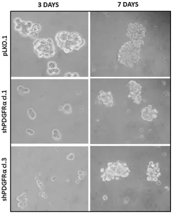
shPDGFRα-GBM CSC display changes in cell morphology and EMT phenotypes, which correlate with modifications in the molecular profile
[image:3.612.92.517.287.623.2]To demonstrate the central role of PDGFRα in CSC proliferation and differentiation, we interfered with PDGFRα gene expression using short hairpin RNA (shRNA) sequences transduced by a lentivirus-based system. We selected two PDGFRα-directed shRNA clones (1 and 3) of p-CSC2 that displayed a robust silencing of
www.impactjournals.com/oncotarget
PDGFRα compared to transduced control cells (pLKO.1) (Figure 2A). Depletion of PDGFRα expression was
associated with a reduction in cell growth as reported previously [23], and with a concomitant impairment of neurosphere formation ability, consistently observed over
the entire seven days of time course (Figure 3). The two
cell clones showed evident morphological changes if
induced to differentiate in presence of 5% FCS. After 2 and 4 days in differentiation conditions, shPDGFRα-CSC
showed an epithelial cell morphology, with formation of adherent cell clusters and few cells with an elongated
phenotype. In contrast, the pLKO.1 control cells showed a flat morphology with a clear mesenchymal phenotype, evenly distributed on the plate (Figure 4). These observations prompted us to explore the gene expression
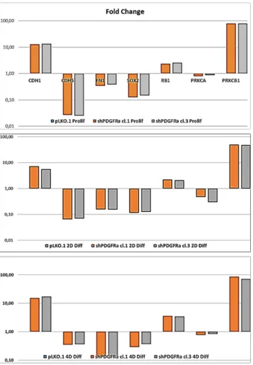
of epithelial cell junction proteins such as E-cadherin (CDH1) and VE-Cadherin (CDH5) or ECM molecules such as fibronectin (FN1) by a RT-qPCR analysis. CDH1 mRNA expression was monitored in PDGFRα-depleted CSCs during cell proliferation. Both clone 1 and 3 showed higher expression compared with control cells, with a
fold change of 13.1 and 13.6 respectively in proliferating
cells and with a peak of activation at 15.2 and 17.3 Fc after 4 days of differentiation (Figure 5). Our finding
were consistent with that published in various cell lines,
reporting an inverse correlation between CDH1 expression and invasiveness [25]. CDH5 mRNA levels were
drastically downmodulated in proliferating cell clones
[image:4.612.96.525.245.610.2]and reached 0.35 and 0.36 Fc in 4 days-differentiated cells respectively (Figure 5). Similarly, FN1 mRNA expression
www.impactjournals.com/oncotarget
was diminished to 0.34 and 0.38 fold in proliferating shPDGFRα-CSC clones compared to pLKO.1, and went down to 0.16 and 0.15 Fc in 2 days-differentiated cells. Similar results were seen at 4 days (Figure 5). As reported by the literature, the combined loss of CDH1, and the gain of mesenchymal markers, such as vimentin, FN1 and N-cadherin favor the EMT program [26].
PDGFRα depletion downmodulates stemness-related and glial markers, induces early
expression of neuronal marker and RB1 but does not affect PKCα in GBM p-CSC
To assess the role of PDGFRα in the regulation of stemness of GBM CSC, we carried out Western blots and RT-qPCR to elucidate the effects of a diminished PDGFRα expression on stemness-related genes. We
[image:5.612.138.477.268.686.2]observed a decrease in protein expression for Nestin, Sox2, Bmi1, but no modulation of Oct4 (Figure 2B). RT-qPCR analysis confirmed the decrease of SOX2 mRNA to 0.11 and 0.13 fold in clone 1 and 3 compared to control cells. Similar results were confirmed at 2 and 4 days-differentiated p-CSC2 (Figure 5). Attenuation of stemness in shPDGFRα-CSC clones correlated with early induction of late neuronal differentiation marker MAP2a/b protein in proliferating cells (Figure 2B), also monitored as mRNA (data not shown). Genetic and pharmacological depletion of PDGFRα triggered the activation of tumor suppressor RB1 gene, a key factor controlling cell cycle exit and differentiation in several cell models. As shown in Figure 5, PDGFRα-depleted CSC clone 1 and 3 displayed the induction of RB1 mRNA up to 2.35 and 2.59 fold respectively compared to control cells. Expression levels raised a peak to 3.59 and 3.45 Fc in 4 days-differentiated
www.impactjournals.com/oncotarget
cells respectively. Western blot analysis revealed the
increase of hypo-Rb1 in proliferating clone 3, or in both differentiated clones compared to pLKO.1 (Figure
2A). Under the same culture conditions, we noticed a
decrease of Stat3 Y705-phosphorylation in both clones in comparison to pLKO.1 cells (Figure 2A). Conversely, we reported a diminished expression of glial fibrillary acidic protein (GFAP) and the chondroitin sulfate proteoglycan NG2 (Figure 2B). The latter is probably the result of lack of a physical association with PDGFRα, which is
an obstacle to the formation of new oligodendroglial
precursors NG2+ve [28]. The reduced PDGFRα expression correlated with higher levels of Bcl-2 and Bcl-xL compared to control cells, suggesting a potential
role of these genes in cell survival in either proliferating or differentiated cells. These results paralleled with an
increase in CycD1 protein in differentiated cells and a
slight decrease in proliferating cells. The latter effect likely correlated to cell proliferation impairment induced
by PDGFRα deletion. Because of the critical role of PKCα in promoting EMT phenotype and malignant progression of glioblastoma cells, we hypothesized that PKCα could regulate stemness of GBM CSC downstream of the PDGFRα signaling cascade. To answer the question, we performed shRNA targeting of PKCα expression in GBM p-CSC2. The results revealed no variation of the multipotent stem cells markers Nestin or Sox2, even with the addition of PDGF-AA for three days (Figure S1). This finding reveals that PKCα is probably not involved in regulation of stemness through the PDGFRα signaling.
PDGFRα depletion inhibits the invasiveness of GBM CSC and triggers EDN1 signaling, PDGF-C and PDGF-D expression
Formerly we reported that GBM p-CSC2 displayed
higher invasive capacity through Matrigel with respect to
[image:6.612.93.521.295.656.2]the counterpart c-CSC2 [23]. Herein, we aim to demonstrate
www.impactjournals.com/oncotarget
the role of PDGFRα/PDGF-AA axis in the regulation of the invasive capacity of p-CSC2. Using an in vitro cell
invasion assay we demonstrated a significant reduction in PDGF-AA stimulated invasion of shPDGFRα-CSC clones compared to pLKO.1. Demonstrating specificity, no difference in the invasive capacity was observed using EGF as chemoattractant (Figure 6A). The EMT leads to a reduced
cellular adhesion, changes in cytoskeletal organization and acquired potential for cell migration, all this process is
[image:7.612.130.487.143.658.2]associated to changes in expression of transcription factors [30]. To corroborate the role of PDGFRα on the invasive phenotype of p-CSC2, we completed the RT-qPCR analysis of critical genes for EMT such as ZEB1, ZEB2, TWIST1 and Vimentin (VIM). Our results displayed a significant
www.impactjournals.com/oncotarget
decrease of VIM, ZEB1, ZEB2, but not TWIST1 in both proliferating cell clones (Figure 6B). Similarly, we noticed a decrease of angiogenic markers (Figure 6B) such as VEGFR2, PECAM1 (CD31), and CDH5 as shown previously (Figure 5). In parallel, Western blot analysis display a significant decrease of CD31, VEGFR2 and VE-Cadherin proteins, corroborating mRNA expression levels (Figure 6C). Unexpectedly, we noticed an increase of mRNA expression for endothelin1 (EDN1), the receptor EDNRB (Figure 6B), its target gene PRKCB1, but not PRKCA (Figure 5). In Figure 5 we reported a significant upregulation of PRKCB1 gene expression in proliferating (79.4 and 78.9 Fc respectively) and differentiated PDGFRα-depleted cell clones 1 and 3 (86.3 and 71.8 Fc respectively) compared with pLKO.1. Instead, PRKCA gene expression was slightly reduced either in proliferating (0.78 and 0.86 Fc respectively) or differentiated PDGFRα-depleted cell clone 1 and 3 (0.7 and 0.8 Fc respectively). Consistently,
PKCα protein expression and phosphorylation of PKCα/ βII isoforms are reduced in differentiation conditions in cell clones compared to pLKO.1 (Figure 2A). In as much as peritumor tissue-derived CSC presented greater
chemoresistance to treatments respect to core tumor
derived-CSC [23], we decided to investigate by RT-qPCR the expression of the main modulators of EMT and/or drug resistance such as TWIST, EDN1 and PRKCA in several samples of GBM CSC (Figure 7B). We observed a slight increase of PRKCA in p-CSC compared to respective c-CSC pools, except for CSC5. EDN1 and TWIST expression levels were upregulated several fold in the majority of p-CSC in comparison to c-CSC (Figure 7A). We examined the mRNA expression profile of PDGF ligand isoforms in PDGFRα-depleted cells. Interestingly, we noticed that PDGF-C and PDGF–D were highly expressed in proliferating PDGFRα-depleted CSC as compared to pLKO.1 cells. PDGF-C reached 2.8 and 3.2 Fc in proliferating clone1 and 3
[image:8.612.98.519.275.603.2]www.impactjournals.com/oncotarget
respectively, while PDGF-D reached a peak of 8.3 and 8.9 Fc respectively. At 2 and 4 days-differentiated cells these values were significantly higher: PDGF-C reached a peak at day 4 with 8.5 and 8.7 Fc in clone 1 and 3 respectively, instead PDGF-D raised up to 13.8 and 16.8 Fc respectively as compared with pLKO.1 (Figure 7B).
DISCUSSION
Our work aimed to assess the role of PDGFRα
signaling in self-renewal, differentiation, invasion
and EMT phenotype in glioblastoma CSC through the knockdown of PDGFRα expression. Previous studies
[image:9.612.125.490.147.676.2]showed that either genetic or pharmacological targeting
www.impactjournals.com/oncotarget
of PDGFRβ or PDGFRα inhibited self-renewal, survival, tumor growth and invasiveness of GBM CSC [7, 23]. Overexpression of PDGFRα in p-CSC of six GBM cases
published previously by our group prompted us to make
the hypothesis that peritumor tissue-derived GBM CSC might be more responsive to inhibition of PDGFR activity
[23]. In fact, we have shown that a highly potent inhibitor
of PDGFR activity, Crenolanib, induced apoptosis in several GBM CSC (p-CSC and c-CSC) which increased in combination with the inhibition of EGFR activity [23].
In order to understand the meaning of PDGFRα overexpression in GBM p-CSC2, we tested the effects of PDGF-AA stimulation on expression of canonical target pathways such as Erk1/2, Stat3, Egr1. Transient Stat3 activation occurred at early time points and, it was subsequently turned off. At later time points, Stat3
activation was once more increased. This latter effect is most likely indirect and it is mediated by progrowth
signals triggered by PDGFRα/PDGF-AA positive loop. In the same experiment, we observed a transient downmodulation of hypo-Rb1, which represents
inactivation of the tumor suppressive function of
Rb1 downstream of the PDGFRα/PDGFAA axis. The subsequent recovery of hypo-Rb1 expression correlated with a decrease in PDGFRα expression. The block of PDGFRα activity by Crenolanib also increased the hypo-Rb1 levels with respect to PDGF-AA alone, and
returned to steady state levels at later time points. This
observation suggests that Rb1 and PDGFR signaling are interconnected to control cell fate, but more experiments
are warranted to elucidate the molecular mechanisms of this relationship.
The induction of the transcription factor Egr1, a member of a zinc-finger transcription factor family, goes
in the direction of an oncogenic regulatory loop directed
by PDGF-AA. Accordingly Sakakini et al. reported that the nuclear expression of Egr1 is restricted to proliferating
cells in high-grade gliomas, and in primary cultures of
glioma stem-like cells, Egr1 contributes to stemness marker expression and proliferation by orchestrating a PDGF-AA-dependent growth stimulatory loop [28].
A prominent effect induced by PDGFRα depletion in GBM CSC was cell growth impairment and an associated reduction in ability of GBM CSC to make neurospheres in vitro. PDGFRα deletion also contributed to a reduced expression of stemness-associated genes as well as the downmodulation of EMT and angiogenic markers. A
schematic representation of biological and molecular
effects triggered by silencing of PDGFRα in GBM CSC is shown (Figure 8). EMT is a physiological process occuring
during embryogenesis that appears to be reinstated under certain pathological conditions, such as cancer. In the
[image:10.612.108.517.383.691.2]EMT process, epithelial cells lose apico-basal polarity and
www.impactjournals.com/oncotarget
gain properties of mesenchymal cells, and become motile and invasive [29–31].
PDGFRα-depleted CSC revealed the concomitant
upregulation of angiogenic and survival pathways apparently in contrast with the downmodulation of
endothelial markers such as VEGFR2, PECAM1 and VE-Cadherin. We hypothesize that GBM CSC counteracted
angiogenic inhibitory signals by activation of progrowth
and prosurvival signals, which comprise PDGF-C, PDGF-D, EDNRB/EDN1/PRKCB1, Bcl-2/Bcl-xL and extracellular signal-regulated kinase (Erk1/2) signaling pathways [8, 32–36]. Several studies have demonstrated that PDGF-C and PDGF-D expression were ubiquitous in
brain tumor cells and tissues but were very low or absent
in normal adult and fetal brain [35]. High levels of those
soluble factors in glioma specimens are associated with more rapid disease recurrence and poorer overall survival
[35]. PDGF-C has been reported to induce angiogenic activity indirectly, via upregulation of VEGF and directly, via activation of PDGFRα. In particular, PDGF-C plays
an important role in glioma vessel maturation and permeability, and can attenuate the response and escape
from anti-VEGF therapy [35]. In vitro studies reported that
inhibition of Bcl-2/Bcl-xL, along with an antitumor agent that induces TRAIL pathway-mediated cell death give a
strong synergistic anti-proliferative effect on pediatric, adult, proneural GBM and glioma stem-like cells [36].
Our data support the existence of autocrine and paracrine signals and implicate for a role of PDGF-C, -D and EDN1 signaling in GBM tumor growth. Liu and colleagues reported that EDN3/EDNRB signaling is involved in maintaining GBM CSC migration, stemness and survival
[33].
Dong et al. reported that the selective inhibition of
PDGFR by the Imatinib, a drug analog of Crenolanib, elicited the activation of Erk1/2 in malignant glioma cells. The activation of Erk1/2 induced by the Imatinib treatment was related to the S-phase re-entry of the cell cycle in one of the three glioma cells tested [38]. Evenly the up regulation of CycD1 observed in PDGFRα-depleted cells compared to pLKO.1, particularly evident at 4th day of
differentiation, would agree with the presence of autocrine and paracrine mitogenic stimuli as argued previously. The overall results reinforce the rationale of using combined anti-angiogenic and multiple approaches to improve the therapeutic response for GBM [39, 35, 9].
Tam and colleagues reported that PKCα overexpression upregulates AP1, which in turn mediates Notch4 activity [40]. Activated Notch4 is closely
associated with the promotion of estrogen-independence and chemotherapy resistance in breast cancer cells. In
different cell contexts such as intestinal, pancreatic and mammary cells, PKCα has anti-proliferative effects [41]. PKCα is a downstream target of PDGFR signaling and
despite the constitutive phosphorylation on T638-641, it does not appear to be modulated at transcriptional level in
proliferation and differentiation conditions. Nevertheless, we hypothesized that PKCα could be involved in stemness regulation in GBM CSC, but unexpectedly we noticed no significant modulation of the multipotency stem cell markers Nestin and Sox2 in PKCα-depleted CSC as compared to parental cells. Further experiments needed to clarify the role of PKCα in the maintenance, invasiveness and differentiation of GBM CSC. Our
data led us to assume a compensatory mechanism as
described formerly because of the increase of PRKCB1 gene expression, however this result is not consistent with the levels of T638-641 phosphorylation of PKCβII
isoform and we think that the issue should be addressed in
details. For PKCβ isoforms there are conflicting reports, as some indicate PKCβ expression, while others not [42]. Clinical studies have evaluated the acceptable tolerability of enzastaurin (an inhibitor of PKCβ and PI3K/AKT) in
patients with recurrent GBM, but the authors reported
that it did not have a superior efficacy of the alkylating agent Lomustine [43, 44]. However, there is a paucity of data on the role of PKCβ isoforms in CSC and further experiments are required to reveal their role in glioma
pathogenesis.
We have reported the up regulation of RB1 at
transcriptional and protein level in its active state
in PDGFRα-depleted CSC either in proliferating or differentiated CSC, which correlated with Stat3 activity decline. Our data reported for the first time that the PDGFRα/PDGF-AA axis is a regulator of the crosstalk between Stat3 and Rb1 signals. In the current study, Stat3 seems to play a dual role as oncogene and tumor suppressor in GBM CSC. Evidences from the literature, report Stat3 persistently phosphorylated and associated
with an unfavorable prognosis in GBM. In the current study, the markers of neural stem cell multipotency
decrease as much as Stat3 activity decreased upon PDGFRα inhibition, suggesting that Stat3 is required
for maintenance of the stem-like features of these cells
[1, 13]. Furthermore, JAK-Stat signaling pathway
selectively enhanced differentiation of neural stem cells towards a glial lineage and inhibited differentiation of neural precursors along a neuronal lineage [11, 12].
Here we show that the upregulation of Stat3 activity during GBM CSC differentiation is consistent with higher levels of GFAP with respect to PDGFRα-depleted cells, which instead revealed a decrease of GFAP and NG2 expression and the early activation of neuronal marker MAP2a/b in proliferating cells [27]. Besides a role of PDGFRα in glial differentiation, the block of PDGFRα activity in vitro promoted the reduction of
NG2 expression and tumor cells growth as reported by Pilkington [45]. Accordingly Chekenya et al. described that the grafting of NG2+ve GBM cell lines displayed
www.impactjournals.com/oncotarget
The Rb1 pathway is altered in almost 70% of human cancer types and this pathway is mutated in 78%
of cases of GBM. Ichimura et al. reported that among
120 GBM, 40% had no wild-type CDKN2A gene, 12% amplified the CDK4 gene, and 14% had no wild-type RB1 gene [47], and the majority of the GMB (64%) had only one of these abnormalities [48]. Nakamura et al. reported that 93% of tumors with Rb1 expression had a normal RB1 gene status [49]. In our paper, we reported higher expression of RB1 gene transcription and hypophosphorylated Rb1 protein in PDGFRα-depleted GBM CSC compared to control cells, thus on the basis of the literature, we could assume that RB1 gene most likely has a normal status. On the other hand, we could not rule out CDKN2A and CDK4 gene alterations. In addition, the complexity of Rb1 pathway is often
accompanied by gain-of-function of p53, resulting in dysregulation of tightly regulated cellular processes such as cell cycle, upregulation of survival and oncogenic
signals as observed in our PDGFRα-depleted GBM CSC [50]. Several preclinical and clinical studies aimed to inhibit the CDK4/CDK6 kinase activity as components that lead to Rb1 inactivation by hyperphosphorylation,
and consequent suppression of cell cycle arrest [51–53].
We assumed that the Rb1 gene activation detected in PDGFRα-depleted GBM CSC clones would promote
anti-proliferative and anti-apoptotic effects and along
with Stat3 might induce the molecular profile change observed in differentiated PDGFRα-depleted GBM CSC. Rb1 is also involved in other cellular processes
such as the induction of terminal cellular differentiation, maintenance of genetic stability, protection from apoptotic insults in GBM and cancer stem cells [19, 48,
50–53].
After all we found that the inhibition of a single molecular target by a genetic approach or the application of receptor tyrosine kinase inhibitor as a single drug could provoke adaptive effects in tumor cells such as the activation of compensatory oncogenic signals [38]. Therefore, combination therapies could be a better way
to kill tumor stem cells. As an example, Ziegler et al., demonstrated that the inhibition of PDGFR in human
glioblastoma cells, is counteracted by inhibitor of apoptosis proteins (IAP), and concomitant inhibition of
PDGFR with inactivation of IAPs resulted in increased
apoptosis [38, 49].
Further experiments should be addressed to unveil
the molecular mechanisms underlying the molecular
crosstalk between Rb1 and Stat3 pathways directed by PDGFRα/PDGFAA axis in GBM CSC. In fact, clinical trials are ongoing to target PDGFR amplification or Stat3
activity in patients with malignant gliomas. Unfortunately, the results of these clinical trials are not yet available, so it is early to draw conclusions. We hypothesize that the
newly discovered PDGFRα/Stat3/Rb1 regulatory axis
might represent an appealing therapeutic target for GBM treatment.
MATERIALS AND METHODS
Ethical statementProcedures for collection of adult human GBM CSC were approved by the Ethical Committee of the Catholic University of Rome as reported previously [24, 25]. The
informed consent was obtained and all patients were fully aware of the aims and scope of this work. The ethical
principles of the declaration of Helsinki, were strictly
followed [24, 25].
Cell culture of human glioblastoma cancer stem cells
We have used the same clinical materials reported
in our previous papers [24]. In brief, the CSC cells were
retrieved from adult patients affected by GBM and
undergoing craniotomy at the Institute of Neurosurgery, Catholic University-School of Medicine of Rome,
Italy. Dissociated cells were cultured in proliferation
medium containing human recombinant EGF (20 ng/ml; PeproTech, Rocky Hill, NJ), human recombinant bFGF (10 ng/ml; PeproTech), in DMEM/F12 (1:1) serum-free medium (Invitrogen, Carlsband, CA) as reported previously [24]. Floating neurospheres were dissociated with Accutase at 37°C (Merck-Millipore). Neurospheres culture were passaged up to passage P30 and the experiments were performed between P20 and P30.
shRNA, transfection and lentivirus production
Experiments of RNA interference are carried out using Mission Lentivirus-based shRNA for PDGFRα (NM_006206-Sigma-Adrich) and PKCα (NM_002737.2-Sigma-Aldrich) as reported previously [24]. We selected several puromycin resistant PDGFRα-directed shRNA cell clones (shPDGFRα-CSC cl.1, 3), but only two significantly targeted PDGFRα expression (TRCN0000195132/ clone 1 and TRCN0000196928/clone 3. Similarly, we selected GBM CSC clones expressing shRNA for PKCα (TRCN0000233511 and TRCN0000233513).
Western blots
GBM CSC are seeded as single cells (1x10⁶/p90
dish) in proliferation medium, and collected 2-3 days later for Western blot analysis. Afterwards, cells were
www.impactjournals.com/oncotarget
Then, cells were sonicated with two pulses of 5 sec at
50% of amplitude (Sonics and Materials, Newtown, CT). Equal amounts (30μg/lane) of total protein extracts, quantified by Bio-Rad protein Assay (Bio-Rad, Munchen, Germany), were loaded on PAGE Bis-Tris gels (FISHER), and transferred on Hybond-P Extra membrane (Amersham Biosciences, GE Healthcare Life Science-Buckinghamshire, UK). Filters were immunoblotted
using the following primary antibodies: rabbit
anti-EGFR, rabbit anti-Y1068-anti-EGFR, goat anti-PDGFRα, rabbit anti-T202/Y204-ERK1/2 and anti-ERK1/2, rabbit anti-Y705-STAT3 and rabbit anti-STAT3, rabbit T638/641-PKCα/βII, rabbit PCKα, rabbit anti-VEGFR2 (all purchased from Cell Signaling, MA-USA), mouse anti-underphosphorylated Rb1 (BD Pharmigen), Rabbit anti-Rb1 (Cell Signaling), mouse anti-BCL-2 (Dako), mouse Nestin (Millipore), mouse anti-GFAP (Covance), rabbit anti-NG2 (Chemicon), mouse anti-MAP2a/b and rabbit anti-OCT4 (Millipore), rabbit anti-BCL-x/L, rabbit anti-CycD1, rabbit anti-SOX2, rabbit BMI1 and rabbit EGR1, rabbit anti-CD31, goat anti-VE-Cadherin (Santa Cruz-USA), mouse anti-β-actin (SIGMA). After three washing with TBS-Tween buffer, immuno-reactive proteins were
detected using rabbit anti-mouse, donkey-anti-rabbit
and donkey anti-goat horseradish peroxidase-conjugated
secondary antibodies directed to the appropriate primary
antibodies (Jackson Immunoresearch Laboratories, West
Grove, PA). The proteins were then visualized using the chemiluminescence system (Millipore). Gels and images
acquisition were done by HP Photosmart Essential Ver. 1.12 and Adobe Photoshop CS5 respectively. The
densitometric analysis of protein bands normalized
against to β-actin protein levels was performed from two independent experiments using the ImageJ software (NIH, USA).
Cell proliferation, differentiation and pharmacological treatment of GBM CSC
To monitor neurospheres formation of shPDGFRα-CSC vs control cells (pLKO.1), cells are dissociated into single cells and 1x10⁴ cells/well were plated in triplicate
on 6-well dishes. Cells are imaged in phase-contrast after 3 and 7 day in growth medium using an Olympus CX41 microscope with 20X/10X objectives. For cell
differentiation, cells were dissociated in single cells and
plated onto matrigel-coated 100 mm dishes in presence of 5% fetal calf serum (FCS) for 1, 2 and 4 days. To assess
the effects of pharmacological treatments, time course
experiments were conducted on cells undergone to growth
factors withdrawal for 2 days and then stimulated with
exogenous PDGF-AA (Peprotech) alone or combined with 10μM of Crenolanib (CP-868,596; Selleckchem), for different time points (5-10-30-120 minutes until 24
hours).
RT-qPCR assay
Total RNA was extracted using Triazol and by RNeasy mini kit (Qiagen, USA). cDNAs were obtained using QuantiTect Reverse Transcription kit (Qiagen, USA). Quantitative Real Time Reverse Transcriptase PCR (RT-qPCR) was conducted in triplicate using SYBR Hi-ROX kit (Bioline, UK). RT-qPCR was performed with a 7900HT instrument equipped with SDS2.2 software (Applied Biosystems, CA). The sequences of oligonucleotides used for RT-qPCR were described in Table 1. The Fold change (Fc) were obtained by normalization of RNAs expression respect to selected housekeeper genes (GAPDH, TBP) and respect to pLKO.1
cells.
Cell invasion assay
For the invasion assay, 1x10⁵ cells resuspended
in 0.3 ml of Stem Medium and placed in triplicate into the top chamber of 150μl matrigel-coated transwell insert (Millipore). The bottom wells contained 0.4 ml of stem medium as control or stem medium with PDGF-AA or EGF (50 ng/ml) as chemoattractant. After 48h, cells on the top surface of the filter are removed with a cottonswab. Thereafter, the filters are fixed and stained with 0.5% of crystal violet and subsequently washed to collect the staining solution. OD values, proportional to
the number of cells, are measured by a plate reader at
590 nm (Bio-Rad). These experiments were performed
twice and each time in triplicate. The absorbance values
were calculated as mean ± SD (n=3) of two different experiments.
Statistical analysis
Statistical analysis was performed with Prism5 (GraphPad) and Microsoft Office Excel 2013. All
data shown are representative of results obtained from
two independent experiments conducted in triplicate. The results were analyzed by Two-way ANOVA and Bonferroni’s post tests. Data are expressed as mean ± SD (n=3) of 2 independent experiments, and P values ≤0.05 (*), ≤0.01(**), ≤0.001 (***) were considered statistically significant.
ACKNOWLEDGMENTS
This work was supported by the Italian Ministry
www.impactjournals.com/oncotarget
Table 1: List of primers for RT-PCR analysis
Gene ID Name Sequences 5’→3’
NM_001128128.2 hZEB1 For ATGACCTGCCAACAGACCAG
Rev TTGCCCTTCCTTTCCTGTGT
NM_001171653.1 hZEB2 For AAGCCTCTGTAGATGGTCCAG
Rev GTCACTGCGCTGAAGGTACT
NM_003380.3 hVIM For AGAGGAAGCCGAAAACACCC
Rev TCAAGGTCAAGACGTGCCAG
NM_000474.3 hTWIST For TCAAGAGGTCGTGCCAATCA
Rev ATGGTTTTGCAGGCCAGTTT
NM_001166055.1 hEDNRA For GAGGTTTTCTGAAGCCGGGG
Rev TGTGCTGCTTTTACACCTTCAC
NM_000115.3 hEDNRB For TGCTTGCTTCATCCCGTTCA
Rev GGCCAATGGCAAGCAGAAAT
NM_001168319.1 hEDN1 For TGAGAGGAAGAAAAATCAGAAGA
Rev TTTCTCATGGTCTCCGACCT
NM_001306132.1 hFN1 For TGACAAGCAGACCAGCTCAG
Rev CTGTCACACGAGCCCTTCTT
NM_001795.3 hCDH5 For ATGCGGCTAGGCATAGCATT
Rev TGTGACTCGGAAGAACTGGC
NM_004360.3 hCDH1 For CGAGAGCTACACGTTCACGG
Rev CTTTGTCGACCGGTGCAATC
NM_000442.4 hPECAM1 For TGATGCCGTGGAAAGCAGAT
Rev GCATCTGGCCTTGCTGTCTA
NM_002253.2 hVEGFR2 For ACAGCAACTTGCAGGACAGT
Rev GAGCTCGATGCTCACTGTGT
NM_003106.3 hSOX2 For AATAGCATGGCGAGCGGGG
Rev CCGTTCATGTGCGCGTAACT
NM_001173531.2 hOCT4 For CTCGAGAAGGATGTGGTCCG
Rev TAGTCGCTGCTTGATCGCTT
NM_000321.2 hRB1 For CTCACCTCCCATGTTGCTCA
Rev GGGTGTTCGAGGTGAACCAT
NM_002737.2 hPRKCA For ATATGTCAACGGTGGGGACC
Rev TCCGATGGAAATCTCTGCCG
NM_002738.6 hPRKCB1 For TCCAGCCCCCTTATAAGCCA
Rev GATGGCGGGTGAAAAATCGG
NM_033023.4 hPDGFA For CGGATACCTCGCCCATGTTC
Rev CGGATGCTGTGGATCTGACT
NM_033016.2 hPDGFB For GGTGAGATGTTTATCATGGGCCT
www.impactjournals.com/oncotarget
CONFLICTS OF INTEREST
The authors declare that they have no conflicts of
interest
REFERENCES
1. De la Iglesia N, Puram SV, Bonni A. STAT3 regulation of glioblastoma pathogenesis. Curr Mol Med. 2009; 9:580-590.
2. Fleming TP, Saxena A, Clark WC, Robertson JT, Oldfield EH, Aaronson SA, Ali IU. Amplification and/or overexpression of platelet-derived growth factor receptors and epidermal growth factor receptor in human glial tumors. Cancer Res. 1992; 52:4550-3.
3. Brennan C, Momota H, Hambardzumyan D, Ozawa T, Tandon A, Pedraza A, Holland E. Glioblastoma subclasses can be defined by activity among signal transduction pathway and associated genomic alterations. PLoS ONE. 2009; Vol. 4, issue 11, e7752.
4. Le Mercier M, Hastir D, Moles Lopez X, De Neve N, Maris C, Trepant AL, Rorive S, Decaestecker C, Salmon I. A Simplified Approach for the Molecular Classification of Glioblastomas. PLoS ONE. 2012; 20 Vol. 7:e45475. 5. Heldin CH. Targeting the PDGF signaling pathway in
tumor treatment. Cell Comm. and Signal. 2013, 11:97. 6. Shih AH, Holland EC. Platelet-derived growth factor
(PDGF) and glial tumorigenesis. Cancer Lett. 2006; 232:139-47.
7. Kim Y, Kim E, Wu Q, Guryanova O, Hitomi M, Lathia JD, Serwanski D, Sloan AE, Weil RJ, Lee J, Nishiyama A, Bao S, Hjelmeland AB, Rich JN. Platelet-derived growth factor receptors differentially inform intertumoral and intratumoral heterogeneity. Genes Dev. 2012; 26:1247-62.
8. Sathornsumetee S, Rich JN. Antiangiogenic therapy in malignant glioma: promise and challenge. Curr Pharm Des. 2007; 13:3545-58.
9. Navis AC, Hamans BC, Claes A, Heerschap A, Jeuken JW, Wesseling P, Leenders WP. Effects of targeting the VEGF and PDGF pathways in diffuse orthotopic glioma models. J Pathol. 2011; 223:626-34.
10. Puram SV, Yeung CM, Jahani-Asl A, Lin C, de la Iglesia N, Konopka G, Jackson-Grusby L, Bonni A. STAT3-iNOS Signaling Mediates EGFRvIII-Induced Glial Proliferation and Transformation. J Neurosci. 2012; 32:7806-18. 11. Bonni, A, Sun Y, Nadal-Vicens M, Bhatt A, Frank DA,
Rozovsky I, Stahl N, Yancopoulos GD, and Greenberg ME. Regulation of gliogenesis in the central nervous system by the JAK–STAT signaling pathway. Science. 1997; 278:477-483.
12. Cao F, Hata R, Zhu P, Nakashiro K, Sakanaka M. Conditional Deletion of STAT3 induces neurogenesis and inhibits astrogliogenesis in neural stem cells. Bioch. Biophis. Com. Res. 2010; 394:843-847.
13. Sherry MM, Reeves A, Wu JK, Cochran BH. Stat3 is required for proliferation and maintenance of multipotency in glioblastoma Stem Cells. Stem Cells. 2009;:2383-92. 14. Benzil DL, Finkelstein SD, Epstein MH, Finch PW.
Expression pattern of alpha-protein kinase C in human astrocytomas indicates a role in malignant progression. Cancer Res.1992; 52, 2951-2956.
15. Kang JH, Mori T, Kitazaki H, Niidome T, Takayama K, Nakanishi Y, Katayama Y. Serum protein kinase Cα as a diagnostic biomarker of cancers. Cancer Biomark. 2013; 13:99-103.
16. Lin CW, Shen SC, Chien CC, Yang LY, Shia LT, Chen YC. 12-O-tetradecanoylphorbol-13-acetate-induced invasion/migration of glioblastoma cells through activating PKCalpha/ERK/NF-kappaB-dependent MMP-9 expression. J Cell Physiol. 2010; 225:472-81.
Gene ID Name Sequences 5’→3’
Rev CGAGTGGTCACTCAGCATCT
NM_016205.2 hPDGFC For TCTGAACCAGGGTTCTGCATC
Rev AAGGGGGTAGCACTGAAGGA
NM_025208.4 hPDGFD For TCGGTATCGAGGCAGGTCAT
Rev GGGAGTGCAACTGTAACGCT
NM_031847.2 hMAP2 For GCTCTGGCTCCCAGTGTATT
Rev GAGCGCTTTTCTGGGCTCTT
NM_001172085.1 hTBP For GAACATCATGGATCAGAACAACA
Rev ATAGGGATTCCGGGAGTCAT
NM_001256799.2 hGAPDH For AGCCACATCGCTCAGACA
www.impactjournals.com/oncotarget
17. Qin L, Yin YT, Zheng FJ, Peng LX, Yang CF, Bao YN, Liang YY, Li XJ, Xiang YQ, Sun R, Li AH, Zou RH, Pei XQ, Huang BJ, Kang TB, Liao DF, Zeng YX, Williams BO, Qian CN. WNT5A promotes stemness characteristics in nasopharyngeal carcinoma cells leading to metastasis and tumorigenesis. Oncotarget. 2015; 6:10239-52. doi: 10.18632/oncotarget.3518.
18. Saito Y, Haendeler J, Hojo Y, Yamamoto K, Berk BC. Receptor heterodimerization: essential mechanism for platelet-derived growth factor-induced epidermal growth factor receptor transactivation. Mol Cell Biol. 2001; 21:6387-94.
19. Indovina P, Pentimalli F, Casini N, Vocca I, Giordano A. RB1 dual role in proliferation and apoptosis: Cell fate control and implications for cancer therapy. Oncotarget. 2015; 6:17873-9. doi: 10.18632/oncotarget.4286.
20. Harbour JW, Luo RX, Dei Santi A, Postigo AA, Dean DC. Cdk phosphorylation triggers sequential intramolecular interactions that progressively block Rb functions as cells move through G1. Cell. 1999; 98:859-69.
21. Baldin V, Lukas J, Marcote MJ, Pagano M, Draetta G. Cyclin D1 is a nuclear protein required for cell cycle progression in G1. Genes Dev.1993; 7:812-21.
22. Schröder LBW and McDonald KL. CDK4/6 Inhibitor PD0332991 in Glioblastoma Treatment: Does It Have a Future? Front Oncol. 2015; 30; 5:259.
23. Cenciarelli C, Marei HE, Zonfrillo M, Pierimarchi P, Paldino E, Casalbore P, Felsani A, Vescovi AL, Maira G, Mangiola A. PDGF receptor alpha inhibition induces apoptosis in glioblastoma cancer stem cells refractory to anti-Notch and anti-EGFR treatment. Mol Cancer. 2014; 13:247.
24. Piccirillo SG, Combi R, Cajola L, Patrizi A, Redaelli S, Bentivegna A, Baronchelli S, Maira G, Pollo B, Mangiola A, DiMeco F, Dalprà L, Vescovi AL. Distinct pools of cancer stem-like cells coexist within human glioblastomas and display different tumorigenicity and independent genomic evolution. Oncogene. 2009; 28:1807-11.
25. Frixen UH, Behrens J, Sachs M, Eberle G, Voss B, Warda A, Löchner D, Birchmeier W. E-cadherin-mediated cell-cell adhesion prevents invasiveness of human carcinoma cells. J Cell Biol. 1991; 113:173-185.
26. Huang L, Fu L. Mechanisms of resistance to EGFR tyrosine kinase inhibitors. Acta Pharm Sin B. 2015; 5:390-401. 27. Nishiyama A, Lin XH, Giese N, Heldin CH, Stallcup WB.
Interaction between NG2 proteoglycan and PDGF alpha-receptor on O2A progenitor cells is required for optimal response to PDGF. J Neurosci Res. 1996; 43:315-30. 28. Sakakini N, Turchi L, Bergon A, Holota H, Rekima S,
Lopez F, Paquis P, Almairac F, Fontaine D, Baeza-Kallee N, Van Obberghen-Schilling E, Junier MP, Chneiweiss H, Figarella-Branger D, Burel-Vandenbos F, Imbert J, Virolle T. A positive feed-forward loop associating EGR1 and PDGFA promotes proliferation and self-renewal in
glioblastoma stem cells. J Biol Chem. 2016; Mar 21. pii: jbc.M116.720698. [Epub ahead of print]
29. Satelli A and Shulin L. Vimentin as a potential molecular target in cancer therapy: an overview and its potential as a molecular target for cancer therapy. Cell Mol Life Sci. 2011; 68:3033-3046.
30. Mikheeva SA, Mikheev AM, Petit A, Beyer R, Oxford RG, Khorasani L, Maxwell JP, Glackin CA, Wakimoto H, González-Herrero I, Sánchez-García I, Silber JR, Horner PJ, Rostomily RC. TWIST1 promotes invasion through mesenchymal change in human glioblastoma. Mol. Cancer. 2010; 194:1-18.
31. Wang Y, Shi J, Chai K, Ying X, Zhou BP. The Role of Snail in EMT and Tumorigenesis Curr Cancer Drug Targets. 2013; 13:963-72.
32. Mazzoleni S, Politi LS, Pala M, Cominelli M, Franzin A, Sergi Sergi L, Falini A, De Palma M, Bulfone A, Poliani PL, Galli R. Epidermal Growth Factor Receptor Expression Identifies Functionally and Molecularly Distinct Tumor-Initiating Cells in Human Glioblastoma Multiforme and Is Required for Gliomagenesis. Cancer Res. 2010, 19:7500-7513.
33. Liu Y, Ye F, Yamada K, Tso JL, Zhang Y, Nguyen DH, Dong Q, Soto H, Choe J, Dembo A, Wheeler H, Eskin A, Schmid I, Yong WH, Mischel PS, Cloughesy TF, Kornblum HI, Nelson SF, Liau LM, Tso CL. Autocrine endothelin-3/ endothelin B receptor signaling maintains cellular and molecular properties of glioblastoma stem cell. Mol Cancer Res. 2011; 9:1668-1685.
34. Kim SJ, Lee HJ, Kim MS, Choi HJ, He J1, Wu Q, Aldape K, Weinberg JS, Yung WK, Conrad CA, Langley RR, Lehembre F, Regenass U, Fidler IJ. Macitentan, in Dual Endothelin Receptor Antagonist in Combination with Temozolomide Leads to Glioblastoma Regression and Long-term Survival in Mice. Clin Cancer Res. 2015; 21:4630-41.
35. Di Tomaso E, London N, Fuja D, Logie J, Tyrrell JA, Kamoun W, Munn LL, Jain RK. PDGF-C induces maturation of blood vessels in a model of glioblastoma and attenuates the response to anti-VEGF treatment. PLoS One. 2009; 4:e5123.
36. Karpel-Massler G, Bâ M, Shu C, Halatsch ME, Westhoff MA, Bruce JN, Canoll P, Siegelin MD.TIC10/ONC201 synergizes with Bcl-2/Bcl-xL inhibition in glioblastoma by suppression of Mcl-1 and its binding partners in vitro
and in vivo. Oncotarget. 2015; 6:36456-71. doi: 10.18632/ oncotarget.5505.
37. Rosanò L, Spinella F, Bagnato A. Endothelin 1 in cancer: biological implications and therapeutic opportunities. Nat Rev Cancer. 2013; 13:637-51.
www.impactjournals.com/oncotarget
39. Lokker NA, Sullivan CM, Hollenbach SJ, Israel MA, Giese NA. Platelet derived growth factor (PDGF) autocrine signaling regulates survival and mitogenic pathways in glioblastoma cells: evidence that the novel PDGF-C and PDGF-D ligands may play a role in the development of brain tumors. Cancer Res. 2002; 62:3729-3735.
40. Tam WL, Lu H, Buikhuisen J, Soh BS, Lim E, Reinhardt F, Wu ZJ, Krall JA, Bierie B, Guo W, Chen X, Liu XS, Brown M, Lim B, Weinberg RA. Protein kinase C α is a central signaling node and therapeutic target for breast cancer stem cells. Cancer Cell. 2013; 24:347-364.
41. do Carmo A, Balça-Silva J, Matias D, Lopes MC. PKC signaling in glioblastoma. Cancer Biol Ther. 2013; 14:287-94.
42. Misra-Press, AP, Fields, Samols D, and Goldthwait DA. Protein kinase C isoforms in human glioblastoma cells. Glia. 1992; 6:188-197.
43. Wick W, Puduvalli VK, Chamberlain MC, van den Bent MJ, Carpentier AF, Cher LM, Mason W, Weller M, Hong S, Musib L, Liepa AM, Thornton DE, Fine HA. Phase III Study of Enzastaurin Compared With Lomustine in the Treatment of Recurrent Glioblastoma Intracranial, J Clin Oncol. 2010; 28:1168-1174.
44. Carducci MA, Musib L, Kies MS, Pili R, Truong M, Brahmer JR, Cole P, Sullivan R, Riddle J, Schmidt J, Enas N, Sinha V, Thornton DE, Herbst RS. Phase I dose escalation and pharmacokinetic study of enzastaurin, an oral protein kinase C beta inhibitor, in patients with advanced cancer. J Clin Oncol. 2006; 24:4092-99.
45. Pilkington GJ. Cancer stem cells in the mammalian central nervous system. Cell Prolif. 2005; 38:423-33.
46. Chekenya M, Hjelstuen M, Enger PØ, Thorsen F, Jacob AL, Probst B, Haraldseth O, Pilkington G, Butt A, Levine JM, Bjerkvig R. NG2 proteoglycan promotes
angiogenesis-dependent tumor growth in CNS by sequestering angiostatin. FASEB J. 2002;16:586-8. 47. Ichimura K, Schmidt EE, Goike HM, Collins VP. Human
glioblastomas with no alterations of the CDKN2A (p16INK4A, MTS1) and CDK4 genes have frequent mutations of the retinoblastoma gene. Oncogene. 1996; 13:1065-72.
48. Michaud K, Solomon DA, Oermann E, Kim JS, Zhong WZ, Prados MD, Ozawa T, James CD, Waldman T. Pharmacologic inhibition of cyclin-dependent kinases 4 and 6 arrests the growth of glioblastoma multiforme intracranial xenografts. Cancer Res. 2010; 70:3228-38.
49. Ziegler DS, Wright RD, Kesari S, Lemieux ME, Tran MA, Jain M, Zawel L, Kung AL Resistance of human glioblastoma multiforme cells to growth factor inhibitors is overcome by blockade of inhibitor of apoptosis proteins. J Clin Invest. 2008; 118:3109-22.
50. Ichimura K, Bolin MB, Goike HM, Schmidt EE, Moshref A, Collins VP. Deregulation of the p14ARF/MDM2/p53 pathway is a prerequisite for human astrocytic gliomas with G1-S transition control gene abnormalities. Cancer Res. 2000;60:417-24.
51. Johnson J, Thijssen B, McDermott U, Garnett M, Wessels LF, Bernards R. Targeting the RB-E2F pathway in breast cancer. Oncogene. 2016; Feb 29. doi: 10.1038/onc.2016.32. [Epub ahead of print].
52. Biasoli D, Kahn SA, Cornelio TA, Furtado M, Campanati L, Chneiweiss H, Moura-Neto V and Borges HL. Retinoblastoma protein regulates the crosstalk between autophagy and apoptosis, and favors glioblastoma resistance to etoposide. Cell Death and Disease. 2013; 4, e767. 53. Nakamura M, Yonekawa Y, Kleihues P, Ohgaki