Review Article
The status and development of
tumor microenvironment simulation platforms
Yunchao Wang1, Yonghua Wang2,3, Yang Zhao2,3, Hao Li2,3, Lingqi Kong2,3, Xuecheng Yang2,3, Jianning Wang1, Haitao Niu2,3
1Department of Urology, Qianfoshan Hospital, Shandong University, Jinan, China; 2Department of Urology, Affliated Hospital of Qingdao University, Qingdao, China; 3Key Laboratory of Urinary System Diseases, Qingdao, China Received November 13, 2015; Accepted November 8, 2016; Epub February 1, 2017; Published February 15, 2017
Abstract: The tumor microenvironment, which is composed of tumor cells and non-malignant cells, plays a crucial role in malignant transformation, local invasion, distant metastasis and drug resistance. Reconstructing the tumor microenvironment in vitro has been used as an indispensable strategy to elucidate the mechanism of tumorigen-esis, provide an early diagnosis and screen drugs. In the past few decades, several simulation platforms have been developed, including spontaneous cell aggregation, cellular scaffolding, the multicellular tumor spheroid model (MCTS), the rotary cell culture system (RCCS), and microfluidic devices. Using these systems, researchers have made significant progress in understanding the regulatory mechanisms of the tumor microenvironment and also in clinical research. These platforms can increase research efficiency, can help achieve individualized diagnoses and treatments and allow for high-throughput drug screening. In this review, we will introduce the current status of tumor microenvironment simulation platforms and their advantages and disadvantages. In addition, we further discuss their applications in their early clinical diagnosis and high-throughput screening of drugs, and their challenges and prospects in the future will be addressed.
Keywords: Tumor microenvironment, tumor microenvironment simulation platform, individualized treatment, drug screening
Introduction
The development and occurrence of tumors are not only associated with genetic alterations but also with the environment around the tumor [1]. This tumor microenvironment, which is com-posed of tumor cells and non-malignant cells, plays a crucial role in malignant transforma-tion, local invasion, distant metastasis and drug resistance [2-4] via a so-called “seed (can-cer)” and “soil (microenvironment)” relationship [5]. Through various biological processes, tumor cells can alter and maintain their own survival conditions to promote their growth and development [6]. Tumor stroma provides con-tinuous support to carcinoma cells throughout different pathophysiological processes in response to molecular signals derived from car-cinoma cells and other host cell types. Moreover, the structural architecture, which is called the extracellular matrix (ECM) and
con-tains collagen, elastin and laminin, provides tis-sues with their mechanical properties and pro-motes communication between the tissue and cells [7].
In vitro mimicking of the tumor microenviron-ment is an indispensable methodology in both basic research and clinical studies. Over the
past few decades, experts from different fields
have designed various cancer models to recon-struct the tumor microenvironment [8, 9], rang-ing from two-dimensional (2D) cell culture sys-tems to 3D cell culture syssys-tems, including spon-taneous cell aggregation, cellular scaffolding, a multicellular tumor spheroid model (MCTS), a
are commonly used as in vitro cell culture mod-els and were developed to study tumor progres-sion in vitro [11-13]. However, due to the limits of technology, there are still advantages and disadvantages to these systems. In this review, we discuss the simulation platform and its application in early clinical diagnosis and high-throughput screening drugs. In addition, chal-lenges and future prospects are discussed.
The importance of the tumor microenviron-ment in cancer research
The tumor microenvironment plays an impor-tant role in tumorigenesis via mechanical or chemical actions. It is a complicated and dynamic system that involves in interactions among cancer cells; non-malignant cells; a
number of stromal cell groups, including fibro
-blasts, immune and inflammatory cells, and
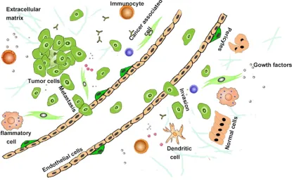
endothelial cells (ECs); blood vessel cells; and networks of cytokines and growth factors [14, 15]. These interactions provide tumor cells with biochemical and biophysical cues [4]. The tumor microenvironment exhibits heterogene-ity,with differing signals based on the types of
tissues, differentiation stage, and pathological conditions [16, 17]. The crosstalk between the cancer cells and the tumor stroma is re- sponsible for tumor progression and meta- stasis via a pyramid-like mechanism. Through autocrine, paracrine and hormonal signaling, tumor cells can alter and maintain their own survival conditions to promote their growth and development [6].
In the microenvironment, fibroblasts interact
with cancer cells and are activated into
hetero-geneous cancer-associated fibroblasts (CAFs) or myofibroblasts [18] (Figure 1). CAFs remo- del components of the ECM by increasing the production of ECM proteins and proteases. Additionally, they suppress the immune res-
ponse by recruiting inflammatory cells (such as
monocytes and macrophages) and modifying immune cell function to create a suitable envi-ronment for tumor growth [19, 20]. CAFs stimu-late tumor proliferation, angiogenesis and metastasis through growth factors and cyto-kines, including vascular endothelial growth
factor (VEGF), transforming growth factor-β (TGF-β), hepatocyte growth factor (HGF), plate
[image:2.612.104.519.78.334.2]let derived growth factor (PDGF), SDF-1, cyclo-oxygenase 2 (COX-2), and several interleukins
(IL-1β, IL-6, IL-8) [21, 22]. VEGF secretion pro -motes multiple processes involved in angiogen-esis, which facilitates cancer pathogenesis by supplying nutrients to enhance tumor growth [23]. In addition, pericytes, classical hallmarks of cancer, directly or indirectly contribute to tumor growth, metastatic spread, and thera-peutic resistance [1].
The tumor microenvironment plays an impor-tant role in tumor metastasis and invasion. Tumor cells invade the surrounding ECM and assemble ECs to form new blood vessels or a similar new reticular structure of blood vessels called tumor vasculogenic mimicry, which is closely associated with the occurrence, devel-opment and metastasis of tumors and poor prognosis [24, 25]. Then, tumor cells enter the lymphatic system and blood circulation to form circulating tumor cells (CTC) [26]. In this
pro-cess, cancer growth and vascularization are
tightly regulated by hypoxia [27], cytokines [23] and a multitude of cell phenotypes and ECM components within the tumor microenviron- ment.
The importance of simulating the tumor mi-croenvironment
Because the tumor microenvironment is a high-ly complicated system, further elucidation of the tumor microenvironment will increase our understanding of tumorigenesis and growth-promoting signaling pathways. Therefore, simu-lating the tumor microenvironment and devel-oping appropriate model systems are indis-pensable method to study the physiology and biochemistry of tumor cells and address many questions. By simulating the tumor microenvi-ronment, we can assess how a malignant state develops from a healthy tissue environment [28], qualify the role of the microenvironment in tumor migration, invasion and metastasis [29], gain insight into the expression of proteins under the regulation of microenvironment [30], and discover the spatial and temporal mecha-nisms of tumor angiogenesis and migration across the vascular system [31].
Constructing the tumor-resident environment to develop antitumor therapy is a useful meth-od. Elucidating the metabolic pathways between cancer cells and antitumor immune
cells could help guide cancer immunotherapy [32]. A better understanding of the relationship between epidermal growth factor (EGF) and the EGF-receptor could be used to inhibit the migra-tion of breast cancer cells. Addimigra-tionally, study-ing how tumor stroma affects tumor cell biologi-cal behavior could help design targeted thera-peutic strategies [19]. Researching the rela-tionship between the tumor and vascular com-partments may facilitate prediction of patient prognosis.
Elucidating tumor metastasis mechanisms by reconstructing the tumor microenvironment will not only identify meaningful therapeutic tar-gets [32] but will also be an important tool for predicting drug metabolism and toxicity in vitro. Presently, several cell-based models have been used to predict drug metabolism and toxicity to obtain more accurate parameters of treatment responses [33].
Approaches for simulating the microenviron-ment
Since Carl Jensen designed the earliest tumor model [34], in which he transplanted mouse sarcomas into healthy mice and measured tumor growth to estimate the vitality of the transplanted cancer, a large variety of tumor cell culture models have emerged, including the conventional monolayer technique, sponta-neous cell aggregation, cellular scaffolding, the
MCTS, the RCCS, and microfluidic devices.
Although each model has its own set of advan-tages and disadvanadvan-tages, the best choice is often a model that is simple and has clinical relevance for human patients [35].
Conventional monolayer technique
Two-dimensional cell culture techniques, such as 2D Petri dishes, 2D multi-well plates or 2D glass slides, have the advantages of conve-nience, operability, low cost, widespread appli-cation and ease of culturing a single cell. These in vitro method are commonly used to study
malignant tumors and analyze antitumor drugs
[36-38]. Cells cultured in these systems could be exposed directly to nutrients, oxygen or
spe-cific environments [39]. Two-dimensional cell
response of the same generation under differ-ent conditions but also observe dynamic chang-es in different generations. In the past decadchang-es, cancer biologists, biomedical researchers, and oncologists have used 2D Petri dishes to study the complicated tumorigenic mechanisms of angiogenesis, invasion, and metastasis [40].
These models offer significant advantages for
preclinical cancer drug discovery efforts given their simplicity and low cost.
The disadvantages of the 2D monolayer cul-tures used as in vitro models were uncovered gradually. In conventional 2D conditions, ECM components and the cell and cell-to-matrix interactions, which are important for dif-ferentiation, proliferation and cellular functions in vivo, are lost [41]. Due to the lack of the ECM components as a structural architecture that supports and connects the cells and alters the
organization and cell physiological activities, specific signaling between tumor cells and the
molecular gradient, which is an important fac-tor for cellular activities, are unavailable [9, 39]. Tumor cells grow in an adherent monolayer and lack a true 3D environment. The activities of
these cells are limited. In addition, trials with these models did not provide information on the chemotherapeutic response mechanism. It
is difficult to predict the effect of drugs in the
body and study the effect of restriction by numerous extracellular barriers in the body
that could otherwise lead to significant reduc
-tions in the infiltration capacity [9, 10]. However,
these models have thus far been inadequate
for discovering definitive cancer treatments. In
addition, these models are unable to accurately simulate the true tumor microenvironment. Spontaneous cell aggregation
[image:4.612.90.524.87.402.2]A deeper understanding of the mechanism and development of clinical treatments for tumors have been hindered by using a traditional monolayer technique [42]. Since Bissell dem-onstrated the different behaviors of cancerous breast cells grown in 3D culture, 3D systems have served as the major in vitro cell culture model for studying tumor progression [11-13]. Early application of the cells in a 3D cell culture model is spontaneous cell aggregation [9]. In this method, tumor cells grow into spheroids or Table 1. Comparison between different materials
Material Advantages Disadvantages
Silk fibroin Good biocompatibility [48] Low hydrophilic property [49] Good biological adhesive [50] Slow degradation [50]
High tensile strength
Collagen High elasticity [51] Lack of flexibility [51, 52]
Low tensile strength [52, 53]
Fibrin glue Small antigen [54] Rapid degradation [54]
Low immunogenicity Poor mechanical strength [54] Good biocompatibility
Chitosan Easy processing [56] Low solubility [55, 56]
Inexpensive Good biocompatibility [55]
Alginate Strong adsorption [57] Instability [57]
Porous Resists degradation
Agarose High hydrophilic property [58] Cannot resist high
Inert Temperature [59]
Stable
Poly glycolic acid Good biocompatibility Low hydrophilic property [60] Mechanical strength [60] Poor biological adhesive Polyethylene glycol Good biocompatibility Cannot be degraded
High mechanical [61]
Poly (lactic-co-glycolic acid) Good biocompatibility [63] Poor biological adhesive [64] Mechanical strength
other 3D forms spontaneously or upon
induc-tion by artificial substrates that induce cellular
differentiation and maintain cellular function. Compared with 2D cell culture techniques, this system closely mimics the complex structures
and organization of tissues in the body [43]
where tumor cells reside histologically as multi-cellular tumor spheroids (MCTS) and provide biochemical as well as physical cues between the ECM and the basement membrane (BM) [44, 45]. This system could be used to co-cul-ture different cell types, facilitating cell-cell interactions and the exchange of growth fac-tors and other biological effecfac-tors; thus, these strategies expand research on the molecular mechanisms of adhesion, migration and
inva-sion [9, 10]. Unfortunately, this method is limit
-ed to specific cell types and the cell interac
-tions and group sizes cannot be controlled.
Cellular scaffolds
To closely simulate a 3D environment, biolo-gists have employed engineered scaffolds to reconstruct the ECM and provide physical/ structural support. An ideal cellular scaffold possesses the following traits: (1) good
biocom-patibility and does not cause inflammation and
abnormal reactions in the body; (2) made of appropriate biodegradable material; (3) lacks immunogenicity and toxicity; (4) maintains cell morphology and phenotype, promotes cell adhesion and proliferation, and induces tissue regeneration; (5) conducive to the diffusion of nutrients. However, in fact, each material has its advantages and disadvantages [46, 47] as noted in Table 1.
MCTS
MCTS systems using in vitro tissue culture methods allow tumor cells to grow into 3D mul-ticellular spherical structures by implanting
tumor cells into a specific scaffold, such as a
collagen scaffold [65], semi-solid medium (agar or agarose) [66], and liquid media [67], and cul-turing the cells to resemble the dimensional effects of the in vivo tumor microenvironment. The tumor cells within spheroids are in close contact or communicate with each other, and this strategy was proposed as a promising method for the maintenance of differentiated functions. Therefore, these systems can bridge the gap between 2D tissue culture models and
animal cell culture systems in the field of study
-ing tumor biology, interactions and drug responses. The major methods of preparing multicellular tumor spheres include the rotating culture method [68] and static culture method [67]. AccBased on their material properties, the systems can be divided into two primary groups: scaffold-based systems, a platform that can be used to investigate the effect of primary exter-nal physical factors on microspheroid growth and signaling [69], and non-adherent, liquid-based systems that allow for the exchange of medium to a certain degree [70].
Given their inherent properties, such as closely arranged cells, hypoxia, and heterogeneity [71, 72], these models have been viewed as ideal tools to provide new insight into phenotypic and cellular heterogeneity and micro-environmental aspects of in vivo tumor growth [70, 73]. These models have also been used to study microen-vironment interactions, particularly intracellu-lar signaling and other functional or celluintracellu-lar processes between exogenous ECM molecules and tumor cell receptors [74-76]. MCTS models can not only be used for elucidation of various mechanisms but have also produced important advances in response to radiation injury, can-cer drug screening and drug discovery [65, 73]. Because the tumor sphere is similar to normal tumor tissues, it can be used to determine
spe-cific tumor tissue sensitivity to chemotherapy
and radiotherapy, which can help identify new anti-cancer drugs and reduce side effects or drug resistance [65]. In addition, multicellular tumor spheres act as pathological cancer cells in the in vitro immersion attack model, which is useful for analysis of multiple factors [77].
Although these models provide significant
advances in the simulation of the tumor micro-environment, some innate limitations still exist. The diffusion of oxygen and nutrients are limit-ed by the spheroid model, which restricts the
size of the spheroid [78].
RCCS
Many tumor cells fail to retain their specialized
sys-tems have disadvantages. National Aeronautics
and Space Administration (NASA) first devel -oped rotary cell culture systems to simulate microgravity, in which cells are cultured in a
dynamic fluid suspension in liquid media mixed
by minimal hydrodynamic forces [9]. RCCS have become a large-scale expansion of the cell cul-ture system.
Compared with traditional methods, RCCS exhibit three distinct characteristics: (1) Tumor cells, gases and nutrients are evenly distribut-ed. In a static training system, tumor cells often are limited to the bottom of the system, and the
interior lacks a sufficient number of cells.
Moreover, gas, nutrients and metabolic wastes are unevenly distributed, resulting in the accu-mulation of waste products and alterations in local pH. These changes may inhibitthe normal tumor microenvironment and prevent the cells
from obtaining sufficient nutrients, leading to
slow growth or even stagnation [80, 81]. However, RCCS are dynamic systems that pro-mote diffusion of oxygen and nutrients, dis-charge more metabolic waste, promote cell growth and have a uniform distribution of the tumor cells. (2) Centrifugation and stir are con-ventional methods to suspended cells in the Petri dishes. However, these generate shear force to damage cells. Thus, cells and tissues spend a considerable amount of time on repair, and tissue differentiation is hindered. However, RCCS use gravity-free, low-shear-force cell cul-tivation [82]. (3) In 3D cell cultures with inert material scaffolds, cells tend to attach to one another on the microcarriers to form complex 3D structures [82]. The following limitations are present: a complicated operation process, high cost and a lack of integration.
Microfluidic devices
Microfluidic devices, a breakthrough in simulat -ing the tumor microenvironment, have pro-foundly affected our understanding of the tumor microenvironment and provided guid-ance for clinical treatment. These approaches were developed based on advances in micro-mechanics, microelectronics, biotechnology and nanotechnology. These approaches allow sample collection, reaction, separation and detection to occur in a basic operation unit at the submillimeter scale and to automatically complete the whole process analysis. Com- pared with traditional experimental technology,
microfluidic devices have the following impor -tant characteristics: (1) These devices accu-rately simulate the microenvironment. Micro-
fluidic devices create a 3D environment that more accurately reflects the human body using
multiple cell types co-cultured with cytokines.
Using new materials, we can even recreate the
tumor microenvironment under hypoxic condi-tions [83, 84]. In addition, multiple types of
cells can be co-cultured on the microfluidic
devices. Thus, the signaling pathway or sequen-tial changes between cells in vitro can be assessed [85]. (2) This technique allows bio-medical research to be conducted in real-time and under controllable conditions. We can observe changes in various processes and obtain valid data in real time. In addition, we can combine required instruments into a
micro-fluidic chip [86]. (3) This method requires a
small amount of sample, which is indispens-able for clinical research. Thus, we can diag-nose diseases with small samples and reduce the cost of drug screening [87]. (4) This
tech-nique is flexible and portable, offering concen -tration of molecules in space and time [88, 89]. Therefore, this system may be useful for
per-sonalized diagnoses and perper-sonalized medi -cine based on drug toxicity screening and dis-ease modeling for drug target discovery. These characteristics satisfy the demand of research-ers for biochemical experiments, given that the characteristics of this methodology (i.e., a small amount of liquid, high-throughput, automation and particularly controllability) are lacking in gene and protein devices [87].
However, a multitude of challenges still exist: (1) Poor reusability, i.e., once polluted, the sam-ple cannot be continually used in the study; (2)
Failure to realize full automation; (3) Low
throughput because the adsorption of antigen and antibody cannot be controlled under the
high flow velocity; thus, we should limit the velocity of the fluid; (4) Lack of popularization,
as this system is exclusively used in the labora-tory and not currently used for clinical purposes.
Application of simulation platforms
What can these simulation platforms be used for? In recent decades, the diagnosis and treat-ment of tumors, drug research and tissue
engi-neering have benefited from these platforms
fluorescent dyes, Western blot analysis, and
polymerase chain reaction (PCR) assays. Based on the results from these platforms,
personal-ized treatment and precision medical diagno
-ses were realized. Complex pharmaceutical
processes have become simple and high-throughput with high content.
Cancer is a major cause of mortality. However, the leading cause of death in patients with
tumors is CTCs [90-93]. Microfluidic devices
can be used to detect and isolate the CTCs [94,
95], allowing for early diagnoses, individualized
treatments and evaluations of prognosis [96].
In addition, microfluidic devices, which are
inherently rapid and sensitive, have been used for point of care testing (POCT), referring to a portable mini test system outside the central laboratory that is close to the test object and offers timely results [97-100]. At present, the
development of a personalized means of analy
-sis is one of the important directions of
micro-fluidic chip POCT research for illness monitoring
and early diagnosis [101].
Although conventional in vitro platforms have made substantial contributions to screening drugs, these methods still have different limita-tions. Because 2D cell culture models are stat-ic states, as time progresses, these models cannot generate the mechanical or chemical stimuli (signaling molecules) that are normally present and simulate the complex internal envi-ronment in the body or the true extracellular mechanical environment [3, 102]. Currently in preclinical stages, 3D cell culture models have been proposed as promising in vitro methods to evaluate and predict tumor responses to chemotherapeutic agents [103-105].
The current trends in drug discovery screening requireboth high-throughput and high content. Thus, 3D cell culture technologies are naturally indispensable tool [106]. MCTS systems are employed for predicting dynamics, screening drugs and reducing the toxicity and side effects
to normal tissues [107]. Microfluidic devices
not only mimic complicated internal environ-ment in vitro but also generate concentration gradients required for physiological activities [108] and high-throughput drug screening [109, 110]. This device would be a simple and
conve-nient but high-efficiency tool to identify drug
targets, study drug metabolism mechanisms and drug response, and assess drug genotoxic-ity and cytotoxicgenotoxic-ity for anti-cancer drug/agent
discovery. Using microfluidic devices to study
the major factors that affect the efficacy of the
anticancer drugs has been previously reported [111, 112].
Conclusion
In this review, the role of the tumor microenvi-ronment has been elucidated with various plat-forms. However, our current knowledge is just the tip of the iceberg. Many detailed
mecha-nisms are uncharacterized. For example, how
does the microenvironment affect
tumorigene-sis specifically? What are the key factors influ
-encing the formation of tumors? Significant
steps forward have been made over the past few years in basic research and clinical applica-tion. Two-dimensional cell culture techniques
have allowed us to recognize the microscopic
world of the tumor. Three-dimensional in vitro tissue culture models enrich or broaden our
horizons. Clearly, the development and applica -tion of 3D tissue culture technology will be the hot spots in the future, especially in terms of
miniaturization, integration and automation.
However, complex production processes and high-throughput features are a major stumbling block to the use of these devices. Our hope is that with interdisciplinary collaboration, we will be able to design high-throughput equipment using 3D printing technology to simplify the pro-duction process.
Acknowledgements
We thank the Qingdao Institute of Biomass Energy and Bioprocess Technology of the Chinese Academy of Sciences for their techni-cal guidance.
Disclosure of conflict of interest
None.
Address correspondence to: Dr. Haitao Niu, Depart- ment of Urology, Affiliated Hospital of Qingdao University, Qingdao, China; Key Laboratory of Urinary System Diseases, Qingdao 266003, China. E-mail: [email protected]; Dr. Jianning Wang, Depart- ment of Urology, Qianfoshan Hospital, Shandong University, Jinan 250014, China. E-mail: doc [email protected]
References
[2] Weber CE, Kuo PC. The tumor microenviron-ment. Surg Oncol 2012; 21: 172-177.
[3] Polyak K, Kalluri R. The role of the microenvi-ronment in mammary gland development and cancer. Cold Spring Harbor Perspect Biol 2010; 2: a003244.
[4] Beck JN, Singh A, Rothenberg AR, Elisseeff JH, Ewald AJ. The independent roles of mechani-cal, structural and adhesion characteristics of 3D hydrogels on the regulation of cancer inva-sion and dissemination. Biomaterials 2013; 34: 9486-9495.
[5] Fidler IJ. The role of the organ microenviron-ment in brain metastasis. Semin Cancer Biol 2011; 21: 107-112.
[6] Ma H, Xu H, Qin J. Biomimetic tumor microenvi-ronment on a microfluidic platform. Biomicro -fluidics 2013; 7: 14.
[7] Abbott A. Cell culture: biology’s new dimen-sion. Nature 2003; 424: 870-2.
[8] Guiro K, Patel SA, Greco SJ, Rameshwar P, Arinzeh TL. Investigating breast cancer cell be -havior using. tissue engineering scaffolds. PLoS One 2015; 10: 22.
[9] Kim JB, Stein R, O’Hare MJ. Three-dimensional in vitro tissue culture models of breast cancer-a review. Brecancer-ast Ccancer-ancer Res Trecancer-at 2004; 85: 281-91.
[10] Kim JB. Three-dimensional tissue culture mod-els in cancer biology. Semin Cancer Biol 2005; 15: 13.
[11] Ravi M, Paramesh V, Kaviya SR, Anuradha E, Solomon FD. 3D cell culture systems-advan-tages and applications. J Cell Physiol 2015; 230: 32.
[12] Szot CS, Buchanan CF, Freeman JW, Rylander MN. 3D in vitro bioengineered tumors based on collagen I hydrogels. Biomaterials 2011; 32: 8.
[13] Worthington P, Pochan DJ, Langhans SA. Pep-tide hydrogels-versatile matrices for 3D cell culture in cancer medicine. Front Oncol 2015; 5: 92.
[14] Kahlert C, Kalluri R. Exosomes in tumor micro-environment influence cancer progression and metastasis. J Mol Med (Berl) 2013; 91: 7. [15] Friedl P, Alexander S. Cancer Invasion and the
Microenvironment: Plasticity and Reciprocity. Cell 2011; 147: 18.
[16] Robertson-Tessi M, Gillies RJ, Gatenby RA, An-derson AR. Impact of metabolic heterogeneity on tumor growth, invasion, and treatment out-comes. Cancer Res 2015; 75: 1567-1579. [17] Hanahan D, Weinberg RA. Hallmarks of
can-cer: the next generation. Cell 2011; 144: 646-674.
[18] Ma H, Liu T, Qin J, Lin B. Characterization of the interaction between fibroblasts and tumor cells on a microfluidic co-culture device. Elec -trophoresis 2010; 31: 1599-605.
[19] Zhang J, Liu J. Tumor stroma as targets for can-cer therapy. Pharmacol Ther 2013; 137: 200-15.
[20] Kalluri R, Zeisberg M. Fibroblasts in cancer. Nat Rev Cancer 2006; 6: 392-401.
[21] Pietras K, Ostman A. Hallmarks of cancer: in-teractions with the tumor stroma. Exp Cell Res 2010; 316: 1324-1331.
[22] Whipple CA, Brinckerhoff CE. BRAFV600E mel-anoma cells secrete factors that activate stro-mal fibroblasts and enhance tumourigenicity. Br J Cancer 2014; 111: 9.
[23] Carmeliet P. Angiogenesis in health and dis-ease. Nat Med 2003; 9: 653-60.
[24] Cao Z, Bao M, Miele L, Sarkar FH, Wang Z, Zhou Q. Tumour vasculogenic mimicry is asso-ciated with poor prognosis of human cancer patients: a systemic review and meta-analysis. Eur J Cancer 2013; 49: 3914-3923.
[25] Seftor RE, Hess AR, Seftor EA, Kirschmann DA, Hardy KM, Margaryan NV, Hendrix MJ. Tumor cell vasculogenic mimicry: from controversy to therapeutic promise. Am J Pathol 2012; 181: 1115-1125.
[26] Kang JH, Krause S, Tobin H, Mammoto A, Kanapathipillai M, Ingber DE. A combined mi-cromagnetic-microfluidic device for rapid cap -ture and cul-ture of rare circulating tumor cells. Lab Chip 2012; 12: 2175-2181.
[27] Bergers G, Benjamin LE. Tumorigenesis and the angiogenic switch. Nat Rev Cancer 2003; 3: 401-410.
[28] [28] Salvatore V, Teti G, Bolzani S, Focaroli S, Durante S, Mazzotti MC, Falconi M. Simulating tumor microenvironment: changes in protein expression in an in vitro co-culture system. Cancer Cell Int 2014; 14: 40.
[29] Reid BG, Jerjian T, Patel P, Zhou Q, Yoo BH, Ka-bos P, Sartorius CA, Labarbera DV. Live multi-cellular tumor spheroid models for high-con-tent imaging and screening in cancer drug discovery. curr chem genom transl med 2014; 8.
[30] Sung SY, Hsieh CL, Wu D, Chung LW, Johnstone PA. Tumor microenvironment promotes cancer progression, metastasis, and therapeutic re-sistance. Curr Probl Cancer 2007; 31: 36-100. [31] Dickinson LE, Lutgebaucks C, Lewis DM, Ge-recht S. Patterning microscale extracellular matrices to study endothelial and cancer cell interactions in vitro. Lab Chip 2012; 12: 4244-8.
[32] McCarthy SA, Mufson RA, Pearce EJ, Rathmell JC, Howcroft TK. Metabolic reprogramming of the immune response in the tumor microenvi-ronment. Cancer Biol Ther 2013; 14: 315-318. [33] Wilding JL, Bodmer WF. Cancer cell lines for
drug discovery and development. Cancer Res 2014; 74: 2377-2384.
[35] Voskoglou-Nomikos T, Pater JL, Seymour L. Clinical predictive value of the in vitro cell line, human xenograft, and mouse allograft preclin-ical cancer models. Clin Cancer Res 2003; 9: 14.
[36] Petrackova D, Halada P, Bezouskova S, Kresin -ova Z, Svobod-ova K. A two-dimensional protein map of Pleurotus ostreatus microsomes-pro-teome dynamics. Folia Microbiol (Praha) 2016; 61: 63-71.
[37] Juvonen H, Maattanen A, Lauren P, Ihalainen P, Urtti A, Yliperttula M, Peltonen J. Biocompat -ibility of printed paper-based arrays for 2-D cell cultures. Acta Biomater 2013; 9: 6704-6710. [38] Wang R, Xu J, Juliette L, Castilleja A, Love J,
Sung SY, Zhau HE, Goodwin TJ, Chung LW. Three-dimensional co-culture models to study prostate cancer growth, progression, and me-tastasis to bone. Semin Cancer Biol 2005; 15: 353-364.
[39] Hutmacher DW, Horch RE, Loessner D, Rizzi S, Sieh S, Reichert JC, Clements JA, Beier JP, Arkudas A, Bleiziffer O, Kneser U. Translating tissue engineering technology platforms into cancer research. J Cell Mol Med 2009; 13: 1417-1427.
[40] Singh M, Close DA, Mukundan S, Johnston PA, Sant S. Production of uniform 3D microtumors in hydrogel microwell arrays for measurement of viability, morphology, and signaling pathway activation. Assay Drug Dev Technol 2015; 8: 1-14.
[41] Mazzoleni G, Di Lorenzo D, Steimberg N. Mod -elling tissues in 3D: the next future of pharma-co-toxicology and food research? Genes Nutr 2009; 4: 13-22.
[42] Justice BA, Badr NA, Felder RA. 3D cell culture opens new dimensions in cell-based assays. Drug Discov Today 2009; 14: 102-107.
[43] Hede K. Environmental protection: studies highlight importance of tumor microenviron-ment. J Natl Cancer Inst 2004; 96: 2.
[44] Schindler M, Nur-E-Kamal A, Ahmed I, Kamal J, Liu HY, Amor N, Ponery AS, Crockett DP, Grafe TH, Chung HY, Weik T, Jones E, Meiners S. Liv-ing in Three Dimensions 3D nanostructured environments for cell culture and regenerative medicine. Cell Biochem Biophys 2006; 45: 13. [45] Knedlitschek G, Schneider F. A tissue-like cul-ture system using microstruccul-tures: influence of extracellular matrix material on cell adhe-sion and aggregation. J Biomech Eng 1999; 121: 5.
[46] Lin CY, Kikuchi N, Hollister SJ. A novel method for biomaterial scaffold internal architecture design to match bone elastic properties with desired porosity. J Biomech 2004; 37: 623-36. [47] Takezawa T. A strategy for the development of
tissue engineering scaffolds that regulate cell behavior. Biomaterials 2003; 24: 2267-2275.
[48] Vepari C, Kaplan DL. Silk as a Biomaterial. Prog Polym Sci 2007; 32: 991-1007.
[49] Rao J, Shen J, Quan D, Xu Y. Property studies on three-dimensional porous blended silk scaffolds. Zhongguo Xiu Fu Chong Jian Wai Ke Za Zhi 2009; 23: 8.
[50] Chang G, Kim HJ, Kaplan D, Vunjak-Novakovic G, Kandel RA. Porous silk scaffolds can be used for tissue engineering annulus fibrosus. Eur Spine J 2007; 16: 1848-57.
[51] Yates KE, Allemann F, Glowacki J. Phenotypic analysis of bovine chondrocytes cultured in 3D collagen sponges: effect of serum substitutes. Cell Tissue Bank 2005; 6: 45-54.
[52] Karna E, Miltyk W, Palka JA, Jarzabek K, Wolc -zynski S. Hyaluronic acid counteracts interleu -kin-1-induced inhibition of collagen biosynthe-sis in cultured human chondrocytes. Pharmacol Res 2006; 54: 275-281.
[53] Ragetly GR, Griffon DJ, Lee HB, Chung YS. Ef-fect of collagen II coating on mesenchymal stem cell adhesion on chitosan and on reacet-ylated chitosan fibrous scaffolds. J Mater Sci Mater Med 2010; 21: 2479-2490.
[54] de la Puente P, Ludena D. Cell culture in autol-ogous fibrin scaffolds for applications in tissue engineering. Exp Cell Res 2014; 322: 1-11. [55] Liskova J, Douglas TE, Beranova J,
Skwarczy-ńska A, Božič M, Samal SK, Modrzejewska Z, Gorgieva S, Kokol V, Bačáková L. Chitosan hy -drogels enriched with polyphenols: Antibacte-rial activity, cell adhesion and growth and min-eralization. Carbohydr Polym 2015; 129: 135-142.
[56] Pradines B, Lievin-Le Moal V, Vauthier C, Pon-chel G, Loiseau PM, Bouchemal K. Cell line-dependent cytotoxicity of poly (isobutylcyano-acrylate) nanoparticles coated with chitosan and thiolated chitosan: Insights from cultured human epithelial HeLa, Caco2/TC7 and HT-29/MTX cells. Int J Pharm 2015; 491: 17-20. [57] Zhang H, Liu X, Yang M, Zhu L. Silk fibroin/so
-dium alginate composite nano-fibrous scaffold prepared through thermally induced phase-separation (TIPS) method for biomedical appli-cations. Mater Sci Eng C Mater Biol Appl 2015; 55: 8-13.
[58] Gorth DJ, Lothstein KE, Chiaro JA, Farrell MJ, Dodge GR, Elliott DM, Malhotra NR, Mauck RL, Smith LJ. Hypoxic regulation of functional ex-tracellular matrix elaboration by nucleus pulp-osus cells in long-term agarose culture. J Or-thop Res 2015; 33: 747-754.
[59] Guo X, Li S, Ji Q, Lian R, Chen J. Enhanced via-bility and neural differential potential in poor post-thaw hADSCs by agarose multi-well dish-es and spheroid culture. Hum Cell 2015; 28: 175-89.
-rameters on alignment of PCL/PGA nanofi -brous scaffold: an artificial neural networks approach. Int J Biol Macromol 2014; 81: 1089-97.
[61] Han C, Takayama S, Park J. Formation and ma-nipulation of cell spheroids using a density ad-justed PEG/DEX aqueous two phase system. Sci Rep 2015; 5: 11891.
[62] Lee EJ, Lee JH, Jin L, Jin OS, Shin YC, Sang JO, Lee J, Hyon SH, Han DW. Hyaluronic acid/ poly(lactic-<I>co</I>-glycolic acid) core/shell fiber meshes loaded with epigallocatechin-3-<I>O</I>-gallate as skin Tissue engineering scaffolds. J Nanosci Nanotechnol 2014; 14: 8458-8463.
[63] Mehrasa M, Asadollahi MA, Ghaedi K, Salehi H, Arpanaei A. Electrospun aligned PLGA and PLGA/gelatin nanofibers embedded with silica nanoparticles for tissue engineering. Int J Biol Macromol 2015; 79: 687-695.
[64] Liuyun J, Lixin J, Chengdong X, Lijuan X, Ye L. Effect of l-lysine-assisted surface grafting for nano-hydroxyapatite on mechanical properties and in vitro bioactivity of poly(lactic acid-co-gly- colic acid). J Biomater Appl 2016 ;30:750-8. [65] Le VM, Lang MD, Shi WB, Liu JW. A
collagen-based multicellular tumor spheroid model for evaluation of the efficiency of nanoparticle drug delivery. Artif Cells Nanomed Biotechnol 2016; 44: 540-4.
[66] Carlsson J. A proliferation gradient in three-di-mensional colonies of cultured human glioma cells. Int J Cancer 1977; 20: 8.
[67] Yuhas JM, Li AP, Martinez AO, Ladman AJ. A simplified method for production and growth of multicellular tumor spheroids. Cancer Res 1977; 37: 6.
[68] Sutherland RM, McCredie JA, Inch WR. Growth of multicell spheroids in tissue culture as a model of nodular carcinomas. J Natl Cancer Inst 1971; 46: 8.
[69] Greiner AM, Richter B, Bastmeyer M. Micro-engineered 3D scaffolds for cell culture stud-ies. Macromol Biosci 2012; 12: 1301-1314. [70] Thoma CR, Zimmermann M, Agarkova I, Kelm
JM, Krek W. 3D cell culture systems modeling tumor growth determinants in cancer target discovery. Adv Drug Deliv Rev 2014; 69-70: 29-41.
[71] Carlss J. A proliferation gradient in three-di-mensional colonies of cultured human glioma cells. Int J Cancer 1977; 20: 8.
[72] Freyer JP, Sutherland RM. Proliferative and clo-nogenic heterogeneity of cells from EMT6/Ro multicellular spheroids induced by the glucose and oxygen supply. Cancer Res 1986; 46: 3513-20.
[73] LaBarbera DV, Reid BG, Yoo BH. The multicel-lular tumor spheroid model for high-through-put cancer drug discovery. Expert Opin Drug Discov 2012; 7: 12.
[74] Ballangrud AM, Yang WH, Charlton DE, McDe-vitt MR, Hamacher KA, Panageas KS, Ma D, Bander NH, Scheinberg DA, Sgouros G. Re-sponse of LNCaP spheroids after treatment with an alpha-particle emitter (213Bi)-labeled anti-prostate-specific membrane antigen anti -body (J591). Cancer Res 2001; 61: 8. [75] Sutherland RM. Cell and environment
interac-tions in tumor microregions: the multicell spheroid model. Science 1988; 240: 8. [76] Carey SP, Starchenko A, McGregor AL,
Rein-hart-King CA. Leading malignant cells initiate collective epithelial cell invasion in a three-di-mensional heterotypic tumor spheroid model. Clin Exp Metastasis 2013; 30: 615-630. [77] Condello S, Morgan CA, Nagdas S, Cao L, Turek
J, Hurley TD, Matei D. beta-Catenin-regulated ALDH1A1 is a target in ovarian cancer spher-oids. Oncogene 2015; 34: 2297-2308. [78] Unsworth BR, Lelkes PI. Growing tissues in mi
-crogravity. Nat Med 1998; 4: 7.
[79] Hammond TG, Hammond JM. Optimized sus -pension culture: the rotating-wall vessel. Am J Physiol Renal Physiol 2001; 281: 15.
[80] Holy CE, Shoichet MS, Davies JE. Engineering three-dimensional bone tissue in vitro using biodegradable scaffolds: investigating initial cell-seeding density and culture period. J Biomed Mater Res 2000; 51: 7.
[81] Vunjak-Novakovic G, Martin I, Obradovic B, Treppo S, Grodzinsky AJ, Langer R, Freed LE. Bioreactor cultivation conditions modulate the composition and mechanical properties of tis-sue-engineered cartilage. J Orthop Res 1999; 17: 9.
[82] Lei XH, Ning LN, Cao YJ, Liu S, Zhang SB, Qiu ZF, Hu HM, Zhang HS, Liu S, Duan EK. NASA-approved rotary bioreactor enhances prolifera-tion of human epidermal stem cells and sup-ports formation of 3D epidermis-like structure. PLoS One 2011; 6: e26603.
[83] Selimovic S, Dokmeci MR, Khademhosseini A. Organs-on-a-chip for drug discovery. Curr Opin Pharmacol 2013; 13: 829-833.
[84] Beebe DJ, Ingber DE, den Toonder J. Organs on Chips 2013. Lab Chip 2013; 13: 3447-3448. [85] Tazawa H, Sato K, Tsutiya A, Tokeshi M,
Ohtani-Kaneko R. A microfluidic cell culture system for monitoring of sequential changes in endothe-lial cells after heat stress. Thromb Res 2015; 136: 328-34.
[86] Marasco CC, Enders JR, Seale KT, McLean JA, Wikswo JP. Real-time cellular exometabolome analysis with a microfluidic-mass spectrometry platform. PLoS One 2015; 10: e0117685. [87] Zheng B, Tice JD, Roach LS, Ismagilov RF. A
[88] Whitesides GM. The origins and the future of microfluidics. Nature 2006; 442: 368-373. [89] Mark D, Haeberle S, Roth G, von Stetten F,
Zengerle R. Microfluidic lab-on-a-chip plat -forms: requirements, characteristics and appli-cations. Chem Soc Rev 2010; 39: 1153-1182. [90] Steeg PS. Tumor metastasis: mechanistic in-sights and clinical challenges. Nat Med 2006; 12: 895-904.
[91] Nguyen DX, Bos PD, Massague J. Metastasis: from dissemination to organ-specific coloniza -tion. Nat Rev Cancer 2009; 9: 274-284. [92] Psaila B, Lyden D. The metastatic niche: ada-
pting the foreign soil. Nat Rev Cancer 2009; 9: 9.
[93] TOS AB, Hynes NE. Staying together on the road to metastasis. Nature 2014; 514: 2. [94] Esmaeilsabzali H, Beischlag TV, Cox ME,
Parameswaran AM, Park EJ. Detection and iso-lation of circulating tumor cells: principles and methods. Biotechnol Adv 2013; 31: 1063-1084.
[95] Stott SL, Hsu CH, Tsukrov DI, Yu M, Miyamoto DT, Waltman BA, Rothenberg SM, Shah AM, Smas ME, Korir GK, Floyd FP Jr, Gilman AJ, Lord JB, Winokur D, Springer S, Irimia D, Na-grath S, Sequist LV, Lee RJ, Isselbacher KJ, Ma-heswaran S, Haber DA, Toner M. Isolation of circulating tumor cells using a microvortex-generating herringbone-chip. Proc Natl Acad Sci U S A 2010; 107: 6.
[96] Friedlander TW, Premasekharan G, Paris PL. Looking back, to the future of circulating tumor cells. Pharmacol Ther 2014; 142: 271-280. [97] Sia SK, Kricka LJ. Microfluidics and
point-of-care testing. Lab Chip 2008; 8: 1982-1983. [98] Yeo LY, Chang HC, Chan PP, Friend JR. Microflu
-idic devices for bioapplications. Small 2011; 7: 12-48.
[99] Huddy JR, Ni MZ, Markar SR, Hanna GB. Point-of-care testing in the diagnosis of gastrointesti-nal cancers: current technology and future di-rections. World J Gastroenterol 2015; 21: 4111-4120.
[100] Su W, Gao X, Jiang L, Qin J. Microfluidic plat -form towards point-of-care diagnostics in infec-tious diseases. J Chromatogr A 2015; 1377: 13-26.
[101] Chin CD, Linder V, Sia SK. Lab-on-a-chip devic-es for global health: past studidevic-es and future opportunities. Lab Chip 2007; 7: 41-57. [102] Hirt C, Papadimitropoulos A, Muraro MG,
Mu-raro MG, Mele V, Panopoulos E, Cremonesi E, Ivanek R, Schultz-Thater E, Droeser RA, Men -gus C, Heberer M, Oertli D, Iezzi G, Zajac P, Eppenberger-Castori S, Tornillo L, Terracciano L, Martin I, Spagnoli GC. Bioreactor-engineered cancer tissue-like structures mimic pheno-types, gene expression profiles and drug resis -tance patterns observed “in vivo”. Biomateri-als 2015; 62: 138-46.
[103] Lovitt CJ, Shelper TB, Avery VM. Miniaturized three-dimensional cancer model for drug eval-uation. Assay Drug Dev Technol 2013; 11: 435-448.
[104] Yang Y, Yang X, Zou J, Jia C, Hu Y, Du H, Wang H. Evaluation of photodynamic therapy effi -ciency using an in vitro three-dimensional mi-crofluidic breast cancer tissue model. Lab Chip 2015; 15: 735-744.
[105] Lovitt CJ, Shelper TB, Avery VM. Evaluation of chemotherapeutics in a three-dimensional breast cancer model. J Cancer Res Clin Oncol 2015; 141: 951-959.
[106] Alonso-Padilla J, Rodríguez A. High throughput screening for anti-Trypanosoma cruzi drug dis -covery. PLoS Negl Trop Dis 2014; 8: 6. [107] Monazzam A, Josephsson R, Blomqvist C,
Carlsson J, Långström B and Bergström M. Ap-plication of the multicellular tumour spheroid model to screen PET tracers for analysis of early response of chemotherapy in breast can-cer. Breast Cancer Res 2007; 9: R45.
[108] Kim SH, Lee GH, Park JY, Lee SH. Micro- plaforms for gradient field generation of vari -ous properties and biological applications. J Lab Autom 2015; 20: 82-95.
[109] Kwapiszewska K, Michalczuk A, Rybka M, Kwapiszewski R, Brzozka Z. A microfluidic-based platform for tumour spheroid culture, monitoring and drug screening. Lab Chip 2014; 14: 2096-104.
[110] Kamei K, Mashimo Y, Koyama Y, Fockenberg C, Nakashima M, Nakajima M, Li J, Chen Y. 3D printing of soft lithography mold for rapid pro-duction of polydimethylsiloxane-based micro-fluidic devices for cell stimulation with concen -tration gradients. Biomed Microdevices 2015; 17: 36.
[111] Young EW. Cells, tissues, and organs on chips: challenges and opportunities for the cancer tumor microenvironment. Integr Biol (Camb) 2013; 5: 1096-1109.