is closely related to the poor prognosis of liver cancer [7]. It also has been detected in colon cancer, and the abnormal expression of PLCB1 is closely related to colon cancer [8].
miRNAs could be tumor suppressors and take part in various cell functions [9, 10]. PLCB1 is reported to be silenced by microRNAs (e.g., miR-1324 and miR-124) [8, 11] and plays a vital role in cell development, proliferation, and ap- optosis [12, 13]. Previous research indicates that miR-423-5p has been identified as a novel biomarker in tumors. miR-423-5p has been demonstrated to increase autophagy in hepato-cellular carcinoma cells [14]. miR-423-5p levels could inhibit osteosarcoma proliferation and invasion by directly targeting STMN1 [15]. Ho- wever, the molecular mechanism that regulates miR-423-5p expression is still unclear for gli- oma.
In this study, we found that PLCB1 was highly expressed in glioma. PLCB1 directly targeted by
Original Article
miR-423-5p inhibits the proliferation and metastasis of
glioblastoma cells by targeting phospholipase C beta 1
Peng Zhao1, Shukai Sun1, Yu’e Zhai1, Qingwu Tian1, Tingting Zhou1, Jing Li2
Departments of 1Clinical Laboratory, 2Nephrology, Affiliated Hospital of Qingdao University, Qingdao, China Received May 7, 2019; Accepted June 25, 2019; Epub August 1, 2019; Published August 15, 2019
Abstract: Glioma is a common brain tumor which is highly invasive, responds poorly to therapy, and has a poor prognosis. There is growing evidence that an abnormal expression of many genes is related to glioma and leads to glioma cell growth and metastasis. Phospholipase C beta 1 (PLCB1) plays critical roles in intracellular transduction and regulating signal activation, which are important to tumorigenesis. Therefore, it could bind to miRNA as a target gene. The purpose of our study was to confirm that PLCB1 plays a critical role in suppressing glioma progression. We found that the expression of miR-423-5p was reduced, but the expression of PLCB1 was increased, in glioma tissues and cells. To explore whether 423-5p affects PLCB1, a bioinformatics approach suggested that miR-423-5p can directly target PLCB1. Moreover, we observed, using luciferase reporter assays, that miR-miR-423-5p could target PLCB1 3’-UTR. Functionally, the overexpression of miR-423-5p could attenuate the proliferation, invasion, and migration and promote the apoptosis of glioma cells. Furthermore, we found that miR-423-5p could enhance p-ERK expression in glioma cells. Taken together, we deduced that miR-423-5p inhibited proliferation and metastasis by targeting PLCB1, and it also promotes apoptosis in glioma cells. These results suggest that miR-423-5p directly targets PLCB1 3’-UTR and could inhibit cell invasion and migration through the ERK-dependent pathway in glioma, and the miR-423-5p/PLCB1 axis may be a potential target for new potential therapeutic strategies to treat glioma.
Keywords: Glioma, miR-423-5p, PLCB1, metastasis
Introduction
Glioma is the most common subtype of primary brain tumor [1, 2], and it is characterized by dif-fuse infiltration and is highly metastatic, which explains its fast recurrence and high mortality. Therefore, it is necessary to require available therapy strategies and find novel treatment approaches. Previous findings indicate that the abnormal expression of many genes is related to glioma and leads to glioma cell growth and metastasis [3-5].
miR-423-5p and PLCB1 promotes the prolifera-tion and migraprolifera-tion of glioma cells. The overex-pression of miR-423-5p suppresses the growth and invasion of glioma. These data suggest that miR-423-5p may be a target for glioma di- agnosis and treatment.
Materials and methods
Clinical sample collection
A total of 30 glioma tissue samples and their adjacent tissues were obtained by surgical resection at the Affiliated Hospital of Qingdao University (Qingdao, Shandong, China) from March 2015 to September 2016. None of the patients received any anti-tumor treatment such as chemotherapy or radiotherapy before their surgeries. The samples were immediately frozen in liquid nitrogen following surgical re- section and stored at -80°C for subsequent study.
Cell line culture
The human glioma U87 cell line was obtained from the American Type Culture Collection (ATCC). The cells were grown in MEM medium (GIBCO, USA) with 10% FBS (GIBCO, USA) and 1% penicillin/streptavidin (GIBCO, USA) and maintained at 37°C in a humidified atmosphere at 5% CO2.
siRNA transfection
For the siRNA transfection, 2 × 10⁵ cells per well were plated in a 6-well plate. After adher-ing for 24 hours, a miR-423-5p duplex mimic, miR-con, PLCB1 siRNA, and control siRNA (Ri- boBio, Guangzhou, China) were added to the transfection medium for 6 hours at 37°C in a CO2 incubator. After transfection, the cells were supplemented with a normal culture medium and cultured at 37°C/5% CO2 for up to 48 hours before harvest.
RNA extraction and quantitative RT-PCR
analy-sis
RNA was isolated from the tissues or cells using a mirVana miRNA Isolation Kit (Ambion, Car- lsbad, CA, USA), following the manufacturer’s instructions. The first strand of cDNA was syn-thesized using a PrimeScript 1st Strand cDNA Synthesis Kit (Takara, Dalian, China). The ex- traction concentration was measured with a
NanoDrop spectrophotometer, with standby preservation at -80°C. The cDNA was amplified using Power SYBR Green PCR Master Mix (Ap- plied Biosystems, Carlsbad, CA, USA) with ap- propriate primers using an ABI 7500-fast ther-mocycler (Applied Biosystems, Foster City, CA). U6 and GAPDH served as internal controls. Relative expression was determined using the 2-∆∆Ct method. PLCB1: forward primer: 5’-GAT-
GAGCCCAGATGGCCG-3’, reverse primer: 5’-AG- TTGAGTCATCATCCCACTTGA-3’, miR-423-5p: fo- rward primer: 5’-ATGGTTCGTGGGTGAGGGGCA- GAGAGCGAGAGCAGGGTCCGAGGTATTCG- 3’, reverse primer: 5’-GTGCAGGGTCCGAGGT-3’ [16].
Immunohistochemical assays
We performed an immunohistochemical assay for each sample. According to the standard pro-tocol, the major steps included fixing, paraf-finizing, dewaxing, dehydrating, antigen retriev-al, blocking, incubation with primary antibodies (PLCB1, Cell Signaling Technology), washing, blocking, incubation with secondary antibodies (Santa Cruz), antigen-antibody reactions with diaminobenzidine (Maixin, Fuzhou, China), and observations.
Western blot analysis
Cell viability assay
Cell proliferation was determined using the Cell Titer-Blue Cell viability Assay (Promega). The cells were seeded in a 96-well plate with the density of the optimized cell number (2,000 cells/well). After 24 hours of seeding, the cells were treated with siRNA or diluted chemicals at the indicated working concentration. The cells were incubated for another 48 hours and then measured using a fluorescence microplate re- ader (Sunrise Remote, Tecan Austria GmbH, Grödig, Austria).
Apoptosis assays
Apoptotic cells were identified using a terminal deoxynucleotidyl transferase dUTP nick end labeling (TUNEL)-based cell detection kit, POD (Roche Applied Science). The cells were fixed on slides with 4% paraformaldehyde for 1 hour, washed with PBS, and then incubated for 10 min with 3% H2O2 in methanol at room tempera-ture. After washing with PBS again, the cells were incubated for 2 min on ice with 0.1% Triton X-100 in 0.1% sodium citrate. The slides were then incubated with the TUNEL reaction mix-ture for 60 min at 37°C and then rinsed with FBS. The stained slides were examined under light microscopy (Motic B1-223A, Motic Deut- schland GmbH, Wetzlar, Germany).
Migration and invasion assay
Migration was performed using a wound heal-ing assay. A linear wound was made by scrapheal-ing the cells using a 200 µl micropipette tip and the debris was washed twice with PBS. At different time points, the cells were photographed with an inverted microscope (Olympus, Japan), and the migration distances were measured. The cell invasion ability was examined using Ma- trigel-coated Transwell chamber inserts with 8mm pores (Corning, USA). Stably transduced cells were trypsinized, adjusted to 10 106/ml in serum-free MEM, and a 200 µl cell suspension (2 × 10⁵ cells) was added to the upper chamber above the Matrigel. The lower chamber was filled with 500 µl 10% FBS-MEM. After 24 h, the upper chamber was removed, and the Matrigel and unmigrated cells were gently scraped with a wet cotton swab. The cells were fixed with 70% ethanol and stained with hematoxylin. Then, the cells were counted under a lighted microscope, randomly selecting three areas for counting.
Software support and statistical analysis
All images were formatted for optimal presenta-tion using Adobe Illustrator CS4 (Adobe Sys- tems). To determine the statistical significance, the P-value from t-statistic was calculated. Results
PLCB1 was significantly highly expressed in
glioma
To examine the level of PLCB1 in tissues and cell lines, we quantified PLCB1 expression in the glioma tissues and the cell lines. qPCR was performed so that the mRNA expression level of PLCB1 had a significantly high expression in the glioma tissues compared with the corre-sponding adjacent tissues (Figure 1A). Similarly, the high protein expression of PLCB1 was veri-fied by immunohistochemistry and Western-blot (Figure 1B and 1C). These results suggest that PLCB1 may play a vital role in glioma. PLCB1 and cell viability evaluation
We detected PLCB1 expression in the glioma U87 cells and the astroglia (AS) cells using qPCR and Western-blot (Figure 2A and 2B). The PLCB1 expression level was clearly higher in the U87 cells compared with the AS cells. To assess the biological role of PLCB1, PLCB1-specific small interfering RNAs (PLCB1 siRNAs) or the corresponding control siRNA (Con siRNA) were introduced into the glioma cells and the efficiency of PLCB1 siRNA was also tested by qPCR and Western-blot (Figure 2C and 2D). As a result, the knockdown of PLCB1 exhibited sig-nificantly decreased cell growth, invasion and migration in theU87 cells (Figure 2E-G). This indicates that PLCB1 plays a vital role in anti-apoptotic activity and in cell proliferation in gli-oma cells.
miR-423-5p directly targets PLCB1 3’-UTR in
glioma cells
mutate-type showed little change in luciferase activity (Figure 2B). These results reflected that PLCB1 directly targets miR-423-5p.
miR-423-5p inhibits cell proliferation and
pro-motes cell apoptosis
To observe the effects of miR-423-5p in glio-ma, we determined the expression level of miR-423-5p in glioma tissues and cell lines using qPCR. The expression of miR-423-5p in the tu- mor samples was lower than in the adjacent tis-sue samples (Figure 4A). Consistent with the cancer tissue results, the miR-423-5p level was lower in the U87 cells compared with the
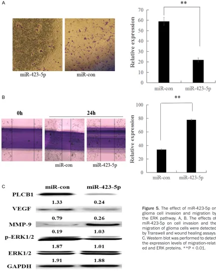
[image:4.612.88.524.73.478.2]cer, glioma and breast can-cer [17-19], but the mecha-nism is not clear in glioma. To reveal whether miR-423-5p suppressed the inva-sion and migration by the ERK-dependent pathway, U87 cells were transfected with miR-423-5p mimics, miR-Con, for 24 h. Western blotting was carried out to analyze the levels of p-E- RK and total ERK, and the results reflected that miR-423-5p inhibited p-ERK (Fi- gure 5C). These results de- monstrate that ERK-depe- ndent signaling mediates the creased levels of inva-cated that the overexpression of miR-423-5p in
U87 cells markedly reduced their tumor grow-ing ability.
Furthermore, a TUNEL assay showed that the apoptosis rate was increased markedly in the U87 cells with miR-423-5p (Figure 4E). It was shown that the accumulation of miR-423-5p could increase the number of apoptotic cells. Taken together, miR-423-5p can inhibit cell pro-liferation and promote cell apoptosis.
miR-423-5p attenuates cell invasion and migration in glioma cells through the ERK-dependent pathway
Cell invasion is a crucial process in cancer metastasis, so a Transwell assay was used to assay the invasion of glioma cells. miR-423-5p at high levels resulted in a significant decrease in cell invasion in transfection with miR-423-5p cells compared with the miR-control group (Figure 5A). The result was consistent with th- ose from a migration assay (Figure 5B). Mo- reover, we detected invasion-associated pro-tein by Western-blot. The results reflected that invasion-associated protein expression was attenuated in U87 cells with miR-423-5p trans-fection. Several studies have suggested that PLCB1 is corrected with the ERK pathway in many kinds of tumor cells, including lung
can-sion and migration in U87 cells. Combined with the above data, these results show that miR-423-5p directly targets PLCB1 3’-UTR and could inhibit cell invasion and migration through the ERK-dependent pathway in glioma cells. Discussion
Glioma, with its low, 5-year survival rates, is the most common and aggressive brain tumor. The main treatment for glioma is surgical, but there is no clear boundary with brain tissue, so it is difficult to remove the tumor. The effect of clini-cal therapy is very poor at present. Therefore, there is a pressing need for better therapies that enable the precise targeting of genes in glioma cells.
Previous studies supported the idea that most cancers result from the accumulation of genet-ic changes (mutations) in cells over time [20, 21]. Previous studies demonstrated that PLCB1 had a higher expression in liver cancer and colon cancer, so it could serve as a biomarker for these cancers [7, 11]. In our glioma sam-ples, a majority of the tumor tissues expressed high levels of PLCB1 mRNA compared to adja-cent tissues. Moreover, we found both high PLCB1 mRNA expression and protein expres-sion in the glioma cells. Furthermore, we deter-mined that high PLCB1 expression was associ-Figure 2. PLCB1 was highly expressed in glioma cells and is essential for cell proliferation, invasion and migration. A, B. Relative mRNA and protein expression of PLCB1 in glioma cells compared with AS cells. C, D. Transfection of PLCB1 siRNA reduced the PLCB1 mRNA and protein levels. E-G. Cell proliferation, invasion, and migration were inhibited by siRNA-PLCB1 in glioma cells. **P < 0.01, *P < 0.05.
[image:6.612.93.386.130.274.2]ated with cell proliferation, invasion, and migra-tion in glioma cells. However, the precise mech-anism and the therapeutic targets of glioma have not yet been fully elucidated.
MicroRNAs (miRNA) are a new class of noncod-ing RNAs, which regulate a variety of biological processes, and their functions are involved in cell proliferation, cell apoptosis, development, and metabolism [22-24]. Nowadays, many stud-ies have reported that miRNAs are associated with glioma. For example, MiR-150-3p targets SP1 and suppresses the growth of glioma cells [25]. MicroRNA-1231 exerts a tumor suppres-sor role by regulating the EGFR/PI3K/AKT axis in glioma [26]. In the present study, we showed that there were few studies finding that miR-423-5p is related to glioma, and even less data indicating the specific mechanism between
[image:7.612.93.521.70.402.2]some cancers, including ovarian cancer, glio-blastomas, and colon cancer [28-30]. In our study, we identified PLCB1 as a direct target gene of miR-423-5p. The enforced expression of miR-423-5p was explored to create low expression levels of PLCB1 in glioma cells and promote their malignant progression. It is
[image:8.612.87.521.72.623.2]with data implicating miR-423-5p induces cell apoptosis in glioma cells by targeting PLCB1, thereby inhibiting colon tumor cell growth, inva-sion, and migration.
In general, these results reveal that miR-423-5p could serve as a potential therapeutic target involved in the regulatory roles of PLCB1 in glio-ma cells. Moreover, our results demonstrated that miR-423-5p plays a vital role in the growth and metastasis of glioma, and affects cell apoptosis. Therefore, these findings may indi-cate that miR-423-5p could be a diagnostic and prognostic marker in the treatment of gli- oma.
Disclosure of conflict of interest
None.
Address correspondence to: Jing Li, Department of Nephrology, Affiliated Hospital of Qingdao Uni- versity, 1667 Wutaishan Road, Qingdao, China. E- mail: starpeng1986@126.com
References
[1] Song J, Ma Q, Hu M, Qian D, Wang B, He N. The inhibition of miR-144-3p on cell proliferation and metastasis by targeting TOP2A in HCMV-positive glioblastoma cells. Molecules 2018; 23.
[2] Mehta S, Lo Cascio C. Developmentally regu-lated signaling pathways in glioma invasion. Cell Mol Life Sci 2018; 75: 385-402.
[3] Han L, Liu D, Li Z, Tian N, Han Z, Wang G, Fu Y, Guo Z, Zhu Z, Du C, Tian Y. HOXB1 is a tumor suppressor gene regulated by miR-3175 in gli-oma. PLoS One 2015; 10: e0142387.
[4] hang J, Cai H, Sun L, Zhan P, Chen M, Zhang F, Ran Y, Wan J. LGR5, a novel functional glioma stem cell marker, promotes EMT by activating the Wnt/beta-catenin pathway and predicts poor survival of glioma patients. J Exp Clin Cancer Res 2018; 37: 225.
[5] Zheng J, Liu X, Wang P, Xue Y, Ma J, Qu C, Liu Y. CRNDE promotes malignant progression of glioma by attenuating miR-384/PIWIL4/STAT3 axis. Mol Ther 2016; 24: 1199-215.
[6] Ngoh A, McTague A, Wentzensen IM, Meyer E, Applegate C, Kossoff EH, Batista DA, Wang T, Kurian MA. Severe infantile epileptic encepha-lopathy due to mutations in PLCB1: expansion of the genotypic and phenotypic disease spec-trum. Dev Med Child Neurol 2014; 56: 1124-8. [7] Li J, Zhao X, Wang D, He W, Zhang S, Cao W,
Huang Y, Wang L, Zhou S, Luo K. Up-regulated expression of phospholipase C, beta1 is
asso-prognosis in hepatocellular carcinoma. Onco Targets Ther 2016; 9: 1697-706.
[8] Zhou L, Li Y, Jiang W, Zhang H, Wen Z, Su Y, Wu F, Zhi Z, Shen Q, Li H, Xu X, Tang W. Down-regulation of circ-PRKCI inhibits cell migration and proliferation in Hirschsprung disease by suppressing the expression of miR-1324 tar-get PLCB1. Cell Cycle 2018; 17: 1092-1101. [9] Rah H, Chung KW, Ko KH, Kim ES, Kim JO,
Sakong JH, Kim JH, Lee WS, Kim NK. miR-27a and miR-449b polymorphisms associated with a risk of idiopathic recurrent pregnancy loss. PLoS One 2017; 12: e0177160.
[10] Li N, Pan X, Zhang J, Ma A, Yang S, Ma J, Xie A. Plasma levels of miR-137 and miR-124 are as-sociated with Parkinson’s disease but not with Parkinson’s disease with depression. Neurol Sci 2017; 38: 761-767.
[11] Lu ML, Zhang Y, Li J, Fu Y, Li WH, Zhao GF, Li XH, Wei L, Liu GB, Huang H. MicroRNA-124 in-hibits colorectal cancer cell proliferation and suppresses tumor growth by interacting with PLCB1 and regulating Wnt/beta-catenin sig-naling pathway. Eur Rev Med Pharmacol Sci 2019; 23: 121-136.
[12] Klenke S, Rump K, Buschkamp K, Engler A, Peters J, Siffert W, Frey UH. Characterization of the PLCB1 promoter and regulation by early growth response transcription factor EGR-1. Eur J Pharmacol 2014; 742: 8-14.
[13] Bakhsh AD, Ladas I, Hamshere ML, Bullock M, Kirov G, Zhang L, Taylor PN, Gregory JW, Scott-Coombes D, Völzke H, Teumer A, Mantripragada K, Williams ED, Clifton-Bligh RJ, Williams NM, Ludgate ME. An indel in phospholipase-C-B-1 is linked with euthyroid multinodular goiter. Thyroid 2018; 28: 891-901.
[14] Stiuso P, Potenza N, Lombardi A, Ferrandino I, Monaco A, Zappavigna S, Vanacore D, Mosca N, Castiello F, Porto S, Addeo R, Prete SD, De Vita F, Russo A, Caraglia M. MicroRNA-423-5p promotes autophagy in cancer cells and is in-creased in serum from hepatocarcinoma pa-tients treated with sorafenib. Mol Ther Nucleic Acids 2015; 4: e233.
[15] Wang X, Peng L, Gong X, Zhang X, Sun R, Du J. miR-423-5p inhibits osteosarcoma prolifera-tion and invasion through directly targeting STMN1. Cell Physiol Biochem 2018; 50: 2249-2259.
[16] Yuan XP, Liu LS, Chen CB, Zhou J, Zheng YT, Wang XP, Han M, Wang CX. MicroRNA-423-5p facilitates hypoxia/reoxygenation-induced ap- optosis in renal proximal tubular epithelial cells by targeting GSTM1 via endoplasmic re-ticulum stress. Oncotarget 2017; 8: 82064-82077.
induces proliferation, survival, and migration in a549 lung cancer cells by activating the ERK signaling pathway through a transcription-in-dependent mechanism. Biomed Res Int 2015; 2015: 404368.
[18] Puustinen P, Junttila MR, Vanhatupa S, Sablina AA, Hector ME, Teittinen K, Raheem O, Ketola K, Lin S, Kast J, Haapasalo H, Hahn WC, We- stermarck J. PME-1 protects extracellular sig-nal-regulated kinase pathway activity from pro-tein phosphatase 2A-mediated inactivation in human malignant glioma. Cancer Res 2009; 69: 2870-7.
[19] Zhao Y, Ma J, Fan Y, Wang Z, Tian R, Ji W, Zhang F, Niu R. TGF-beta transactivates EGFR and fa-cilitates breast cancer migration and invasion through canonical Smad3 and ERK/Sp1 sig-naling pathways. Mol Oncol 2018; 12: 305-321.
[20] Peralta-Rodríguez R, Valdivia A, Mendoza M, Rodríguez J, Marrero D, Paniagua L, Romero P, Taniguchi K, Salcedo M. Genes associated to cancer. Rev Med Inst Mex Seguro Soc 2015; 53 Suppl 2: S178-87.
[21] Sonnenschein C, Soto AM. Cancer and the elu-sive cancer genes. Med Sci (Paris) 2014; 30: 688-92.
[22] Li J, Jin H, Yu H, Wang B, Tang J. miRNA1284 inhibits cell growth and induces apoptosis of lung cancer cells. Mol Med Rep 2017; 16: 3049-3054.
[23] Eichelmann AK, Matuszcak C, Hummel R, Hai- er J. Role of miRNAs in cell signaling of cancer associated fibroblasts. Int J Biochem Cell Biol 2018; 101: 94-102.
[24] Cui Z, Liu G, Kong D. miRNA27a promotes the proliferation and inhibits apoptosis of human pancreatic cancer cells by Wnt/beta-catenin pathway. Oncol Rep 2018; 39: 755-763. [25] Tan Z, Jia J, Jiang Y. MiR-150-3p targets SP1
and suppresses the growth of glioma cells. Biosci Rep 2018; 38.
[26] Zhang J, Zhang J, Qiu W, Zhang J, Li Y, Kong E, Lu A, Xu J, Lu X. MicroRNA-1231 exerts a tumor suppressor role through regulating the EGFR/ PI3K/AKT axis in glioma. J Neurooncol 2018; 139: 547-562.
[27] Singh NK. miRNAs target databases: develop-mental methods and target identification tech-niques with functional annotations. Cell Mol Life Sci 2017; 74: 2239-2261.
[28] Li S, Zeng A, Hu Q, Yan W, Liu Y, You Y. miR-423-5p contributes to a malignant phenotype and temozolomide chemoresistance in glio-blastomas. Neuro Oncol 2017; 19: 55-65. [29] Tang X, Zeng X, Huang Y, Chen S, Lin F, Yang G,
Yang N. miR-423-5p serves as a diagnostic in-dicator and inhibits the proliferation and inva-sion of ovarian cancer. Exp Ther Med 2018; 15: 4723-4730.