Original Article
High expression of HAX-1 protein is associated with
tumor growth in papillary thyroid carcinoma
Yiwei Wang1, Xiaochen Xie2, Zhenxiang Sun1, Jin Zang1, Xuren Sun3, Shiqiao Peng2, Xiaohui Yu2, Xiaojie Wang1
1Department of Anatomy, Shenyang Medical College, Shenyang, Liaoning Province, P. R. China; 2Department
of Endocrinology and Metabolism, Institute of Endocrinology, Liaoning Provincial Key Laboratory of Endocrine Diseases, The First Affiliated Hospital of China Medical University, China Medical University, Shenyang, Liaoning, P. R. China; 3Department of Gastroenterology, The First Affiliated Hospital of China Medical University, China
Medical University, Shenyang, Liaoning, P. R. China
Received January 16, 2018; Accepted February 23, 2018; Epub April 1, 2018; Published April 15, 2018
Abstract: Papillary thyroid carcinoma (PTC) is the most common type of endocrine malignancy. HS1-associated pro-tein X-1 (HAX-1) is an anti-apoptotic factor involved in the development of many types of cancer. However, its func-tional role in human PTC remains unclear. Here we investigated HAX-1 overexpression in human PTC samples and correlated with tumor size and TNM stage. Decreased expression of HAX-1 significantly inhibited proliferation, mi -gration, and invasion in papillary thyroid cancer cell lines TPC1 and K1. Furthermore, we found that down-regulation of HAX-1 induced cell apoptosis. Our results suggest that HAX-1 plays a significant role in regulating the biological behavior in PTC cells and may contribute to PTC target therapy.
Keywords: HAX-1, papillary thyroid carcinoma, apoptosis, migration, invasion
Introduction
PTC is the most common type of all thyroid cancers [1]. Despite most of the patients suf-fering from PTC having satisfactory long term prognosis with approximately 95% 5-year sur-vival rate, about 20% of PTC patients exhibit distant metastasis or local recurrence [2]. Lymph node metastasis develops in the early stages of PTC and enhances the rate of mortal-ity and recurrence [3, 4]. Mechanisms regard-ing tumorigenesis to therapeutic means remain unclear, indicating the need for more studies to identify novel treatments for PTC.
HCLS1-associated protein X-1 (HAX-1) was first
considered as a protein, which contributes to B cell signaling transformation by interplaying
with HS-1 (hematopoietic lineage cell-specific
protein-1) [5]. HAX-1 can exert its function by interacting with diverse structural proteins, in- dicating its contribution in intracellular signal-ing [5-7]. HAX-1 is widely expressed in human and mouse tissues and has been suggested to be positioned in cell membranes and the
HAX-1 in papillary thyroid carcinoma
Materials and methods
Tissue specimen acquisition
This study was performed with the approval of
the Ethics Committee of First Affiliated Hospital
of China Medical University, and informed con-sent was obtained from all the patients with PTC whose tissue specimens were used in this study. None of the patients included suffered from other concurrent malignancies or were anaplastic or had other poorly differentiated follicular carcinomas of the thyroid or under-went any treatment prior to surgery. Fresh-frozen tumor tissues (n=12) and adjacent normal tissues (n=12) were obtained from patients with PTC undergoing surgical
resec-tion at the First Affiliated Hospital of China
Medical University. Samples were stored at
-80°C for further experiments. Paraffin section
specimens from the thyroid were obtained from 102 Chinese patients who had PTC. All patients had a clinical duration of less than 3 years and had been admitted to the hospital for standard thyroidectomies between 2011
and 2013. Diagnoses were confirmed through
histopathological examination. None of the patients had received radiotherapy or chemo-therapy before undergoing surgical. TNM stage was assessed according to the tumor, node,
and metastasis system classification proposed
by the American Joint Committee on Cancer (7th edition).
Cell culture and transfection
Human papillary thyroid cancer cell lines TPC-1 and K1 were kind gifts from Dr Wei Sun. TPC-
1 was cultured Dulbecco’s modified Eagle’s
medium (DMEM, Gibco, NY, USA) and 10% fetal bovine serum (FBS, Hyclone, UT, USA). Cells
were cultured in Dulbecco’s modified Eagle’s
medium (DMEM): Ham’s F12: MCDB105 (2:1:1) plus 2 mM glutamine and 10% fetal bovine serum. All cells were cultured under 5% CO2 atmosphere 37°C. The medium was changed 2 days and sub-cultured when the cell
popula-tion density reached to 70-80% confluence.
HAX-1- and HAX-1-RNAi-lentiviral vectors were purchased from Shanghai GeneChem Com- pany (Shanghai, China). The HAX-1 sequence was 5’-AGCCCAAATCCTATTTCAA-3’ and the shRNA control sequence was 5’-TTCTCCGAA- CGTGTCACGTTT-3’.
Western blot assay
Tissues and cells were lysed in radioimmuno-precipitation (RIPA) buffer and total protein concentration was determined with a bicincho-ninic acid (BCA) assay (Beyotime, China). Twenty micrograms of total protein were separated by 10% SDS-PAGE and then transferred to
polyvinylidene fluoride (PVDF) membranes. The
membranes were washed, blocked, and
incu-bated sequentially with specific primary anti -bodies, namely: rabbit polyclonal anti-HAX1 (1:1000, Abcam, Cambridge, UK), rabbit mono-clonal anti-Cleaved-caspase 3 (1:1500, Abcam, Cambridge, UK), rabbit monoclonal anti-cleav- ed-caspase 9 (1:1500, Abcam, Cambridge, UK), rabbit monoclonal anti-BAX (1:1500, Ab- cam, Cambridge, UK), anti-DR5 (1:500, Abcam, Cambridge, UK) mouse polyclonal anti-Tubulin (1:3000, Santa Cruz, USA). Incubation in pri-mary antibodies was followed by goat anti-rab-bit secondary antibody or goat anti-mouse secondary antibody (1:5000, Origene Co.,Ltd. Beijing, China). The reactions were detected by enhanced chemiluminescence assay. Each experiment was performed in triplicate.
Cell viability assays
The 3-(4,5-dimethylthiazol-2-yl)-2,5-diphenylte- trazolium bromide (MTT) assay was used to determine the cell viability. thyroid cancer cell were seeded on 96-well plates at 1 d, 2 d and 3 d after transfection, the samples were rinsed twice with sterile phosphate buffered saline (PBS) and the adherent cells were incubated in medium containing 100 mg/0.1 ml of MTT (Sigma Aldrich) for 4 h. After carefully removing the medium, the blue crystal layer attached to the surface of the material was then dissolved using dimethyl sulfoxide (DMSO). The optical density (OD) was measured at 490 nm using a spectrophotometer.
Cell migration assay and invasion assay
Migration assays were performed using
atran-swell chamber with an 8-μm pore size for
24-well plates (Corning). Cells transfected with shHAX-1 and shNC were seeded in the upper chamber at 2-5×10⁵ in DMEM serum free mediumand DMEM with 10% FBS was added in the bottom of the chamber. After 24 h, the
cells on the upper surface of the filter were
were fixed in 95% ethanol 15 min stained with 0.1% crystal violet. Three fields per chamber
were randomly selected and the average cells numbers were counted. Cell invasion assays were performed in plates were coated by Matrigel (BD Bioscience, San Jose, CA, USA) and with similar conditions for the migration assay.
Results
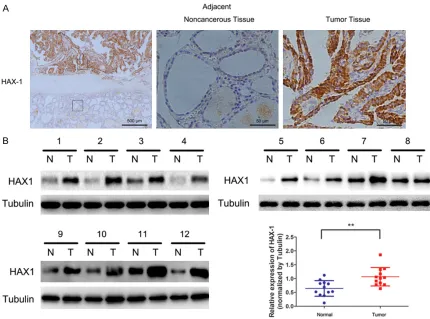
HAX-1 is upregulated in PTC and correlated with clinical characteristics
To evaluate the role of HAX-1 in the progression of PTC, immunohistochemistry was used to test in 102 PTC specimens and their adjacent normal tissues (Figure 1A). The association between HAX-1 expression levels and patient clinical characters is shown in Table 1. High
expression of HAX-1 was significantly associat -ed with tumor size (P<0.05) and tumor TNM stage (P<0.05). No correlations were found
with gender, patient age, or lymph node
metas-tasis. These findings suggest that levels and
activity of HAX1 correlate with the stage of PTC. Furthermore, Western blot assay was employed for analysis of HAX-1 protein expression in 12 PTC tissues samples. As shown in Figure 1B, HAX-1 in PTC tissues was higher than that in the matched adjacent normal tissues (P<0.01).
Knockdown of HAX1 in PTC cell lines inhibits cellular proliferation and colony formation
In order to characterize the role of HAX-1 in
TPC1 and K1 cell proliferation, we first used a
shRNA targeted to HAX1 (shHAX-1) markedly decreased the HAX-1 level compared to the control sequence (shNC) in both TPC1 and K1 cells (Figure 2A). Next, to assess cells proli- feration rate we used MTT assay. The result showed that the proliferation rate of shHAX-1
group was significantly reduced than that of
[image:3.612.92.522.71.390.2]shNC group in both cell lines (Figure 2B). Furthermore, clonogenic assays showed that
HAX-1 in papillary thyroid carcinoma
HAX1 knockdown significantly inhibited cell
colony formation (Figure 2C). These findings
suggest that silencing HAX-1 could impair PTC cell growth.
Knockdown of HAX1 inhibits PTC cell migration and invasion
To explore whether knockdown of HAX-1 could affect cell invasion and migration capacities, we performed transwell assay in both cell lines transfected with shHAX1 and shNC. The results showed that, compared to the shNC group, the migration and invasion cell number of TPC1 and K1 in shHAX1 group was dramatically down-regulated (Figure 2D and 2E). Moreover,
the results confirmed that down-regulation of
HAX-1 could reduce PTC cell migration and invasion ability.
HAX-1 knockdown induced PTC cells apoptosis
To further study the effect of HAX-1 inhibition on the apoptosis of PTC, cells transfected with shHAX-1 and their control group cells were
stained with Annexin V and PI. The flow cytom -etry results suggested that the apoptosis in-
duced by shHAX-1 was significantly increased
in both TPC1 and K1 cell lines. In addition, the
expression levels of cleaved caspase 3 and 9 were dramatically increased through suppres-sion of HAX-1 expressuppres-sion in PTC cells (Figure 3B). Moreover, we examined whether BAX and death receptor 5 (DR5) were regulated by HAX-1 silencing. The result showed that both BAX and DR5 were up-regulated in TPC and K1 cells. Discussion
In the present study, our results shown that HAX-1 expression is up-regulated and correlat-ed with clinical pathological features in PTC. Additionally, knockdown of HAX-1 in two PTC cell lines (K1 and TPC-1) inhibited proliferation, migration and invasion. In addition, HAX-1 also exhibited apoptosis in TPC-1 and K1 cell lines since repression of HAX-1 can caused cell apoptosis.
PTC is the most regular thyroid cancer in clini- cal symptoms. Most PTC has no obvious early symptoms, hence the unobvious symptom
and the difficulties lying in the early diagnosis
[image:4.612.89.295.95.335.2]largely affect the effect of the treatment [12]. In this study, we found that expression of HAX-1 was up-regulated in human PTC samples compared to normal thyroids by Western blot and immunohistochemistry. On the basis of Oncomine [13], a cancer microarray database, HAX-1 was over-expressed in several types of carcinoma, such as hepatoma, lung cancer, myeloma, and lymphoma. HAX-1 plays a crucial role in carcinogenesis and has been demon-strated to be up-regulate in multiple tumor cell lines, which provides a strong evidence of its role in tumorigenesis and metastasis. In our results, there was a positive correlation be- tween HAX-1 expression and TNM stage. HAX-1 was also closely associated with tumor size. These results are in accordance with the results of other carcinomas. Wang et al. reported that up-regulation of HAX-1 is positively correlated with cancer grade in hepatocellular carcinoma [14], suggesting that HAX-1 may contribute to the tumorigenesis and progression of PTC. According to our in vitro study, silencing HAX-1 can remarkably repress cell proliferation, mig- ration, and invasion in two PTC cell lines, TPC1 and K1. HAX-1 has been suggested to exert a vital role in the mediation of cell migration [7, 8]. The HAX-1 chaperone HS1 is isogenous to cortactin, which is a cytoskeletal protein mostly elevated in cancer [5]. Due to the homology Table 1. Clinicopathologic features of 102
thy-roid carcinomas patients
Characteristics Expression of hax1 P value
Low High
Number=39 Number=63 Gender
Male 15 35 0.093
Female 24 28
Age (years)
<50 18 30 0.885
≥50 21 33
LNM
Yes 17 37 0.115
No 22 25
TNM Stage
I+II 25 25 0.017*
III+IV 14 38
Tumor size (cm)
<2 23 23 0.027*
≥2 16 40
LNM: lymph node metastasis. Statistical analyses were
HAX-1 in papillary thyroid carcinoma
between HS1 and cortactin [15], it is possible that HAX-1 can also bind with cortactin [8]. Several research studies had demonstrated that cortactin induces the invasion and metas-tasis in tumor cells [16]. HAX-1 can bind with cortactin by forming complex, thus promotes migration, while activates cell adhesion due to the lack of HAX-1 [11, 17]. A recent report has suggested that HAX-1 plays a vital role in the mediation of cancer cell migration and invasion by clathrin-regulated endocytosis [8]. Silencing
of the HAX-1 gene significantly inhibited cell
proliferation, migration, and invasion, which suggests that HAX-1 is involved in oncogenesis and development of PTC, which may repress differentiation and lymph node metastasis of this malignancy.
[image:6.612.90.518.70.446.2]In our experiment, the expression of cleaved caspase 3 and 9 were also up-regulated when treating with shHAX-1. It has been previously reported that HAX-1 could repress caspase 9 activity. Moreover, it could induce accumulation of BAX (BCL2 associated X, apoptosis media-tor)by interacting with HtrA2 (high temperature regulated A2), thus initiating the apoptotic pathway. HAX-1 may exert vital anti-apoptotic effects through modulating the mitochondrial pathway [5, 18-20]. It has been demonstrated that HAX-1 could prevent cell from mitochon-drial impairment caused by environmental and other factors and reducesecretion of pro-apop-totic signals from the mitochondria membrane [21, 22]. In this study, BAX was increased through silencing HAX-1, and the apoptotic cell
death was highly increased as well. Consistent with our studies, it has been suggested that high expression of BAX was represent in human PTC tissues and cell lines. Functional inhibition of HAX-1 increases apoptosis in two PTC cell lines. We also examined whether the
expres-sion of death receptor, DR5 was influenced
by shHAX-1 supplement by Western blot. This result proves that elevated expression of DR5 by shHAX-1 treatment may contribute to cell death in PTC cell lines. This study also analyzed the cleavage of caspase-3 and 9 in response to treat with shHAX-1, in accordance with a role of apoptosis in HAX-1 regulated cell death. In addition, we measured expression by Western blotof DR5, a death receptor by silencing HAX-1 gene, in two PTC cell lines. Cell death caused by apoptosis is initiated by co-action between
death receptor 5 and knockdown HAX-1 signifi -cantly increased protein expression of DR5 in both cell lines. Apoptosis is a vital mediator of tumorigenesis, which may be promoted by acti-vation of agonists such as the tumor necrosis factor (TNF) family [23]. TNF-related apoptosis inducing ligand (TRAIL) is a potential death receptor (DR) ligands leading to apoptosis via binding with DR in multiple carcinoma cell lines [24, 25]. This combination results in the conse-quent activation of caspase 9, which mediates downstream caspases activationand expres-sion of pro-apoptotic protein BAX. The role of DR signaling pathway during tumor initiation and malignant progression has made it an important regulator in cancer treatment. Conclusion
Our results suggest that HAX-1 is over-express- ed in PTC tissues and that up-regulation of HAX-1 is related to histological grade, as well as tumor size. Moreover, silencing HAX-1 promot-ed apoptosis, while inhibiting cell proliferation, migration and invasion. Our study provides novel insight that HAX-1 may function as a new orientation for PTC therapy.
Acknowledgements
The Science and Technology Project of Liaoning Province (grant numbers 201005036), Science and Technology Project of the Technology Divi- sion of Shenyang city (grant numbers F13-221-9-33), Natural Science Foundation of Liaoning Province (grant numbers 20170520040, 2014- 021039), the National Natural Science Found-
ation of China (grant numbers 81702738), the
Scientific Program for Doctoral initial funding
(grant 201501042).
Disclosure of conflict of interest
None.
Address correspondence to: Xiaojie Wang, Depart- ment of Human Anatomy, Shenyang Medical Colle- ge, Huanggu District, Shenyang 110034, Liaoning Province, P. R. China. Tel: 0086 13840484058; Fax: 0086 24 62216816; E-mail: wangxiaojie_ symc@163.com
References
[1] Kleihues P and Sobin LH. World health organi-zation classification of tumors. Cancer 2000; 88: 2887.
[2] Mazzaferri EL and Jhiang SM. Long-term im-pact of initial surgical and medical therapy on papillary and follicular thyroid cancer. Am J Med 1994; 97: 418-428.
[3] Leboulleux S, Rubino C, Baudin E, Caillou B, Hartl DM, Bidart JM, Travagli JP and Schlum- berger M. Prognostic factors for persistent or recurrent disease of papillary thyroid carcino-ma with neck lymph node metastases and/or tumor extension beyond the thyroid capsule at initial diagnosis. J Clin Endocrinol Metab 2005; 90: 5723-5729.
[4] Dionigi G, Dionigi R, Bartalena L, Boni L, Rovera F and Villa F. Surgery of lymph nodes in papillary thyroid cancer. Expert Rev Anticancer Ther 2006; 6: 1217-1229.
[5] Suzuki Y, Demoliere C, Kitamura D, Takeshita H, Deuschle U and Watanabe T. HAX-1, a novel intracellular protein, localized on mitochon-dria, directly associates with HS1, a substrate of Src family tyrosine kinases. J Immunol 1997; 158: 2736-2744.
[6] Kawaguchi Y, Nakajima K, Igarashi M, Morita T, Tanaka M, Suzuki M, Yokoyama A, Matsuda G, Kato K, Kanamori M and Hirai K. Interaction of Epstein-Barr virus nuclear antigen leader pro-tein (EBNA-LP) with HS1-associated propro-tein X-1: implication of cytoplasmic function of EBNA-LP. J Virol 2000; 74: 10104-10111. [7] Radhika V, Onesime D, Ha JH and
Dhanase-karan N. Galpha13 stimulates cell migration through cortactin-interacting protein Hax-1. J Biol Chem 2004; 279: 49406-49413.
inte-HAX-1 in papillary thyroid carcinoma
grin alphavbeta6. Cancer Res 2007; 67: 5275-5284.
[9] Banerjee A, Saito K, Meyer K, Banerjee S, Ait-Goughoulte M, Ray RB and Ray R. Hepatitis C virus core protein and cellular protein HAX-1 promote 5-fluorouracil-mediated hepatocyte growth inhibition. J Virol 2009; 83: 9663-9671.
[10] Lee AY, Lee Y, Park YK, Bae KH, Cho S, Lee DH, Park BC, Kang S and Park SG. HS 1-associated protein X-1 is cleaved by caspase-3 during apoptosis. Mol Cells 2008; 25: 86-90.
[11] Cilenti L, Soundarapandian MM, Kyriazis GA, Stratico V, Singh S, Gupta S, Bonventre JV, Alnemri ES and Zervos AS. Regulation of HAX-1 anti-apoptotic protein by Omi/HtrA2 protease during cell death. J Biol Chem 2004; 279: 50295-50301.
[12] Baldane S, Ipekci SH, Sozen M and Kebapcilar L. Mean platelet volume could be a possible biomarker for papillary thyroid carcinomas. Asian Pac J Cancer Prev 2015; 16: 2671-2674. [13] Rhodes DR, Yu J, Shanker K, Deshpande N,
Varambally R, Ghosh D, Barrette T, Pandey A and Chinnaiyan AM. Oncomine: a cancer mi-croarray database and integrated data-mining platform. Neoplasia 2004; 6: 1-6.
[14] Wang Y, Huo X, Cao Z, Xu H, Zhu J, Qian L, Fu H and Xu B. HAX-1 is overexpressed in hepatocel-lular carcinoma and promotes cell prolifera-tion. Int J Clin Exp Pathol 2015; 8: 8099-8106. [15] Uruno T, Zhang P, Liu J, Hao JJ and Zhan X.
Haematopoietic lineage cell-specific protein 1 (HS1) promotes actin-related protein (Arp) 2/3 complex-mediated actin polymerization. Biochem J 2003; 371: 485-493.
[16] Li Y, Tondravi M, Liu J, Smith E, Haudenschild CC, Kaczmarek M and Zhan X. Cortactin poten-tiates bone metastasis of breast cancer cells. Cancer Res 2001; 61: 6906-6911.
[17] Chuma M, Sakamoto M, Yasuda J, Fujii G, Nakanishi K, Tsuchiya A, Ohta T, Asaka M and Hirohashi S. Overexpression of cortactin is in-volved in motility and metastasis of hepatocel-lular carcinoma. J Hepatol 2004; 41: 629-636. [18] Shaw J and Kirshenbaum LA. HAX-1 represses
postmitochondrial caspase-9 activation and cell death during hypoxia-reoxygenation. Circ Res 2006; 99: 336-338.
[19] Yan J, Ma C, Cheng J, Li Z and Liu C. HAX-1 in-hibits apoptosis in prostate cancer through the suppression of caspase-9 activation. Oncol Rep 2015; 34: 2776-2781.
[20] Chao JR, Parganas E, Boyd K, Hong CY, Opferman JT and Ihle JN. Hax1-mediated pro-cessing of HtrA2 by Parl allows survival of lym-phocytes and neurons. Nature 2008; 452: 98-102.
[21] Trebinska A, Hogstrand K, Grandien A, Grzybo- wska EA and Fadeel B. Exploring the anti-apop-totic role of HAX-1 versus BCL-XL in cytokine-dependent bone marrow-derived cells from mice. FEBS Lett 2014; 588: 2921-2927. [22] Jiang X and Wang X. Cytochrome c promotes
caspase-9 activation by inducing nucleotide binding to Apaf-1. J Biol Chem 2000; 275: 31199-31203.
[23] Thompson CB. Apoptosis in the pathogenesis and treatment of disease. Science 1995; 267: 1456-1462.
[24] Ozoren N and El-Deiry WS. Cell surface death receptor signaling in normal and cancer cells. Semin Cancer Biol 2003; 13: 135-147. [25] LeBlanc HN and Ashkenazi A. Apo2L/TRAIL