Original Article
MiR-181a regulates the chondrogenic differentiation
in pig peripheral blood mesenchymal stem cells
Daohong Zhao1, Yanlin Li2, Yan Li3, Zhaowei Jiang3, Duo Shen4, Zhi Zhao1, Fuke Wang2
1Department of Orthopedics, Second Affiliated Hospital of Kunming Medical University, Kunming, China; 2
Depart-ment of Sports Medicine, First Affiliated Hospital of Kunming Medical University, Kunming, China; 3Department
of Orthopedics, People Hospital of Dehong State, Yunnan Province, China; 4Department of Orthopedics, People
Hospital of Longchuan County, Yunnan Province, China
Received May 14, 2017; Accepted August 9, 2017; Epub February 1, 2018; Published February 15, 2018
Abstract: Articular cartilage injury and therapy are important clinical issues around the world. Mesenchymal stem cells (MSCs) have the ability to differentiate into chondrocytes, which makes MSCs good candidates for use in cartilage repairing. However the regulation and the mechanism of chondrogenesisin MSCs is still unclear. To clarify the factor and mechanism which contribute to the process of chondrogenic differentiation, we focus on miRNAs. Considering the role of 181a in chondrogenesis and osteoblast formation, we tested the expression of miR-181a in the induced chondrogenic differential pig PBMSCs by using qRT-PCR. And we identified miR-miR-181a as an up-regulated miRNA in the TGF-β3-induced pig PBMSCs chondrogenic differentiation from the early stages and maintained elevated throughout the whole process. After inhibition of the endogenesis miR-181a expression by transfecting the miR-181a inhibitor, the western-blot results and immunofluorescence results indicated that the expression of differentiation-related protein COL2A1, BMP2 were decreased, together with the Alcian blue assay, proving the process of differentiation was inhibited significantly. Taken together, our results demonstrated that miR-181a might be necessary in chondrogenesis of MSCs. Even so, the mechanism of miR-181a on regulating the chondrogenesis still needed to be investigated in future work. And our data would provide an experimental evidence for the research of tissue engineering.
Keywords: Chondrogenesis, miR-181a, mesenchymal stem cells
Introduction
Mesenchymal stem cell (MSCs) were first iden
-tified and described by Friedenstein as a type of plastic-adherent, fibroblast-like cells and iso -lated from bone marrow [1]. Following the initial discovery, various studies have demonstrated that MSCs possess the potency of self-renewal [2] and multipotential differentiation, such as fat, tendon, cartilage, and bone [3-7]. Fu et al successfully isolated MSCs from mobilized peripheral blood (PB) of New Zealand White rabbits and found that PBMSCs share certain similar biological characteristics in vitro and chondrogenesis in vivo as BM MSCs, which makes PBMSCs a new source of seed cells used in articular cartilage repair [8].
Chondrogenesis is an essential process con-trolled by numerous environmental and endo-crine factors in cartilage and bone
develop-ment [9-14]. Although various signaling path-
ways, such as TGF-β, fibroblast growth factor,
and Indian hedgehog, involved in
chondrogen-esis have already been defined, the other
important factor and mechanisms promoting chondrogenesis process are worth to be eluci-dated, continuously.
microRNAs have been experimentally validated as key regulators in chondrogenesis. And there are an increasing number of studies have focused on the mechanisms of microRNAs reg-ulation in chondrogenic differentiation of MSCs. miR-140 plays an important role in both carti-lage development and homeostasis via regulat-ing its downstream target genes, HDAC4 and Smad3 [24-27]. Large scale miRNA screening
identifies that miR-574-3p up-regulated during
chondrogenesis in MSCs. Furthermore, MiR-574-3p expression increases at early stage of chondrogenesis, and maintains at an elevated level throughout differentiation which exhibited a similar expression pattern to that of miR-140 [28]. Paik et al discovermiR-449 negatively reg-ulates chondrocyte differentiation of MSCs [29]. Some miRNAs and their target genes may form a feedback loop, as miR-335 decreases-Rock1 and Daam1 to increase Sox9, which in turn increases Mest and miR-335 transcription by suppressing miR-29a and miR-29b [30]. There are some other microRNAs such as miR-24, miR-199b, miR-101, miR-124a, miR-199a, miR-18, miR-96 [31, 32], and miR-145 [33] were proved to regulate lineage determination during MSC differentiation. However, more evi-dences of the roles of miRNAs in regulating chondrogenic differentiation in PBMSCs are needed.
Previous studies show that miR-181a is in- volved in bone formation. miR-181a is upregu-lated upon osteoblast differentiation [34] and Bhushan et al provide evidence that miR-181 miRNAs (miR181a, b, c and d) promote
osteo-blast differentiation by downregulating TGF-β
signaling [35]. It was also reported that chicken chondrocytes abundantly express miR-181a. MiR-181a can repress expression of Ccna2 (encoding for cyclin A2) and Acan, which may act as a negative feedback for cartilage homeo-stasis [36].
So in this study, to determine the role of miR-NAs in chondrogenic differentiation of PBMSCs, we focused on miR-181a. Here, we showed that miR-181a had an important role in promoting chondrogenic differentiation of the pig periph-eral blood mesenchymal stem cells. Impor- tantly, suppression of miR-181a resulted in inhibiting chondrogenic differentiation. Accor-
dingly, we identified for the first time that
miR-181a acts as a key mediator to promote early chondrogenic differentiation.
Methods
Isolation of peripheral blood-derived mesen-chymal stem cells (PBMSCs)
Peripheral blood (30 ml) was harvested from small-ear pigs (12-15 kg) which were provided by the center of laboratory animal science of Kunming University, China, collected in 5 ml vacuum collection tubes with sodium heparin, and diluted immediately with D-Hank’s solution (Sigma) in a 1:1 proportion. The diluted blood was gently loaded onto Ficoll density gradient (GE Health care) in 10 ml tubes and centrifuged for 30 min at 1600 g at room temperature. Mononuclear cell fraction was collected and rinsed three times with D-Hank’s solution, and then cultured in serum-free medium (Advcell) and incubated at 37°C with 5% CO2 in a
humid-ified incubator. The medium was replaced every
three days. This study protocol was approved by the Animal Ethics Committee of Kunming University, China.
Flow cytometric analysis of the immunopheno-typing of PBMSCs
The following antibodies conjugated to
differ-ent fluorochrome were used to perform flow
cytometric analysis on P3 PBMCs: PE-anti-CD44 (BD Biosciences), FITC-anti-CD90 (BD Biosciences), Biotin-anti-CD105 (BD Bioscien- ces), APC-anti-CD45 (BD Biosciences), and PerCP-anti-CD34 (BD Biosciences). The har-vested P3PBMSCs were washed with cold PBS, blocked with 1% BSA (Amresco), and then incu-bated with antibodies at 4°C for 30 min. After washing by PBS three times, all cells were
ana-lyzed on FACScan flow cytometer.
In vitro chondrogenic differentiation of PBM-SCs
For chondrogenesis, P3 PB-MCSs were plated at 2×10⁴ cells/cm2 in 24-well plates and in- duced underosteogenic conditions (Advcell serum-free medium with 10-7 M
dexametha-sone, 50 μM L-ascorbic acid-2-phosphate, 10 ng/ml TGF-β3, 1% insulin-transferrin-selenium, 5 mM sodium pyruvate, 40 μg/ml L-proline, and
of absorbance at 450 nm using a microplate reader.
Transfection assay
To demonstrate the function relevance ofmiR-181a, miR-181a inhibitor or its negative control (GenePharm, Shang) was transfected, respec-tively, into induced-differentiation PBMSCs with Lipofectamine 2000 transfection agent follow-ing the manufacturer’s instruction.
Alcian blue stain
To demonstrate the deposition of cartilage ma- trix proteoglycans, representative cultures we- re collected at indicated time points (day 3, day 7 and day 14) of induction and sulfated car- tilage glycosaminoglycans (GACs) were mea-sured by alcian blue staining. The pellets for
alcian blue staining were routinely fixed by 4% paraformaldehyde, dehydrated and paraffin imbedded. 5 μm sections were stained by 0.5%
alcian blue for 20 min. The stained pellet sec-tions were mounted and evaluated microsco- pically.
Immunofluorescence staining
Cells were fixed in 4% paraformaldehyde for 20
min at room temperature, subsequently was- hed twice with PBS, blocked with 5% BSA and 0.1% Triton X-100 in PBS and proceeded to immunocytochemistry with primary antibodies against BMP2 (Abcam) or COL2A1 (Abcam). Alexa-647-conjugated secondary antibodies (RICKY) were used. Nuclei were counter-stained
with DAPI (Thermo Fisher Scientific) and visual -ized using the confocal microscope (OLYMPUS). Isolation of RNA and quantitative RT-PCR
Total RNA was isolated using Trizol Agent (Invitrogen), and miRNAs were reverse tran-scribed using MirXTM microRNA First-Strand Synthesis kit (Clontech). cDNA were
[image:3.612.90.520.64.403.2]sized from miRNAs was quantified using SYBR
Green qPCR master Mix (Bestar). The primer sequences were 5’-CTCAACTGGTGTCGTGGAGT- CGGCAATTCAGTTGAGGTGAGTT-3’ and 5’-ACA- CTCCAGCTGGGAACATTCAACGCTGTCGG-3’. The relative abundance of miR-181a was normal-ized to the expression of a U6 and calculated
using the ΔΔCt method.
Western blot analysis
Cells lysates were prepared using RIPA buffer (Beyotime Biotechnology) for 30 minutes on
The freshly cultured pig PB-MCSs appeared spindle shape after the initial 3 days. After the initial 3 days, the PBMSCs changed to typical
polymorphic fibroblast-like morphology. After
being subcultured every 3 days, the cells appeared to be a relatively homogeneous mor-phology (Figure 1A). To confirm whether
[image:4.612.92.370.69.459.2]PBMSCs cultured up to passages 3 have char-acteristics of general MSCs, the proliferation of PBMSCs was analyzed using CCK-8 assay. And it showed that PBMSCs at passage 3 grew quickly during the initial 7 days, after the initial
Figure 2. MiR-181a is up-regulated during TGF-β3-induced pig PBMSCs chondrogenic differentiation. Pig PBMSCs were treated with TGF-β3. A. After 3 days, 7 days and 14 days of treatment, the expression of chondrogenic differentiation markers, such as BMP2, COL2A1, and AGR were measured via western-blot. B. After 3 days, 7 days and 14 days of treatment, the ex-pression ofMiR-181a was measured via qRT-PCR. C. After 14 days of treat-ment, the expression of chondrogenic differentiation markers COL2A1 was measured via immunofluorescence. D. After 14 days of the treatment, the differentiated cells were measured by alcian blue staining.
ice, and the protein
concen-tration was quantified using
BCA protein assay kit (The-
rmo). The samples (30-50 μg
protein) were separated by 10% polyacrylamide gel elec-trophoresis and transferred to a PVDF membrane (Millipore, USA). The membranes were blocked with 5% BSA (Am- resco, USA), and incubated
with specific antibodies fol -lowed by incubation with HRP-conjugated secondary immu- noglobulin antibodies (BOS- TER). The primary antibodies used in the studies are as fol-lows: GAPDH (Abcam), BMP2 (Abcam), COL2A1 (Abcam), AGR (Abcam). ECL chromogen-ic substrate (Millipore) was used and signals were
record-ed on X-ray film. GAPDH anti -body was taken as loading control.
Statistical analysis
The statistical analysis for the results was carried out using the Student’s t test, and the data were expressed as the mean ± standard deviation. Values of P<0.05 or 0.01 were considered statistically signi-
ficant.
Results
7 days the PBMSCs stop growing (Figure 1B).
The PBMSCs analyzed using flow cytometry. It
is well known that MSCs express CD44, CD90 and CD105, whereas do not express CD34 and CD45, hematopoietic stem cell marker. Immun-
ophenotypic analyses by flow cytometry indi -cated that the cells at P3 were strongly positive for CD44, CD90 and CD105, while negative for CD34 and CD45 (Figure 1C).
miR-181a is up-regulated during TGF-β3-induced pig PBMSCs chondrogenic differentia-tion
Chondrogenesis of the PBMSCs acquired by the previously mentioned methods. After induc-tion of chondrogenic differentiainduc-tion for 14 days,
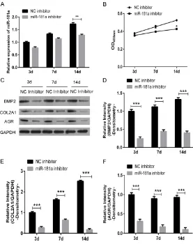
sion of BMP2, COL2A1 and AGR protein in NC inhibitor, and miR-181a inhibitor-transfected PBMSCs. MiR-181a inhibitor suppressed the expression of miR-181a in PBMSCs throughout the transfected process (Figure 3A). And the down-regulated miR-181a inhibited the prolif-eration of PBMSCs in 3 days (72 hours) (Figure 3B). Meanwhile miR-181a inhibitor decreased the protein expression of BMP2, COL2A1 and AGR compared with the NC inhibitor-transfect-ed PBMSCs, which showinhibitor-transfect-ed that miR-181a may increase protein expression of BMP2, COL2A1 and AGR during chondrogenic differentiation of PBMSCs by testing with weatern-blot and
immunofluorescence (Figures 3C-F and 4A, 4B). We further examined the chondrogenic differentiation potential of miR-181a
inhibitor-Figure 3. MiR-181a increases chondrogenic differentiation in PBMSCs. Pig PBMSCs were treated with TGF-β3 together with transfected with miR-181a inhibitor or its NC control. A. After 3 days, 7 days and 14 days of treatment, the expression of miR-181a was measured via qRT-PCR. B. The proliferation assay of treated PBMCs within 72 hours. C-F. After 3 days, 7 days and 14 days of treatment, the expression of chondrogenic differentiation markers, such as BMP2, COL2A1, and AGR were measured via western-blot.
western-blot analysis showed
a significant increase in the
protein expression levels of chondrogenesis markers incl- uding BMP2, COL2A1 and AGR after induction for 3 days, 7 days and 14 days (Figure 2A). And we confirmed that
miR-181a increased in the third day of chondrogenesis and up-regulation maintained for 2 weeks during differentia-tion by using qRT-PCR (Figure 2B). Additionally, immunofluo -rescence and confocal imag-ing showed the same result as western-blot analysis (Figure 2C). As shown in Figure 2D, the differentiation cells were positively stained for alcian blue staining.
MiR-181a was reported as a bone formation-relevant mi- RNA. These results showed that increased expression of miR-181a was associated wi- th the differentiation of MSCs towards chondrocytes.
miR-181a promotes chondro-genic differentiation in PBM-SCs
[image:5.612.93.370.71.421.2]expres-transfected PBMSCs by alcian blue staining. Inhibition of endogenous miR-181a expression in PBMSCs by transfection of miR-181a inhibi-tor, under the same induction conditions as above, resulted in suppressing chondrogenic
differentiation as shown by a significant
de-crease in alcian blue staining intensity (Figure 4C). Collectively, our data demonstrated that miR-181a act as a key positive regulator of chondrogenic differentiation.
Discussion
Expounding the mechanism of chondrogenesis is distinctly important resulting from the grow-ing importance of articular cartilage injury and repair. In this study, we investigated the func-tion of miRNA-181a in the process of chondro-genic differentiation in pig PBMSCs.
we focused on the role of miR-181a in the pro-cess of chondrogenic differentiation in MSCs. The previous study indicated that miRNAs would induce the process of chondrogenic dif-ferentiation. In our study, we found that
miR-181a was up-regulated during TGF-β3-induced
pig PBMSCs chondrogenic differentiation from the early stages and maintained elevated throughout the whole process, while the
west-ern-blot results and immunofluorescence
re-sults indicated that the expression of differenti-ation-related protein COL2A1, BMP2 and AGR were decreased, together with the Alcian blue assay proving the process of differentiation
was inhibited significantly after inhibiting the
[image:6.612.93.372.73.386.2]endogenesis miR-181a. All these results dem-onstrated that miR-181a act as a key positive regulator of chondrogenic differentiation in vi- tro.
Figure 4. MiR-181a increases chondrogenic differentiation in PBMSCs. Pig PBMSCs were treated with TGF-β3 together with transfected with miR-181a inhibitor or its NC control. A, B. After 14 days of treatment, the expression of chondrogenic differentiation markers COL2A1 and BMP2 was measured via immunofluorescence. C. After 14 days of the transfection of anti-miR-181a or its negative NC control, the differentiated cells were measured by alcian blue staining.
PBMSCs were reported to have the potency of multipo-tential differentiation and self-renew. Manipulation the gen-eration of desired cell types differentiated from PBMSCs
was noticed in the field of
cell-based therapies of articular cartilage injury or tissue engi-neering. There are some clas-sical signaling pathway invo- lved in the process of PBMSCs chondrogenic differentiation,
including fibroblast growth
factor (FGF) signaling pathway
[37, 38], TGF-β/BMP signaling pathway [39-41] and
Wnt/β-catenin signaling pathway [42-46]. Recently, a large number of novel factors including
Although we identified a novel miRNA on
re-gulating the chondrogenic differentiation of MSCs, the mechanism of miR-181a on modu-lating the process of chondrogenic differentia-tion was still unclear. As reported, three major target genes including C/EBPb, Sox9 and Adam9 have been implicated in mediating the effects of miRNAs in regulating chondrogenesis [47].
Considering defect of articular cartilage is an unique challenge on clinic, more and more researchers focus on the tissue engineering
field as the therapeutic strategy of articular
cartilage injury. PBMSCs are appropriate cells for cartilage tissue engineering with the advan-tage of amount and the ability to differentiate into functional cartilage and maintain a chon-drocyte phenotype long-term. Our results proved that miR-181a plays an important role in the process of chondrogenic differentiation from PBMSCs, suggesting that miR-181a has the potential to be the novel target to induce
the generation of cartilage artificially.
In summary, we present evidences for the important role of miR-181a on the regulation of MSCs chondrogenesis, also suggest that the up-regulation of miR-181a during MSC differen-tiation might be required for chondrocyte lin-eage maintenance. And the up-regulated
miR-181a might influence the expression of some
differentiation process protein via post-tran-scriptional regulation, resulting in the pro- motion of chondrogenic differentiation. Such hypothesis will be investigated in the future work.
Acknowledgements
We thank the center of laboratory animal sci-ence of Kunming Medical University for provid-ing the peripheral blood from the small-ear pigs, the National Natural Science Foundation (No. 8140340); Yunnan Province innovation team project (No. 2014HC018); the Yunnan Province Natural Science Key Project (No. 2017FE467 (-007)).
Disclosure of conflict of interest
None.
Abbreviations
PBMSCs, Peripheral blood Mesenchymal stem cells; COL2A1, collagen type II alpha 1 chain;
BMP2, bone morphogenetic protein 2; qRT-PCR, quantitative real-time polymerase chain
reaction; TGF-β3, transforming growth factor
beta 3; AGR, aggrecan; HDAC4, (histone dea- cetylase 4); Smad3, (SMAD family member 3); Sox9, SRY-box9; Adam9, ADAM metallopepti-dase domain 9.
Address correspondence to: Yanlin Li, Department of Sports Medicine, First Affiliated Hospital of Kunming Medical University, 295 Xichang Road, Kunming 650032, China. E-mail: yanlinliedu@yeah. net; 852387873@qq.com
References
[1] Friedenstein AJ, Petrakova KV, Kurolesova AI and Frolova GP. Heterotopic of bone marrow. Analysis of precursor cells for osteogenic and hematopoietic tissues. Transplantation 1968; 6: 230-247.
[2] Sacchetti B, Funari A, Michienzi S, Di Cesare S, Piersanti S, Saggio I, Tagliafico E, Ferrari S, Robey PG, Riminucci M and Bianco P. Self-re-newing osteoprogenitors in bone marrow sinu-soids can organize a hematopoietic microenvi-ronment. Cell 2007; 131: 324-336.
[3] Beyer Nardi N, da Silva Meirelles L. Mesenchy-mal stem cells: isolation, in vitro expansion and characterization. Handb Exp Pharmacol 2006; 249-282.
[4] Delorme B and Charbord P. Culture and char-acterization of human bone marrow mesen-chymal stem cells. Methods Mol Med 2007; 140: 67-81.
[5] Javazon EH, Beggs KJ and Flake AW. Mesen-chymal stem cells: paradoxes of passaging. Exp Hematol 2004; 32: 414-425.
[6] Martin DR, Cox NR, Hathcock TL, Niemeyer GP and Baker HJ. Isolation and characterization of multipotential mesenchymal stem cells from feline bone marrow. Exp Hematol 2002; 30: 879-886.
[7] Pittenger MF, Mackay AM, Beck SC, Jaiswal RK, Douglas R, Mosca JD, Moorman MA, Sim-onetti DW, Craig S and Marshak DR. Multilin-eage potential of adult human mesenchymal stem cells. Science 1999; 284: 143-147. [8] Fu WL, Zhou CY and Yu JK. A new source of
mesenchymal stem cells for articular cartilage repair. Am J sports Med 2013; 42: 592. [9] Mackie EJ, Ahmed YA, Tatarczuch L, Chen KS
and Mirams M. Endochondral ossification: how cartilage is converted into bone in the develop-ing skeleton. Int J Biochem Cell Biol 2008; 40: 46-62.
[11] Goldring MB, Tsuchimochi K and Ijiri K. The control of chondrogenesis. J Cell Biochem 2006; 97: 33-44.
[12] Koay EJ and Athanasiou KA. Hypoxic chon- drogenic differentiation of human embryonic stem cells enhances cartilage protein synthe-sis and biomechanical functionality. Osteoar-thritis Car-tilage 2008; 16: 1450-1456. [13] Liu F, Kohlmeier S and Wang CY. Wnt signaling
and skeletal development. Cell Signal 2008; 20: 999-1009.
[14] Wu X, Shi W and Cao X. Multiplicity of BMP sig-naling in skeletal development. Ann N Y Acad Sci 2007; 1116: 29-49.
[15] Bartel DP. MicroRNAs: genomics, biogenesis, mechanism, and function. Cell 2004; 116: 281-297.
[16] Hutvagner G and Zamore PD. A microRNA in a multiple-turnover RNAi enzyme complex. Sci-ence 2002; 297: 2056-2060.
[17] Lim LP, Glasner ME, Yekta S, Burge CB and Bartel DP. Vertebrate microRNA genes. Sci-ence 2003; 299: 1540.
[18] Chen Y and Stallings RL. Differential patterns of microRNA expression in neuroblastoma are correlated with prognosis, differentiation, and apoptosis. Cancer Res 2007; 67: 976-983. [19] Thompson BJ and Cohen SM. The Hippo
path-way regulates the bantam microRNA to control cell proliferation and apoptosis in Drosophila. Cell 2006; 126: 767-774.
[20] Wu H, Neilson JR, Kumar P, Manocha M, Shan-kar P, Sharp PA and Manjunath N. miRNA pro-filing of naive, effector and memory CD8 T cells. PLoS One 2007; 2: e1020.
[21] Cheng AM, Byrom MW, Shelton J and Ford LP. Antisense inhibition of human miRNAs and in-dications for an involvement of miRNA in cell growth and apoptosis. Nucleic Acids Res 2005; 33: 1290-1297.
[22] Chen CZ, Li L, Lodish HF and Bartel DP. MicroR-NAs modulate hematopoietic lineage differen-tiation. Science 2004; 303: 83-86.
[23] O’Rourke J, Georges S, Seay H, Tapscott S, Mc-manus M, Goldhamer D, Swanson M and Harfe B. Essential role for Dicer during skeletal mus-cle development. Dev Biol 2007; 311: 359-368.
[24] Wienholds E, Kloosterman WP, Miska E, Alva-rez-Saavedra E, Berezikov E, de Bruijn E, Hor-vitz HR, Kauppinen S and Plasterk RH. Mic- roRNA expression in zebrafish embryonic de -velopment. Science 2005; 309: 310-311. [25] Tuddenham L, Wheeler G, Ntounia-Fousara S,
Waters J, Hajihosseini MK, Clark I and Dalmay T. The cartilage specific microRNA-140 targets histone deacetylase 4 in mouse cells. FEBS Lett 2006; 580: 4214-4217.
[26] Kobayashi T, Lu J, Cobb BS, Rodda SJ, McMa-hon AP, Schipani E, Merkenschlager M and Kronenberg HM. Dicer-dependent pathways regulate chondrocyte proliferation and differ-entiation. Proc Natl Acad Sci U S A 2008; 105: 1949-1954.
[27] Pais H, Nicolas FE, Soond SM, Swingler TE, Clark IM, Chantry A, Moulton V and Dalmay T. Analyzing mRNA expression identifies Smad3 as a microRNA-140 target regulated only at protein level. RNA 2010; 16: 489-494.
[28] Guerit D, Philipot D, Chuchana P, Toupet K, Brondello JM, Mathieu M, Jorgensen C and Noel D. Sox9-regulated miRNA-574-3p inhibits chondrogenic differentiation of mesenchymal stem cells. PLoS One 2013; 8: e62582. [29] Paik S, Jung HS, Lee S, Yoon DS, Park MS and
Lee JW. miR-449a regulates the chondrog- enesis of human mesenchymal stem cells through direct targeting of lymphoid enhancer-binding factor-1. Stem Cells Dev 2012; 21: 3298-3308.
[30] Lin X, Wu L, Zhang Z, Yang R, Guan Q, Hou X and Wu Q. MiR-335-5p promotes chondrogen-esis in mouse mesenchymal stem cells and is regulated through two positive feedback loops. J Bone Miner Res 2014; 29: 1575-1585. [31] Suomi S, Taipaleenmaki H, Seppanen A,
Ripat-ti T, Vaananen K, Hentunen T, Saamanen AM and Laitala-Leinonen T. MicroRNAs regulate osteogenesis and chondrogenesis of mouse bone marrow stromal cells. Gene Regul Syst Bio 2008; 2: 177-191.
[32] Laine SK, Alm JJ, Virtanen SP, Aro HT and Laitala-Leinonen TK. MicroRNAs 96, miR-124, and miR-199a regulate gene expression in human bone marrow-derived mesenchymal stem cells. J Cell Biochem 2012; 113: 2687-2695.
[33] Yang B, Guo H, Zhang Y, Chen L, Ying D and Dong S. MicroRNA-145 regulates chondrogen-ic differentiation of mesenchymal stem cells by targeting Sox9. PLoS One 2011; 6: e21679. [34] Bakhshandeh B, Soleimani M, Hafizi M, Pay
-lakhi SH and Ghaemi N. MicroRNA signature associated with osteogenic lineage commit-ment. Mol Biol Rep 2012; 39: 7569-7581. [35] Bhushan R, Grunhagen J, Becker J, Robinson
PN, Ott CE and Knaus P. miR-181a promotes osteoblastic differentiation through repression of TGF-beta signaling molecules. Int J Biochem Cell Biol 2013; 45: 696-705.
[36] Sumiyoshi K, Kubota S, Ohgawara T, Kawata K, Abd El Kader T, Nishida T, Ikeda N, Shimo T, Yamashiro T and Takigawa M. Novel role of miR-181a in cartilage metabolism. J Cell Bio-chem 2013; 114: 2094-2100.
cells for enhanced chondrogenesis. PLoS One 2011; 6: e22887.
[38] Solchaga LA, Penick K, Porter JD, Goldberg VM, Caplan AI and Welter JF. FGF-2 enhances the mitotic and chondrogenic potentials of hu-man adult bone marrow-derived mesenchymal stem cells. J Cell Physiol 2005; 203: 398-409. [39] Fischer L, Boland G and Tuan RS. Wnt-3A en-hances bone morphogenetic protein-2-medi- ated chondrogenesis of murine C3H10T1/2 mesenchymal cells. J Biol Chem 2002; 277: 30870-30878.
[40] Hartmann C. A Wnt canon orchestrating osteo-blastogenesis. Trends Cell Biol 2006; 16: 151-158.
[41] Zhou S, Eid K and Glowacki J. Cooperation be-tween TGF-beta and Wnt pathways during chondrocyte and adipocyte differentiation of human marrow stromal cells. J Bone Miner Res 2004; 19: 463-470.
[42] Hartmann C and Tabin CJ. Dual roles of Wnt signaling during chondrogenesis in the chick-en limb. Developmchick-ent 2000; 127: 3141-3159. [43] Enomoto-Iwamoto M, Kitagaki J, Koyama E,
Tamamura Y, Wu C, Kanatani N, Koike T, Okada H, Komori T, Yoneda T, Church V, Francis-West PH, Kurisu K, Nohno T, Pacifici M and Iwamoto M. The Wnt antagonist Frzb-1 regulates chon-drocyte maturation and long bone develop-ment during limb skeletogenesis. Dev Biol 2002; 251: 142-156.
[44] Hartmann C and Tabin CJ. Wnt-14 plays a piv-otal role in inducing synovial joint formation in the developing appendicular skeleton. Cell 2001; 104: 341-351.
[45] Kawakami Y, Wada N, Nishimatsu SI, Ishikawa T, Noji S and Nohno T. Involvement of Wnt-5a in chondrogenic pattern formation in the chick limb bud. Dev Growth Differ 1999; 41: 29-40. [46] Lako M, Lindsay S, Bullen P, Wilson DI, Robson
SC and Strachan T. A novel mammalian wnt gene, WNT8B, shows brain-restricted expres-sion in early development, with sharply delim-ited expression boundaries in the developing forebrain. Hum Mol Genet 1998; 7: 813-822. [47] Green JD, Tollemar V, Dougherty M, Yan Z, Yin